Contribution to the study of the links
between consciousness and swallowing
Under the supervision of
Steven Laureys & Jean-François Kaux
Thèse présentée en vue de l’obtention du grade de
Docteur en Sciences Médicales
Evelyne Mélotte
February 2021
Promotor: Prof. Dr. Steven Laureys
Co-promotor: Prof. Dr. Jean-François Kaux Jury Members
Prof. Dr. Didier Ledoux (President) Prof. Dr. Aude Lagier (Secretary) Dr. Olivia Gosseries
Prof. Dr. Audrey Maudoux Prof. Dr. Dominique Morsomme Prof. Dr. Gwen Van Nuffelen Dr. Audrey Vanhaudenhuyse Prof. Dr. Virginie Woisard
c ⃝ Evelyne Mélotte, 2021
The studies presented in this thesis have been financially supported by:
University and University Hospital of Liège, the Belgian National Funds for Scientific Research (F.R.S-FNRS), Luminous project (EU-H2020-fetopen-ga686764), the European Union’s Horizon 2020 Framework Program for Research and Innovation [Human Brain Project SGA3, Grant Number 945539] and the BIAL Foundation.
1
A Bijan et tous les autres patients présentant des troubles de la conscience ainsi qu’à leurs proches qui les accompagnent
au quotidien.
3
CONTENT
Abstract 7 Résumé 9 Remerciements 11 Scientific Publications 13 Glossary 15 List of tables and figures 17General Introduction 19
Chapter 1. Literature Review: links between swallowing and consciousness:
insight from behavioral and neuroimaging studies
27 Abstract 29 Introduction 30 Section 1: Swallowing from the perspective of volition 34 Section 2: Nonpathological modifications of consciousness: sleep and anesthesia
46 Section 3: Links between consciousness and swallowing in brain-injured patients
49 Section 4: Evaluation and treatment of swallowing in patients with DOC
63 Discussion 70 Conclusion 74
Chapter 2. Is oral feeding compatible with an unresponsive wakefulness
syndrome? 77 Abstract 79 Background 80 Method 81 Results 81 Case Report 1 82 Case Report 2 83 Discussion 86 Conclusion 90
Chapter 3. Swallowing in individuals with disorders of consciousness: A
cohort study
93 Abstract 95 Introduction 96 Participants and methods 97 Participants 97 Diagnosis of consciousness 99 Respiratory status, type of feeding and swallowing assessments
99 Statistical analysis 100 Results 101 Discussion 104
4
Chapter 4. The development and validation of the SWADOC: A study
protocol for a multicenter prospective cohort study
109 Abstract 111 Introduction 111 Method and analysis 114 Development of the SWADOC 114 Presentation of the tool 116 Study Design 119 Population and recruitment 119 Validation study procedure 119 Study Hypotheses 122 Data Analysis 122 Discussion 124 Strengths and opportunities 124 Limitations and pitfalls 125 Ethics and dissemination 125
General Discussion and Prospects 129
References 149
Appendices 173
Appendix 1. Additional information related to General Introduction 175 Appendix 1.1. CRS-R administration and scoring
guidelines
175 Appendix 1.2 Hypotheses concerning which other behaviors can be considered as linked to level of consciousness and which can be seen as signs of consciousness based on results of the studies
192 Appendix 2. Additional information related to Chapter 1 194 Appendix 2.1 Search strategy and selected criteria 194 Appendix 2.2 Description and results of the 31 identified studies with healthy human adults without dysphagia who performed a swallowing task with a neuroimaging technique
196 Appendix 2.3 Description of the consciousness scales used in the studies identified in Section 3
204 Appendix 3. Additional information related to Chapter 3 206
STROBE Statement – Checklist of items that should be included in reports of cohort studies
Appendix 4. Additional information related to Chapter 4 208 Appendix 4.1 English version of the SWADOC’s Administration Guide
208 Appendix 4.2 French version of the SWADOC’s Administration Guide
226 Appendix 4.3 SWADOC-scored (French version) 244
5
Appendix 4.4 The FOTT-SAS (table adapted from Mortensen et al., 2016)
245 Appendix 5. Original published articles 246
Appendix 5.1 Is oral feeding compatible with an unresponsive wakefulness syndrome
246 Appendix 5.2 Swallowing in individuals with disorders of consciousness: A cohort study
7
ABSTRACT
Following severe brain injuries (e.g., traumatic or anoxic brain injury, stroke), a small proportion of patients will remain in an altered state of consciousness. Patients with prolonged (> 28 days post-insult) disorders of consciousness (DOC) can open their eyes (sometimes showing electrophysiological sleep/wake cycles) and the majority no longer need invasive ventilation. However, most of them receive artificial feeding, suggesting that consciousness affects swallowing capacities.
The aim of this thesis is to contribute to the study of the links between consciousness and swallowing. The hypotheses are that swallowing capacities are linked to level of consciousness, and that the presence of some components of swallowing constitutes a possible sign of consciousness.
Based on a literature review, we show that the sequencing of the components of swallowing falls on a continuum of voluntary to reflex behaviors. Components of the oral phase may be considered as voluntary behaviors because they are controllable and suppressible (although largely automated), components of the pharyngeal phase as somatic reflexes, and components of the esophageal phase as autonomic reflexes. The triggering of the swallowing reflex inhabits the border region between voluntary behaviors and somatic reflexes, while the opening of the upper esophageal sphincter divides somatic from autonomic reflexes. If voluntary behaviors are considered possible signs of consciousness, the presence of components of the oral phase of swallowing should be considered as revealing conscious behaviors. Moreover, we show that a range of cortical areas (mainly the primary sensorimotor cortex, premotor cortex and supplementary motor area, anterior part of the cingulate cortex, insula and cerebellum) are involved in both volitional and non-volitional swallowing tasks.
In two retrospective studies analyzing swallowing in patients with DOC diagnosed by means of repeated behavioral assessment and neuroimaging, we demonstrate that almost all such patients present at least one dysfunction in the oral and/or pharyngeal phase. Patients who do not show behavioral signs of consciousness (unresponsive wakefulness syndrome – UWS) do not present an efficient oral phase of swallowing allowing oral feeding with solid food. Consequently, the preservation of components of the oral phase of swallowing should be
8 considered as a sign of consciousness and be one of the diagnostic criteria for consciousness. The absence of an efficient oral phase of swallowing in patients with UWS and its presence in only a small proportion of minimally conscious (MCS) patients explain why no “typical” patients with UWS (i.e., with behavioral and neuroimaging assessments pointing in the same direction) can be fed entirely orally while no patients with MCS can eat ordinary textured food.
Furthermore, in the studied group, level of consciousness is linked to components of the pharyngeal phase (reflected by the absence of a tracheostomy, pharyngo-laryngeal secretions or saliva aspiration) and to the cough reflex. Indeed, more patients with MCS than UWS present efficient spontaneous saliva management and a cough reflex, although these components are present in some patients with UWS. For that reason, these components seem to represent cortically mediated behavior but do not constitute signs of consciousness as such.
Finally, this work highlights the lack of appropriate tools to assess and treat swallowing for patients with DOC. A protocol study for the validation of a swallowing assessment tool for patients with DOC is therefore proposed.
9
RESUME
A la suite d'atteintes cérébrales sévères telles que des accidents vasculaires cérébraux, des traumatismes crâniens ou anoxies cérébrales, une petite proportion de patients conserve une altération de l’état de conscience. Les patients avec des états de conscience altérée (ECA) dits « prolongés » (>28 jours post-accident) ouvrent leurs yeux (certains présentant des cycles éveil/sommeil) et la majorité d’entre eux ne bénéficient plus d’une ventilation invasive. En revanche, la plupart des patients ECA sont nourris artificiellement, suggérant un impact de la conscience sur les capacités de déglutition.
L’objectif de ce travail de thèse est de contribuer à l'étude des liens entre conscience et déglutition. Nos hypothèses sont que les capacités de déglutition sont en lien avec le niveau de conscience et que la présence de certains composants de la déglutition constitue un signe possible de conscience.
Sur base d’une revue de littérature, nous montrons que l'enchaînement des composants de la déglutition s'inscrit sur un continuum allant de comportements volontaires à réflexes. Les composants de la phase orale peuvent être considérés comme volontaires car davantage contrôlables et suppressibles (bien que largement automatisés), les composants de la phase pharyngée comme des réflexes somatiques et les composants de la phase œsophagienne comme des réflexes autonomes. Le déclenchement du réflexe de déglutition se situe quant à lui à la frontière entre comportement volontaire et réflexe somatique et l’ouverture du sphincter supérieur de l’œsophage sépare réflexe somatique et autonome. Si nous considérons les comportements volontaires comme de possibles signes de conscience, la présence de composants de la phase orale de la déglutition peut être considéré comme un comportement conscient. Nous montrons en outre qu’un grand nombre d’aires corticales (principalement le cortex sensorimoteur primaire, le cortex prémoteur, l’aire motrice supplémentaire, le cortex cingulaire antérieur, l’insula et le cervelet) sont impliquées à la fois dans des tâches de déglutition dites « volontaires » et « non-volontaires ».
Nous démontrons dans deux études rétrospectives analysant la déglutition de patients ECA diagnostiqués à l’aide d’évaluations comportementales répétées et d’examens de neuroimagerie, que la très grande majorité des patients ECA présentent une atteinte de la phase orale et/ou pharyngée.
10 Les patients ne montrant pas de signes comportementaux de conscience (état d’éveil non-répondant – EENR) ne présentent pas non plus de phase orale efficace autorisant une alimentation en texture solide. En conséquence, la préservation de composants de la phase orale de la déglutition devrait donc être envisagée comme un signe de conscience et faire partie des critères diagnostiques des patients ECA. L’absence de phase orale efficace chez les patients EENR et sa présence chez seulement une faible proportion de patients en état de conscience minimal (ECM) expliquent pourquoi aucun patient EENR « typique » (examens comportementaux et de neuroimagerie pointant dans la même direction) ne peut être nourri exclusivement per os ainsi que le fait qu’aucun patient ECM n’est nourri oralement en texture ordinaire.
En outre, dans le groupe de patients étudiés, le niveau de conscience est en lien avec certains composants de la phase pharyngée (reflété par l’absence de trachéotomie, de sécrétions pharyngo-laryngées ou de fausses-routes salivaires) ainsi qu’avec le réflexe de toux. En effet, il y a davantage de patients ECM comparés aux patients EENR qui présentent une gestion salivaire efficace et une toux réflexe bien que ces composants soient aussi présents chez certains patients EENR. Pour cette raison, ces éléments semblent constituer des « comportements sous contrôle cortical » sans pour autant constituer des signes de conscience en tant que tel. Enfin, ce travail souligne le peu d’outils et de techniques de prise en charge de la déglutition adaptés aux patients ECA. Un protocole de validation d’un outil d’évaluation de la déglutition pour les patients ECA est dès lors proposé.
11
Remerciements
J’adresse mes remerciements à Steven Laureys pour m’avoir ouvert les portes du domaine des troubles de la conscience et m’avoir donné l’opportunité de réaliser ce travail. Mes remerciements vont également à Jean-François Kaux pour m’avoir permis de me lancer dans ce défi passionnant et exigeant qui est de combiner recherche et travail clinique.
Dans la team du Coma Science Group, je remercie chaleureusement Olivia, pour ses conseils avisés tout au long de mon parcours de thèse ainsi que pour son analyse critique de mes travaux qui m’ont encouragée à me dépasser. Merci à Audrey Maudoux qui a accepté de présider mon comité de thèse pendant ces quatre années et qui m’a accompagnée sur le chemin de la recherche. Je remercie également Audrey Vanhaudenhuyse pour les nombreux coups de pouce lors de mes débuts, son écoute et son soutien bienveillant. Mes remerciements vont enfin à toute l’équipe du Coma Science Group qui a contribué de près ou de loin à l’aboutissement de ce travail.
Je tiens également à remercier les autres membres de mon comité d’accompagnement et membres du jury de thèse, les Professeurs Didier Ledoux, Dominique Morsomme et Aude Lagier. Merci à vous de m’avoir guidée tout au long de ces quatre années et merci pour les échanges toujours constructifs lors de mes comités d’accompagnement. Aude, ton arrivée à Liège a changé notre travail du quotidien. Je te remercie chaleureusement pour ton écoute, ton soutien et tes enseignements ces quatre dernières années. Une complicité et une amitié sont nées.
Je souhaite aussi remercier le Professeur Virginie Woisard ainsi que Dr Gwen Van Nuffelen d’avoir accepté de faire partie de mon jury externe.
Merci à mes collègues ORL du CHU de Liège, particulièrement Sabrina Delhalle pour avoir partagé avec moi son travail clinique impressionnant auprès des patients en état de conscience altérée.
Je remercie chaleureusement Marion et Roxane. Une collaboration solide et une complicité se sont installées, qui je l’espère, se poursuivront vers d’autres projets et aventures.
Je ne serais rien sans ma bouée de sauvetage. Celle qui a toujours été là, dans les bons moments comme dans les moins bons. Merci à vous Christine, Barbara, Kelly, Charlotte.
A toi mon cher Papa, qui nous a appris que dans la vie tout est possible, que nous sommes capables de tout et même encore plus. A toi Maman, qui m’a transmis ta force, ton courage et ta détermination. Merci à Raphaël et Magali pour leur intérêt et soutien vis-à-vis de mon travail tout au long de ces années.
12
Enfin, merci à Antoine, mon partenaire, graphiste privé, coach personnel, de m’avoir soutenue dans cette aventure.
13
Scientific publications
The present thesis is based on the following publications:
Links between swallowing and consciousness: Insight from behavioral and neuroimaging studies
Mélotte, E., Maudoux, A., Panda, R., Kaux, J.-F., Lagier, A., Herr, R., Belorgeot, M., Laureys, S., & Gosseries, O. (submitted).
Is oral feeding compatible with an unresponsive wakefulness syndrome?
Mélotte, E.+, Maudoux, A.+, Delhalle, S., Martial, C., Antonopoulos, G.,
Larroque, S. K., Wannez, S., Faymonville, M.-E., Kaux, J.-F., Laureys, S.*, Gosseries, O.*, & Vanhaudenhuyse, A.* (2018). Journal of Neurology, 265(4), 954‑961. https://doi.org/10.1007/s00415-018-8794-y
Swallowing in individuals with disorders of consciousness: A cohort study
Mélotte, E., Maudoux, A., Delhalle, S., Lagier, A., Thibaut, A., Aubinet, C., Kaux, J.-F., Vanhaudenhuyse, A., Ledoux*, D., Laureys, S.*, & Gosseries, O*. (2020). Annals of Physical and Rehabilitation Medicine (in press). https://doi.org/10.1016/j.rehab.2020.04.008
The development and validation of the SWADOC tool: A study protocol for a multicentric prospective cohort study
Mélotte, E.+, Belorgeot, M. +, Herr, R. +, Simon, J., Kaux, J.-F., Laureys,
S., Sanz R.D., L., Morsomme, D., Lagier, A., Pellas, F.*, & Gosseries, O*. Frontiers in Neurology (accepted).
Other publications: Article (as co-author)
Swallowing function after severe COVID-19: early videofluoroscopic findings
Lagier, A., Mélotte, E., Poncelet M., Remacle S., Meunier P. (2020). European Archives of Oto-Rhino-Laryngology
Book chapter (as co-author)
Feasibility of Oral Feeding in Patients with Disorders of Consciousness
14 Maudoux, A., Breuskin, I., Gosseries, O., Mélotte, E., Schnakers, C., & Vanhaudenhuyse, A. (2017). In Schnakers C. & Laureys, S. (eds),
Coma and Disorders of Consciousness (p. 137‑153). Springer International Publishing.
+ contribute equally to the work as first author * contribute equally to the work as last author
15
Glossary
ABI Acquired Brain Injury
CAMMRI Comprehensive Measure for the Minimally Responsive Individual
CIs Confidence Intervals CPG Central Pattern Generator CRS-R Coma Recovery Scale-Revised DOC Disorders Of Consciousness DOCS Disorders Of Consciousness Scale DTI Diffusion Tensor Imaging
EEG Electroencephalography
EMCS Emergence from the Minimally Conscious State ENT Ear, Nose and Throat
FDG-PET Fluorodesoxyglucose Positron Emission Tomography FILS Food Intake Level Scale
fMRI Functional Magnetic Resonance Imaging FEES Fiberoptic Endoscopic Evaluation of Swallowing FOIS Functional Oral Intake Scale
FOTT Facial Oral Tract Therapy
FOTT-SAS Facial Oral Tract Therapy Swallowing Assessment of Saliva
FOUR Full Outline of UnResponsiveness GCS Glasgow Coma Scale
IDDSI International Dysphagia Diet Standardisation Initiative LIS Locked-In-Syndrome
MCS Minimally Conscious State MRI Magnetic Resonance Imaging NVOST Non-volitional swallowing task
ORs Odds Ratios
PDOC Prolonged Disorders of Consciousness RLA Ranchos Los Amigos Scale
SECONDs Simplified Evaluation of CONsciousness Disorders SMART Sensory Modality Assessment and Rehabilitation
Technique
SS Spontaneous swallowing
SLT Speech and Language Therapists
STROBE Strengthening the Reporting of Observational studies in Epidemiology
SWADOC SWallowing Assessment in Disorders of Consciouness TBI Traumatic Brain Injury
UES Upper Esophageal Sphincter
16
VFSS VideoFluoroscopic Swallowing Study VOST Volitional swallowing task
VS Voluntary swallowing WHIM Wessex Head Injury Matrix
17
List of tables and figures
Tables
Chapter 1
Table 1. Characteristics of voluntary behavior and somatic and autonomic
reflexes
35
Table 2. Description of the different types of swallowing tasks 42
Table 3. Characteristics of studies exploring the relation between level of
consciousness and swallowing-related abilities in patients with severe brain injury
50
Table 4. Summarized results of studies analyzing the link between level of
consciousness and swallowing components
61
Table 5. The Facial Oral Tract Therapy Swallowing Assessment of Saliva
(FOTT-SAS) (adapted from Mortensen et al., 2016)
65
Table 6. Indications concerning swallowing adapted from the National Italian
Consensus Conference
67
Table 7. Percentage of agreement of 40 speech and language therapists
with several items linked to the assessment and treatment of dysphagia in patients with prolonged disorders of consciousness (extracted from Roberts & Greenwood, 2019, p. 7-8)
69
Chapter 3
Table 8. Descriptive statistics of the whole sample 101
Table 9. Descriptive and statistical analysis of the 10 criteria in the UWS and
MCS groups
103
General discussion and prospects
Table 10. Hypotheses concerning which components of swallowing can be
considered to be linked to level of consciousness according to the literature and the main findings of our studies
139
Figures
Introduction
Figure 1. CRS-R and possible other signs of consciousness along the
continuum from coma to recovery of consciousness
24
Chapter 1
Figure 2. Summary of the research fields explored in this review 33
Figure 3. Classification of the oral, pharyngeal and esophageal phases of
swallowing on the continuum of voluntary to reflexive behaviors
40
Figure 4. Brain areas activated by spontaneous and voluntary swallowing
tasks
18
Figure 5. Neuroimaging results (sagittal and axial views) of the two UWS
patients who were able to swallow and of healthy controls
57
Chapter 2
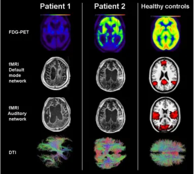
Figure 6. Neuroimaging results (PET, fMRI Default mode Network, fMRI
Auditory Network and DTI) of the two patients and healthy controls.
85
Chapter 3
Figure 7. Flowchart of the study 98
Figure 8. Percentage of UWS and MCS patients for the 10 criteria 104
Chapter 4
Figure 9. Description of the SWADOC 117
Figure 10. SWADOC-scored (English version) 118
Figure 11. Study protocol 121
General discussion and prospects
PAGE DE GARDE
INTRODUCTION
General Introduction - 21 Humans swallow several hundred times per day (Tanaka et al., 2013). Swallowing occurs spontaneously during wakefulness and sleep, and most of the time, without our being aware of it. Swallowing is one of the most essential physiological mechanism since it allows us to manage saliva and secretions.
We can all remember the sensation of food or liquid going down the wrong way, although the incidence of choking episodes is actually very low in the healthy population (Hemsley et al., 2019). In contrast, several acquired and chronic neurological conditions lead to swallowing problems (Hemsley et al., 2019). Depending on the disease, the incidence of dysphagia can be very high1 (Arnold et al., 2016; Dunn & Rumbach, 2019; Guan et al.,
2015; Kalf et al., 2012; Michel et al., 2018; Takizawa et al., 2016) and different components of the swallowing sequence can be affected. After an acquired brain injury, we can reasonably assume that, the more severe the brain injury, the more severe the dysphagia (Formisano et al., 2004; Mackay et al., 1999a, 1999b). However, the question of what factors (e.g., lesion localization and volume, type of brain injury, consciousness) most affect the severity of dysphagia following brain injury has not yet been completely elucidated.
Level of consciousness has an impact on a variety of abilities such as language (Aubinet et al., 2018), motor function (Thibaut et al., 2015), sphincter function (Foxx-Orenstein et al., 2003) and feeding (Brady et al., 2006). Most patients with disorders of consciousness (DOC) are fed by enteral feeding tube. However, the true impact of consciousness on swallowing abilities remains poorly understood.
Consciousness allows us to experience things inside and outside us (Damasio & Meyer, 2009) and is classically defined by two components: arousal and awareness. Modifications of consciousness can be pathological (DOC) or non-pathological (sleep or anesthesia).
DOC represent different states along a continuum from coma (no arousal and no awareness) to being conscious and awake (preserved arousal and 1 Stroke: 20.7% (Arnold et al., 2016), 9.1%–80% (Takizawa et al., 2016);
Non-traumatic subarachnoid hemorrhage: 31.6% (Dunn & Rumbach, 2019); Multiple sclerosis: 36% (Guan et al., 2015); Parkinson’s disease: 35% with subjective measurements and 82% with objective measurements (Kalf et al., 2012), 11%– 81% (Takizawa, 2016); Older patients with dementia: 86.6% (Michel et al., 2018); Traumatic brain injury: 27%–30% (Takizawa et al., 2016); Community-acquired pneumonia: 91.7% (Takizawa et al., 2016).
General Introduction - 22 awareness). Between the two extremes, unresponsive wakefulness syndrome (UWS, previously termed vegetative state) is defined by recovery of arousal in the absence of any sign of awareness (Laureys et al., 2010), whereas minimally conscious state (MCS) refers to preserved arousal and reproducible but inconsistent signs of consciousness (Giacino et al., 2002). The MCS entity can be subdivided into minimally conscious MINUS (MCS–) and PLUS (MCS+) based on the presence (MCS+) or absence (MCS–) of behaviors indicating at least partial preservation of language abilities (Bruno et al., 2011; Thibaut et al., 2020). When patients recover the ability to functionally communicate or to use two objects appropriately, we consider that they are emerging from the minimally conscious state (EMCS) (Giacino et al., 2002). Patients with locked-in syndrome (LIS) have woken from their coma and are fully conscious but are unable to show behavioral signs of consciousness except by eye movements (Gosseries et al., 2009).
Misdiagnosis can have serious medical and ethical consequences for patients and their families. Indeed, functional outcomes and prognoses are better for MCS than UWS (Luaute et al., 2010; Noé et al., 2012). Moreover, response to treatment seems to be better in patients with MCS (Thibaut et al., 2014). Regarding pain management, noxious stimuli seem to elicit a larger cerebral response in patients with MCS than in UWS, suggesting that patients with MCS may be more likely to feel pain than those with UWS (Boly, Faymonville, et al., 2008; Demertzi et al., 2009, 2013). Finally, level of consciousness influences end-of-life decisions (Bernat, 2008; Demertzi et al., 2011)
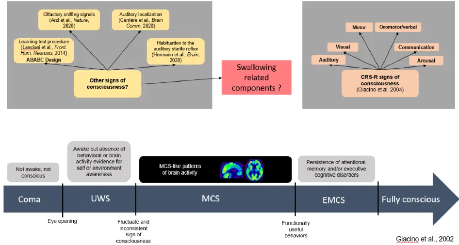
Recent guidelines for the diagnosis of patients with DOC recommend that one use valid, reliable standardized neurobehavioral assessments of consciousness (Giacino et al., 2018a, 2018b; Kondziella et al., 2020). The Coma Recovery Scale – Revised (CRS-R) is the reference standard for the clinical bedside evaluation of consciousness (Giacino et al., 2004), as it fulfills all the Aspen Neurobehavioral Workgroup criteria (Seel et al., 2010). The diagnostic criteria for consciousness in the CRS-R are classified in six categories (auditory, visual, motor, oromotor/verbal, communication, arousal) (see Appendix 1.1). Beyond behaviors assessed using the CRS-R, some authors have identified other criteria linked to level of consciousness (Chatelle et al., 2018; van Ommen et al., 2018) and other possible signs of consciousness (Arzi et al., 2020;
General Introduction - 23 Carrière et al., 2020; Hermann et al., 2020; Lancioni et al., 2014) (see Figure 1 & Appendix 1.2).
General Introduction - 24
Figure 1. CRS-R and possible other signs of consciousness along the continuum from coma to recovery of consciousness
Note: CRS-R=Coma Recovery Scale-Revised; UWS=Unresponsive Wakefulness Syndrome; MCS=Minimally Conscious State; EMCS=Emergence of minimally
General Introduction - 25 Hermann et al. (2020) emphasized the importance of expanding the range of signs of consciousness that can be tested at the bedside.
Until now, swallowing capacities have not been included in the diagnostic criteria for consciousness (Giacino et al., 2002) and it has not been clearly established whether the level of consciousness is related to the presence or absence of certain components of swallowing.
Our hypotheses are that swallowing capacities are linked to level of consciousness, and that the presence of some components of swallowing constitutes a possible sign of consciousness.
As clinicians, at the beginning of this thesis project, our ambition was to work on the management of dysphagia in patients with DOC. However, there was no real understanding of the impact of consciousness on swallowing or the pathophysiology of swallowing in patients with DOC, nor were there any assessment tools. For that reason, we chose to devote this work to establishing the basis of the links between swallowing and consciousness.
To lead off, Chapter 1 presents a theoretical introduction in the form of a literature review on the links between swallowing and consciousness. In that section, we identify several thematic areas addressing the relationships between swallowing and consciousness.
In Chapter 2, we retrospectively explore the possibility of oral feeding in a group of 68 patients without behavioral signs of consciousness.
To extend our understanding of the possibility of oral feeding and, more broadly, the pathophysiology of swallowing in all patients with DOC, in Chapter 3, we retrospectively document the incidence and characteristics of dysphagia in 92 patients with DOC (UWS and MCS), and describe the links between level of consciousness and different components of swallowing.
Finally, Chapter 4 presents the protocol study for the development and validation of a new instrument: the SWallowing Assessment in Disorders Of Consciousness (SWADOC).
This thesis takes the form of a collection of scientific papers. The figures and tables have been renumbered and all references are now presented in a single list at the end of the thesis.
PAGE DE GARDE
Chapter 1 - 29
ABSTRACT
This literature review focuses on swallowing and associated brain mechanisms from the perspective of consciousness. Swallowing phases operate on a continuum ranging from voluntary (oral components) to somatic (pharyngeal) and autonomic (esophageal) reflex behaviors. Moreover, cortical activations seem to be similar in volitional and non-volitional swallowing tasks, although non-volitional swallowing activates a larger cortical area than non-volitional swallowing. These cortical areas are, however, not specific to swallowing but common to related motor tasks and consciousness networks. The efficacy of the oral phase seems to be the most robust sign of consciousness related to swallowing. Components of the pharyngeal phase (in terms of abilities of saliva management) and cough reflex are cortically mediated behaviors but not necessarily signs of consciousness. This review also highlights the critical lack of tools and techniques to assess and treat dysphagia in patients with disorders of consciousness.
Chapter 1 - 30
INTRODUCTION
Consciousness is a complex phenomenon. In the field of clinical science, researchers define consciousness based on two components: wakefulness (arousal) and awareness (subjective experience) (Laureys, 2005). Consciousness allows us to be aware of objects and events, inside and outside our body (Damasio & Meyer, 2009; Laureys, 2005). Wakefulness and awareness are generally correlated. Although healthy people are aware when they are awake, during coma and in most cases during general anesthesia, patients are neither awake nor aware. Modifications of consciousness can be pathological (disorders of consciousness) or nonpathological (sleep or anesthesia) and can alter cognitive, language and motor functions, and also swallowing capacities (Schnakers, 2017).
In healthy individuals, swallowing is such an automated sensorimotor mechanism that, apart from episodes of food “going down the wrong way” due to distraction, no one consciously experiences their swallowing. An exception exists with mindfulness and we can consciously experience our swallowing if we decide to voluntarily pay attention to it. Depending on the disease, the prevalence of dysphagia can be very high in neurological populations (Arnold et al., 2016; Dunn & Rumbach, 2019; Guan et al., 2015; Kalf et al., 2012; Michel et al., 2018). In acquired brain injury, we can reasonably assume that, the more severe the brain injury, the more severe the dysphagia (Formisano et al., 2004; Mackay et al., 1999b). The severity of brain injury is classically defined, among other things, according to the Glasgow Coma Scale (GCS) (Teasdale & Jennett, 1974), on admission and coma duration (Asikainen et al., 1998). Some comatose patients die because of the severity of their brain injuries or subsequent complications, while others open their eyes and initially present disorders of consciousness (DOC) before recovering partial or complete consciousness (Schnakers, 2017).
Based on the presence of reflexive or conscious behaviors, level of consciousness can reflect three different clinical entities (Gosseries et al., 2014): coma, which is characterized by no arousal and no awareness;
unresponsive wakefulness syndrome (UWS, previously termed vegetative
state), characterized by eye opening and reflexive movements only (Laureys et al., 2010); and minimally conscious state (MCS), identified by reproducible but inconsistent signs of consciousness, such as visual
Chapter 1 - 31 pursuit or response to commands (Giacino et al., 2002). MCS can be subdivided into minimally conscious MINUS (MCS–) and PLUS (MCS+) based on the presence (MCS+) or absence (MCS–) of behaviors indicating at least partial preservation of language abilities (Bruno et al., 2011; Thibaut et al., 2020). When patients recover the ability to functionally communicate or to use two objects appropriately, they are emerging from MCS (EMCS) (Giacino et al., 2002). UWS and MCS are usually transitional states between coma and higher levels of consciousness. However, some patients present prolonged, chronic DOC.
Recent guidelines for the diagnosis of patients with DOC recommend using valid, reliable standardized neurobehavioral assessments of consciousness (Giacino et al., 2018a, 2018b; Kondziella et al., 2020). Behavioral assessments of consciousness in brain-injured patients aim to identify the presence or absence of voluntary behaviors versus reflexive behaviors (Fischer & Truog, 2015). In the last 20 years, an increasing number of researchers have focused their work on the identification of signs of consciousness. In 2002, the Aspen Neurobehavioral Workgroup outlined the clinical features associated with coma, UWS, MCS and
Locked-in syndrome across seven categories (consciousness,
sleep/wake, motor, auditory and visual functions, communication and emotion) and defined the diagnostic criteria for MCS (Giacino et al., 2002). The development of diagnostic criteria for the MCS led to the revision of the Coma Recovery Scale (CRS) and the development of its current version, the Coma Recovery Scale – Revised (CRS-R) (Giacino et al., 2004). The CRS-R is considered the gold standard for clinical bedside evaluation of signs of consciousness (Giacino et al., 2004), as it fulfills all the Aspen Workgroup criteria (Seel et al., 2010). Beyond the behaviors assessed using the CRS-R, other possible signs of consciousness include the learning test procedure (Lancioni et al., 2014), resistance to eye opening (van Ommen et al., 2018), olfactory sniffing signals (Arzi et al., 2020), auditory localization (Carrière et al., 2020) and auditory habituation (Hermann et al., 2020).
It is relatively clear to therapists working in dysphagia rehabilitation that level of consciousness influences swallowing abilities. However, the links between swallowing and consciousness have not yet been examined to any great extent.
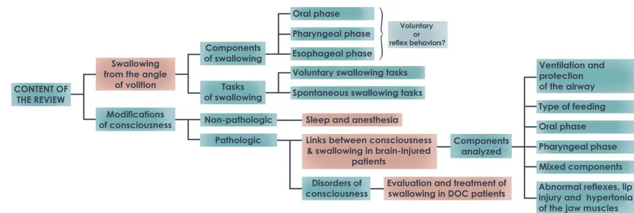
Chapter 1 - 32 Because of the scarcity of studies directly related to swallowing and consciousness, in this article we chose to explore a wide range of themes addressing the links between swallowing and consciousness rather than focusing on one topic or answering one specific question (see figure 2). For this reason, we present the results of our research in the form of a literature review instead of a systematic review.
Chapter 1 - 33
Figure 2. Summary of the research fields explored in this review
Chapter 1 - 34
SECTION 1: Swallowing from the perspective of volition
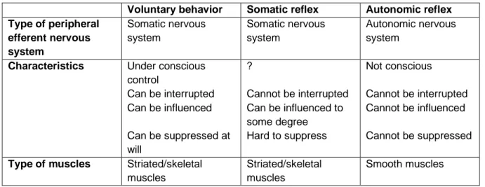
The approach historically used to determine whether or not a patient is conscious consists in the comparison of reflexive and voluntary behaviors (Giacino et al., 2002). However, the difference between conscious and reflexive behaviors remains ambiguous (Fischer & Truog, 2015). In fact, there are no empirical characteristics that allow us to reliably distinguish reflexive behaviors from conscious behaviors (Fischer & Truog, 2015). Prochazka et al. (2000) demonstrated that the distinction between
voluntary and reflex differs depending on the approach. The
Prochazka/Loeb/Rothwell position (Prochazka et al., 2000) describes voluntary behaviors as those that proceed under conscious control (Loeb) and that we can interrupt, influence (Rothwell) and suppress at will (Prochazka) and reflex behaviors as those that are automatic and hard to suppress (Prochazka) and that cannot be modified voluntarily. Some researchers (D’Ostilio & Garraux, 2012; Sumner & Husain, 2008) also agree that all voluntary behaviors contain automatic processes contributing to their rapidity and flexibility. Moreover, two types of reflexes are involved in swallowing: somatic and autonomic reflexes (see below) (Miller, 2002). Somatic reflexes implicate striated/skeletal muscles, and autonomic reflexes target smooth muscles.
Based on these characteristics (see table 1), we will analyze the different components of swallowing and try to distinguish voluntary from reflex behavior.
Chapter 1 - 35
Table 1. Characteristics of voluntary behavior and somatic and autonomic reflexes
Voluntary behavior Somatic reflex Autonomic reflex Type of peripheral efferent nervous system Somatic nervous system Somatic nervous system Autonomic nervous system
Characteristics Under conscious control Can be interrupted Can be influenced Can be suppressed at will ? Cannot be interrupted Can be influenced to some degree Hard to suppress Not conscious Cannot be interrupted Cannot be influenced Cannot be suppressed
Type of muscles Striated/skeletal muscles
Striated/skeletal muscles
Chapter 1 - 36 Swallowing is classically divided into three phases: oral, pharyngeal and esophageal (Shaker, 2013). The oral phase has two stages. Stage I consists of the transport of ingested food from the incisal area to the molar region and the reduction of food with mastication to a “swallowable” condition. Stage II consists of the movement of the bolus from the oral cavity through the pillars of the fauces to the oropharynx (Hiiemae & Palmer, 1999). The duration of the oral phase depends on several parameters, such as the consistency and volume of the bolus (Hiiemae et al., 1996)..
In the pharyngeal phase, the bolus is transferred from the base of the tongue to the esophagus with successive rapid mechanisms (nasopharyngeal closure, elevation of hyoid bone and pharynx with laryngeal elevation, laryngeal closure, downward movement of the epiglottis and esophageal sphincter relaxation).
Finally, during the esophageal phase, the bolus is pushed into the stomach through a series of peristaltic contractions.
1.1. Components of swallowing
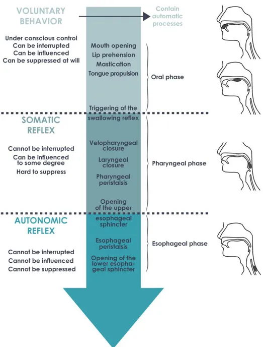
As we have seen, swallowing is divided into three phases (oral, pharyngeal, esophageal), each one comprising several components. The oral phase is classically described as the voluntary phase of swallowing while the pharyngeal and esophageal phases are the reflexive phases (Ertekin & Aydogdu, 2003). To confirm this assumption, we will now discuss the different components in each phase of swallowing in light of the characteristics of voluntary versus reflex behaviors (figure 3, page 40).
1.1.1. Oral phase
Although we chew and transport food without consciously controlling each orofacial movement, the oral phase is the only phase of swallowing that can be entirely interrupted and consciously controlled. In that respect, the modifiable and suppressible character of the oral phase categorizes this phase as voluntary behavior. In addition, several studies have demonstrated that consciously controlling the oral phase modifies its sequencing (Ashiga et al., 2019; Furuya et al., 2014; Palmer et al., 2007). Indeed, the chewing sequence can be significantly lengthened (almost twice as long) with volition (e.g., chewing with a conscious effort, or a
Chapter 1 - 37 specific number of chews) than without volition (i.e., eating normally). These data emphasize the role of automatic processes in natural feeding conditions. In other words, most of the time, the various lip and tongue movements occur without volition but rather as semiautomatic periodic or rhythmic movements, which explains why, in “controlled” conditions, the oral phase lasts longer.
The notion of semiautomatic periodic or rhythmic movements is not a recent one (Dubner et al., 1978). Many studies, especially those by Sessle’s team (Martin et al., 1997; Sessle et al., 2005, 2007; Yao et al., 2002), have explored the neural control of orofacial movements in primates using intracortical microstimulations. They indicated that the primary motor cortex dedicated to the orofacial area is involved in voluntary movements but also in the control of semiautomatic movements, such as tongue and mastication movements. Studies of oral reflexes also showed that diffuse stimuli to the palate in decerebrated and anesthetized cats elicited rhythmic tongue activity (Miller, 1982). Moreover, in the field of epilepsy, one study showed that electrical stimulation of the right inferior frontal gyrus (fronto-opercular cortex) leads to oroalimentary automatisms (lip movements, chewing) (Maestro et al., 2008).
The oral phase of swallowing can be classified as a voluntary behavior but, like any another motor activity, it includes some automatic processes. As described by Humbert and German (2013), during feeding, the different components of the oral phase can moved along the continuum of low to high voluntary control depending on the degree of attention dedicated specifically to them. The pattern-generating circuits for chewing and licking are located in the brainstem but receive direct cortical inputs (Moore et al., 2014).
1.1.2. Pharyngeal phase
The triggering of what is commonly called the “swallowing reflex” heralds the end of the oral phase and the beginning of the pharyngeal phase. This reflex is a somatic reflex because it involves striated/skeletal muscles. In “natural” conditions, the swallowing reflex occurs in response to saliva accumulation or to the presence of liquid or food in the oropharyngeal space (i.e., area of the soft palate, faucial pillars, pharyngeal surface of the epiglottis, dorsal pharyngeal wall). Indeed, when a sensory input (presence of saliva or a liquid or solid bolus) reaches a certain threshold,
Chapter 1 - 38 it triggers the swallowing reflex, which elicits the start of the sequence leading to protection of the airway and transportation of the bolus to the esophagus (Nishino, 2013). The timing of the initiation of the swallowing reflex is influenced by the waking state (see section 2), type of bolus (shortest with liquids) (Palmer et al., 1992) and cognitive functions (Dodderi & Larisa, 2016; Dodderi et al., 2018; Troche et al., 2014).
Although the swallowing reflex is usually triggered without conscious perception, it can be evoked voluntarily. Moreover, the swallowing reflex can be artificially initiated in humans by air pulses (Theurer et al., 2009) or electrical stimulations (Takatsuji et al., 2012; Tsukano et al., 2012) of the pharyngeal area. Whereas the execution of the oral phase can be stopped at any time, the swallowing reflex is hard to suppress for a long time during feeding or at rest.
If we refer to the definitions of Prochazka et al. (2000), the swallowing reflex can be triggered voluntarily but is usually automatic and is hard to suppress. It is thus on the borderline between a voluntary behavior and a reflex. Moreover, we can postulate that the transition between a voluntary behavior and a somatic reflex takes place somewhere between the beginning of the stage II oral phase transport and the triggering of the swallowing reflex.
On the other hand, the proceedings occurring after the trigger of the swallowing reflex (pharyngeal phase) cannot be suppressed voluntarily, unlike the oral phase and, to a lesser extent, the triggering of the swallowing reflex. However, some studies have shown that the pharyngeal phase can be influenced voluntarily to some extent; for example, patients can learn maneuvers that change swallowing physiology and help to reduce aspirations (e.g., Mendelsohn maneuver or effortful swallow) (Humbert & German, 2013; Shaker, 2013; Wheeler-Hegland et al., 2008). The process of the pharyngeal phase is mainly a somatic reflex.
1.1.3. Esophageal phase
The opening of the upper esophageal sphincter (UES) marks the end of the pharyngeal phase and the start of the esophageal phase. The UES is also called the inferior pharyngeal sphincter (Shaker, 2013). Muscles involved in the upper third of the esophagus (mainly the UES) are striated muscles under the control of vagal cholinergic motoneurons in the nucleus ambiguus of the brainstem (partly with the vagus cranial nerve X). In the
Chapter 1 - 39 lower two thirds of the esophagus, which is composed of smooth muscles, neural control switches to the autonomic/vegetative (enteric) nervous system through motoneurons situated in the ganglia (Broussard & Altschuler, 2000; Musgrove et al., 2020; Richards & Sugarbaker, 1995; Shaker, 2013).
The esophageal phase cannot be voluntarily triggered or suppressed. The only possible influence on the esophageal phase is a passive effect on the UES. In fact, Shaker et al. (2002) showed that head-raising exercises improve the UES, among other things. The mechanism at play is a passive stretch of the UES and/or an improvement of pharynx propulsion, which facilitates the opening of the UES.
For these reasons, the esophageal phase can be considered as an autonomic reflex. Given anatomical considerations (Meyer et al., 1986), we can assume that the transition between a somatic and an autonomic reflex (and consequently between striated and smooth muscles) takes place somewhere in the upper third of the esophagus.
Chapter 1 - 40
Figure 3. Classification of the oral, pharyngeal and esophageal phases of swallowing on
the continuum of voluntary to reflexive behaviors
Note. The transition between a voluntary behavior and a somatic reflex takes place
somewhere between the beginning of the stage II oral phase transport and the triggering of the swallowing reflex. The transition between a somatic and an autonomic reflex (and consequently between striated and smooth muscles) takes place somewhere in the upper third of the esophagus.
Chapter 1 - 41 1.2. Swallowing tasks
In the last 20 years, several researchers have explored the different stages of swallowing in healthy participants and in patients with dysphagia. Swallowing has been studied in several conditions (saliva, liquid or food swallowing) and, in addition to the voluntary or reflexive nature of each component of swallowing, some authors also distinguish swallowing tasks depending on the influence of volition.
Ertekin et al. (2001) distinguished between reflexive swallows (water introduced to the back of the tongue with a syringe), nasopharyngeal swallows (water introduced through a canula at the level of the uvula), spontaneous swallows (accumulation of saliva in the mouth that triggered spontaneous swallowing) and voluntary swallows (1–3 ml of water swallowed voluntarily) in an electrophysiological study. They showed, among other things, that the time interval between the onset of submental EMG and the onset of upward deflection of the larynx was significantly shorter for reflexive, nasopharyngeal and spontaneous swallows than for voluntary swallows. Kern, Jaradeh, et al. (2001) compared reflexive (rapid injection of water into the pharynx) and voluntary swallows (cued to swallow saliva volitionally once every 30 s by a tactile cue) in a neuroimaging study. While reflexive swallowing was associated with bilateral activity concentrated in the primary sensory/motor regions, volitional swallowing was represented bilaterally in the insula, prefrontal, cingulate, and parietooccipital regions in addition to the primary sensory/motor cortex.
One decade later, Ertekin (2011) dedicated a literature review to a comparison of spontaneous swallowing (SS) and voluntary swallowing (VS). He described SS as a “type of protective reflex action that occurs to ensure safety of the upper airway tract against any escape of food particles or saliva, or as an emotion-related reflex activity occurring during stressful conditions” (2011, p. 184). SS occurs without awareness while one is awake or asleep. The oral phase is bypassed in most cases, although there may be partial excitation. SS is also sometimes called “reflexive swallowing” or “non-nutritive swallowing.” On the other hand, he described VS (also called “conscious swallowing”) as sequential eating or drinking voluntarily initiated or facilitated by the cerebral cortex during the awake and aware state (Ertekin, 2011).
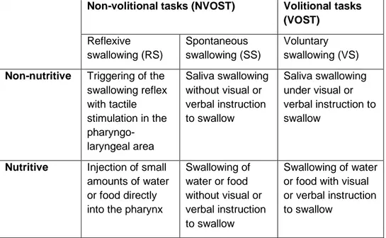
Chapter 1 - 42 In table 2, we describe several types of volitional (VOST) and non-volitional swallowing tasks (NVOST) related to the concept of reflexive (RS), spontaneous (SS) and voluntary swallowing (VS). Because of the potentially different brain activations and different physiological mechanisms at play during nutritive compared to non-nutritive swallowing, we also make a distinction between these two types of swallowing tasks. To examine this distinction in light of neuroimaging data, we did a qualitative analysis of studies exploring cerebral areas activated during RS, SS or VS task in healthy subjects (see Appendix 2.1 for research strategy and selected criteria).
Table 2. Description of the different types of swallowing tasks
Non-volitional tasks (NVOST) Volitional tasks (VOST) Reflexive swallowing (RS) Spontaneous swallowing (SS) Voluntary swallowing (VS) Non-nutritive Triggering of the
swallowing reflex with tactile stimulation in the pharyngo-laryngeal area Saliva swallowing without visual or verbal instruction to swallow Saliva swallowing under visual or verbal instruction to swallow
Nutritive Injection of small amounts of water or food directly into the pharynx
Swallowing of water or food without visual or verbal instruction to swallow Swallowing of water or food with visual or verbal instruction to swallow
Note. Reflexive swallowing refers to triggering of the swallowing reflex by an external
stimulus (tactile or with the injection of a bolus). In this case, the participation of the oral phase is diminished but not completely bypassed considering the involvement of the tongue in any swallowing process. Non-nutritive spontaneous swallowing refers to the management of saliva and secretions that are produced spontaneously by all healthy humans, while nutritive spontaneous swallowing is associated with eating and drinking. Volitional swallowing tasks refer to tasks occurring further to an internal or external request.
In the 31 studies identified (see Appendix 2.2 for description of studies and results), the majority (n=28) focused on VS tasks using water or saliva and compared the induced brain activity with that induced by other tasks,
Chapter 1 - 43 such as motor imaging of swallowing, speaking out loud, tongue movement or finger tapping. One author compared RS with VS (Kern, Jaradeh, et al., 2001), another compared SS with VS (Martin et al., 2001) and one study (Paine et al., 2011) focused on SS task but compared their results with another study analyzing VS tasks (Malandraki, 2009).
Only four studies (Hamdy & Rothwell, et al., 1999; Harris et al., 2005; Zald & Pardo, 1999, 2000) used positron emission tomography (PET); the majority used functional magnetic resonance imaging (fMRI) (n=27). Regarding brain location, the majority of the studies (n=24) focused on whole brain analysis whereas five targeted regions of interest (Lowell et al., 2012; Luan et al., 2013; Malandraki et al., 2010; Mihai et al., 2014; Toogood et al., 2017), and one focused specifically on the brainstem (Komisaruk et al., 2002). The small number of studies focusing on the brainstem may reflect the technical challenges of imaging this area (Sclocco et al., 2018).
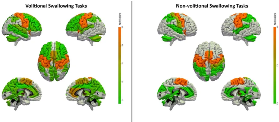
We did a qualitative analysis of areas involved in volitional (VS) compared to non-volitional (RS and SS) swallowing tasks, based on the brain areas and Brodmann areas mentioned in studies. Each brain area identified was linked with brain areas in the AAL2 Atlas (Rolls et al., 2015), implemented in Surf Ice (https://www.nitrc.org/projects/surfice/) (figure 4).
Numerous cortical and subcortical areas are involved in the control of VOST tasks (figure 4). Based on the 30 studies using VOST tasks, we found that the main brain regions (identified in at least 50% of the studies) involved in VOST tasks are the primary motor cortex (n=26), insula (n=24), primary somatosensory cortex (n=22), premotor cortex and supplementary motor area (n=19) and anterior cingulate cortex (n=19) (right>left). Regions identified in at least 25% of studies are the cerebellum (n=13) (left>right), superior temporal gyrus (n=11), posterior cingulate cortex (n=10), thalamus (n=10), inferior parietal lobule (n=9), precuneus (n=9), putamen (n=9) (left>right) and superior frontal gyrus (n=8). The other regions (identified in less than 25% of studies) are the frontal operculum (n=7), inferior frontal gyrus (n=7), cuneus/lingual gyrus (n=7), middle frontal gyrus (n=5), middle cingulate gyrus (n=5) and middle temporal gyrus (n=5) (figure 4).
The three studies exploring NVOST tasks (Kern, Jaradeh, et al., 2001; Martin et al., 2001; Paine et al., 2011) showed that the activated regions were not specific to NVOST as they were all also activated in VOST tasks.
Chapter 1 - 44 However, some brain areas (i.e., precuneus, inferior, middle and superior frontal cortex, frontal operculum, putamen and middle cingulate cortex) were activated during VOST tasks but not NVOST tasks.
In the two studies directly comparing VOST and NVOST tasks (Kern, Jaradeh, et al., 2001; Martin et al., 2001), the authors reported that some areas were activated in VOST tasks and not in NVOST tasks; the insula and the prefrontal, anterior cingulate, and parieto-occipital regions in the Kern study and the anterior cingulate cortex in the Martin study. Kern et al. also argued that NVOST (RS) is characterized by greater cortical activation in the left hemisphere, whereas VOST shows greater volume activation in the right hemisphere.
Paine et al. (2011) compared their results obtained with naive saliva swallowing (SS) with the results of another study (Malandraki et al., 2009), which used voluntary water swallowing (VS). They showed that regions activated in both tasks were almost identical. Indeed, regions related to motor control, sensory input and somatosensory integration were significantly activated in both SS and VS conditions. However, the authors reported that the significant activations from the SS study were much more localized in motor control areas.
Finally, swallowing tasks and specific oral (e.g., jaw clenching, tongue movements) or pharyngeal (i.e., throat clearing) tasks share areas of activation. Indeed, the main brain areas activated during swallowing were also activated during isolated oral or pharyngeal tasks.
All these studies were done during wakefulness, but we will see now what impact sleep and anesthesia have on swallowing.
Chapter 1 - 45
Figure 4. Brain areas activated by volitional and non-volitional swallowing tasks
Note. Colors refer to the number of studies that mentioned the depicted area. For studies using volitional tasks, brain areas mentioned in at least five studies are
illustrated in the figures. For the non-volitional tasks, all areas are depicted because of the low number of studies (n=3). The main regions activated in volitional
tasks are the bilateral primary somatosensory cortex and bilateral insula, followed by the bilateral supplementary motor area and the right anterior cingulate cortex.
The other regions activated at least in five studies are the bilateral cerebellum (left>right), bilateral superior temporal cortex, bilateral posterior cingulate cortex, bilateral thalamus, bilateral inferior parietal cortex, bilateral precuneus and cuneus, bilateral inferior frontal cortex and frontal operculum, left putamen, bilateral superior frontal cortex, left middle frontal cortex, bilateral middle cingulate cortex and right middle temporal cortex. Brain areas activated in non-volitional tasks are the primary sensorimotor cortex, supplementary motor cortex and insula, followed by the bilateral anterior and posterior cingulate cortex, bilateral cerebellum, left cuneus, bilateral thalamus, bilateral inferior and superior parietal cortex and bilateral superior temporal cortex.
Chapter 1 - 46
Section 2: Nonpathological modifications of consciousness:
sleep and anesthesia
Investigating swallowing in nonpathological modifications of consciousness such as sleep or anesthesia allows to explore swallowing without the ambiguity of conscious control. Indeed, sleep and anesthesia are associated with reduced consciousness and lack of volition, enabling volitional versus nonvolitional swallowing to be distinguished.
Sleep is classically divided into three stages of nonrapid eye movement (NREM) sleep (N1, N2 and N3) and rapid eye movement (REM) sleep. As described by Sanders et al. (2012), in NREM sleep individuals are generally considered unconscious, disconnected and not responsive, but people recall dreams after being woken from NREM sleep in 23% to 74% of cases. In contrast, during REM sleep, individuals are sometimes considered conscious (in approximately 80% of REM sleep awakenings) and report vivid dreams, but they do not experience their environment. They are disconnected and not responsive.
During sleep and in the case of DOC, the absence of consciousness does not lead to a complete absence of swallowing. Several studies have explored spontaneous saliva swallowing in healthy adults during sleep (Guiu Hernandez et al., 2019; Kelly et al., 2007; Lichter & Muir, 1975; Okuno et al., 2016; Pohl et al., 2013; Sato et al., 2011, 2016; Sato & Nakashima, 2006). During sleep, swallowing is episodic, absent for long periods and influenced by sleep stage (Sato et al., 2011, 2016; Sato & Nakashima, 2006). The deeper the sleep stage, the lower the mean swallowing frequency. Swallowing occurs almost in association with movement arousals in both REM and NREM sleep (Lichter & Muir, 1975; Sato et al., 2016; Sato & Nakashima, 2006). Some authors reported no (Sato et al., 2016) or very few (Sato & Nakashima, 2006) swallows during deep sleep (NREM stage N3). Regarding the efficacy of the pharyngeal phase, healthy adults have lower velopharyngeal and hypopharyngeal swallowing pressures when asleep (Guiu Hernandez et al., 2019). In their study, Kelly et al (2007) showed that breathing-swallowing coordination differed between volitional (saliva swallowing on command) and nonvolitional swallowing (spontaneous saliva swallowing without cuing) conditions but not between their two nonvolitional conditions (spontaneous saliva swallowing during waking and sleep). Moreover, during a functional test (instillation of water in the pharynx), more aspirations after swallowing
Chapter 1 - 47 were observed during sleep than during wakefulness, as well as more repetitive swallowing and coughing after swallowing (Okuno et al., 2016). Patients with neurological impairments (cerebral atrophy or lacunar infarct) demonstrated a delayed response between the delivery of water in the pharynx and the triggering of swallowing when asleep, compared to when awake, while the healthy group showed no significant difference between wakefulness and sleep (Pinto et al., 1994). In Parkinson’s disease patients, the mean duration of sleep decreases while the number of spontaneous saliva swallowings increases compared to healthy subjects (Uludag et al., 2016). Moreover, patients present more multiple swallows than healthy subjects.
Anesthesia can also be considered as a way of exploring consciousness but cannot be considered simply as an “absence of consciousness” (Bonhomme et al., 2019). Different consciousness states can be observed during general anesthesia, depending on the anesthetic agent and dose: (1) a complete absence of subjective experience (unconsciousness); (2) conscious experience without perception of the environment (disconnected consciousness, as in dreaming); or (3) episodes of oriented consciousness with awareness of the environment (connected consciousness) (Bonhomme et al., 2019).
Some authors (D’Angelo et al., 2014; D’Honneur et al., 1994; Gemma et al., 2016; Rimaniol et al., 1994; Taylor et al., 1972) have shown that general anesthetics (e.g., propofol, sevoflurane, ketamine and midazolam), which generally cause some form of unconsciousness (Sanders et al., 2012), can alter swallowing. Thus, during general anesthesia, the frequency of spontaneous saliva swallowing decreases and the number of pathological swallows (characterized by inspiration or followed by an inspiration) increases (D’Angelo et al., 2014). Moreover, studies analyzing the efficacy of swallowing after the injection of a liquid at the back of the tongue or the pharynx during anesthesia showed that the latency between the injection and the initiation of the swallowing reflex (D’Honneur et al., 1994; Nishino et al., 1987; Rimaniol et al., 1994), and the number of aspirations (Gemma et al., 2016) increase while laryngeal reflexes are depressed (Taylor et al., 1972). Moreover, coordination between respiration and swallowing can change with deep sedation or during the recovery period from general anesthesia (Cedborg et al., 2015; Nishino & Hiraga, 1991).
Chapter 1 - 48 All this information (see Appendix 2.1 for research strategy and selected criteria) shows that the frequency, speed of initiation of the swallowing reflex, efficacy of the pharyngeal phase of swallowing (mainly the number of aspirations) and coordination between respiration and swallowing are influenced by the level of consciousness during sleep and general anesthesia.
In the next section, we will see how consciousness affects swallowing in patients with brain injuries.
Chapter 1 - 49
Section 3: Links between consciousness and swallowing in
brain-injured patients
The prevalence of dysphagia further to severe brain injury is very high (Mélotte et al., 2020), mainly due to the large number of brain areas dedicated to swallowing (see above), any of which can be severely damaged by a brain injury. A large majority of patients with DOC require artificially delivered hydration and nutrition, mainly through a gastrostomy feeding tube (Mélotte et al., 2020; Royal College of Physicians, 2003a). The aim of this section is to examine the extent, variety and characteristics of swallowing disabilities in patients with acquired brain injury (ABI), and to identify which swallowing components are related to consciousness. To better understand these links, we reviewed studies analyzing swallowing in relation to consciousness level (see Appendix 2.1 for research strategy and selected criteria).
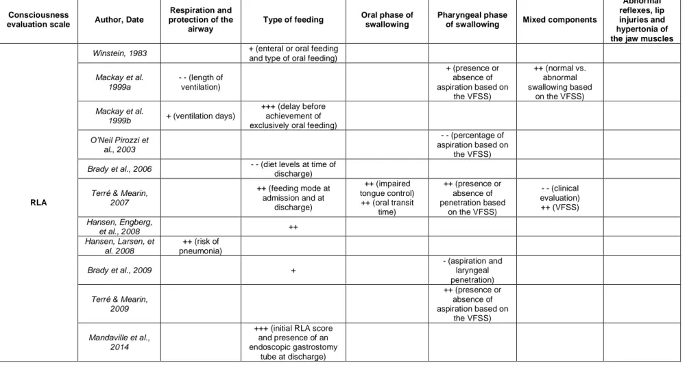
We found 18 studies that describe a link between consciousness and swallowing abilities (see table 3 for characteristics of the studies). Nine studies explored swallowing abilities for all etiologies (Brady et al., 2006, 2009; Bremare et al., 2016; Godet et al., 2017; Kjaersgaard et al., 2015; Mélotte et al., 2018, 2020; Millwood et al., 2005; Wang et al., 2019, while nine focused solely on traumatic brain injury (TBI) (Hansen, Engberg, et al., 2008; Hansen, Larsen, et al., 2008; Mackay et al., 1999a, 1999b; Mandaville et al., 2014; O’Neil-Pirozzi et al., 2003; Terré & Mearin, 2007, 2009; Winstein, 1983).
The studies are quite heterogeneous in terms of the swallowing components described, the consciousness scales used, the range of consciousness level considered, and the design of studies.
Twelve studies reported the results of swallowing in patients diagnosed with the Rancho Los Amigos (RLA) Scale (Hagen et al., 1987), four with
the Coma Recovery Scale – Revised (CRS-R) (Giacino et al., 2004), one
with the Wessex Head Injury Matrix (WHIM) (Shiel et al., 2000) and one with the Full Outline of UnResponsiveness (FOUR) (Wijdicks et al., 2005) (see Appendix 2.3 for description of scales).
We will present the results based on the type of swallowing components studied (i.e., ventilation and protection of the airway, type of feeding, oral phase, pharyngeal phase, abnormal reflexes, lip injury and hypertonia of the jaw muscles, and mixed components of the oral and pharyngeal phase). Table 4 summarizes the results of the different studies.