Direction des bibliothèques
AVIS
Ce document a été numérisé par la Division de la gestion des documents et des archives de l’Université de Montréal.
L’auteur a autorisé l’Université de Montréal à reproduire et diffuser, en totalité ou en partie, par quelque moyen que ce soit et sur quelque support que ce soit, et exclusivement à des fins non lucratives d’enseignement et de recherche, des copies de ce mémoire ou de cette thèse.
L’auteur et les coauteurs le cas échéant conservent la propriété du droit d’auteur et des droits moraux qui protègent ce document. Ni la thèse ou le mémoire, ni des extraits substantiels de ce document, ne doivent être imprimés ou autrement reproduits sans l’autorisation de l’auteur.
Afin de se conformer à la Loi canadienne sur la protection des renseignements personnels, quelques formulaires secondaires, coordonnées ou signatures intégrées au texte ont pu être enlevés de ce document. Bien que cela ait pu affecter la pagination, il n’y a aucun contenu manquant.
NOTICE
This document was digitized by the Records Management & Archives Division of Université de Montréal.
The author of this thesis or dissertation has granted a nonexclusive license allowing Université de Montréal to reproduce and publish the document, in part or in whole, and in any format, solely for noncommercial educational and research purposes.
The author and co-authors if applicable retain copyright ownership and moral rights in this document. Neither the whole thesis or dissertation, nor substantial extracts from it, may be printed or otherwise reproduced without the author’s permission.
In compliance with the Canadian Privacy Act some supporting forms, contact information or signatures may have been removed from the document. While this may affect the document page count, it does not represent any loss of content from the document.
Inhibition of respiratory syncytial virus
by nasaIly administered si
RNA
moditied with F -ANA.
par
Julie Juan Wang
Programme de Sciences Biomédicales
Faculté de Médecine
Mémoire présenté à la Faculté des études supérieures
en vue de l'obtention du grade de Maîtrise
en Sciences Biomédicales
Décembre 2007
Université de Montréal
Faculté des études supérieures
Ce mémoire intitulé:
Inhibition of respiratory syncytial virus
by nasally administered siRNA modified with F -ANA.
présenté par:
Julie Juan Wang
Programme de Sciences Biomédicales
Faculté de Médecine
a été évalué par un jury composé des personnes suivantes:
Dr. Karim Maghni Dr. Paolo Renzi Dr. Céline Bergeron Président -rapporteur Directeur de recherche Membre du jury 11
RÉSUMÉ
Le virus respiratoire syncytial humain (VRS), un pneumovirus, est URe cause importante de maladie des voies respiratoires inférieures. L'infection au VRS pendant la petite enfance est associée au développement de la bronchiolite et de l'asthme. Il n'existe aucun médicament ou vaccin. qui prévienne l'infection à VRS. La seule thérapie antivirale disponible estla ribavirine, réservée. aux personnes à haut risque de maladie grave. Des études récentes ont démontré que des 'smaU interfering RNAs' (siRNAs) dirigés contre les ,protéines virales P et NSI inhibaient la réplication le VRS. Par ailleurs, on a aussi '. cherché à prolonger l'activité des siRNAs en leur apportant des modifications chimiques. Une de ces modifications est le F-ANA (acide 2'deoxy-2'-tluoro-f3-D-arabinonucléique). Le présent travail étudie l'efficacité des siRNAs modifiés au F-ANA à inhiber la . réplication dU VRS. In vitro, les siRNAs F-ANA ciblant les protéines viralesP .et NS 1 inhibent plus efficacement la réplication virale. In vivo, à plus forte concentration, les siRNAs modifiés. ou non au F-ANA et la ribavirine inhibent la synthèse des protéines, virales avec une efficacité comparable (inhibition de 44% ± 6 pour le siRNA non-modifié, 47% ± 8 pour le siRNAF-ANA et 41% ± 9 pour la ribavirine). Cependant, à tàible dose, l'efficacité du siRNA F-ANA est 84% supérieure à celle du si RNA non-modifié, un taux qui surpasse l'efficacité de la ribavirine de 57%. Les modifications F-ANA ont donc le potentiel d'améliorer les performances des siRNAs en tant qu'agent prophylactiques ou thérapeutiques conn:e le VRS.
Mots clés : Virus respiratoire syncytial, agents antiviraux, ARN d'interférence, smaU interfering RNA, modifications avec :.:'-ANA, phosphoprotéines virales, protéines non structurale virales, ribavirine, modèle de souris.
IV
ABSTRACT
Human respiratory syncytial,virus (hRSV), a pneumovirus, is the major cause of severe lower respira tory tract disease in young children, the elderly and the immunosuppressed~ Additionally, hRSV infection during infancy has been associated with the development of bronchiolitis andasthma. Despite extensive research, no reliable drug or vaccine currently exists that can prevent RSV infection. The only antiviral therapy available is ribavirin, whose use is reserved for persons at high risk for severe disease. Recent studies have shown that smàll interfering RNAs (siRNAs), targeting viral P prote in and also viral NSI protein inhibited RSV growth in culture and in vivo. Recently, efforts have been made to enhance stability and duration of ~ctivity of siRNAs through chemical ,modifications. One such modificationis F-ANA (2'deoxy-2'-fluoro-~-D;.arabinonucleic
acid). The efficacy of siRNAs modified with F-ANA at inhibiting hRSV replication was investigated both in vitro and in vivo: In vitro screening demonstrated greaterefficacy of siRNA targeting viral P protein than NS 1 protein. In vivo, both unmodified and F-ANA modified siRNAs targeting viral P gene inhibited hRSV protein levels equally weil at high doses (44% ± 6 for unmodified siRNA and 47% ± 7 for F-ANA modified siRNA),
comparable to 41 % ± 8 inhibition achieved by ribavirin. At lower doses, the F-ANA: modified siRNA demonstrated 84% increase in efficacy to inhibit hRSV replication compared to unmodified sequence, exceeding ribavirin by 57%. In summary, F-ANA modifications have the potential to optimize siRNAs as prophylactic and/or therapeutic agents against hRSV infection.
Key words: Respiratory syncytial VIruS, antiviral agents, RNA interference small interfering RNA, F-ANA modifications, viral phosphoroproteins, viral' nonstructural proteins, ribavirin, mou se modd.
TABLE OF CONTENTS
IDENTIFICATION OF THE mRY ... ; ... , ... ii
RÉSUMÉ .... · ... ~ ... iii
. -ABSTRACT ... ; ... iv
·TABLE OF CONTENTS ... ; ... ·.~ ... ; ... v
LIST OF TABLES ... viii
LIST OF FIGURES ... .ix
LIST OF ABBREVIATIONS ... ' ... xii
DEDICATION ... xiv·
ACKNOWLEDGEMENTS .... ~ ... ; ... xv
·CHAPTER 1: GENE~L INTRODUCTION ... 1
CHAPTER II: RESPIRA TORY SYNCYTIAL VIRUS AND DISEASE. ... : 4
2.1 HISTORY ... 5
2.2 EPIDEMIOLOGY ... 5
2.3 VIRAL STRUCTURE, REPLICATION, AND INFECTION ... 6
2.3.1 Virion structure ... ; ... 6
232 . . G enome organlzatIon ... : ... . . . . r 8 2.3.3 Viral proteins ... 9
2.3.4 Viral replicative cycle ... · ... ; ... , ... 12
2.4 DISEASE AND CLINICAL RELEV ANCE ... 15
.2.4.1 RSV infection and transmission ... : ... ; ... 15
2.4.2 Clinical symptoms ... ; ... 16
2.5 THE IMMUNE RESPONSE TO RSV ... 17
2.5.1 Theinnate immune response to RSV~ ... 18
2.5.2 Humorallantibody-mediated immunity to RSV ... 19
2.5.3 Cell-mediated immunity to RSV ... 19
VI
CHAPTER III: RNA INTERFERENCE AND siRNAs FOR THE THERAPY OF
RSV INFECTION ... 24
3.1 RNA INTERFERENCE ... 25
,
.
3.1.1 Nucleic acid drugs ... , ... ' ... 253.1.2 Discovery of RNA interference ... : ... 27
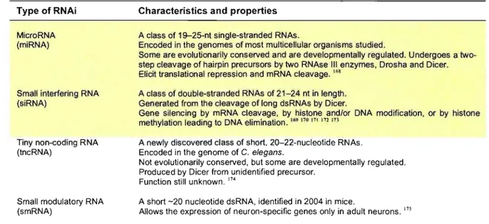
3.1.3 Types of RNA interference .. , ... · ... ; ... : .. 28
3.2 SMALL INTERFERlNG RNA ... 29
3.2.1 Silencing pathway... 29
3.2.2 Non-specific off-target effects ... 31
3.2.3 SiRNA selection and design ...
32,-3.3 DELIVERY AND MODIFICATIONS OF siRNAS ... 33
3.3.1 . Delivery of siRNAs in vitro and in vivo .. ... 34
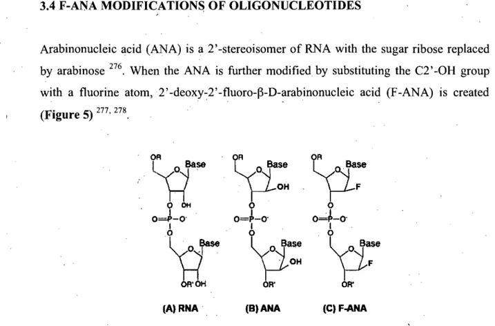
3.3.2 Modifications of siRNAs ... i . • • • • • • • • • • • • • • • • • • • • • • • • • • • • • • 36 3A F-ANA MODIFICATIONS OF OLIGONUCLEOTIDES ... 39
3.5 APPLICATIONS OF siRNA IN RSV DISEASE ... 40
) CHAPTER IV: MATERIALS AND METHODS ... .42
-4.1
CELL CULTURE AND MAINTENANCE ... -· ... 434.2 VIRUS PROPAGATION AND PURIFIèATION ... 43
4.3 SiRNA AND siF-ANA SEQUENCES AND ANNEALING ... 45
4A SiRNATRANSFECTION AND VIRUS INFECTION IN VITRO .. ... 47
4.5 IMMUNOST AINING PLAQUE ASSAy ... ... A7 4.6 ANIMALS ... : ... 48
4.7 NASAL ADMINISTRATION OF siRNA AND hRSV ... .49
4.8 BRONCHOAL VEOLAR LA V AGE,WHOLE LUNG COLLECTION AND CELL COUNT ... 49
4.9 ANAL YSIS OF INTRACELLULAR CYTOKINE PRODUCTION IN CELL . SUPERNAT ANT AND BAL ... 50
4.10 PROTEIN EXPRESSION ANAL YSIS BY hRSV PROTEIN ELISA ... 50
4.11 DETECTION OF VIRAL mRNA IN CELL L YSATES AND LUNG HOMOGENATES ... 51
4.12 STATISTICAL ANALYSIS ... : ... ~ .. 52
.) CHAPTER V: RESULTS ... · ... ,· ... 53
5.1 ESTABLISHMENT AND VALIDATION OF MEASUREIvIENT METHODS ... ' ... : ... 54
5.1.1 Establishing and maintenance of hRSV culture in hum an epithelial HEp-2 ceUs ... 54
5.1.2 Inhibition ofRSV infection with ribavirin ... 56
5.1.3 Establishing infection of hRSV in A549 ceUs ... 58
5.1.4 Determination of readout methüds for measuring infection ... " 59 , 5.2 CHOICE OF APPROPRIATE siRNAS ... 63
5.2.1 Designing siRNAs and their mismatches ... ,. 63
5.2.2 In vitro screening ofall four siRNAs ... : ... 64
5.2.3 In vitro screening ofsiRSV-Pl and -P2 ... 69
5.2.4 In vitro screening of siRSV-P2 and aU its F-ANA combinations ... 71
5.3 V ALIDA TION OF IN VIVO MODEL. ... : ... 74
5.3.1 Developing an in vivo mouse hRSV mode!.. ... , ... 75
5.3.2 Optimizing the in vivo mouse hRSV mOdei. ... : ... 81
5.3.3 Validation ofmurine hRSV model via treatment with ribavirin ... 85
5.4 DETERMINATION OF EFFICACY OF siRNA IN AN ANIMAL MODEL OF hRSV INFECTION ... : ... 89·
5.4.1 Determination of in vivo efficacy ofunmodified siRSV-P2, preliminary . experiments ... : .... '.' ... , ... 89
5.5 COMPARISON OF IN VIVO EFFICACY OF UNMODIFIED VS F-ANA MODIFIED siRNA ... ~ ... 93 .
5.5.1 Comparison ofin vivo efficacy ofunmodified siRSV-P2 0/0 vs. F-ANK modified siRSV-P2 01F4~ pooled analysis ... ,' ... 93
CHAPTER VI: GENERAL DISCUSSION ... ~ ... 98
V III
LIST OF TABLES
CHAPTERII·
Table 1: The RSV genome encoded viralproteins and functions ... , ... 9 Table II: Current therapies for RSV infections and 'their functions ... 22
'.
CHAPTERIII
Table 1: The five major classes of nucleic acid drugs ... ; ... 26 Table II: Various types of RNA interference and their main characteristics ... 28
CHAPTERIV
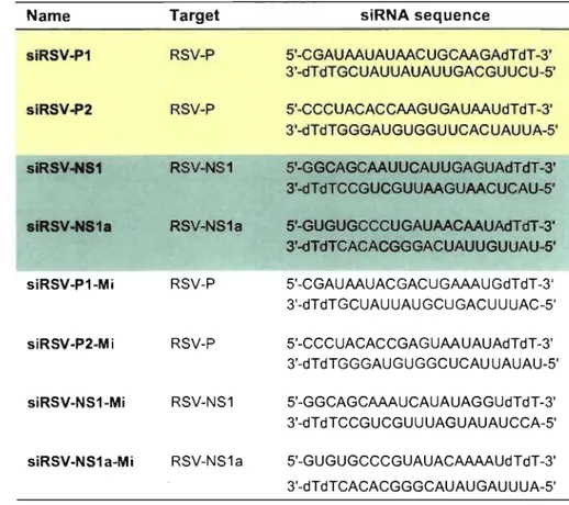
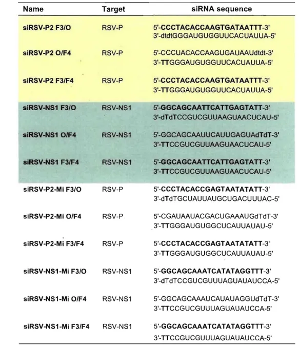
Table 1: Sequences of unmodified siRNAs ... 45 Table II: Sequences of siRNAs modified with F-ANA ... 46 Table III: Primer sequences ... ; ... : ... 52
CHAPTERV
Table 1: Titer measured by plaque assay on day 3 PI from supematant ... 58 Table II: SiRNA name and sequences .... ; ... 63 rable III: F-ANA modified siRNA names and sequences ... : ... 72
LIST OF FIGURES
CHAPTERII
Figure 1: Respiratory syncytial virus structure ... : ... 7
Figure 2: RSV genes and the sizes oftheir mRNA and encoded proteins ... 8
Figu re 3:. The schematic representation of the replicative cycle of RSV ... 13
Figure 4: Structural formula offibavirin ... : ... 21
CHAPTERIII Figure 1: Silencing pathway of synthe tic siRNA ... : ... : ... 30
Figure 2: Schematic illustration of important features of siRNA structure ... 32
\ Figure 3: Delivery of small interfering RNAs ... ;.: ... 34
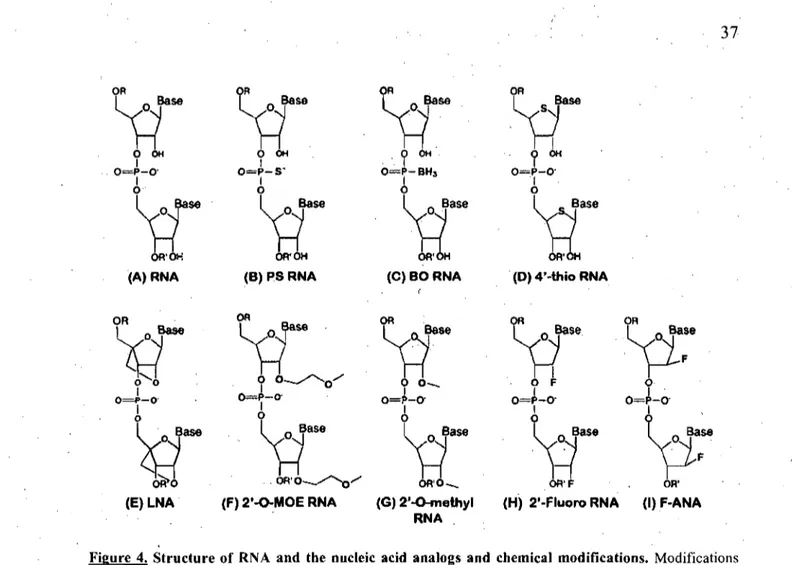
Figure 4: Structure of RNA and the nucleic acid analogs and chemical modifications. 37 Figure 5: Chemical structure of Ribonucleic and arabinonucleic acids ... 39
Figure 6: Example of the modifications ofsiRNA with F-ANA chemistries along either or both the sense and antisense strands ... " ... 40 .
CHAPTERV Figure 1: Detection ofhRSV on HEp-2 monolayer by method ofimmunostaining ... 54
Figure 2: Propagation ofUV-treated hRSV ... ~ ... 55
Figure 3: hRSV titer levels detected in A549 cells treated with ribavirin ... : ... 57
Figure 4: hRSV protein levels detected in A549 cells treated with ribavirin ... 57
Figure 5: Viral titer measured by plaque assay on day 3 PI from supematant. ... 59
Figure 6: hRSV protein levels detected with hRSV ELISA over time ... , ... 60
Figure 7:IL-8 secretion levels detected by IL-8 ELISA over time ... -61
Figure 8: MIP-1a section levels detected by MIP-Ia ELISA over time ... 61
Figure. 9: mRNA levels detected by real-time RT-PCR ... 62
Figure 10: Inhibition ofhRSV titer by immunostaining plaque assay ... 64
Figure Il: % Inhibition ofhRSV protein in hRSV infected A549 cens ... 67
x
Figure 13: % Inhibition ofIL-8 in hRSV infected A549 cells ... 69
Figure 14: % Inhibition ofhRSV P mRNA in hRSV infected A549 cells .... : ... 70
Figure 15: % Inhibition ofhRSV proteins in hRSV infected A549 cells ... ~ .. 70
Figure 16: % Inhibition ofhRSV P mRNA in hRSV infected A549 cells ... , ... 72
Figure 17: % Inhibition ofhRSV proteins in hRSV infected A549 cells ... 73
Figure 18: % Inhibition ofMIP-la in hRSV infected A549 cells ... ~ .... : ... 74
/ Figure 19: Timeline ofhRSV infection ... 75
Figure 20: Mouse weight difference in percentage measured over time ... ; ... 76
Figure 21: Total cells in BAL ofhRSV infected BALB/c mice over time ... 77
Figure 22: Percent differential cells in BAL ofhRSV infected mice over time ... 78
Figure 23: Absolute number of differential cells in BAL of hRSV infected mice over time ... 78
Figure 24: Determining hRSV infection in BAL and lung homogenates ofhRSV infected BALB/c mice over time ... ~ ... 79
Figure 25: Timeline ofhRSV infection ... 81'
Figure 26: Total cells in BAI: ofhRSV infected BALB/c mice over time ... 82
Figure 27: Percent differential cells in BAL ofhRSV infected mice over time ... 82
Figure 28: Absolutenumber ofdifferential cells in BAL of hRSV infected'jmice over time .... : ... 83
Figure 29: Determining hRSV infection in BAL and lung homogenates of hRSV infected BALB/c mice over time ... ' ... 84
,Figure 30: Timeline ofhRSV infection ... , ... ' ... 85
Figure 31: Totalcells in BAL ofhRSV infected BALB/c rriice over time ... 86
-Figure 32: Percent di(ferential cens in BAL ofhRSV infected mice over time ... ' .... 86
Figure 33: Absolute number of differential cens in BAL of hRSV infected mice bver time ... 87
Figure 34: Detennining hRSV infection in BAL and lung homogenates ofhRSV infected BALB/c mice over time ... 88
Figure 35: Timeline ofhRSV infection ... 89
Figure 36: Total cens in BAL ofhRSV infected BALB/c mice over time ... 90
Figure 38: Absolute number oflymphocytes in BAL ofhRSV infected mi ce over time ... : ... ~ .. 91
Figure 39: Determining hRSV infection in BAL and lung homogenates ofhRSV infected BALB/c mice over time ... 92 Figure 40: Timeline ofhRSV infection ... : ... 93 Figure 41: % Inhibition of total ceUs in BAL ofhRSV infected BALB/c mice over
titne ... : ... ' ... , ... : ... 94 Figure 42: % Inhibition of ceUs released in BAL of hRSV infected BALB/c mi ce
(~easured by total and differential ceU count~) ... 95 Figure 43: % Inhibition of hRSV proteins in BAL of hRSV infected BALB/c mice
(measured by hRSV. ELISA) ... 96 Figure 44: % Inhibition ofhRSV P mRNA levels detected in lung homogenates ofhRSV
in BALB/c mice (measure by real-time Rt-PCR) ... 96 Figure 45: % Inhibition of mouse MIG levels .in BAL of hRSV infected BALB/c
AS ON
BAL bp bRSV BSA CTL dNTP dsRNA DTT F-ANA FBS HRP hRSV IFN-Ig IL-kDa L Mi MIP-la miRNA mMIG MOI N NS nt ORF-P LIST OF ABBREVIATIONS antisense oligonucleotide bronchoalveolar lavage base pairbovine respira tory. syncytial virus . bovine serum albumin
cytotQxic T ceU
desoxyribonucleoside triphosphates double-stranded RNA
dithiothreitol
2'deoxy-2'-fluoro':'f)-D-arabinonucleic acid fetal bovine. serum
horseradish peroxidase
human respiratory syncytial virus. inteiferon immunoglobulin interleukin-kiIodalton polymerase mismatch
macrophage inflammatory protein-l alpha microRNA
mouse monokine induced by gamma-interferon multiplicity of infection
nucleoprotein
nonstructtiral protein nucleotide
open reading frame phosphoprotein
PBS phosphate-buffered saline
PBS-t phosphate-buffered saline/O.05% Tween
PCR polymerase chain reaction
RdRP RNA-dependent RNA polymerase
PFU plaque forming units
PI post infection
RISC RNA-induced silencing complex RNAi RNA interference
RNase ribonuc1ease
RNP ribonucleoprotein
RPMI Roswell Park Memorial Institute
RSV respiratory syncytial virus
RT-PCR reverse transcription PCR
SEM standard error of mean
siRNA small interfering RNA
TBS tris-buffered saline
Th! T helper 1
Th2 T helper 2
TLR toll-like receptor
TMB tetra methyl henzidine
TNF-a tumor necrosisfactor-alpha
XIV
ACKNOWLEDGEMENTS
First, 1 would like to express my sincere thanks to Dr. Paolo M. Renzi, my mentor and my supervisor, ·for having given me the opportunity to pursue gradua te studies in his research laboratory. 1 am very grateful for having worked under your supervision and for receiving aIl the support throughout my masters' experience.
1 would like to thank Dr. Rosanne Seguin for your kind support, incisive critiques and remarkable patience: 1 have leamt a great deal from working with you and 1 am appreciative for your unfailing encouragements through aIl the ups and downs. "
\
1 would like to thank everyone at Topigen, especially Ann Brasey, with whom 1 spentmy every days with. Thank you for aIl your guidance and especiaIly, your friendships.
To those who were courageous enough to have read my thesis, you must have a patient and brave soul. Thank you.
To my family and friends, especially Mike, 1 seriously do not know how 1 would have gotten this far' without you. Thanks for aIl your love and understanding. You are my rock, my foundation and my inspiration in life.
J
CHAPTERI
. )
Ruman Respiratory Syncytial ViruS (hRSV), an RNA ViruS of the Paramyxoviridae
family, is recognized as the most frequent cause of severe lower respiratory tract disease in infants and young children worldwide 1. hRSV infects approximately 50% of infants . during the first year of life and almost all children by age of two. Lower respiratory tract infections, such as bronchiolitis and pneumonia, result in an ·estimated 51,000-82,000 hospitalizations annually among young children in the United States 2. Bronchiolitis is associated with long-term impairment of pulmonary function and often hyperresponsiveness of the airways after an RSV-infection during infancy'may presage the development of asthma later on in children and adults 3.
Acquired immunity provides neither an efficient protectionagainst re-infections nor reduction in viral shedding after infection, thus acquired immunity is incomplete and not durable. Despite extensive research, no reliable drug or vaccine currently exists that can prevent RSV infection. Two forms of anti-RSV antibody prophylaxis are available on the market, RSV-IGIV and palivizumab, both of which are questionable in their cost-effectiveness 4, 5, 6. Currently treatment is supportive and the sole antiviral therapy available is Ribavirin, whose use is reserved for persons at high risk for severe disease, and still remains controversial in efficacy and expensive 6. In addition, due to its very .. labor-intensive administration methocis and its potential hazardous effects on the caregivers 7, its use has been questioned on economic and safety grounds.
Recent studies have shown that small interfering RNA duplexes (siRNAs) targeting either the viral P prote in 8, an essential subunit of the RSV replication machinery, or the viral NS 1 prote in 9, another essential component of viral repli cation, strongly inhibited all RSVgrowth in culture and also in vivo, when administered intta-nasally. The problem remains of enhancing siRNA stability so that it can find its way into cel1s. Intetestingly, an increase in activity and an enhancement of seruin stability of siRNAs has been observed when modified with F-ANA (2'deoxy-2' -fluoro-~-D-arabinonuc1eic acid) at specific regions of the sens~ or antisense strands in culture 10, 11, however has nbt been fully examined in an animal model. The efficacy of siRNA modified with F-ANA to
3
inhibit hRSV replicaiion in vitro and also in vivo in a murine model of RSV infection was . therefore investigated.
f."
r
~.CHAPTERII
RESPIRA TORY SYNCYTIAL VIRUS ANDDISEASE
5
2.1 HISTORY
In 1956, a group of young chimpanzees in a colony outside Washington,
De
(USA) were demonstrating respiratory symptoms of a cold-like illness 12. Morris, Blount and Savage recovered the cytopathic agent from one of these chimpanzees, with an upper respiratory tract illness, wlÎich induced symptoms of coryza, runny nose, and malaise. They named, .
this agent "chimpanzeecoryza agent", which was responsible fot an outbreak of severe coryza illness in adolescent monkeys in the previous year 12, 13. Upon examination of the entire colony nearly all of the chimpanzees were found to be infected. Ruman contacts working with the chimpanzees were also shown to be infected and had exhibited mild upper respiratory tract illness and coryza, however at levels less severe than observed in the chimpanzees; Subsequent studies identified two major isolates of the virus recovered from other patients with upper respiratory tract illnesses 12. In 1957, the initial name of "chimpanzee coryza agent" was changed to respiratory syncytial virus (RSV) because of its selectivity for the respiratory tract and the characteristic tendency of infected cells to fuse together and form giant cell syncytia in tissue culture 13, 14.
2.2 EPIDEMIOLOGY
Severe respiratory infection in infants and young children was first recognized over 150 years ago and termed "congestive catarrhal fever." Based on clinical descriptions published at the time, there is a high probability that the disease described can be attributed to RSV 15._ Following the discovery and identification of RSV in the late 1950s and early 1960s, epidemiological studies clearly established it as the most important cause of serious respiratory tract infection in infants and young children 16, 17, Approximately 25 years later, RSV is recognized as the leading cause of infection of the lower respiratory tract in infants and young children 1. It is the single most important cause Of hospitalization during the first year of life 18, infecting around 50% of Infants during the first year oflife and almost all children by the age of two 19. It is the leading , cause of death due to viral illness in children younger than 1 year with a mortality rate in
this age group 10 times gteater than that from influenza 20. Unlike the common cold or
flu, RSV continues to be the leading kiUer among infectious diseases with an annual 'death toU of about one mÜlion worldwide 21, 22. Although RSV infection was reported in
1
adults with pneumonia in the 1960s, it has only been during the past two decades that the
potential for widespread occurrence with serious clinical impact in this population has been recognized 23, 24, 25, 26. RSV has a global distribution and it is a seasonal infection,
with peaks around winter and/or spring 27.
Two major antigenic subgroups of respiratory syncytial virus (designated A and B) have been described, differentiated mostly on the basis of reactivity patterns between monoclonal antibodies and the viral glycoprotein
(0'
protein) 28, 29. Both subgroups are infectious: one strain tends to dominate during an individual epidemic in an individual location, although at times both strains can be isolated from patients in the same area 30.In addition, a number of studies suggest that Strain Amay result in more virulent
infections than Strain B 31, however there is great vaiiability, possibly due to viral burden
and individual host factors. Along with these 2 strains, several subtypes of each strain have been identified 32, 30. In the United States and United Kingdom, ,Strain A i~ found more commonly, although it has been observed to alternate with Strain B in a somewhat
. 1 fi 33 S . B h
lrregu ar pattern rom year to year . tram , owever, appears more common1y in
epidemics in Europe 34.
2.3 VIRAL STRUCTURE, REPLICATION AN~ INFECTION
2.3.1 Virion structure
Respiratory syncytial viruses, both bovine (bRSV) and human (hRSV), are enveloped
RNA viruses and members of the Pneumovirus subfamily, of the family Paramyxoviridae 35. The Paramyxoviridae family includes two genera, Morbillivirus (measles virus) and the paramyxoviruses (containing, e.g., parainfluenza virus, types l, 2, and 3, and mumps virus) 32. The RSV virion is about 150-300 nm in diameter consisting of a nucleocapsid
7
core that is packaged within a lipid envelope 36 (Figure 1). Each of the viral mRNAs
encodes a major viral protein 37.
F protein SH protein . - - - G protein Negative ssRNA M (matrix) prote in N, P, L proteins
Figure 1. Respiratory syncytial virus structure. The RSV virion is about 200 nm in size and consists of a nuc1eocapsid within a lipid bilayer. The major nuc1eocapsid protein N, the phosphoprotein P, the large polymerase subunit L, the anti-termination factor M2 and a single-stranded RNA genome of negative polarity compose the RNP complex. The attachment protein G, the fusion protein F and the sm ail hydrophobie protein SH are the three transmembrane surface glycoproteins present on the viral surface. The matrix protein M forms a scaffold on the inner layer of the envelope. Figure modified from (Hacking, 2002) 37.
The symmetrically helical nucleocapsid (RNP complex) is 12-15 nm in Slze and is composed of the nucleocapsid protein N, the phosphoprotein P, the large polymerase subunit L and the M2-1 protein. The envelope is a lipid bilayer derived from the ho st plasma membrane and containing on its surface virally encoded transmembrane glycoproteins (G) responsible for attachment, the fusion protein (F) and the small hydrophobie (SH) protein, whose function is unknown however speculated to also be involved in cell fusion 38. Inside the lipid bilayer is a scaffold formed by the matrix protein (M), which connects the viral membrane with the RNP complex 30, 32 (Figure 1).
2.3.2 Genome organization
The RSV genome is a 15,200 nucleotide-long non-segmented single-stranded negative-sense RNA containing 10 genes that are transcribed into Il major sub-genomic mRNAs
39,40,41,42 (Figure 2), AlI mRNAs are capped at the 5'-end and polyadenylated at the
3'-end by the viral polymerase, however genomic and anti-genomic RNA are neither capped nor polyadenylated and are components of the nucleocapsids. Transcription and replication is initiated from the 3' -end in an obligatory sequential manner with only a fraction of the virally encoded RNA-dependent RNA polymerase (RdRP), minimally composed of the large protein (L) and the phosphoprotein (P), moving to the next gene. This mechanism creates a gradient of transcription al attenuation with distance from the transcriptional start-site indicating the required relative abundance of the encoded proteins (Figure 2). The overall strategy of RSV gene expression is common to aIl members of the negative-strand RNA virus super-family 43, 44. The initial rounds of
transcription, known as "primary" transcription, are carried out by the RdRP activity associated with the incoming viral genome. The transcribed mRNAs are translated into
de nova viral proteins including more RdRP, which boosts new rounds of viral gene expression.
3' NS1 NS2 N P M SH G F Ml L 5'
t , 4;f (lSSj . . . . , . . . ... . . . ,
S1t
of"
1191 "6 ,JI oIOS 91' 'm1.19 I~ .191 }of' 1}6 60f lSI8 S'of
''''
1165Figure 2. RSV genes and the sizes of their rnRNA and encoded proteins. The RSV genome is a single stranded negative sense RNA of a 15,200 nuc1eotides that is transcribed into II major sub-genomic mRNAs. Transcription is initiated from the 3'-end with only a fraction of the polymerase moving on to the next gene. This mechanism creates a gradient of transcription al attenuation with distance from the transcriptional start-site indicating the required relative abundance of the encoded proteins Figure modified from (Hacking, 2002) 37.
9
2.3.3 Viral proteins
Nine of the 11 major proteins, N, P, M, SH, G, F, M2, and L, are structural proteins (present in infected cells and virions) and surface glycoproteins 30, 32 (Figure 2). Three of nine structural proteins are transmembrane surface glycoproteins~ (G, F, and SH), of 11-20nm in size, embedded in the lipid bilayer derived from the host cell membrane, at, intervals of 6-10 nm 37. The viral nuc1eocapsid core is a symmetrical helix with a helical diameter of 12-15 nm 37,38 and composed of N, P, L proteins (which together comprise the viral replicase) and M2 open reading frame 1 (M2 ORF-l) (Table 1). FinalIy, two proteins (NS 1 and NS2) are non structural viral products that accumulate in infected cells but are present in only scarce amounts in mature virions 39, 45.
Genome 3' NS1 NS2 N P M SH G F M2 L 5' Proteins NS1 NS2 N P M SH G F M2-1 Î M2-2 L Functions
Non-structural protein: Unknown function (Essential in replication?) Non-structural protein: Unknown function (Anti-IFN a and J3 activity?) Nucleocapsid protein: structural protein essential for transcriptional activity Phosphoprotein protein: essential structural protein and cofactor of the polymerase Matrix protein: viral assembly
Small hydrophobie protein: function unknown; not required for replication nor syncytium formation
Transmembrane protein: mediates viral attachment; membrane bound and secreted forms; neutralizing antigen
Fusion glycoprotein: mediates syncytial formation, penetration, neutralization and protection
M2-1 protein (upstream ORF): transcription and elongation factor
M2-2 protein (downstream ORF): regulation of transcription and RNA replication Major polymerase subunit
Table I. The RSV genome, encoded viral proteins and functions. The RSV genome is a single stranded negative sense RNA of a 15,200 nucleotides that is transcribed into II major sub-genomic mRNAs. Each viral protein encodedby the genes play a key function in the repli cation and transcription of the virus. Nine are structural proteins and the remaining two are non-structural proteins. '
The RSV F protein is a transmembrane glycoprotein essential in the role of viral entry, by ,mediating surface fusion of the virion envelopewith the host's cellular plasma membrane and also in the formation of syncytium, late in the infectio~. The F protein (70 kDa) Is a type l transmembrane glycoprotein with a cleaved N-terminal signal sequence and a transmembrane anchor near the C terminus, and is similar to that of the, other Paramyxoviruses iti structure and function 41, 46, 47, 48. The F protein is synthesized as a precursor F 0 that is activated by cellular protease( s) and cleaved into two subunits, FI and
F2, linked by disulfide bonds, after the synthesis and modification by the addition of N-linked sugars 49. The fusion protein is protein that induces in the human host the most important responses,. including humoral and cytotoxic T -lymphocyte responses 50.
The RSV G prote in is a heavily N- and O-glycosylated and large type II transmembrane glycoprotein, involved in mediating viral attachment but not essential for propagation 51. The G protein (90 kDa) has a number of unusual structural features, including a large number of proline, setine andthreonine residues and a significant content of O-linked oligosaccharides 52, 53 and it also as the lacks neuraminidase and hemagglutinin 36. Both glycoproteins have been shown to be major antigenic determinants of the virus 36 especially the G prote in, dividing the virus into the two major subgroups, A and B, as described above 54.
The small hydrophobie SR prote in (7.5-30 kDa) isa short Integral transmembrane protein and is present in glycosylated and non-glycosylated species in the form of oligomers. Its function is still unkllown, however it has been established that it is not required for viral replication or syncytium fonnation in vitro 37, whereas attenuation has been achieved by targeting the SR protein in the lower respiratory tract in vivo 55.
The matrix prote in M is a non-glycosylated protein that forms a scaffold on the inner side of the viral enve1ope. It plays an important role in virus assembly and, budding by mediating the association between RNP complex and cell plasma membrane 56, 57. In early infection, M prote in localizes the nucleoprotein into the nucleus where it possibly inhibits host-cell transcription, and also inactivates transcription activity of the RNP
Il
before packaging 57. RSV M protein has also exhibited sorne RNA-binding capacity however the actual function of this interaction still remains unc1ear.
The N (44 ~a), P (34 kDa), and L (~200 kDa) proteins, co-purified with the nuc1eocapsid, are all. essential for RNA replication and transcription 37. The major nuc1eocapsid protein is the nuc1eoprotein. It binds to genomic and antigenomic RNA
, .
conferring RNase resistance to the nuc1eocapsids.' The exact positioning of the RNA relative to the N molecules is not yet determined 5~. It has also been suggested that the N protein itselfmight ~:e a comportent of the promoter 59. The phosphoprotein P is the major phosphorylated species, in which it acts as a chaperone for N protein and is essential tùgether with N protein for encapsidation. The phosphoprotein P is also a polymerase cofactor which functions by converting initiated polymerase into st~ble complex and its phosphorylation of itself is imperative for its function 60. Bitko et al. have derilonstrated the importance of P protein in viral repli cation and propagation, through achieving attenuation of viral repli cation by inhibiting the viral P mRNA 8. The large protein L is the major RNA-depe?dent RNA polymerase subunit and it is bound to its cofactors, the phosphoprotein P and M2-l by the Nprotein 61. The L protein together with the p'protein fomis the L-P complex and is proposed to be responsible for the recognition of the promoter, RNA synthesis, capping and methylation of the 5' termini of the mRNAs and polyadenylation of their 3' ends. Evidence has shown that the L protein contains the enzymic domains required for these processes and its multifunctional nature tallies with its size, with t~e L gene accounting for almost half of the RSV coding potential 62.
M2 ORF-l and M2 ORF-2 are two open reading frame products of·the M2 (22 ~a) protein that are important in the production of full-Iength mRNA by polymerase and in the regulation of transcription 41. The M2-l protein is a transcription elongation factor whichcomplexes with N, P, and L to ensure efficient production offull-Iength mRNA 37. The M2-l protein is encoded by the upstream translational ORF of the M2mRNA, whereas· the M2-::2 is a second, distinct prbtein also transcribed from the M2 gene, with defined properties in transcriptional regulation. The M2-2 protein overlaps with the first and encodes a putative negative regulatory activity on viral transcription and thus appears
to be an Il th RSV gene. The SR, M, and M2 proteins are all envelope-associated proteins 36
The two non-structural proteins (~14-l5 kDa), NS 1 and NS2 proteins, are encoded by the first two genes in the viral genome and it is this presence that is perhaps what distinguishes the Pneumo virus genus from the rest of the Paramyxoviridae family. The proteins are so named because they are not packaged into the mature virion. N onetheless, both genes are expressed in the infected cell and the mRNAs are rel,atively abundant due to the promoter-proximal (3' end of thenegative-stranded genome) location ofthe genes 63. The NSI and NS2 proteins each containthe same found carboxy-terminal amino acids but otherwise are unrelated. While the function of both nonstructural proteins is still unknown, research has shown an anti-interferon a and
B
activity of these proteins 37,64. The NS 1 protein has been suggested to be a potent inhibitor of intracellular RSV transcription and RNA replication 9, 65, while the NS2 protein decreased Stati levels in airway epithelial cells, and thus may lliodulate type 1 interferon signaling and gene expression 66. Furthermore, the deletion of ei~her NS 1 or NS2 severely attenuated RSV infection both in vivo and in vitro, suggesting that the NS proteins play a vital role in viral replication 55, 67, 68, 69.2.3.4 Viral replicative cycle
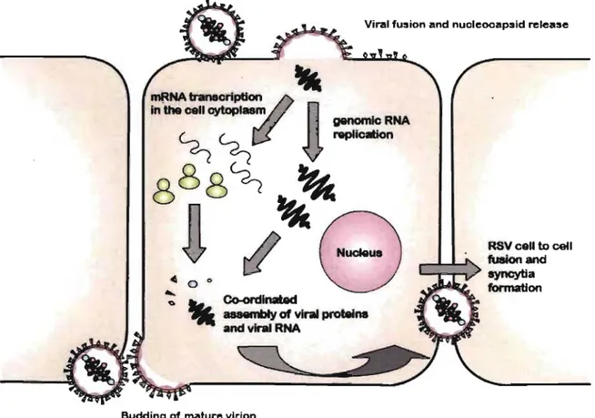
The viral cycle of RSV begins with the attachment and entry into the host ce li , predominantly airway epithelial cells~ which line the noseas well as the large and small airways. Binding and entry of RSV into these target cells is mediated by the interaction between the G and the F proteins, with the host cell (Figure 3). The specifie cellular receptor(s) for the G protein has not yet been identifièd however cell surface glycosaminoglycans, such as heparin sulfate and chrondroitin sulfate B, have been shown to have an important function in in vitro infection through a putativeheparin binding domain in the G protein 70. Interestingly, recombinant RSV virus es with either mutated G protein or simply lacking the G prote in are still infectious in cell cultures, whereas attenuation of infectivity is demonstrated in vivo 71, 72. This observation suggests that the
13
G protein is non-essential for cell attachment but has other functions that may influence the efficiency of the process, as it con tains a motif at 182-186 amino acids common to the CX3C chemokine fractalkine 53. lt has been shown that the G prote in binds to the CX3C receptor (CX3CR1) and can facilitate RSV infection 73.
The F protein is absolutely required by RSV and can mediate attachment on its own. The fusion of the viral envelope or infected cell membranes with uninfected cell membranes and the fonnation of syncytium is an essential step in the virus life cycle 74. Viral uptake
appears to occur by fusion rather than endocytosis. Once incorporated into the target cell membrane, the nucleocapsid (RNP) is released into the cytoplasm, where the genome is transcribed and replicated (Figure 3).
Viral fusion and nucleocapsid release
--
genomlcRNAmRNA
traneCriptlon2f
in 'the cell cytopla8m
n
~ ~ ~ication
888~ ~
:~o r1~8
o . . . Co-ordlnatad
'II
aaaembIy of virai proteineand virlll RNA
Budding of mature virion
RSV ceU ta celi fuaionand ayncytia formation
Figure 3. The schematic representation of the replicative cycle of RSV. The attachment protein Gand fusion protein F mediate RSV entry and uptake respectively. After the viral enveJope has been incorporated with the ho st cell membrane, the nuc1eocapsids (RNPs) are released into the cytoplasm, where the en tire replication occurs. RNA transcription and replication is followed by encapsidation and budding on the cell surface ofnewly fonned virions. Figure modified from (Hacking, 2002) 37.
The entire RSV replicative cycle takes place in the cytoplasm beginning with the transcription of the genome into ten monocistronic, 5' -capped, methylated and 3'-polyadenylated mRNAs by the viral RNA-dependent polymerase complex 61 (Figure 3).
RNA syrtthesis occurs in a sequential manner from the 3' -~nd of the genomewith the polymerase complex terminating and reinitiating mRNA transcription at each junction.
, '
Re-initiating can be occasionally inefficient and this results in a gradient of mRNA decrea~ing proportionally to the distance of the gene from the 3' -end of the genome.
The polymerase complex is also responsible for the synthesis of ahtigenomic RNA, resulting in a complementary positive-sense copy of the viral genome. The antigenomic RNA represents a replicativè intermediate that is less abundant in comparison to the genomic RNA. However, both RNAs are packed into virions in equal proportions. Antigenomic and genomic RNA synthesis correlates with protein translation suggesting a need for co-synthetic encapsidation. Where RSV differs from other Paramyxoviruses is in the switch between RSV transcription and RNA replication, as it seems to involve the
(
M2-2 protein and is not dependent on the intracellular levels ofN and P proteins 75. Thus the M2-2 gene oversees this transition from transcription to production of genomic RNA. r,?uring the earlier stages of infection, low leve1s of M2-2 corre1ates with a high transcriptional 'rate, whereas afterwards, when the intracellular levels of M2-2protein increase, transcription is inhibited in favor of replication 76. Replication involves generation of a complete, positive-sense RNA complement of ,the genome, the antigenome, which in tum acts as a template for genome synthesis. The genomes are then incorporated into nucleocapsids as they are synthesized and recycled through the RNA-synthesis pathways, or are transported to the plasma membrane for assembly into virus particles (Figure 3):
Assembly of the nucleocapsids is entirely intra-cytoplasmic. The N protein associates first with genomic and antigenomic RNA followed by P and L. Association of the nucleocapsids with the nascent envelope is mediated by the M protein, through a series of interactions by coordinating the assembly of the envelope proteins F and G, via their cytoplasmic domains, with the nucleocapsid proteins N P and M2-l 77. Once ass~mbled,
15
nucleocapsids are transported close to the ceU surface and bud at specific plasma membrane patches where glycoprotein G clusters. Budding appears to be the reverse of penetration and occurs in vitro on the apical ceU surface (Figure 3).
,
2.4 DISEASE AND CLINICAL RELEV ANCE
2.4.1 RSV infection and transmission
. The virus undergoes replication in the nasopharynx, and is then spread to the lungs via the respiratory epithelium or aspiration of secretions. Inoculation is via the upper respiratory tract and the virus is spread downwards to the lower respiratory tract along the . respiratory mucosa through syncytial formation and epithelial ceUsloughing 78. Lower respiratory infection follows 1-3 days later 79. There is however no systemic spread and the infection is confined to the respiratory mucosa 78.
Respiratory syncytial virus is very contagious and' is thought to spread by large droplets of secretions (i.e. from sneezes) from an infected person. This oœurs via direct,contact with infectious secretions on envlronmental surfaces or through close contact with a person who has an active infection 80, 81, 82. An example of this close contact may be hand-to-eye or hand-to-rtasàl-epithelium following han~ contact with infectious secretions or even hand-to-hand transfer. However, studies have shown that no infection . oœurs if a person is at a distance of greater than six feet from an infected individual, thus the theory of small particle aerosol spread is rejected 83. The virus is highly resistant and can survive on surfaces long after it has been in contact with an infected persôn. Nasal secretions on tissue or cloth are infectious for up to 30 min, whereas those on hard surfaces such as countertops, stethoscopes or silverware are infectious for at least 6-12 hours. After the initial contact, an incubation period of 2-8 days follows 15, in which, the
infected person will conimonly exhibit mild to moderate nasal congestion and low-grade fever and a productive cough with mucous secretion. However, these sy~ptoms may persist as an upper respiratory infection for several weeks and then resolve without
further incident, particularly in patients who have had a previous RSV infection 30.
Due to the extremely contagious nature of the virus, an infected person will remain contagious as long as the virus is being shed. Shedding of the virus begins within a day or so of infection, often before the onset of major signs of illness 81. Shedding of the virus is highly variable and appears to be roughly paralleled with the age of the person, the severity of the infection, and whether the infected person is immunocompromised. Infected adults would typically shed the virus for 3-7 days following infection 84. Since age seems to affect the shedding, infants would normally shed for up to 14 days in lighter infections, however ipfants less than 6 months of age with severe infections may shed for up to 3 to 4 weeks, thus increasing its contagious nature. OIder children and adults may only shed the virus for 3 or 4 days 80, 85. Immunocompromised individuals may shed for several months following an infection 86.
2.4.2 Clinical symptoms
RSV often causes an upper respiratory tract infection in infants and children. It is characterized by symptoms of rhinorrhea and cough. Lower respiratory tract infections are generally associated with expiratory wheezing (often referred to as bronchiolitisor wheezy bronchitis), pneumonia cart occur with RSV infection and bacterial acute otitis media can follow 30, 3,6. Also, approximately a third of RSV-infected children develop acute otitis media 87. Acute otitis media is characterized by the presence of fluid, typically pus, in the middle ear with symptoms ofpain, redness of the eardrum, and possible fever .. RSV is the causative agent in about 50% of infant pneumonia and 10:·-30% of pediatric bronchitis 88.
The clinical profile of RSV infection differs according to age. In neonates, RSV infections differ from those in older children and apnoea (the cessation ofbreathing) may . be the onlysymptom· of infection. Almost aIl infants (6 weeks to 2 years of age) will be infecfed with RSV, the primary infection being often asymptomatic. However, RSV infection in infant· will involve the lqwer respiratory tract and cause bronchiolitis and pneumonia. Although this occurs in a minority of infants, it becomes the number one
17 .\
cause of hospitalization in this age group. In oIder children, infections areusually less se~ere. The mortality in healthy children is extremely low, but in immunocompromised patients (including children infected with human immunodeficiency virus, children with combined immunodeficiency syndrome, children undergoing chemotherapy for leukemia, and children who have undergone transplantation), in patients with cardiac abnormalities and in cystic fibrosis patients, RSV infection is often life threatening 88, 89, 90, 91. In the
elderly, RSV infection of the lower respiratory tract can manifest itself in the formof pneumol1ia 30, 36. In adults and the elderly, infection of the upper respiratory tract
manifests commonly with symptoms such as rhinorrhea, nasal congestion, pharyngitis, and cough 30, 36.
In addition, RSV infection may trigger acute respiratory distress syndrome with
. . '- )
substantial morbidity and mortality
,
80. RSV infèctions leading to ARDS appear to havelong-term negative consequences similar to that of patients with other pulmonary diseases 30. Infected patients hospitalized for RSV lowef respiratory tract infection may
develop recurrent wheezing during a l-year follow up. The occurrence of wheezing was . significantly higher in infants with airflow limitations than in those without airflow limitation during the acute infection 92, 93. Although the persistence of wheezing has been
established 94, the relationship between RSV and subsequent asthma and atopy is not
nearly as· clear 95. A number ~f studies appear to have found at· least a statistical
connection between RSV infection and asthm~ in youngchÙdren 3,96,97,92,.
2.5 THE IMMUNE RESPONSE TO RSV
The immune response to RSV infection is similar tothat of any other infection, in thatit is comprised of an innate response, and subsequently by activation of humoral and cell-mediated immunity. However, natural infection with RSV does not provide efficient protection against re-infections, indicating that the acquired immunity is incomplete and unstable. Ineffectiveness of the immune response against RSV infection has hindered the development of effective vaccines against both the bRSV and hRSV. The other' major
1
during subsequent natural infection 98, 99, 100, 101. Humoral immunity plays a key role in limiting the severity of subsequent infections, however does not offer a complete protection against RSV infection.
2.5.1 The innate immune response to RSV
Respiratory epithelial cells, are not only the principle target for RSV infection but are
\
also the tirst line of defense in the innate immune response'to the virus, followed by the 1
involvement of antigen presenting ce Us, namely macrophages and dendritic ~ells. Not only do respiratory epithelial ceUs release nitricoxide (NO) upon infection and produce opsonins and coUectins (which are important in the clearance of the virus), but also sècrete a rànge of 'inflammatory mediators, su ch as chemokines, leukotrienes and cytokines 37,102,103,-104, such as intraceUular adhesion molecule (ICAM)~I 105, interleukin (IL)-8 106 and RANTES (Regulation upon activation normal T cell-expressed and secreted) 107. The release of such inflammatory mediators initiatesmaturation and chemotaxis of macrophages and induces neutrophils, eosinophils, and CD4+ T helper cells. In addition to the action of epithelial c'eUs, alveôlar macrophages are very important in the innate immune defense against R~V as they rnay regulate the immune response 108, and when infected release pro-inflammatory cytokines 109, such as tumor necrosis factor (TNF) alpha, IL-10, IL-6 and IL-8 which are involved in the regulation of inflammation.
In both the macrophages and epithelial ceUs, RSV has been shown to induce the activation of nuclear factor kappa-B (NF-KB), which in turn stimulates transcription of genes affiliated with the antiviral response 110, Ill, NF-KB 'also plays a crucial role in the . early stages of innate immune response, mainly via the Toll-like receptor (TLR) signaling pathway 112, 1I3. TLRs are type 1 transmembrane pro teins and are evolutionary conserved.
pattern recognition receptors, which respond to pathogen-associated molecùlar patterns. PAMPs include Iipopolysaccharides, nonmethylated CpG DNA and dsRNA 114, 115, 116. Cytokine and chemokine production in RSV-infected ceUs involve the Toll-like receptor-. signaling pathwayreceptor-.' Recently, glycoprotein F of RSV was shown to activatereceptor-. TLR4,
19
on resting monocytes/macrophages 117, 118 The role of several TLRs is still cUITently under extensive examination.
2.5.2 Humorallantibody-mediated immunity to RSV
During the first 2 months of the life of a newborn, protection against infection is passively acquired via maternaI immunoglobulins. Despite this fonn of "acquired" immunity, the presence of maternaI antibodies decreases gqldually during the first 6 months of life, leaving most infants un-protected against RSV between 2 and 4 months of age 119. Thus the humoral response is stronger if primary infection with RSV occurs after 6 months of age. These responses are also enhanced after each subsequent episode of re-infection throughout life. However, not aIl humoral responses are favorable. For example RSV-specific IgE may also play a role in the increase of severity of the disease 120, 121, 122. McIntosh and colleagues detÀonstrated that secretory antibodies (IgA)were defective in neutralizing the virus in vitro, which may explain the failuTe of immunity during the early stages of an infant's life 123. Similarly, increased serum IgG titers do not entirely guarantee that a person will be protected from acquiring a new RSV infection or that a future disease will be mild. Thus. humoral immunity does not appear to pro vide a complete protection against RSV infection and CUITent evidence in the literature has focused on the importance of cell-mediated immune response in viral clearance.
2.5.3 Cell-mediated immunity to RSV
The activation of cellular immunity following an RSV infection is due mainly to epithelial cells and alveolar macrophages. Infants with a primary RSV infection often develop a cellular immune response within 10 days 124. Human cytotoxic T cell lymphocytes (CTLs) recognize the N, SH,F, M, M2 and NS2 proteins but not the G prote in 50. In mouse models, specifically in BALB/~ mice, the first cells to appear are the natural killer (NK) cells' followed by CD8+ cytotoxic T ce Ils, which may further modulate the immunity by secreting cytokines, especially IFN-y 124, 125,126. These CTLs predominantly target the M2 prote in followed by the Fand Nproteins 127,128. Both CD4+
and CD8+ T cells have been demonstrated to have antiviral activity on passÎve transfer to RSV infected mice andreduce pulmonary shedding of the virus. However both cell types
1 . d '1 d 129
a so cause lllcrease pu monary amage .
The response to RSV by helper T (Th) cells has also been evaluated in the airWays of RSV-infected patiènt,s 130, 131. T cells pro duce pro-inflam~atory mediators c1assified as type 1 (Thl) or type 2 (Th2) cytokines. Thl cells secrete IL-2, IFNyand lymphotoxin, whereas Th2 cells produce IL-4, IL-5, IL-6 and IL-13 m. Increase in disease severity is associated with enhanced' T cell responses 133, as observed in the mouse model for RSV bronchiolitis, where prior sensitization with a formalin.,.inactivated RSV vaccine leads to a helper Th2-driven augmented disease, contrasting with the usual Thl response seen in primary viral infections 134. Individuals who have deficiencies in cell-mediated immune responsiveness have been shown to develop an unusual'but severe form of RSV infection with prolonged proliferation of the virus in the respiratory tract and progressive pneumonia 135. Of note, wheezing was not a major part of the clinical manifestations in
these immunodeficient patients. These crucial data provide evidence that cell-mediated responses are important in terminating RSV infection and that cellular immunity may contributeto the development of wheezing after RSV infection and the severity of the disease. It is apparent that cell-mediated immunity is ari important component in a patient's response to RSV infection, as well as in his/her recovery.
2.6 CURRENT TREATMENTS AND PROPHYLAXIS
Despite growing concems over this problem, to this day, after almost five decades of effort, no effective and r.eliable vaccine or preventive antiviral against RSV exists 6, 21,136. Currently, treatment of RSV infection is supportive which involves ensuring adequate fluid intake, providing supplemental oxygen, and sometimes, administering bronchodilators. In some cases of severe illness and/or high-risk patients, the broad-spectrum antiviral drug ribavirin (Virazole) may be used in aerosolized form. Ribavirin, a synthetic guan6sine analog with the empirical formula CsH12N40S (Figure 4), was
approved and licensed III 1985 to be delivered Via small-particle aerosol for use in hospitalized children in the United States 137. The mechanism of action of its antiviral properties remains unknown, however it was shown that ribavirin inhibits of the synthesis of viral structural proteins, thereby slowing viral· repli cation and also resulting in a reduced IgE response 138. Although ribavirin is the only anti-viral preparation approved for RSV infections, the use of this teratogen is· still controversial as it has not demonstrated significant reduction in either hospitalization or mortality rates 139, 140. Furthermore, due to its very labor-intensive administration methods and its potential hazardous effects on the caregivers 7, its use has been questionedon economic and safety grounds.
Figure 4. Structural formula of ribavirin. Ribavirin is a synthetic guanosine analog. It has an empirical formula of C8H12N405. Figure modified from (Domachowske, 1999) 35.
Prevention of RSV infection by passive administration of antibodies on the other hand is tremendously expensive thus is restricted solely to high-risk infants (defined ,by prematurity, immunocompromised status, or pre-existing pulmonary disease). There are currently two forms of anti-RSV antibody prophylaxis available on the market: RSV-IGIV and palivizumab. RSV-RSV-IGIV (RespiGam; Medlmmune, Gaithersburg, MD) is produced with pooled serum fromhuman donors with high titers of antibody against RSV 4. It has been shown to significantly reduce hospitalization rates for high-risk infants when administered intravenously once a month at a dose of 750 mg/kg for each month of the peak RSV season. However due to the high cost and the complications associated with the intravenous administration, the cost-effectiveness of this product has become questionable 4, 5. In 1998, Medlmmune introduced palivizumab (Synagis), a humanized monoclonal antibody directed against the F protein. Palivizumab is administered
intramuscularly, at a dose of 15mg/kg, 50 fold lower th an RSV-IGIV, yet appears tohave similar efficacy in the reduction of RSV hospitalization rates. However, lik~ RSV -IGIV, the co st for the injection of palivizumab is high. With this basis,. revised guidelines for RSV prophylaxis limits its administration to infants with chronic lung disease, infants born earlier th an 33 weeks gestational age, or later-term premature infants with at least one other risk factor 6.
Therapeutics Oxygen ~2 agonist Racemic Epinephrine Aerosolized recombinant human DNase (Pulmozyme)
Inhaled and systemic corticosteroids
Nasopharyngeal suctioning
. Helium-Oxygen gas mixtures (Heliox)
Inhaled Nitric Oxide
Extracorporeal membrane oxygenation Ribavirin
Function & Results
Used in cases where patients exhibit the typical signs of lower respiratory tract infection. The supplemental oxygen should be administered to maintain a saturation of >92%.
Used to treat wheezing, frequently present in HSV infections. However, despite numerous clinical trials, the effectiveness ofthis therapy rerriains questionable. Given either via injection or nebulized to ameliorate the symptoms of RSV infection. Racemic administration may improve oxygenation, respiratory distress score, and pulmonary function measures in majority of the cases. . After DNase administration, chest radiograph scores improved significantly, however, other measures such as improvement in respiratory rate, wheezing, and retractions during hospitalization did not. Thus concluding that the effectiveness of this treatment is doubtful.
Prescribed for the treatment of bronchiolitis, both during the acute phase and during the period of recurrent wheezing that follows RSV infection in many infants. Studies have shown that the anti-inflammatory action of the corticosteroids should provide effective therapy for infections, however its efficacy Jor RSV remains still unclear.
Defined as extending a suction catheter up through the nose, passing the tip to the hypopharynx, and then applying suction as the catheter is withdrawn. An exceptionally effective palliative measure for infants with RSV by clearing the secretions present in the nose, pharynx and lower airways to improve the resistance of breathing and provide some symptom reJief ..
Functions by decreasing the work of breathing and by improving gas exchange in obstructive conditions (i.e. croup and chronic obstructive pulmonary disease).· Studies have shown that this may be useful in avoiding respiratorY failure and intubation in RSV-infected infants. . Currently approved by the United States Food and Drug Administration for the treatment of persistent pulmonary hypertension of newborns only. Given to treat severe RSV pneumonia and bronchopulmonary dysplasia and may improve oxygenation and respiratory system resistance.
A suitable option for patients with severe RSV disease who cannot be supported on mechanical ventilation.
A synthetic nucleoside (guanosine analog) and the sole anti-viral preparation currently approved for the treatment of RSV infections. While the mechanism of action of its antiviral properties remains unknown, it may act by inhibiting RSV replication during the active replication phase of the virus.
Table II. Current therapies for RSV infections and their functions. Despite almost five decades of research, no effective and reliable vaccine or preventive antiviral against RSV exists. Currently, treatment
23
of RSV infection is supportive, such as the administration of supplemental oxygen, ~2 agonist, racemic epinephrine, aerosoli:.;:ed recombinant human DNase, inhaled and systemic corticosteroids, helium-oxygen gas mixtures, inhaled nitric oxide, suctioning of the nasopharyngeal regions, and oxygenation of the extracorporeal membrane oxygenation. The sole antiviral drug available is Ribavirin, whose use is reserved for persons at high risk for severe disease 95.
Different approaches have been developed for RSV infections (Table II), however no effective treatment beyond palliative measures has yet appeared· 95. With very few
therapies available for RSV infectioI?, more research is needed to find effective measures to prevent or treat RSV infection. Although the disease is self-limited by the body's own . immune response, the persons most susceptible to severe infection which inc1ude immunocompromised patients, infants, elderly, and bone marrow transplanted patients are those individuals who are the least capable of coping with an infection. Ultimately, prophylaxis is the most effective way to handle the infection and a novel technology , termed RNA interference may be the answer to treat an infection during epid,emics or once established.
CHAPTERIII
RNA INTERFERENCE AND siRNAS FOR THE THERAPY OF RSV INFECTION
25
3.1 RNA INTERFERENCE
The discovery of the structure ofDNA by Watson and Crick in 1953 is considered to be one of the most important scientific breakthroughs in the last century 141. Through their model of the DNA structure, genetic information was described as being coded along the
.
-polymeric DNA molecule composed of only four types ofmonomeric units, which were 'orga~ized into genes, and that DNA m'olecules are the basis of heredity 142, 143. Genes were described as controlling the synthesis of various types of RNA, most of which were involved in the process of protein synthesis 144.
Since a11 genomes of complex organisms are pote,ntial tatgets of invasion by viruses and transposable elements, gene therapy seems to hold tremendous potential in the treatment of the many genetic and acquired diseases. Although there are many nucleic acid drugs currently on the market and in production, recently a growing interest has been placed on . , RNA interference (RNAi) as a potential therapeutic agent for silencing numerous viral infectious (gene~ r~quired for viral repli cation) and cancer genes (oncogenes).
3.1.1 Nucleic acid drugs
The majority of cutrently employed pharmacological approaches for modulation of gene function rely upon the interaction of low molecular weight chemical compounds, such as nucleic acids, targeting proteins so as to qown-regulate the function of the( proteins themselves. Nuc1eic acidsmay be defined as. the macromolecliles in which the nucleotides remain linked to each other by phosphodiester bonds between the 3' - and 5'-positions of the sugars 145. These macromolecules control metabolism throughout the life of a cell by directing and regulating prote in and enzyme synthesis and are key elements in transferring genetic information from one offspring to another. These charaçteristics can be taken advantage when developing drugs targeting cancer or infectious dis'eases.