HAL Id: pastel-00605911
https://pastel.archives-ouvertes.fr/pastel-00605911
Submitted on 4 Jul 2011HAL is a multi-disciplinary open access
archive for the deposit and dissemination of sci-entific research documents, whether they are pub-lished or not. The documents may come from teaching and research institutions in France or
L’archive ouverte pluridisciplinaire HAL, est destinée au dépôt et à la diffusion de documents scientifiques de niveau recherche, publiés ou non, émanant des établissements d’enseignement et de recherche français ou étrangers, des laboratoires
Quantitative metabolism of natural and antiviral
nucleosides and nucleotides in human cells by
LC-MS/MS
Emilie Fromentin
To cite this version:
Emilie Fromentin. Quantitative metabolism of natural and antiviral nucleosides and nucleotides in human cells by LC-MS/MS. Pharmacology. AgroParisTech, 2010. English. �NNT : 2010AGPT0040�. �pastel-00605911�
!"#$$%&&'$(!)*$++++$ $ $ ! ! ! ! ! ! ! ! ! ! ,-./012$34$56-78-.94:$;<9=47>912$5-79>?@/3AA!
présentée et soutenue publiquement par
Emilie FROMENTIN
le 1er Juin 2010Quantitative metabolism of natural and antiviral nucleosides and
nucleotides in human cells by LC-MS/MS
$
Doctorat ParisTech
T H È S E
pour obtenir le grade de docteur délivré par
L’Institut des Sciences et Industries
du Vivant et de l’Environnement
(AgroParisTech)
Spécialité : Chimie analytique et Pharmacologie
Directeur de thèse : Pr. Raymond F. SCHINAZI Enseignant responsable : Pr. Douglas N. RUTLEDGE Jury : Président du jury : Dr. Gilles GOSSELIN
M. Luigi AGROFOGLIO, Pr., ICOA UMR-CNRS, Université d’Orléans Rapporteur
Mme Arlette BAILLET, Pr., Faculté de Pharmacie, Université Paris-Sud11 Rapporteur
M. Thierry BELTRAN, Dr., SANOFI-AVENTIS Examinateur
Quantitative metabolism of natural and
antiviral nucleosides and nucleotides in
human cells by LC-MS/MS
This work was performed at the Emory University
School of Medicine and Veterans Affairs Medical Center
and was supported by Emory University, CFAR and NIH
funding.
Acknowledgments
I would like to thank Professor Rutledge and Mme Launay for accepting my application to the ABIES doctoral school and for supporting this collaboration between AgroParisTech and Emory University.
I would like to express my deepest gratitude to the thesis committee members who kindly accepted our invitation to evaluate the research presented in this dissertation and the oral presentation.
Dr Raymond. F. Schinazi has trusted my work and me since October 2006, when he offered me a position in his laboratory. Thanks to his scientific advices, help and patience I have been able to learn day after day the basics not only in nucleoside analytical chemistry, but also in virology, toxicology and cell biology. I would like to thank him for encouraging me to explore new areas, such as pharmacology and pharmacokinetics. He supported my ideas and contributed tremendously to my scientific growth. This thesis represents a great team effort, which could not have been born without Dr Schinazi’s supervision. Importantly, I would like to thank Dr Schinazi for allowing me to attend numerous International and local conferences since I joined the laboratory in order to broaden my horizons.
Working in the pharmacology team has been a tremendous experience. I would like to thank Aleksandr Obikhod for teaching me chromatography, mass spectrometry, cell culture and for his help on instruments maintenance. We have worked together on several projects and established a functional teamwork. Selwyn Hurwitz taught me pharmacokinetics and encouraged me to be critical with the interpretation of my experiments. His advices were very important for my scientific training. Christina Gavegnano has taught me cell culture as well as helped me with the understanding of the basic virology and immunology of HIV infection. She trusted me with the analysis of her precious macrophages, which represented weeks of work. I also would like to thank her for her contagious enthusiasm, which cheered me up more than once. I would also like to thank Ghazia Asif and especially Judy Matthew for their help. I am especially grateful for Claire de la Calle who has been a great student during the summer 2009.
Importantly, I am thankful to the virology team, Mervi Detori, Kim Rapp, Sadie Solomon and Cathy Montero for providing me with cells when I needed and for their precious teamwork. I would also like to thank all the biologists and chemists at RFS Pharma, LLC for their encouragement, support and friendship.
Carol Jeffcoat has been a great support during these four years. She created an enjoyable atmosphere in the laboratory, for everyone to feel comfortable.
I could not thank enough Brenda Hernandez-Santiago for making me feel so welcome in her house for my first year in Altanta. She not only trusted me scientifically, with work that we published together, but she also personally made my arrival to this new country extremely comfortable and enjoyable. The year that we shared together was a privileged experience to me and I will never forget it.
I would like to thank Dr Schinazi, Judy Mathew, Selwyn Hurwitz and Franck Amblard for their careful reading of this dissertation and for making the suggestions and corrections that were necessary to clarify and improve this manuscript.
I would like to thank Barbara Stoll, the chair of the pediatrics department, Dr Schinazi and Emory University for giving the financial support that was necessary to accomplish this work. I met during those 4 years in Atlanta extraordinary people who have changed the way I see the world. Merci Maïra pour ton soutien quotidien et ta joie de vivre. I specially would like to thank Ivana, Meta, Adriana, Niko, Feda, Helder, Omar, Jessica, Thandi, Leda, Matthew, Tami… Je n’oublierai pas la French team Ethel, Franck, Véro, Vincent, Ugo pour les fameux diners à la française et randonnées en Géorgie. Votre amitié m’a rendue plus forte et m’a accompagné tout au long de cette merveilleuse expérience. Gracias, obrigado, thank you, merci.
Cette longue histoire ne serait pas arrivée sans l’aide de Thierry Beltran qui a non seulement été à l’origine de cette opportunité d’emploi aux Etats-Unis mais aussi de cette thèse. Il avait déjà compris durant mon stage de fin d’études d’ingénieur que j’aurai la volonté et l’enthousiasme nécessaire pour poursuivre une formation doctorale et m’a aidé à le réaliser.
Si cette décision de quitter mon pays pour aller travailler aux Etats-Unis fut riche, elle ne fut pas sans peine. Quitter ma famille pendant ces quatre années a représenté pour moi un sacrifice important. Mais le soutien qu’ils m’ont apporté dans cette décision initiale ainsi que dans la transformation de cette expérience en doctorat a été crucial. Même si mes choix vous ont parfois un peu dépassés, je vous suis reconnaissante à tous les trois, Angélique, papa, maman pour la confiance que vous m’avez accordée et tout l’amour que vous m’avez donné.
David, porque estuviste a mi lado todo el tiempo, haciendo mejores mis días. Porque me diste la confianza para progresar y porque tuviste la paciencia para enseñarme los intrincados caminos de la ciencia. Porque cambiaste mi vida a un mundo sin nubes, por esto es que quiero decirte
“Chronologiquement, aucune connaissance ne précède en nous l’expérience et c’est avec elle
To remember those who have fought.
“In June of 1987, a small group of strangers gathered in a San Francisco storefront to document
the lives they feared history would neglect. Their goal was to create a memorial for those who had died of AIDS, and to thereby help people understand the devastating impact of the disease. This meeting of devoted friends and lovers served as the foundation of the NAMES Project AIDS Memorial Quilt. The Quilt was conceived in November of 1985 by long-time San Francisco gay rights activist Cleve Jones, who helped organize the annual candlelight march honoring Harvey Milk and Mayor George Moscone. At the end of the 1985 march, Jones and others stood on ladders taping placards bearing the names of friends and loved ones who had died of AIDS to the walls of the San Francisco Federal Building. The wall of names looked like a patchwork quilt. On October 11, 1987, The Quilt included 1,920 panels and was displayed for the first time on the National Mall in Washington, D.C. during the National March on Washington for Lesbian and
Gay Rights. The Quilt returned to Washington, D.C. in October of 1988, when 8,288 panels
were displayed on the Ellipse in front of the White House. The entire Quilt was again displayed on the National Mall in 1992 and 1996, when it contained approximately 37,440 individual panels. The Quilt was nominated for a Nobel Peace Prize in 1989, and is today the largest community art project in the world. The Quilt has been the subject of countless books, films, scholarly papers, articles, and theatrical, artistic and musical performances. In 2001 The NAMES Project Foundation headquarters and The AIDS Memorial Quilt moved from San Francisco to Atlanta, GA, and today displays continue being held across the country every day, new panels continue to be accepted and incorporated into The Quilt, and a new, solid financial foundation continues to be built beneath The Quilt, guaranteeing its ongoing work and securing
List of publications related to this work (2007-2010)
ManuscriptsIn preparation
1. Study of 9-!-D-(1,3-dioxolan-4-yl)guanine phosphorylation alone and in combination with other nucleosides analogs in human peripheral blood mononuclear cells. Emilie Fromentin, Claire De La Calle, Mervi Detorio, Aleksandr Obikhod, Selwyn J. Hurwitz, Raymond F. Schinazi.
2. Cellular Pharmacology and Potency of HIV-1 Nucleoside Analogues in Primary Human Macrophages: Implications for HIV-1 Eradication. Christina Gavegnano, Emilie Fromentin, Leda Bassit, Mervi Detorio, and Raymond F. Schinazi.
3. In vivo and in vitro studies supporting the use of reduced dose regimens for d4T. Selwyn J. Hurwitz, Emilie
Fromentin, Raymond F. Schinazi.
4. Biochemical Simulation of Human Immunodeficiency Virus Type 1 Reverse Transcription with Physiological Nucleotide Pools found in Macrophages and CD4+ T cells. Edward M. Kennedy, Christina Gavegnano, Laura Nguyen, Rebecca Slater, Amanda Lucas, Emilie Fromentin, Raymond F. Schinazi and Baek Kim.
Published
1. Simultaneous quantification of intracellular and antiretroviral nucleosides and nucleotides by liquid chromatography-tandem mass spectrometry. Emilie Fromentin, Christina Gavegnan, Aleksandr Obikhod, Raymond F. Schinazi. Anal Chem. 2010 Mar 1;82(5):1982-9.
2. Lack of pharmacokinetic interaction between amdoxovir and reduced and standard dose zidovudine in HIV-1 infected individuals. Selwyn J. Hurwitz, Ghazia Asif, Emilie Fromentin, Phillip M. Tharnish, Raymond F. Schinazi. Antimicrob Agents Chemother. 2010 Mar;54(3):1248-55.
3. Synthesis and evaluation of 3’-azido-2’,3’-dideoxypurine nucleosides as inhibitors of human immunodeficiency virus. Hong-wang Zhang, Steven J. Coats, Lavanya Bondada, Franck Amblard, Mervi Detorio, Emilie Fromentin, Sarah Solomon, Aleksandr Obikhod, Tony Whitaker, Nicolas Sluis-Cremer, John W. Mellors, Raymond F. Schinazi. Bioorg Med Chem Lett. 2010 Jan 1;20(1):60-4.
4. Simultaneous quantification of 9-(!-D-1,3-dioxolan-4-yl)guanine, amdoxovir and zidovudine in human plasma by liquid chromatography-tandem mass spectrometric assay. Emilie Fromentin, Ghazia Asif, Aleksandr Obikhod, Selwyn J. Hurwitz, Raymond F. Schinazi. J Chromatogr B Analyt Technol Biomed Life Sci. 2009 Nov 1;877(29):3482-8.
5. Synthesis, antiviral activity, and stability of nucleoside analogs containing tricyclic bases. Franck Amblard, Emilie Fromentin, Mervi Detorio, Alexander Obikhod, Kimberly L. Rapp, Tamara R. McBrayer, Tony Whitaker, Steven J. Coats and Raymond F. Schinazi. Eur J Med Chem. 2009 Oct;44(10):3845-51.
6. Acyclovir is activated into a HIV-1 reverse transcriptase inhibitor in herpesvirus-infected human tissues. Andrea Lisco, Christophe Vanpouille, Egor P. Tchesnokov, Jean-Charles Grivel, Angélique Biancotto,Beda Brichacek, Julie Elliott, Emilie Fromentin, Robin Shattock, Peter Anton, Robert Gorelick, Jan Balzarini, Christopher McGuigan, Marco Derudas, Matthias Götte, Raymond F. Schinazi and Leonid Margolis. Cell Host Microbe. 2008 Sep 11;4(3):260-70.
7. Short communication cellular pharmacology of 9-(!-D-1,3-dioxolan-4-yl) guanine and its lack of drug interactions with zidovudine in primary human lymphocytes. Brenda I Hernandez-Santiago, Aleksandr Obikhod, Emilie Fromentin, Selwyn J Hurwitz, Raymond F Schinazi. Antivir Chem Chemother. 2007;18(6):343-6.
Conference Communications
1- Elevated rNTP incorporation by HIV-1 RT with dNTP/rNTP pools found in human macrophages. Edward Kennedy, Raymond Schinazi, Christina Gavegnano, Emilie Fromentin, Baek Kim. Palm Springs Symposium on HIV/AIDS, CA. March 11-13, 2010.
2- Synthesis of 2’-C-methyl purine modified nucleosides as a potential anti-HCV agents. H-W Zhang, SJ Coats, L Zhou, M Detorio, T Whitaker, T McBrayer, PM Tharnish, A Obikhod, E Fromentin, and RF Schinazi. HEP DART, Hawaii, Dec 6-10 2009. Global antiviral journal, volume 5 supplement 1, p92.
3- Novel purine dioxolane protides are potent inhibitors of hepatitis B virus. L Bassit, L Bondada, A Obikhod, E Fromentin, SJ Coats, and RF Schinazi. HEP DART, Hawaii, Dec 6-10, 2009. Global antiviral journal, volume 5 supplement 1, p91.
4- Anti-HIV-1 nucleoside analogue triphosphate levels are significantly lower in primary human macrophages than in lymphocytes. C Gavegnano, E Fromentin and RF Schinazi. 4th HIV-1 Persistence Workshop, St. Martin, French West Indies. Dec 8-11, 2009. Global antiviral journal, volume 5 supplement 2, p43.
5- HIV-1 nucleoside analogue triphosphate levels are significantly lower in primary human macrophages than in human lymphocytes. Christina Gavegnano, Emilie Fromentin and Raymond F. Schinazi. XVIII International Drug Resistance Workshop, Fort Meyers, FL. June 9-13, 2009. Global antiviral journal, volume 4, supplement 1, p77. 6- Metabolism of highly active and selective 3’-azido-2’,3’-dideoxypurine nucleosides in primary human lymphocytes and MT-2 cells. Aleksandr Obikhod, Emilie Fromentin, Mervi Detorio, Steven J. Coats, Nicolas Sluis-Cremer, and John W. Mellors, and Raymond F. Schinazi. HIV DART, Rio Grande, Puerto Rico - Dec 9-12, 2008. Global antiviral journal, volume 4, supplement 1, p79.
7- Cellular pharmacology of HIV-1 nucleoside analogues in primary human macrophages compared to human lymphocytes. Christina Gavegnano, Emilie Fromentin and Raymond F. Schinazi. Georgia Life Sciences Summit, Atlanta, GA. Sept 24, 2008.
8- Synthesis and antiviral activity of tricyclic 3,9-dihydro-9-oxo-5H-imidazo[1,2-A]purine nucleosides: toward new nucleoside prodrugs. F Amblard, E Fromentin, A Obikhod, TR McBrayer, KL Rapp, M Detorio, T Whitaker, SJ Coats, RF Schinazi. Georgia Life Sciences Summit, Atlanta, GA. Sept 24, 2008.
9- Potent activity and lack of pharmacokinetic interaction between amdoxovir when co-administered with low dose zidovudine. Selwyn J. Hurwitz, Emilie Fromentin, Ghazia Asif, Phillip Tharnish, Judy Mathew, Nancy M. Kivel,Carlos Zala, Claudia Ochoa, Robert Murphy, and Raymond F. Schinazi. XVII International AIDS Society, Mexico City, Mexico, Aug 3-8, 2008.
10- A common anti-herpetic drug acyclovir is converted into a nucleoside reverse transcriptase inhibitor that suppresses HIV-1 in herpesvirus infected human tissues. A. Lisco, C. Vanpouille, E. Tchesnokov, J.-C. Grivel, A. Biancotto, B.Brichacek, J. Elliott, E. Fromentin, R.Shattock, P. Anton, R.Gorelick, M. Derudas, J. Balzarini, C. McGuigan, M. Götte, R.Schinazi, L. Margolis. XVII International AIDS Society, Mexico City, Mexico, Aug 3-8, 2008. 11- Pharmacokinetics and potent anti-HIV-1 activity of amdoxovir plus zidovudine in a randomized double-blind placebo-controlled study. Robert Murphy, Carlos Zala, Claudia Ochoa, Phillip Tharnish, Judy Mathew, Emilie Fromentin, Ghazia Asif, Selwyn J. Hurwitz, Nancy M. Kivel, Raymond F. Schinazi. 15th CROI, Boston, MA. Feb 3-6, 2008. Abstract 794.
12- Simultaneous quantification of (-)-!-D-dioxolane guanosine (DXG), its prodrug amdoxovir, and zidovudine in human plasma by LC-MS/MS. Emilie Fromentin, Ghazia Asif and Raymond F. Schinazi. LCMS Montreux Symposium, Hilton Head, SC. October 10-12, 2007.
Abbreviations
Biological termsAIDS: Acquired Immunodeficiency Syndrome AUC: Area under the curve versus time CI: Synergistic index
Cl: Drug clearance
CNT: Concentrative Na+ or H+-dependent nucleoside transporter
Cmax: Maximal concentration
Cmin: Minimal concentration
DEC: Dead end complex DNA: Deoxyribonucleic acid
dNTP: 2’-deoxyribonucleoside-5’-triphosphate EC50: 50% effective concentration
ENT: Equilibrative nucleoside transporter F: Bioavailability
FBS: Fetal bovine serum
FDA: Food and Drug Administration
HAART: Highly active antiretroviral treatment t1/2: Half-life
HIV: Human immunodeficiency virus HBV: Hepatitis B virus
IC50: 50% inhibitory concentration
IL-2: Interleukin-2
Ki: HIV-1 RT inhibition constant against a DNA/RNA primer/template Km: Apparent binding constant
MRP: Multidrug resistance protein Mt: Mitochondrial
OAT-OCT: Organic anions or cations transporter PBM cells: Peripheral blood mononuclear cells PBS: Phosphate-buffered saline
PHA: Phytohemagglutinin PK: Pharmacokinetics RNA: Ribonucleic Acid
rNTP: Ribonucleoside-5’-triphosphate RT: Reverse transcriptase
SIV: Simian immunodeficiency virus TAMs: Thymidine analogue mutations Tmax: Time at Cmax
Vd: Volume of distribution
WHO: world health organization Drugs and metabolites
FI/EI: Fusion/Entry inhibitor INI: Integrase inhibitor
NRTI: Nucleoside reverse transcriptase inhibitor
NRTI-MP: Nucleoside reverse transcriptase inhibitor monophosphate NRTI-DP: Nucleoside reverse transcriptase inhibitor diphosphate
NRTI-TP: Nucleoside reverse transcriptase inhibitor triphosphate NtRTI: Nucleotide reverse transcriptase inhibitor
NNRTI: Non-nucleoside reverse transcriptase inhibitor PI: Protease inhibitor
ABC: Abacavir, (1S,4R)-4-[2-amino-6-(cyclopropylamino)-9H-purin-9-yl]-2-cyclopentene-1-methanol succinate
CBV-TP: Carbovir-triphosphate, (-)-carbovir-triphosphate, 2-amino-9-[(1R,4S)-4-(hydroxymethyl)cyclopent-2-en-1-yl]-3H-purin-6-one -5’-triphosphate
DAPD: Amdoxovir, (-)-!-D-(2R,4R)-1,3-dioxolane-2,6-diaminopurine DPD: 2,6-diaminopurine-2’-deoxyriboside
ddATP: 2’,3’-dideoxyadenosine triphosphate ddC: Zalcitabine, 2',3'-dideoxycytidine ddI: Videx, 2',3'-dideoxyinosine
DXG: 9-!-D-(1,3-dioxolan-4-yl)guanine d4T: Stavudine, 3'-deoxythymidinene
(-)-FTC: Emtricitabine, (-)-!-3 -thia-2 ,3 -dideoxy-5-fluorocytidine PMPA, TFV: Tenofovir, (R)-9-(2-phosphonylmethoxypropyl)adenine RBV: Ribavirin, 1-!-D-ribofuranosyl-1,2,4-triazole-3-carboxamide TDF: Tenofovir disoproxil fumarate
ZDV: Zidovudine, 3’-Azido-3’-deoxythymidine 3TC: Lamivudine, (-)-!-3 -thia-2 ,3 -dideoxycytidine
Liquid chromatography and mass spectrometry:
CRM: Charge Residue Model ESI: Electrospray Ionization HA: Hexylamine
IEM: Ion Evaporation Method IS: Internal Standard
LC: Liquid chromatography MS: Mass spectrometry m/z: mass to charge
N,N-DHMA: N,N-dimethylhexylamine Enzymes
ADA: Adenosine deaminase, EC:3.5.4.4 AMPDA: Adenylate deaminase, EC:3.5.4.6 AMPK: Adenylate kinase, EC:2.7.4.3
APRT: Adenine phosphoribosyl-transferase, EC:2.4.2.7
ADSS/L: Adenylosuccinate synthase/lysase EC:6.3.4.4/EC:4.3.2.2 Ca-NT: Calcium activated nucleotidase, EC:3.6.1.6
CDA: Cytidine deaminase, EC:3.5.4.5
CMPK: Cytidine monophosphate (UMP-CMP) kinase 2, mitochondrial, EC:2.7.4.14 CTPS: CTP synthase, EC:6.3.4.2
dCK: 2’-deoxycytidine kinase, EC 2.7.1.74 dGK: 2’-deoxyguanosine kinase, EC:2.7.1.113
dCMPDA: dexoycytidine monophosphate deaminase, EC:3.5.4.12 EDP : Ectonucleoside triphosphate diphosphohydrolase, EC:3.6.1.5 GDA: Guanine deaminase, EC:3.5.4.3
GMPS: Guanylate synthase, EC:6.3.5.2
HPRT: Hypoxanthine phosphoribosyl-transferase, EC:2.4.2.8 IMPDH: Inosinate dehydrogenase, EC:1.1.1.205
NDPK : Nucleoside diphosphate kinase, EC:2.7.4.6 PNP: Purine nucleoside phosphorylase, EC:2.4.2.1 RNR : Ribonucleotide reductase, EC:1.17.4.1 TK1 or TK2: Thymidine kinase 1 or 2, EC:2.7.1.21 TMPK: Thymidylate kinase, EC:2.7.4.9
TMPS: Thymidylate synthetase, EC:2.1.1.45 TP: Thymidine phosphorylase, EC:2.4.2.4 UCK: Uridine-cytidine kinase, EC:2.7.1.48 UP: Uridine phosphorylase, EC:2.4.2.3 UTG: UDP-glucuronyltransferase, EC 2.4.1.17 XDH: Xanthine dehydrogenase, EC:1.17.1.4
5’-NT : High Km 5’-nucleotidase, cytosolic II, EC:3.1.3.5
Endogenous nucleobase, nucleosides and nucleotides
A, Ade: Adenine, Ado: adenosine, dAdo: 2’-deoxyadenosine AMP: Adenylate, adenosine monophosphate
ADP: Adenosine diphosphate ATP: Adenosine triphosphate
C, Cyt: Cytosine, Cyd: cytidine, dCyd: 2’-deoxycytidine CMP: Cytidylate, cytidine monophosphate
CDP: Cytidine diphosphate CTP: Cytidine triphosphate
G, Gua: Guanine, Guo: guanosine, dGuo: 2’-deoxyguanosine GMP: Guanylate, guanosine monophosphate
GDP: Guanosine diphosphate GTP: Guanosine triphosphate
IMP: Inosinate, inosine monophosphate OMP: Orotate
PPi: Pyrophosphate
PRPP: Phosphoribosyl pyrophosphate RMP: "-D-ribose-5-phosphate
T, Thy: Thymine, Thd: Thymidine
TMP: Thymidylate, thymidine monophosphate TDP: Thymidine diphosphate
TTP: Thymidine triphosphate
U, Ura: Uracil, Urd: Uridine, dUrd: 2’-deoxyuridine UMP: Uridine monophosphate
UDP: Uridine diphosphate UTP: Uridine triphosphate 3PG: 3-phosphoglycerate
Table of Content
!! "#$%&'()$*&#++++++++++++++++++++++++++++++++++++++++++++++++++++++++++++++++++++++++++++++++++++++++++++++++++++++++++++++++++++++++++++++++++++ ,! !+!! -./0123&%2$&%40&506*&)./7*)2809.2%72)&8&:4;01<69++++++++++++++++++++++++++++++++++++++++++++++++++++++++++++++,! !+=! >(72#0"77(#&'/5*)*/#)40?*%(@0A>"?B ++++++++++++++++++++++++++++++++++++++++++++++++++++++++++++++++++++++++++++++++++++++ !C! "#$#"! %&'()*+,-!./0!(1.,.(2+,&3.2&)/!)4!567#################################################################################################"8! "#$#$! 9:&0+;&)<)=-!)4!567#####################################################################################################################################"8! "#$#>! 7&,)<)=-!)4!567################################################################################################################################################""! "#$#?! 567!:,)=,+''&)/!./0!:+,'&'2+/(+############################################################################################################"@! !+D! E#$*%/$%&F*%280$%/2$7/#$+++++++++++++++++++++++++++++++++++++++++++++++++++++++++++++++++++++++++++++++++++++++++++++++++++++++++++++++++ !G! "#>#"! AB(<+)'&0+!C+*+,'+!D,./'(,&:2.'+!6/1&E&2),'!FACD6G####################################################################"H! "#>#$! I+2.E)<&';!./0!;+(1./&';!)4!.(2&)/!)4!ACD6 #################################################################################$$! "#>#>! %,B=!();E&/.2&)/!./0!2,+.2;+/2!<&;&2.2&)/'####################################################################################$J! !+H! I#'&:/#&(@0#()8/&$*'/0J&&8++++++++++++++++++++++++++++++++++++++++++++++++++++++++++++++++++++++++++++++++++++++++++++++++++++++++++ DD! "#?#"! 9/0)=+/)B'!/B(<+)2&0+!'-/21+'&' ##########################################################################################################>?! "#?#$! 6/2+,.(2&)/'!E+2K++/!ACD6!./0!+/0)=+/)B'!/B(<+)2&0+!:))<###################################################>L! "#?#>! 6;E.<./(+'!&/!21+!+/0)=+/)B'!/B(<+)2&0+!:))<!(./!(.B'+!2)M&(&2-##########################################>H! "#?#?! 9/0)=+/)B'!/B(<+)2&0+!:))<!*.,-!K&21!(+<<!2-:+!./0!.(2&*.2&)/###############################################?8! !+K! L/$/)$*#'0M(2#$*5*)2$*�&50#2$(%2802#'0@4#$./$*)0#()8/&@A$B*'/@++++++++++++++++++++++++++++ HD! "#@#"! N1+;&(.<!:,):+,2&+'!)4!/B(<+)'&0+'!./0!/B(<+)2&0+'#####################################################################?>! "#@#$! 9<+(2,)':,.-!&)/&3.2&)/!2./0+;!;.''!':+(2,);+2,- #####################################################################??! "#@#>! 5&=1!O,+''B,+!P&QB&0!N1,);.2)=,.:1-!F5OPNG ###############################################################################?H! "#@#?! RB./2&4&(.2&)/!)4!/B(<+)'&0+'!./0!/B(<+)2&0+'!E-!PNSITUIT####################################################@"! =! <3N/)$*F/@ ++++++++++++++++++++++++++++++++++++++++++++++++++++++++++++++++++++++++++++++++++++++++++++++++++++++++++++++++++++++++++++++++++++++KD! D! O2$/%*2802#'07/$.&'@++++++++++++++++++++++++++++++++++++++++++++++++++++++++++++++++++++++++++++++++++++++++++++++++++++++++++++++++KH! D+!! P./7*)28@02#'0Q/2:/#$@ ++++++++++++++++++++++++++++++++++++++++++++++++++++++++++++++++++++++++++++++++++++++++++++++++++++++++++++++++++ KH! D+=! "@&82$*&#;0'*55/%/#$*2$*#'0)(8$(%/0&50)/88@+++++++++++++++++++++++++++++++++++++++++++++++++++++++++++++++++++++++++++ KK! >#$#"! OVI!(+<<'!&')<.2&)/!./0!'2&;B<.2&)/#######################################################################################################@@! >#$#$! %&44+,+/2&.2&)/!)4!OVI!(+<<'!&/!;.(,):1.=+'#####################################################################################@@! D+D! P/88(82%0J.2%72)&8&:40@$('*/@++++++++++++++++++++++++++++++++++++++++++++++++++++++++++++++++++++++++++++++++++++++++++++++++++++++ KR! >#>#"! T2B0&+'!&/!OVI!(+<<'!./0!&/!;.(,):1.=+' ###########################################################################################@L! >#>#$! %WX!:1)':1),-<.2&)/!'2B0&+'!&/!1B;./!OVI!(+<<' #########################################################################@L! >#>#>! %WXUY%7!();E&/.2&)/' ##############################################################################################################################@Z! >#>#?! %WXU>DN!./0!%WXUFSGS[DN!();E&/.2&)/'##########################################################################################@Z! >#>#@! %WX!0+(.-##########################################################################################################################################################@Z! D+H! P&88/)$*�&50@27J8/@05&%0)8*#*)280$%*28 ++++++++++++++++++++++++++++++++++++++++++++++++++++++++++++++++++++++++++++++++++++++++ K,! D+K! 9%/J2%2$*�&50)8*#*)280@27J8/@ ++++++++++++++++++++++++++++++++++++++++++++++++++++++++++++++++++++++++++++++++++++++++++++++++++++ KS! >#@#"! 6/2,.(+<<B<.,!+M2,.(2&)/ ###############################################################################################################################@J! >#@#$! O<.';.!+M2,.(2&)/ ##########################################################################################################################################@J! D+R! 1PTOUVOU++++++++++++++++++++++++++++++++++++++++++++++++++++++++++++++++++++++++++++++++++++++++++++++++++++++++++++++++++++++++++++++++++++++++++++++ RC! >#L#"! \<2&;.2+!>888!./0!DTR!RB./2B;!\<2,. ###############################################################################################L8! >#L#$! PNSITUIT!4),!/B(<+)2&0+' #########################################################################################################################L8! >#L#>! PNSITUIT!4),!/B(<+)'&0+'#########################################################################################################################L?!H! P>E9-IQ0!W0X(2#$*5*)2$*�&50#()8/&@*'/@02#'0#()8/&$*'/@0*#02)$*F2$/'02#'0 (#2)$*F2$/'0.(72#0J%*72%4096O0)/88@02#'072)%&J.2:/@ ++++++++++++++++++++++++++++++++++++++++++++++++++G!! H+!! U*7(8$2#/&(@0M(2#$*5*)2$*�&50*#$%2)/88(82%0#2$(%2802#'02#$*%/$%&F*%280#()8/&@*'/@0 2#'0#()8/&$*'/@03408*M(*'0).%&72$&:%2J.40$2#'/7072@@0@J/)$%&7/$%4++++++++++++++++++++++++++++++++++ G!! ?#"#"! N)<B;/!./0!2+;:+,.2B,+!'+<+(2&)/########################################################################################################Z>! ?#"#$! 6)/S:.&,!;+21)0!0+*+<):;+/2 #################################################################################################################Z?! ?#"#>! I+21)0!'+/'&2&*&2-!./0!:.,2&.<!*.<&0.2&)/# ##########################################################################################ZH! H+=! EJJ8*)2$*&#W0O/2@(%/7/#$0&50YQ-"T-902#'0/#'&/:/#&(@0#()8/&$*'/@0*#096O0)/88@02#'0 *#072)%&J.2:/@ +++++++++++++++++++++++++++++++++++++++++++++++++++++++++++++++++++++++++++++++++++++++++++++++++++++++++++++++++++++++++++++++++++++++++++++ ,D! ?#$#"! I+21)0!(,)''S*.<&0.2&)/!E-!();:.,&')/!2)!:,+*&)B'!:BE<&'1+0!,+'B<2'################################H>! ?#$#$! D1+!<+*+<'!)4!ACD6SDO!.,+!<)K+,!&/!;.(,):1.=+'!21./!&/!OVI!(+<<'#######################################H>! H+D! P&#)8(@*&#@ ++++++++++++++++++++++++++++++++++++++++++++++++++++++++++++++++++++++++++++++++++++++++++++++++++++++++++++++++++++++++++++++++++++++++++ ,,! K! P>E9-IQ0=W0U$('40&50LZ[0J.&@J.&%482$*#'0/#'&:/#&(@0#()8/&$*'/0J&&80*#0 .(72#0J/%*J./%28038&&'07&#&#()8/2%0)/88@ ++++++++++++++++++++++++++++++++++++++++++++++++++++++++++++++++++++++++++++,S! K+!! LZ[0J.&@J.&%482$*�*@08*7*$/'0340)&7J/$*$*�\*$.02880:(2#*#/0#()8/&@*'/@++++++++++++ SC! K+=! 12)]0&50*#$/%2)$*/$\//#0^L?02#'0LZ[05&%0J.&@J.&%482$*�*#096O0)/88@03($0 '/)%/2@/'0/#'&:/#&(@0'Y-908/F/8@ +++++++++++++++++++++++++++++++++++++++++++++++++++++++++++++++++++++++++++++++++++++++++++++++++++++++ S=! K+D! 12)]0&50*#$/%2)$*/$\//#0D-P0&%0ATBT_-P02#'0LZ[05&%0J.&@J.&%482$*�*#096O0)/88@000 `````+ +++++++++++++++++++++++++++++++++++++++++++++++++++++++++++++++++++++++++++++++++++++++++++++++++++++++++++++++++++++++++++++++++++++++++++++++++++++++++++ SK! K+H! LZ[0J.&@J.&%482$*�*@0@$%&#:840*#.*3*$/'0340:(2#*#/0#()8/&@*'/@03($0#&$0340&$./%0 YQ-"0++++++++++++++++++++++++++++++++++++++++++++++++++++++++++++++++++++++++++++++++++++++++++++++++++++++++++++++++++++++++++++++++++++++++++++++++++++++++++++++++++ SR! K+K! L&0YQ-"0255/)$0$./0/#'&:/#&(@0#()8/&$*'/0J&&8a +++++++++++++++++++++++++++++++++++++++++++++++++++++++++++++++++++ SS! K+R! P&#)8(@*&#@ ++++++++++++++++++++++++++++++++++++++++++++++++++++++++++++++++++++++++++++++++++++++++++++++++++++++++++++++++++++++++++++++++++++++++!CC! R! P>E9-IQ0DW0X(2#$*5*)2$*�&50#()8/&@*'/@0*#0.(72#0J82@7203401PTOUVOU02@0J2%$0 &5020)8*#*)280@$('405&%0LE9L0)&73*#2$*�\*$.0%/'()/'0'&@/0&50^L?++++++++++++++++++++++++++++ !C!! R+!! U*7(8$2#/&(@0M(2#$*5*)2$*�&50STAbTLT!;DT'*&c&82#THT48B:(2#*#/;027'&c&F*%02#'0 d*'&F('*#/0*#0.(72#0J82@7203401PTOUVOU02@@24+++++++++++++++++++++++++++++++++++++++++++++++++++++++++++++++++++++++++++!C!! L#"#"! PNSITUIT!(1.,.(2+,&'2&('####################################################################################################################### "8$! L#"#$! N.<&E,.2&)/'!(B,*+'!./0!<&;&2!)4!QB./2&4&(.2&)/############################################################################## "8?! L#"#>! 7.<&0.2&)/!,+'B<2'######################################################################################################################################## "8@! L#"#?! %&'(B''&)/!)4!21+!./.<-2&(.<!.0*./(+'!E-!21+!:,+'+/2!;+21)0################################################ "8Z! R+=! EJJ8*)2$*&#W09.2%72)&]*#/$*)@0&50LE9L0J8(@0^L?+++++++++++++++++++++++++++++++++++++++++++++++++++++++++++++++!CS! L#$#"! A)/S();:.,2;+/2.<!O]!./.<-'&'########################################################################################################## "8J! L#$#$! 9*&0+/(+!4),!21+!<.(^!)4!&/2+,.(2&)/!E+2K++/!%_O%!./0!Y%7################################################# ""8! R+D! P&#)8(@*&#@ ++++++++++++++++++++++++++++++++++++++++++++++++++++++++++++++++++++++++++++++++++++++++++++++++++++++++++++++++++++++++++++++++++++++++!!!! G! P&#)8(@*&#@02#'0J/%@J/)$*F/@++++++++++++++++++++++++++++++++++++++++++++++++++++++++++++++++++++++++++++++++++++++++++++++ !!=! ,! Q/5/%/#)/@ ++++++++++++++++++++++++++++++++++++++++++++++++++++++++++++++++++++++++++++++++++++++++++++++++++++++++++++++++++++++++++++++++++ !!K!
1 Introduction
1.1 The Laboratory of Biochemical Pharmacology, LOBP
The research presented in this dissertation was performed in the Laboratory of Biochemical Pharmacology (LOBP) at the Veterans Affairs Medical Center/Emory University. LOBP is a drug discovery and preclinical drug development laboratory directed by Dr. Raymond F. Schinazi. The objective of the laboratory is to discover and develop new drugs for the treatment of human immunodeficiency virus-1 (HIV-1) and hepatitis infections, focusing on nucleoside analogues. Work performed at LOBP is independent from a biotech company in Tucker, GA, called RFS Pharma, LLC (RFSP; also founded by Dr. Raymond F. Schinazi, www.rfspharma.com, last consulted on March 24th 2010). RFSP has in-licensed a series of anti-HIV drugs including Amdoxovir™ (AMDX, DAPD) from Emory University and the University of Georgia Research Foundation.
LOBP is organized around five cross-functional teams that work together to complete the preclinical process of drug discovery (Figure 01). The organic chemists design and synthesize new antiviral compounds. The virology group evaluates the potency of these compounds in vitro using peripheral blood mononuclear (PBM) cells infected with various strains of HIV, including resistant mutations. Toxicity is initially assessed against a panel of cells grown in culture, subsequently tested in animal models provided the compound has favorable therapeutic window and pharmacology profile in vitro. Pharmacology studies are undertaken to understand the metabolism of potential nucleosides, intracellular phosphorylation to active triphosphate, which is responsible for viral inhibition and the activity at the enzyme level. Pharmacokinetic (PK)
distribution, metabolism and elimination of these agents in animals. These results contribute towards predicting a starting dose for clinical testing in humans. The pharmacology team is comprised of cellular biologists, bioanalytical chemists, pharmacological and pharmacodynamic modelists, pharmacometricians and clinicians. Part of the accomplishments on amdoxovir, which is currently in phase II clinical development will be presented in this dissertation along with in vitro studies of approved anti HIV drugs. Accumulation of safety and efficacy data on drugs post marketing can provide useful information for new drug design.
Figure 01: Schematic of the drug development process.
Adaptated from http://www.medscape.com Preclinical testing: laboratory and animal studies - safety and biological activity - 1 to 2 years Phase I: 20 to 100 healthy volunteers - safety and dosage - 3 years -70% success Phase II: 100-300 patient volunteers - efficacy and safety - 4 to 5 years - 33% success! Phase III: 1,000-3,000 patient volunteers - efficacy and long
term safety/ tolerability - 6 to 8 years - 27% success! File New Drug Application (NDA) Chemical synthesis of new compounds In vitro testing: Antiviral activity and toxicology In vitro drug resistance and pharmacology screening!
LOBP LOBP LOBP
RFS pharma
FDA
1.2 Human Immunodeficiency Virus (HIV)
1.2.1 Discovery and characterization of HIV
In the summer of 1981, clinicians in New York and California reported cases of men who were diagnosed with Pneumocystic carnii pneumonia, a rare opportunistic infection1 or with the rare skin cancer Kaposi’s sarcoma.2 This condition, characterized by low CD4+ T cell counts, lymphadenopathy and infection by opportunistic diseases, was soon named Acquired Immunodeficiency Syndrome (AIDS). The transmission of this infectious microorganism was suggested to occur through sexual activity or blood.3 In 1983 the virus was isolated from infected patients by two different teams: Gallo and Montagnier. Gallo showed that this virus morphology was similar to human T cell leukemia and other type-C retroviruses.4 Montagnier characterized the retroviral activity using [3H]-uridine and named it lymphadenopathy-associated virus.5 Later, phylogenetic studies and replication cycle analysis showed that the AIDS virus was related to members of the lentivirus genus of the retroviridae family of viruses, and was named human immunodeficiency virus (HIV).6, 7
The natural ancestor of HIV is likely simian immunodeficiency virus (SIV), which infects naturally chimpanzees in west central Africa. A common ancestor would have appeared in the 1920’s, and spread as the main cities such as Congo-Kinshasa were expanding.8, 9
1.2.2 Epidemiology of HIV
In 2008, there were an estimated total of 33.4 million (31.1-35.8 million) people who were living with HIV in the world (Figure 02), with about 2.7 million of newly infected adults and children and 2 million deaths. The number people living with the virus has increased by 40% since 1990. The number of deaths due to AIDS has decreased by about 10% since 2004 and the new infections are 30% less frequent than in the mid 90’s. 10
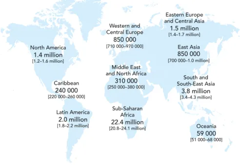
Figure 02: Adults and children estimated to be living with HIV in 2008.10
The epidemic is evolving with HIV prevention strategies, availability of drugs and improved access to treatment having a positive impact on disease burden. However, because the incidence of new infections continues to increase in limited resource countries, HIV/AIDS remains a global health priority. Since no vaccine is available, the treatments need to be improved and universalized.10
1.2.3 Virology of HIV
1.2.3.1 Features of HIV replication cycle
Lentiviruses comprise a separate genus of the family retroviridae; they cause immune deficiencies, disorders of the hematopoietic and central nervous system, and have a larger genome than those of simple retroviruses.11 The genome of HIV-1 consists of two identical 9.2 kb single stranded RNA molecules within the virion, which codes for 15 proteins, whereas the persistent form of the HIV-1 genome is a proviral double-stranded DNA within infected cells (Figure 03 and 04).12
82
M A P S | 2 0 0 9 A I D S E P I D E M I C U P D A T E
Adults and children estimated to be living
with HIV, 2008
Total: 33.4 million (31.1–35.8 million)
Sub-Saharan Africa 22.4 million [20.8–24.1 million] Latin America 2.0 million [1.8–2.2 million] Caribbean 240 000 [220 000–260 000] North America 1.4 million [1.2–1.6 million] Middle East and North Africa
310 000 [250 000–380 000] Western and Central Europe 850 000 [710 000–970 000] Oceania 59 000 [51 000–68 000] East Asia 850 000 [700 000–1.0 million] South and South-East Asia 3.8 million [3.4–4.3 million] Eastern Europe and Central Asia
1.5 million
Figure 03: Stucture of HIV virions. 12
Source: http://www3.niaid.nih.gov/topics/HIVAIDS/Understanding/Biology/hivVirionLargeImage.htm
HIV virions have spherical morphology and consist of a lipid bilayer membrane that surrounds a dense truncated cone-shaped nucleocapsid, which contains the genomic RNA molecules, the viral proteases (not represented), reverse transcriptase, integrase.12
Three coding regions of the genome are the env, pol and gag genes, producing structural polyproteins that are common to all retroviruses. The env gene encodes the envelope proteins that are necessary for viral fusion to the host cells. It encodes two glycoproteins, gp41 anchors the structure of the viral envelope and gp 120 binds to the CD4 molecule on the surface of CD4+ T cells.13 This binding exposes a secondary site for binding to a chemokine co-receptor on the
host cell, CCR5 or CXCR4.6
Pol and gag genes exert control over host-cell machinery by encoding a gag-pol precursor protein that is subsequently cleaved by proteases to generate various essential proteins, including: structural nucleocapside, capside and matrix proteins, as well as the viral enzymes: protease, reverse transcriptase (RT) and integrase (Figure 04).6
Figure 04: Steps in the HIV Replication Cycle.
Source: http://www3.niaid.nih.gov/topics/HIVAIDS/Understanding/Biology/hivReplicationCycle.htm
Lentiviral genomes encode for auxiliary proteins, tat and rev, which perform regulatory functions. The long terminal repeats promote transcriptional activators (NF-"B or NFAT), which activate T-cells.11
Finally, four accessory proteins, Nef, Vif, Vpr and Vpu (or Vpx for HIV-2) modulate viral replication. They are not necessary for viral reproduction in immortalized cell lines, but are necessary for viral replication in vivo11 since they may protect the virus against cellular restriction factors.14
1.2.3.2 Reverse transcription and genome integration
The viral RT uses both RNA and DNA as templates to copy the single-stranded HIV-1 RNA genome to either a double-stranded DNA genome or a RNA-DNA heteroduplex.15, 16 RT from retroviruses has two catalytic activities: DNA polymerization and associated RNase H activity (RNA/DNA heteroduplex RNA degradation). The enzyme is a heterodimer formed by a regulator subunit of p51 (in grey Figure 05) and a catalytic subunit of p66 (coloured Figure
05).17 The p66 subunit resembles a right hand, where the subdomains are designated fingers, palm and thumb (Figure 05). The palm acts as a clamp to position the template-primer to the catalytic motif. 18-20
Figure 05: Crystal structure of the HIV-1 RT enzyme coupled with primer (backbone structure in pink) and template (backbone in blue).12
One major property of the HIV-1 RT is its high rate of base-substitution, addition and deletion errors in the HIV-1 genome. The error rate in the HIV-1 genome has been reported as 1 in 2,000 to 5,000 nucleotides in vitro,21, 22 and 3.4 x 10⁻⁵ mutation per base pair per cycle in
vivo.23-25 The induced nucleotide mismatches are inherited as replication continues.26 This high
rate of mutation can be attributed to the lack of HIV-1 RT 3’#5’ exonucleolytic proofreading capacity,27 the fluctuations in the nucleotide pool levels,28 the incorporation of dUTP 29 and the G#A hypermutations.30 This hypermutability of new virions leads to the emergence of a
complex mixture of “quasispecies”, which are related but genetically distinct forms of HIV-1 in infected individuals, which renders treatment challenging.27
Following reverse transcription, the double stranded DNA viral copy is integrated into the host cell’s genome, by the viral integrase. The viral genome is then transcribed and translated into protein products by the viral protease, which generates a new batch of proteins that will form the new virion (Figure 04).31
1.2.4 HIV progression and persistence 1.2.4.1 The course of HIV infection
HIV infection is characterized by an acute and a chronic phase. During the acute phase of infection, HIV viral replication is widespread and extensive in lymphoid and intestinal tissues. CCR5+ CD4+ T cells are the initial targets and are rapidly depleted. They are located in mucosal surfaces of the intestinal, respiratory and reproductive tract.32 Subsequent targets are CD4+ T cells bearing CXCR4+ co-receptor in peripheral blood, lymph nodes, and spleen.33, 34 This phase of profound depletion is followed by a phase of CD4+ T cell renewal. However, the restoration of memory CD4+ T creates additional targets for HIV-1 leading to exhaustion of the immune system.34 The HIV infected individuals enter a phase of clinical latency characterized by a slow, continuous decrease in CD4+ T cells and maintenance of CD8+ T cells. The HIV infection becomes chronic (Figure 06).33
During progression to AIDS, both CD4+ and CD8+ T cell counts drop dramatically. The increased viral replication in the lymph nodes eventually lead to their destruction and the patient becomes susceptible to opportunistic infections that ultimately cause death (Figure 06).11
Figure 06: Pathogenic events in untreated HIV mediated disease.7, 35
1.2.4.2 HIV latency
The latent reservoirs are established early during the infection by infected cells, which do not divide or produce virions (little production of HIV mRNA). One infected CD4+ T cell in a million reverts back to a resting state.36 Macrophages also exhibit the co-receptor CCR5 causing susceptibility to infection by HIV-1 but might be less susceptible to viral cytopathicity.37 Even though monocytes and macrophages produce ~ 10 to 60-fold fewer virus particles per cell than CD4+ T cells during each infectious cycle, they can secrete virus over a longer period and contribute to the viral burden 38 sufficiently to be considered a reservoir.37 Macrophages and dendritic cells also play an important role in virus dissemination to CD4+ T cells.31, 33
CO M M E N TA RY
tured T cells essential to the propagation of
large quantities of HIV in the laboratory18.
tinues to the present time. Three structural and six regulatory genes, which together encode at least 15 viral proteins, were identified and their relationship to the complex mechanisms of
HIV replication soon unfolded7. These
find-ings were crucial to an understanding of the replication cycle of HIV and its relationship to the pathogenic mechanisms of HIV disease. In addition, they provided an avenue to iden-tify important targets for the development of effective antiretroviral drugs.
The study of the molecular virology of HIV also opened the door to the study of the
molecular epidemiology of HIV19. The
sci-ence of molecular epidemiology was essential in defining the evolving heterogeneity of HIV throughout the world, including the presence of circulating recombinant forms of the
virus20and the origin of HIV in the human
species. With regard to the latter, the zoonotic nature of HIV was established by the close phylogenetic relationship between HIV-2, first identified in West African individuals in 1986 (ref. 21), and the simian immunodefi-ciency virus in sooty mangabeys. In 1999, it was shown that HIV-1 had probably origi-nated from the Pan troglodytes troglodytes species of chimpanzees, in which the virus
coevolved over centuries22. Because
chim-panzees are killed for food in parts of sub-Saharan Africa, the species jump probably occurred by accident.
A blood test for HIV
The next critical advance after the identifica-tion of HIV was the development of a sensi-tive and specific test for antibodies to HIV that could be used for diagnosing individuals (with confirmation by immunoblot analysis)
and for large-scale screening23. This
funda-mental scientific advance had immediate and profound implications for public health. With an ELISA to detect antibodies to HIV, the blood supplies in the United States and other developed countries were screened for HIV and rendered extremely safe by 1985 (ref. 24), thereby preventing millions of potential transfusion-related infections. HIV antibody tests have subsequently been used in numerous epidemiological and natural history studies to clarify the global scope and
evolution of the epidemic25. Only with the
availability of this simple screening was the real and potential scope of the AIDS pan-demic fully appreciated.
Before the ELISA for HIV, clinicians were generally seeing individuals who were in the
infected with HIV, to describe more accu-rately the true clinical course of HIV disease, and to follow the natural history of the dis-ease prospectively in individuals for whom a time of seroconversion could be determined.
HIV pathogenesis
The pathogenesis of HIV disease, from a virological and immunological standpoint, has been studied intensively and defined
pro-gressively over the past 20 years6,8. The
path-ogenic mechanisms of HIV disease are
extremely complex and multifactorial27
(Fig. 1). Even before HIV was identified, it was recognized that an apparent paradox existed whereby the immune system was aberrantly activated at the same time that the individual was experiencing immune
defi-ciency5. This was later shown to be due to a
combination of the aberrant secretion of var-ious cytokines, many of which could upregu-late virus expression, and the intensive cell
signaling induced by the viral envelope28.
Depletion of CD4+T cells was recognized as
a hallmark of disease early on11,12, even
before the classic demonstration in 1984 that the CD4 molecule was the primary receptor for the virus on a subset of T cells and
mono-cytes29,30. In addition, much evidence
sug-gested that other factors were necessary for HIV fusion and entry, but these putative ‘coreceptors’ remained elusive for several
years31.
In the mid-1990s, a number of diverse areas of investigation elucidated the roles of the chemokine receptors CXCR4 and CCR5 in the efficient binding and entry of two dif-ferent strains of HIV-1 called X4 and R5,
respectively6,31. Indeed, RANTES, MIP-1!
and MIP-1", the ligands for CCR5, were shown to potently inhibit the binding of virus to its target cell. This recognition that HIV could use different coreceptors also helped to explain the occurrence of syncytial (CXCR4-using) and nonsyncytial
(CCR5-using) variants of HIV6. The importance of
the CCR5 coreceptor in the pathogenesis of HIV infection was proven by the finding that cells from individuals homozygous for a deletion of 32 base pairs in the CCR5 gene could not be infected in vitro with R5 viruses and that such individuals, who comprise about 1% of white populations, are extremely resistant to HIV infection even
when repetitively exposed to virus32.
Studies of lymphoid tissue in individuals infected with HIV revealed the disseminated
Primary infection Massive viremia Wide dissemination to lymphoid organs HIV-specific immune
response Trapping of virus and establishment of chronic, persistent
infection Establishment
of infection in lymphoid tissue
Immune activation mediated by cytokines and HIV envelope– mediated aberrant cell signaling
Accelerated virus replication Partial immunological control of virus replication Rapid CD4+ T cell turnover Destruction of immune system CD44+ Lymph Lymph Lymph e nodee
Figure 1 Pathogenic events in untreated
HIV-mediated disease. HIV (pink) enters the body and binds to Langerhans or dendritic cells (orange), which carry the virus to CD4+T cells.
Infected CD4+T cells home to lymphoid tissue,
where the infection is established. Virus replication accelerates, and massive viremia leads to the wide dissemination of virus throughout the body’s lymphoid tissue. An HIV-specific immune response occurs and virus is trapped on the follicular dendritic cells of germinal centers in the lymphoid tissue. At this point, chronic, persistent infection is established despite an immunological response to the virus. Immune activation is an important driver of HIV replication and is mediated by the secretion of various cytokines and by aberrant cell signaling caused by interaction of the viral envelope with cellular receptors. Because there is usually only partial immunological control of virus replication, continual and accelerated production of virus ensues. This is associated with a rapid turnover of CD4+T cells. Ultimately, lymphocyte
depletion occurs, along with destruction of the architecture of lymphoid tissue. Adapted with permission from ref. 6.
Katie Ris © 2003 Nature Pub lishing Gr oup http://www .nature .com/naturemedicine
To date, HIV-1 latency established in resting memory CD4+ T cells,39 macrophages 31, 38,
40, 41 or mononuclear cells 42 represents a major barrier to virus eradication.43 Highly active
antiretroviral treatment (HAART) is capable of suppressing HIV, even to undetectable levels in the blood, but they cannot eliminate the virus hiding in these latent reservoirs. Studying the mechanism of viral persistence to existing HAART remains a focus of research, and may lead to the devlopment of new strategies to purge these viral reservoirs.44
1.2.4.3 Predominant sites of HIV infection in vivo
In addition to the sites of infection mentioned previously, CNS/brain is targeted by HIV-1 primarily mediated by infected macrophages and is responsible for CNS dysfunction in infected individuals (Figure 07).45
Figure 07: HIV can hide in the brain, lymph nodes, skin, peripheral blood, reticuloendothelial system, bone marrow, and gastrointestinal cells.
Source: http://www3.niaid.nih.gov/topics/HIVAIDS/Understanding/Biology/hidesImmuneSystem.htm
1.3 Antiretroviral treatment
Modern drug discovery has transformed HIV-1 infection into a treatable chronic infectious disease.46 Before the availablity of drugs, patients diagnosed with AIDS would die
replication, and prevent the decline of CD4+ T cells by targeting several steps in the virus replication. Increased access to treatment and care has raised the life expectancy of HIV-1 infected individuals to over five years in resource-limited countries, and even longer in high-income countries (Figure 08).10
Figure 08: AIDS related death with and without antiretroviral therapy in the world.10
To date, the US Food and Drug Administration (FDA) has formally approved 24 drugs for the treatment of HIV infections. However, not all are used widely due to cytotoxicity and poor response. These antiretroviral agents can be classified in 5 categories according to the step of the HIV replicative cycle they target (Figure 04): nucleoside reverse transcriptase inhibitors (NRTI), non-nucleoside reverse transcriptase inhibitors (NNRTI), protease (maturation) inhibitors (PI), integrase inhibitors (INI) and fusion/entry inhibitors (FI/EI).48
1.3.1 Nucleoside Reverse Transcriptase Inhibitors (NRTI)
The replication of HIV-1 is dependent on a viral reverse transcriptase enzyme not expressed by mamalian cells, thus, making it an attractive therapeutic drug target. The first antiretroviral agent was zidovudine (ZDV, AZT), and since then many more potent and safer NRTI have been developed, and this class of drug remains the backbone of modern HAART regimens especially because of its high genetic barrier to resistance.49 The LOBP focuses on the development of new NRTI including the recently developed NRTI amdoxovir™, which will be
cited throughout this dissertation. Please refer to Figure 9 for structures of FDA approved NRTI that will be described below.
1.3.1.1 FDA approved NRTI
The first HIV inhibitor 3’-azido-3’-deoxythymidine (Zidovudine, ZDV, AZT), was approved by the FDA for anti-HIV usage in 1987. ZDV showed antiviral activity in cell lines at concentrations as low as 50-500 nM.50 Soon after the approval of ZDV, a panel of dideoxynucleosides was evaluated 51 and 2',3'-dideoxyinosine (ddI)52 and 2',3'-dideoxycytidine (ddC) 53 were approved by the FDA in 1991 and 1992, respectively; ddC is no longer marketed because of its toxicity.48 Modifications of 2’,3’-deoxythymidine to form an unsaturated derivative, namely 2’,3’-didehydro-3’-deoxythymidine led to the development of stavudine (d4T).54
The first four nucleosides approved were D-enantiomers, which is the configuration of natural nucleosides. However, it was also discovered that certain L-nucleosides, such as lamivudine, (3TC) or emtricitabine [(-)-FTC], were more potent and less toxic than the (+) isomer.55 FTC may offer improved activity compared to 3TC, due to better affinity of (-)-FTC-TP for the HIV-1 RT, probably due to tighter hydrogen bonding with the the 5-fluorine.56 Furthermore, the 5-fluorine moiety on the base increases its lipophilicity and may increase penetration of the nucleoside analogue into the CNS, which is an important reservoir for HIV-1.
Abacavir (ABC) was the first guanosine analogue approved. The carbocyclic sugar moiety decreases the lability of the glycosidic bond cleavage that is typical of 2’,3’-dideoxynucleosides.57
The acyclic nucleotide reverse transcriptase inhibitor tenofovir disoproxil fumarate (TDF DF) is a salt of (R)-9-(2-phosphonylmethoxypropyl)adenine (PMPA) and was approved in 2001
by the FDA. This drug was formulated as the disoproxil fumarate salt to mask the two negative charges on the phosphonate moiety, which limits the gastrointestinal absorption of PMPA.58 The vast majority of the drug is observed as PMPA in the systemic circulation. TDF was initially suggested for pre- and post-exposure prophylaxis and is now marketed as a fixed dose combination with (-)-FTC (Truvada®) and a triple coformulation with (-)-FTC and efavirenz (Atripla®).59
Figure 09: Structures of the currently FDA approved NRTI.
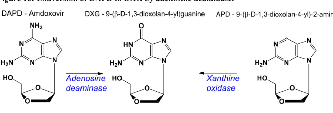
1.3.1.2 Amdoxovir
DXG [9(-!-D-1,3-dioxolan-4-yl)guanine] (Figure 10), is a guanosine nucleoside with a potent and selective activity against HIV-1, HIV-2 and hepatitis B virus (HBV). Since DXG is poorly soluble, a prodrug, amdoxovir, [(-)-!-D-2,6-diaminopurine dioxolane or DAPD] (Figure
10) was designed and resulted in improved oral bioavailability. DAPD is deaminated by the
Thymidine analogues 2’-deoxycytidine analogues
2’-deoxyadenosine analogues 2’-deoxyguanosine analogue O N3 HO N HN O O ZDV - Zidovudine S O HO N N NH2 O 3TC - Lamivudine O HO N HN O O S O HO N N NH2 O F d4T - Stavudine (-)-FTC - Emtricitabine O HO N N NH2 O ddC - Zalcitabine HN N N N O O HO ddI - Didanosine N N N NH H2N N HO ABC - Abacavir N N N N NH2 O P O O O O O O O O O
TDF - Tenofovir Disoproxil Fumarate
COOH COOH
ubiquitous adenosine deaminase (ADA) to DXG, before undergoing intracellular phosphorylation.60 DXG-TP has a Ki of 20 nM versus HIV-1 RT making it one of the most
potent inhibitors of this enzyme. DAPD is currently in phase II clinical testing for the treatment of HIV-1 infection, and has been safely administered to 206 individuals 61 (www.rfspharma.com, accessed on March 24th, 2010).
Figure 10: Conversion of DAPD to DXG by adenosine deaminase.
Following oral administration of DAPD to woodchucks or rhesus monkeys, plasma concentrations of DXG are significantly higher than DAPD. These data suggest that DAPD is rapidly absorbed and converted into DXG in vivo. None of the currently approve NRTI contains a dioxolane sugar motif. In addition, DXG does not cause cellular toxicity up to 500 #M in primary cells and in cell lines. Together, these preliminary results make DXG an attractive molecule for future drug development.62
The major toxicity of amdoxovir in extensive animal toxicology studies was obstructive nephropathy at high doses, secondary to the precipitation of DAPD/DXG in the renal tubules. This was probably due to the poor aqueous solubility of DXG, similar to that observed for the related nucleoside analogues acyclovir. In a 52-week study, five cynomolgus monkeys, receiving DAPD 800 or 1200 mg/kg per day, developed obstructive nephropathy and associated uremia after 26 weeks of treatment, which was reversible after early identification and discontinuation of
Adenosine deaminase Xanthine oxidase HN N N O H2N N O O HO DXG - 9-(!-D-1,3-dioxolan-4-yl)guanine N N N NH2 H2N N O O HO DAPD - Amdoxovir N N N H2N N O O HO APD - 9-(!-D-1,3-dioxolan-4-yl)-2-aminopurine
medication. In addition, islet cell atrophy and hyperglycemia occurred, and lens opacities were observed,63 which were thought to be secondary to hyperglycemia, and not a direct effect of DAPD. No individuals enrolled in clinical trials of DAPD have developed any renal abnormalities attributable to the drug.64, 65 Consequently, various prodrugs have been designed to improve DXG solubility and its pharmacological profile.66 APD [(-)-!-D-2-aminopurine-dioxolane] (Figure 10) is an alternative produg of DXG in preclinical development at LOBP. Although preclinical studies are promising, APD is yet to undergo clinical testing.67
1.3.1.3 Other NRTI in development
There are currently, 3 other NRTI undergoing clinical evaluation, (-)-2'-deoxy-3'-oxa-4'-thiocytidine (apricitabine) developed by Avexa, Ltd., (±)-!-2',3'-dideoxy-5-fluoro-3'-thiacytidine (racivir) by Pharmasset, Inc. and (-)-!-2',3'-didehydro-2',3'-dideoxy-5-fluorocytidine (elvucitabine)by Achillion Pharmaceuticals, Inc (Figure 11).68
Figure 11: Structures of other NRTI in development
1.3.2 Metabolism and mechanism of action of NRTI 1.3.2.1 Plasma pharmacokinetics
Following oral absorption, generally a nucleoside is absorbed in the gastro-intestinal tract and rapidly through the gut wall via a facilitative (non-ATP dependent) nucleoside transporter into the hepatic circulation. After passing through the liver, where some metabolism can occur
N N NH2 O F S O HO (+)-FTC N N NH2 O O S HO Apricitabine Racivir Elvucitabine N N NH2 O F S O OH (-)-FTC N N NH2 O F O OH
Figure 12: Drug metabolism during the absorption process.69
The extent of the drug distribution to tissues and organs depends on its physico-chemical properties and the expression of nucleoside transporters on their surface. Nucleosides are relatively hydrophilic and generally do not bind to plasma proteins and are not extensively metabolized by the cytochrome CYP450 enzymes, with the exception of ZDV and ABC.70, 71
Pharmacokinetic studies are performed in order to quantify the drug adsorption, distribution, metabolism and excretion (ADME) characteristics, mainly using drug concentration versus time profiles measured in blood plasma and urine, since these compartments are easily sampled. Drug concentrations are usually measured in plasma, which correlated with the proportion of drug in equilibrium with target cells, rather than in whole blood.
Since HIV infected individuals are often given several drugs in combinations, the appropriate clinical trials should be designed to detect potential drug-drug interactions and evaluate the pharmacokinetics for each drug alone and in the combination to be used in the clinic.
1.3.2.2 Non-compartmental analysis
Non-compartmental pharmacokinetic analysis is a descriptive regression analysis, which does not include any assumption regarding tissue compartments, providing a detailed modeling of absorption or distribution processes of the drug or metabolites. The area under a plot of the plasma concentration versus time curve (AUC) is a measure of drug exposure, and is often correlated with drug efficacy and toxicity. For single dose measurements, the component of the AUC between dose and the last data point (AUCobs) is calculated using a modification of the
trapezoidal rule (Figure 13).
Figure 13: Graphical representation of the trapezoidal rule.
The remaining portion of the AUC between the last point measured and infinity (AUCextrap), is extrapolated assuming exponential decay. The total AUC (AUCtot) is then AUCobs
+ AUCextrap.
With repeated dosing, equilibrium is reached when the AUC between doses (AUC$) are
reproducible. At equilibrium the amount of drug entering and leaving the plasma are equal. It follows that AUCtot from the first dose is equal to AUC$ at steady-state. Maximal plasma
concentrations (Cmax) are of interest, since drug exposure may correlate with toxicity. Cmin
represents minimal drug concentrations between doses, when the tissues of the person are most
0 500 1000 1500 2000 0 2 4 6 8 10 12 drug concentration, ng/mL time, hr !"# !"$%# &"# &"$%# !!t"# Cmax tmax AUC0tn = 0 i=1 Ci + Ci+1 2 . !t