Scintimammography with
technetium-99m methoxyisobutylisonitrile:
results of a prospective European multicentre trial
H. Palmedo1, H.J. Biersack1, S. Lastoria2, J. Maublant3, E. Prats4, H.E. Stegner5, P. Bourgeois6, R. Hustinx7, A.J.W. Hilson8, A. Bischof-Delaloye9
1Department of Nuclear Medicine, University of Bonn, Germany
2Department of Nuclear Medicine, National Cancer Institute of Naples, Italy
3Department of Nuclear Medicine, Centre Jean Perrin in Clermont Ferrand, France
4Department of Nuclear Medicine, University Hospital of Zaragoza, Spain
5Department of Gynecology, University of Hamburg, Germany
6Department of Nuclear Medicine, Hospital St. Pierre in Bruxelles, Belgium
7Department of Nuclear Medicine, University Hospital in Liège, Belgium
8Department of Nuclear Medicine, NHS Trust Hospital in London, United Kingdom
9Department of Nuclear Medicine, University of Lausanne, Switzerland
&misc:
Received 11 October and in revised form 12 December 1997
&p.1:
Abstract. The aim of the trial was to determine the
diag-nostic accuracy of scintimmammography with
techne-tium-99m methoxyisobutylisonitrile (
99mTc-MIBI) in the
detection of primary breast cancer and to verify its
clini-cal usefulness. A total of 246 patients with a suspicious
breast mass or positive mammogram were included in
this prospective European multicentre trial. At 5 min and
60 min (optional) p.i. two lateral prone images were
ac-quired for 10 min each; 30 min p.i. one anterior image
was acquired for 10 min. There were 253 lesions (195
palpable and 58 non-palpable), in respect of which
his-tology revealed 165 cancers and 88 benign lesions.
Insti-tutional and blinded read results were correlated to core
laboratory histopathology results obtained during
exci-sional biopsy. Diagnostic accuracy for the detection of
breast cancer was calculated per lesion. The overall
sen-sitivity and specificity of blinded read
scintimammogra-phy were 71% and 69%, respectively. For palpable
le-sions, the sensitivity of blinded read and institutional
read scintimammography was 83% and 91%,
respective-ly. Sensitivity was not dependent on the density of the
breast tissue. Invasive ductal and invasive lobular
can-cers showed similar sensitivity. The sensitivity and
spec-ificity of mammography were 91% and 42%,
respective-ly, and did not depend on the tumour size. In 60% of
false-negative mammograms,
99mTc-MIBI was able to
diagnose malignancy (true-positive). High-quality
imag-ing with
99mTc-MIBI has a high diagnostic accuracy for
the detection of primary breast cancer. Used as a
com-plementary method, scintimammography with
99mTc-MIBI can help to diagnose breast cancer at an earlier
stage in patients with dense breasts.
&kwd:
Key words: Technetium-99m methoxyisobutylisonitrile –
Scintimammography – Breast cancer – Dense breasts –
Mammography
Eur J Nucl Med (1998) 25:375–385
Introduction
Breast cancer accounts for the highest proportion of
can-cer-related deaths among women [1, 2]. Over recent
de-cades, it has been shown that the incidence of this
malig-nant disease has increased and is still increasing [3]; this
is especially true in younger age groups. It seems
possi-ble that mortality might be reduced by therapeutic
ap-proaches as well as by efficient diagnostic methods. A
significant benefit for the survival of breast cancer
pa-tients who are older than 50 years has been
demonstrat-ed using mammography as a screening method [4–6].
However, for patients younger than 50 years, a
signifi-cant reduction of mortality could not be proven. Yet, it is
in this patient group that major difficulties and frequent
delays in the diagnosis of breast malignancies are often
experienced [7]. The main reason for the diagnostic
problems is dense or hyperproliferative glandular breast
tissue which is typical for the premenopausal woman.
Therefore, lumpy and mammographically dense breasts
are frequent in this age group, and the sensitivity of
pal-pation and mammography is significantly decreased [8].
Correspondence to: H. Palmedo, Department of Nuclear
Medi-cine, University of Bonn, Sigmund-Freud-Strasse 25, D-53127
Table 2. Patient population of the trial&/tbl.c:&tbl.b:
Data collection: 246 patients Centre Number
Italy 71 France 64 Spain 42 Germany 37 Pooled centres 32 Efficacy population
Patients: 232 patients, after exclusion of
14 patients
Lesions: 253 lesions (21 patients with
2 examined lesions 195 palpable and 58
non-palpable lesions
Mean age: 54.5 years
Mean weight. 64.0 kg
Post-/peri-/premenopausal: 62%/8%/30%
&/tbl.b:
Studies have shown that tumour size correlates with
the frequency of axillary and distant metastases [9, 10].
For a cancer size of 1.5 cm, the rate of axillary disease
has been calculated to be about 30%, whereas cancers
with a size of 3 cm showed an increased rate of 48% [9].
Furthermore, 8 years after primary therapy of breast
can-cer, distant metastases occur with a probability of about
20% in the case of cancers of between 1 and 2.5 cm but
with a probability of 40% when cancers are between 3.5
and 4 cm in size [11]. It has been shown that distant
me-tastases correlate with mortality [12]. Thus, it is clear
that the earlier tumours are detected, the better will be
the survival rate of patients.
Recently, encouraging results have been obtained by
means of nuclear breast imaging using different
radio-pharmaceuticals such as fluorine-18 fluordeoxyglucose,
technetium-99m methoxyisobutylisonitrile (
99mTc-MIBI), thallium-201 chloride,
99mTc tetrofosmin,
99mTc
methylene diphosphonate, radiolabeled antibodies and
iodine-123 oestradiol [13–40]. A number of studies have
demonstrated high diagnostic accuracy of
99mTc-MIBI
for the detection of breast cancer [22–32]. Therefore,
this multicentre trial was set up to confirm the value of
scintimammography using
99mTc-MIBI.
Material and methods
Study design
The study has been a prospective open-label multicentre trial to
de-termine the diagnostic accuracy of 99mTc-MIBI scintigraphy for the
identification of malignant breast lesions in two groups of patients (Fig. 1): (1) patients with mammographically detected, non-palpa-ble breast abnormalities; (2) patients with breast abnormalities de-tected by palpation. Further objectives have been: (3) to compare the diagnostic accuracy of sestamibi imaging with that of mam-mography, (4) to establish whether the diagnostic performance of
the imaging technique can be improved by modifying the method of interpretation and (5) to acertain whether use of the two tech-niques, scintigraphy and mammography, in conjunction provides a better predictive capability than either technique used alone.
Axillary tracer uptake had been documented on the case report
forms. Since only a few patients showed axillary uptake of 99m
Tc-MIBI, these data are not presented in this paper. It was not the aim
of the trial to evaluate 99mTc-MIBI for the detection of lymph
node metastases.
Patients
Inclusion and exclusion criteria for entry into the trial are listed in Table 1. Data were received for 246 patients (Table 2), from a to-tal of nine sites (Ito-taly 71 patients, France 64 patients, Spain 42 pa-tients, Germany 37 papa-tients, Belgium including two centres, 21 patients, Great Britain 7 patients, Switzerland 4 patients). The da-ta of centres recruiting less than 20 patients were pooled. Since no
Subjects who have undergone mammography and are positive for (a) at least one palpable breast abnormality detected
by physical examination OR
(b) at least one breast abnormality detected by mammography within the previous 3 weeks and are scheduled to undergo
excisional biopsy are ENROLLED ⇓
Bolus injection of 99mTc-MIBI administered within 3 weeks
of mammography and physical examination ⇓
5 min p.i. start of 10-min lateral image acquisitions, followed by anterior image
⇓
10-min lateral acquisition repeated 1 h p.i. (optional) ⇓
EXCISIONAL BIOPSY performed within 6 weeks
following 99mTc-MIBI study
Fig. 1. Flow chart of the prospective trial with 99mTc-MIBI.&/fig.c:
Table 1. Inclusion and exclusion criteria for entry into the trial&/tbl.c:&tbl.b:
INCLUSION CRITERIA
1. Female older than 21 years, non-pregnant, non-lactating 2a. Suspicious lesion of the breast detected by physical
examination and scheduled for mammography within the next 3 weeks
2b. Suspicious lesion detected by mammography in the previous 3 weeks
3. Recommendation for excisional biopsy, after mammography,
but within 6 weeks following 99mTc-MIBI study
4. Informed consent.
EXCLUSION CRITERIA
1. Previous mastectomy (modified) of breast with suspicious lesion
2. Local tumour recurrence
3. Fine-needle biopsy within 1 week prior to scintimammography 4. Receipt of an investigational drug within 10 physical
half-lives prior to 99mTc- MIBI
excisional biopsy had been performed, 14 patients were excluded from the efficacy population, thus leaving 232 patients in this pop-ulation. The overall ratio of palpable to mammographically de-tected lesions was about 3:1. The number of lesions in the “by le-sion” efficacy population was 253 (195 lesions detected by paltion and 58 lesions detected by mammography). In 21 cases, pa-tients had two pathological lesions of the breast (ten bilateral can-cers).
The mean age and weight for patients with palpable lesions and mammographically detected lesions were 53.4 years and 63.1 kg and 56.4 years and 65.5 kg, respectively (range 21–87 years and 40–159 kg). The majority (95.5%) of patients were Caucasian. A history of pregnancy was indicated for 74% of pa-tients, with age at first pregnancy varying between 15 and 39 years (mean 24.5). In 18% of patients, a family history of breast cancer was present. Oral contraceptive usage and hormone re-placement therapy were indicated in 10% and 7%, respectively. Sixty percent of women were postmenopausal. Pre- and peri-menopausal status was documented in 30% and 8% of patients, respectively. Surgical biopsy, found to be benign, had been previ-ously conducted in 14% of patients. Mastectomy and lumpectomy of the contralateral breast had been carried out in 4% and 6% of patients.
Scintigraphy
Radiopharmaceutical. &p.2:The radiolabelling and quality control
pro-cedures for the preparation of 99mTc-MIBI (Dupont Pharma) were
carried out according to the manufacturer’s instruction. The vial
preparation requires reconstitution with sodium 99mTc
pertechne-tate followed by heating in a water bath. In order to be used, the radiochemical purity of the radiopharmaceutical had to be greater than or equal to 90%.
Patient preparation and administration. &p.2:Each patient received an intravenous injection into the arm on the side contralateral to the breast lesion. A “cold” injection with 10 ml saline solution was
administered after the injection of 99mTc-MIBI. The average dose
was 20 mCi (range 18–30 mCi). No meal was consumed between injection and imaging; water intake was unrestricted, however. When both breasts had a palpable or mammographically deter-mined abnormality, the injection was given in a dorsalis pedis vein.
The subject was initially examined in the prone position with the arms raised above the head, the shoulders flat against the ble, and the head turned to one side. For lateral views, a special ta-ble overlay was used to provide maximal separation of breast tis-sue from the myocardium and the liver. This overlay consisted of a foam cushion with two cut-offs at the lateral side. Then the pa-tient was imaged in the supine position.
Imaging. &p.2:Planar imaging was started 5 min after the injection of
99mTc-MIBI. The imaging sequence was as follows: (1) 10-min
lateral view 90° acquisition of the breast with the suspected le-sion, (2) 10-min lateral view 90° acquisition of the other breast, following repositioning of the subject, (3) 10-min anterior view with the subject positioned supine and her arms raised behind her head. Delayed imaging 1 h post-injection was optional. Planar im-ages were performed with a 256×256 matrix, a 10% window and an energy peak of 140 keV. A low-energy high-resolution colli-mated gamma camera without zoom was used. The camera was positioned as close as possible to the breast. A minimum number of 500 000 counts (field of view) per 40 cm head standardized had to be acquired. For the first two subjects enrolled in the study at
each centre, 50 pixels in the breast were measured to allow com-parative quantification.
Mammography
The analysis of mammograms is based on the institutional read. A standard mammographic examination had to be applied to all pa-tients. The mammographer assigned a probability of malignancy (PM) for each lesion detected. If there was more than one lesion in a breast, the PM was taken as the maximum recorded level for that breast. When the probability was not given as a percentage, but descriptively instead, probabilities were assigned according to the following classification:
Description Percentage
Low 20%
Medium/suspicious 50%
High 70%
Very suspicious 80%
If no lesion was detected mammographically in that breast, the PM was assigned as zero. For analysis, PMs were grouped into or-dered categories: 0%–24%, 25%–49%, 50%–74% and 75%– 100%. For the calculation of sensitivity/specificity statistics, mammography was taken as indicating malignancy if the assessed PM was 50% or greater.
Institutional and blinded read scintigraphy
For each lesion, a maximum of four images were assessed, an ini-tial and a delayed image for both lateral and anterior views. The delayed image was optional. Each set of images was assessed by an institutional reader and a panel of four blinded readers. The in-stitutional reader used both the initial and delayed images if avail-able (but for the delayed image scored only the lateral view). The blinded readers scored the initial and the delayed views separately without knowing that they were from the same patient. For each view, the image was assessed in each of a number of segments [six per breast for the lateral view, four per breast for the anterior view plus the axillary nodes (one score per breast)]. Each segment was assessed using the following scale (Figs. 2 and 3):
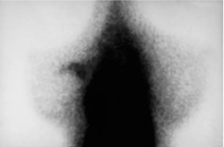
Fig. 2. Lateral scintimammography with 99mTc-MIBI in a
54-year-old patient with a palpable lesion in the left breast. Focal accumu-lation is observed in the upper part of the left breast, correspond-ing to a histopathologically confirmed invasive ductal cancer (di-ameter 2.0 cm). This scan was scored as 2 by the blinded read and
0: Normal 1: Equivocal
2: Focal uptake – low intensity 3: Focal uptake – medium intensity 4: Focal uptake – high intensity
The blinded read was blinded in the sense that the assessors were ignorant of the centre at which the images were taken, and of any other ancillary medical information about the patient. Each asses-sor scored the images independently.
For the primary assessment of diagnostic accuracy, any breast with a maximum segment score of 2 or greater was interpreted as a positive result (definition 1). A secondary assessment was also made, in which a positive result was assumed if the maximum score was 1 or greater (definition 2). For the blinded read, the as-sessment based on the delayed view was made separately from that using the early views, but only the information from the early views was used for making comparisons with other assessment methods.
For the interpretation of the blinded read panel results, a breast was deemed to be positive if at least two of the four assessors scored the breast as such. As only in 3% of the blinded read scintigrams did two readers each score a scan positive and nega-tive, there was no further consensus reading. Data had to be avail-able for at least three of the assessors for this procedure to be ap-plied; otherwise, the diagnosis was regarded as missing.
The blinded read assessors also scored the images for quality; each of the four images (early and delayed, lateral and anterior) was scored separately on a four-point scale: 1, excellent; 2, good; 3, fair; 4, poor. To obtain an overall assessment for each reader, an average value for the four scores was calculated. To obtain an av-erage across readers, the numerical avav-erage of 16 observations (four readers by four views) was calculated.
Histopathology
An excisional biopsy was taken in all evaluated patients. This was diagnosed by the institutional pathologist and the diagnosis was later confirmed (in all but three cases) by the core centre patholo-gist. The measurement of the tumour size was based on the
insti-tutional results. Analysis by tumour size was done for the largest dimension given. Where two lesions were excised from the same breast, the size of the larger lesion was used. The core centre diag-nosis was taken as indicating malignancy (for the primary analy-sis) if either of the following description boxes was ticked: (1) type of invasive cancer, (2) ductal carcinoma in situ (DCIS). For a secondary analysis, lobular carcinoma in situ (LCIS) and ductal and/or lobular hyperplasia as indicators for an increased breast cancer risk were also regarded as true-positive results.
Core centre histopathology diagnosed 109 invasive ductal and 29 invasive lobular cancers, 11 DCIS, two metastases, six tubular carcinoma, two medullary carcinomas and two papillary carcino-mas. Furthermore, one mucinous cancer, one case of Paget’s dis-ease, one malignant cystosarcoma phylloides and one sarcoma have been revealed. Among the benign alterations, there were 37 fibroadenomas, 25 fibrocystic changes, six fat necrosis, five in-flammatory processes, four LCIS, three scleradenosis, three nor-mal breasts, two scars, two adenomas and one hemangioma.
The tumour sizes reported at the main participating centres are shown in Fig. 4. The average maximum tumour dimension ranged from 1.3 cm (pooled centres) over 1.5 cm (France, Germany) and 2.0 cm (Italy) to 2.4 cm (Spain). Forty-one cancers had a maximal diameter of below 1 cm, 37 a maximum diameter between 1.0 cm and 1.5 cm and 87 a maximum diameter of more than 1.5 cm.
Data analysis
All data were collected on case report forms (CRFs) which had been distributed to all participating centres. All CRF data were en-tered into a database and converted to SAS datasets for delivery and into STATA datasets for analysis.
The safety population of the trial consisted of all patients who
received the 99mTc-MIBI imaging agent. To be included in the
ef-ficacy population patients had to meet the following criteria: (1) have had a biopsy for which a core centre microscopic diagnosis exists, (2) have blinded read data for the scintigraphy. The so called by lesion efficacy population consisted of all lesions for which a core centre microscopic diagnosis exists. Some classes of protocol violators were identified which were, however, not con-sidered sufficient to warrant exclusion from the efficacy popula-tion. These were: (1) no mammography (two patients), (2) mam-mography more than 2 months prior to the scintigraphy (six pa-tients who had no discrepancy between mammographic and scinti-graphic results), (3) biopsy more than 6 weeks after the scintigra-phy (19 patients, of whom four showed false-positive scintigrams
Fig. 3. Lateral scintimammography with 99mTc-MIBI in a
61-year-old patient with a palpable lesion in the left breast. Slightly in-creased tracer uptake is observed in the upper part of the left breast. Histopathology revealed an invasive ductal cancer with a maximum diameter of 1.1 cm. This scan was scored as 1 by the blinded read and considered as a true-positive scintigram for
defi-nition 2&/fig.c:
Fig. 4. Tumour size (mean maximum tumour dimension and range
of tumour size) according to the site of the participating centre
and one a false-negative scintigram). Among these 27 patients (12 malignant and 15 benign lesions), there were three false-negative and five false-positive blinded read scintigrams referring to defini-tion 2. The exclusion of these patients did not result in any altera-tion in sensitivity and specificity as shown in the Results secaltera-tion. All efficacy analyses were made separately for each target group of patients: (1) mammographically detected breast abnormalities, (2) breast abnormalities detected by palpation.
The measurement of agreement between any two assessment methods was based on a two-by-two table, in which one of the classifying factors was the gold standard (e.g. core centre histo-pathological results). From the tables, the following statistics were derived: sensitivity, specificity, overall agreement, kappa, positive predictive value (PPV) and negative predictive value (NPV). For the assessment of whether one technique is better than another, four-by-four tables were created indicating false-negative and false-positive, and true-negative and true-positive results of the methods. Several different types of assessment were evaluated: (1) institutional centre sestamibi read (definition 1 and definition 2) compared to core centre histopathology, (2) blinded sestamibi read (definition 1 and definition 2) compared to core centre histo-pathology, 3. comparison between institutional and blinded read scintigraphy, (4) comparison between blinded read scintigraphy and mammography.
Safety assessment
The numbers of patients experiencing any adverse event were tab-ulated. Adverse events had to be classified by type of event and summarized accordingly. Any serious event had to be tabulated separately.
Results
Scintigraphic blinded read
When the results of the blinded read were compared
with the histopathological results of the core centre,
scintigraphy was true-positive in 117 of 165 cancers
(definition 2). This resulted in an overall sensitivity of
71% (Table 3). In this group, sensitivity for palpable and
non-palpable cancers was 83% and 30%, respectively.
For tumours bigger than 1.5 cm, sensitivity was 90%.
For tumours between 1.0 and 1.5 cm and for those
smaller than 1 cm, sensitivity was 65% and 40%,
respec-tively.
If only scintigrams scored 2 or more were taken into
account (definition 1), overall sensitivity was only 61%.
In this group, sensitivity was 72% for the palpable
(n = 127) and 21% for the non-palpable cancers (n = 38;
Tables 3 and 4). For tumours bigger than 1.5 cm,
sensi-tivity was higher, with a value of 80%.
Among 88 benign alterations of the breast, there were
68 palpable and 20 non-palpable lesions (Table 5).
Over-all specificity was 69% (definition 2, 27/88
false-posi-tives scans) and 81% (definition 1, 17/88 false-positive
scans). For palpable lesions, specificity was 75%
(defint-ion 2) and 79% (definit(defint-ion 1), respectively, and for
le-sions bigger than 1.5 cm, specificity was 72% (definition
2) and 78% (definition 1).
When the results of the blinded read were separated
out by main sites, the sensitivities and specificities were
as follows (definition 1, Table 6): 87% and 84% (Spain),
87% and 77% (Italy), 61% and 78% (Germany), 46%
and 100% (pooled centres) and 40% and 71% (France).
If the pooled smaller centres were excluded, the overall
sensitivity improved slightly to 63.5% but the specificity
remained constant at 80% (definition 1).
If the 27 patients with “acceptable” protocol
viola-tions were excluded, the overall sensitivity and
specifici-ty did not change, with values of 70.6% (108/153
true-positives) and 70% (51/73 true-negatives), respectively
(definition 2).
Institutional read
If the histopathological results were correlated to the
in-stitutional read the overall sensitivity and specificity, the
PPV and the NPV were 88%, 66%, 84% and 72%,
re-spectively (for definition 2; Table 7). In this group,
sen-Table 3. Sensitivities (Sens) and specificities (Spec) of blinded
read scintimammography according to the scoring mode&/tbl.c:&tbl.b:
All Palpable Non-palpable
(Sens/Spec) (Sens/Spec) (Sens/Spec)
Definition 1 61%/81% 72%/79% 21%/85%
Definition 2 71%/69% 83%/75% 30%/50%
Definition 1 = all lesions scored as 2 or more considered positive,
definition 2 = all lesions scored as 1 or more considered positive&/tbl.b:
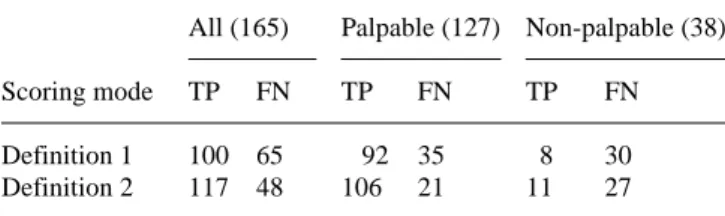
Table 4. Results of blinded read scintimammography in respect of
malignant lesions (numbers in parentheses), according to the
scor-ing mode&/tbl.c:&tbl.b:
All (165) Palpable (127) Non-palpable (38)
Scoring mode TP FN TP FN TP FN
Definition 1 100 65 92 35 8 30
Definition 2 117 48 106 21 11 27
Definition 1 = all lesions scored as 2 or more considered positive; definition 2 = all lesions scored as 1 or more considered positive;
TP, true-positives; FN, false-negatives&/tbl.b:
Table 5. Results of blinded read scintimammography in respect of
benign lesions (numbers within parentheses), according to the
scoring mode&/tbl.c:&tbl.b:
All (88) Palpable (68) Non-palpable (20)
Scoring mode TN FP TN FP TN FP
Definition 1 71 17 54 14 17 3
Definition 2 61 27 51 17 10 10
Definition 1 = all lesions scored as 2 or more considered positive; definition 2 = all lesions scored as 1 or more considered positive;
sitivity for palpable and non-palpable lesions was 91%
and 60%, respectively. For tumours bigger than 1.5 cm,
sensitivity was 95%. For tumours between 1.0 and
1.5 cm and for those smaller than 1 cm, sensitivity was
74% and 55%, respectively.
If definition 1 was used the overall sensitivity and
specificity, the PPV and the NPV were calculated to be
81%, 74%, 86% and 65%, respectively. In this group,
sensitivity and specificity for palpable and non-palpable
lesions of the breast were 86% and 69%, and 63% and
81%, respectively.
The institutional results by main sites were as
fol-lows: sensitivity and specificifity were 93% and 83% for
the Italian centre, 87% and 80% for the Spanish centre,
83% and 56% for the German centre, 73% and 43% for
the French centre and 71% and 100% for the pooled
cen-tres (definition 1, Table 6). If the pooled smaller cencen-tres
were excluded, the overall sensitivity improved slightly
to 83% with a minimal decrease in specificity to 72%
(definition 1).
Mammography
When comparing mammographic results with the core
centre microscopy, mammography was true-positive in
152 of 165 cancers and true-negative in 37 of 88 benign
alterations of the breast. This results in a sensitivity and
specificity of 91% and 42%, respectively. Sensitivity
was not dependent on the size of the breast tumour (for
tumours >1.5 cm, 90.7%, for those between 1.0 and
1.5 cm, 96%, and for those <1.0 cm, 87%; non-palpable
88% and palpable 92%). In the category of probability
of malignancy of 50%–74%, mammography
underesti-mated the real cancer probability for palpable breasts but
overestimated it for non-palpable breasts. If
mammogra-phy and blinded read scintigramammogra-phy were compared, eight
cancers scored true-positive by the blinded read
scintig-raphy had been indicated by mammogscintig-raphy to be
negative (Table 8). This means that in 61% of all
false-negative mammograms scintigraphy could diagnose the
cancer. The aforementioned eight cancers (seven
palpa-ble, one detected by ultrasonography) were studied by
different centres participating in the trial. Except for one
breast, the corresponding mammograms showed dense
breast tissue. In five of the eight malignant tumours, no
suspicious mass could be detected, and
microcalcifica-tions were present only in two breasts (Table 9). Just one
patient was postmenopausal.
Centre Italy France Spain Germany Pooled centre
Blinded read 87%/77% 40%/71% 87%/84% 61%/78% 46%/100%
(Sens/Spec)
Institutional 93%/83% 73%/43% 87%/80% 83%/56% 71%/100%
(Sens/Spec)
Sens, Sensitivity; Spec, Specificity&/tbl.b:
Table 6. Sensitivities and specificities of
blinded read and institutional scintimam-mography separated by participating
cen-tres (referring to definition 1)&/tbl.c:&tbl.b:
Table 7. Sensitivities (Sens) and specificities (Spec) of
institution-al read scintimammography depending on the scoring mode&/tbl.c:&tbl.b:
All Palpable Non-palpable
(Sens/Spec) (Sens/Spec) (Sens/Spec)
Definition 1 81%/74% 86%/69% 63%/81%
Definition 2 88%/66% 91%/65% 60%/93%
Definition 1=all lesions scored as 2 or more considered positive;
definition 2=all lesions scored as 1 or more considered positive&/tbl.b:
Table 8 Comparison of mammographic and blinded read
scinti-graphic results (referring to definition 2)&/tbl.c:&tbl.b:
Mammographic Scintigraphic results
results TP FP FN TN TP 109 – 43 – FP – 15 – 36 FN 8 – 5 – TN – 12 – 25
TP, True-positives; FP, false-positives; FN, false-negatives; TN,
true-negatives&/tbl.b:
Table 9. Results of mammography, scintigraphy and histology in
patients showing a false-negative mammogram and a true-positive
scintigram&/tbl.c:&tbl.b:
Patient Breast MX PM SMM Histology
density
1 HeD MC, no mass 20% Acc,2 Inv. duct, G2
2 ExD MC, no mass 30% Acc, 3 Medullary, G3
3 HeD Mass 20% Acc, 3 Inv. duct, G2
4 HeD Mass 10% Acc, 2 Metastasis
5 ExD No mass 0% Acc, 2 Inv. lobular G1
6 ExD No mass 0% Acc, 2 Inv. duct, G2
7 NvD Mass 20% Acc, 2 Inv. duct, G1
8 HeD No mass 0% Acc, 3 Inv. duct, G1
MX, Mammography; SMM, scintigraphy; PM, probability of ma-lignancy; HeD, heterogeneously dense; ExD, extremely dense; NvD, numerous vague densities; MC, microcalcifications; Acc 2/3, focal accumulation with score 2 or 3; inv. duct, invasive
Density of breasts
Sensitivity of blinded read scintigraphy was not
depen-dent on the density of breast tissue. Of 165 cancers, 118
(72%) were categorized as located in a breast of
mam-mographic grade I or II density (group 1) and 47 (28%)
as located in a grade III/IV density breast (group 2). The
mean tumour diameter in groups 1 and 2 was 1.9 and
1.8 cm, respectively. Overall-sensitivity of the blinded
read scintigraphy (definition 2) for the first and second
groups was 70% (83 true-positives from 118 cancers)
and 72% (34 true-positives from 47 cancers),
respective-ly.
Time of imaging
As delayed images were optional, comparison of the
ear-ly and delayed blinded read scintigrams was confined to
176 breasts. Using definition 1, 30% of the breasts were
found to be positive on the early reads as compared with
26% on the delayed reads. Using definition 2, reads of
the early views yielded positive results in 38% compared
with 31% for the delayed reads. Only in three cases did
the early view give a false-negative result while the
de-layed read was true-positive.
Kappa-statistics
Overall, the agreement between the readers of the
blind-ed scintigraphy read did not show a significant
differ-ence if definition 1 was compared with definition 2.
Overall agreement referring to all breasts was high, at
0.812 and 0.793, respectively. In 97% of the cases, three
or more readers agreed in scoring a scintigram either
positive or negative (definition 1). The reader agreement
was somehow better for palpable lesions than for
mammographically detected lesions. This difference in
kappa values was more significant if definition 1 was
used. With decreasing quality of images, the kappa value
fell slightly but always remained above 0.7. In each
quality class, the same high percentage of reader
agree-ment (three or more readers scoring either positive or
negative) could be found with values of 94%–96%. One
main reason for disagreement between the readers was
the way axillary nodes have been handled. These were
entered as axillary nodes by some readers but as being in
one of the breast segments by others.
When comparing the blinded and the institutional
read, the overall kappa value for the agreement was 0.63.
For palpable lesions, the agreement was better, with a
value of 0.7. The decrease in kappa values with the
qual-ity score was more significant than mentioned before
be-tween the blinded readers. The main reason for the
dif-ferences in diagnoses of blinded and institutional reads
was that the institutional readers assigned higher scores
to lesions than did the blinded readers, especially where
the lesion was indistinct.
Histopathology
With regard to the histopathological characterization of
the cancer, the sensitivity of the blinded read (definition
2) for invasive ductal cancers (80 true-positives from
109), invasive lobular cancers (20 true-positives from
29) and ductal carcinoma in situ (seven true-positives
from 11) was 74%, 69% and 64%, respectively. The
blinded read correctly diagnosed two metastases, two
medullary cancers, one sarcoma and one malignant
cys-tosarcoma phylloides. Among six tubular and two
papil-lary cancers, blinded read scintigraphy was true-positive
in three and one cases, respectively, but could not
diag-nose one mucinous carcinoma and one case of Paget’s
disease (Table 10).
If the histopathological results of the benign
altera-tions are taken into account, specificity for
fibroadeno-mas and fibrocystic disease was 68% and 88% (definition
2), respectively. Among the six cases of fat necrosis, five
inflammations of the breast, two scars and one
haemangi-oma, blinded read scintigraphy was true-negative in four,
two, one and one cases, respectively. In two adenomas
and three cases of normal breast tissue, results were
true-negative in two cases each. All three breasts with
sclerad-enosis showed false-positive scintigrams (Table 11).
Table 10. Results of scintimammography (SMM; definition 2;
blinded read) according to the histological type of breast cancer&/tbl.c:&tbl.b:
Histology No. TP-SMM FN-SMM Inv. ductal 109 80 29 Inv. lobular 29 20 9 DCIS 11 7 4 Tubular 6 3 3 Others 10 7 3 All 165 117 48
Inv., Invasive; DCIS, ductal carcinoma in situ; TP-SMM,
true-pos-itive SMM; FN-SMM, false-negative SMM&/tbl.b:
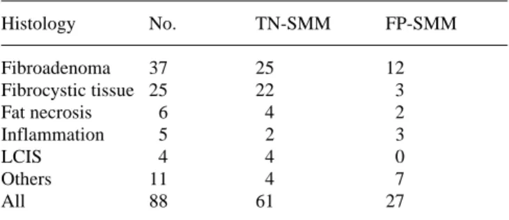
Table 11. Results of scintimammography (SMM; definition 2;
blinded read) according to the histological type in benign lesions&/tbl.c:&tbl.b:
Histology No. TN-SMM FP-SMM Fibroadenoma 37 25 12 Fibrocystic tissue 25 22 3 Fat necrosis 6 4 2 Inflammation 5 2 3 LCIS 4 4 0 Others 11 4 7 All 88 61 27
LCIS, Lobular carcinoma in situ; TN-SMM, true-negative SMM;
Discussion
Recently, nuclear breast imaging has gained significant
interest as several radionuclides have demonstrated
promising results in the diagnosis of breast cancer.
99m
Tc-MIBI is a cationic complex which can accumulate
in tumour cells [41]. The accumulation of
99mTc-MIBI in
the tumour is dependent on the quantity of mitochondria
in the tumour cell, on the electric membrane potential
and on the expression of the multidrug resistance (MDR)
gene [42–46].
Recent studies using
99mTc-MIBI for breast cancer
detection have shown high sensitivity and specificity for
palpable cancers [22–32]. For non-palpable breast
le-sions, diagnostic accuracy has been less favorable [25,
27, 29, 30]. In this trial, scintigraphy with
99mTc-MIBI
read blinded achieved an overall sensitivity of 71%. For
palpable tumours and tumours bigger than 1.5 cm,
sensi-tivity was 83% and 90%, respectively. Specificity in
these two groups was clearly over 70%. These values are
slightly below those reported by previous studies
[23–30]. Generally, it can be expected that it will be
more difficult to obtain similar results if the method is
assessed by a blinded multicentre study. Consequently,
in this trial, the institutional read demonstrates an
in-crease in the overall sensitivity in all centres up to a
val-ue of 88% when compared with the blinded read. It must
be supposed that the clinical data such as size of lesion,
location and probability of malignancy are important
factors for the reader of scintigraphy and their
knowl-edge will increase sensitivity. In the case of palpable
le-sions and lele-sions bigger than 1.5 cm, sensitivity
in-creased to 91% and 96%, respectively, by the
institution-al read. These results are in agreement with a
multicen-tre trial conducted in the United States and Canada
which revealed an institutional sensitivity of 95% for
palpable lesions [47].
In this study, it has been shown that sensitivity can be
significantly increased if lesions which are scored as
equivocal are considered as a malignant process. Even if
this definition was used, specificity did not fall below a
level of 70%. This means that any abnormality must be
considered as suspicious for malignancy. In this way, a
high sensitivity of scintimammography in association
with an acceptable specificity can be achieved (Fig. 5).
As demonstrated by the ROC curves, there will be a
learning process for the interpretation of scintigrams
re-sulting in an increase in sensitivity.
There are still significant limitations to the use of
mammography for the detection of breast cancer, and
these limitations persist in spite of technical
improve-ments facilitating dianosis. Especially in younger
wom-en (less than 50 years old) with dwom-ense breasts, the
diag-nostic benefit of mammography is less favourable [7, 8].
Thus, there is a need for a non-invasive method to
com-plement mammography and to help differentiate benign
and malignant breast lesions in dense breasts. Such a
method should be reliable and have a high sensitivity
and a high predictive value. In this trial, mammographic
results were within the range of the values reported in
the literature [48]. Overall sensitivity and specificity
were 91% and 40%, respectively. Sensitivity of
mam-mography was not dependent on the tumour size, and,
therefore, no decrease in sensitivity in patients with
non-palpable cancers was observed. In this trial, overall
sen-sitivity of mammography was superior to that of
scintig-raphy, demonstrating that scintimammogscintig-raphy, in its
current state, is not suitable for breast cancer screening.
For the mammograms, only an institutional read has
been performed. This means that the mammographic
re-sults of this trial correspond to those of the clinical
ev-eryday practice of the radiologist.
However, this trial has shown that scintigraphy can
provide additional information to mammography and
help to detect breast cancer earlier in a subgroup of
pa-tients. In 60% of patients with a false-negative
mammo-gram, scintigraphy could diagnose the breast cancer.
This group consisted of younger patients with
mammo-graphically dense breast tissue resulting in a
false-nega-tive mammogram. As this study was able to prove, the
diagnostic accuracy of scintimammography is not
de-pendent on the density of breast tissue. This has also
been reported by Khalkhali et al. [49, 50]. Consequently,
premenopausal patients whose mortality from breast
cancer cannot be decreased significantly by screening
mammography will benefit most from
scintimammogra-phy.
In the majority of the cases, the woman herself
de-tects an alteration of the breast for which she consults
the physician [51]. Sensitivity of breast palpation is not
satisfactory, and often lumpy breasts make it difficult to
characterize sufficiently a palpable nodule or mass in the
breast [51]. If mammography performed in the further
diagnostic work-up is indeterminate and suspicion of
malignancy not high, the patient will be advised to
re-turn for a control mammography in 3–6 months. In this
Fig. 5. ROC curves of blinded and institutional (institut) read
scintigraphy for all and for palpable (palp) lesions. The first value of each curve refers to definition 1 and the second value of the
patient group, the majority of which comprises
premeno-pausal women, scintigraphy could help to diagnose
breast cancer at an earlier point in time when it is used
as a complementary method to mammography (Fig. 6).
It is important for a breast cancer imaging modality
that sensitivity is not dependent on the histological type
of the cancer. For invasive ductal and lobular
carcino-mas, the sensitivity of scintimammography did not show
a significant difference. This group of malignant
tu-mours represent 80%–85% of all breast cancers [52]. For
DCIS, sensitivity was slightly lower than for invasive
cancers. Furthermore, less frequent cancers such as
med-ullary and tubular carcinomas and metastases could also
be diagnosed by scintimammography. This makes
scin-tigraphy suitable as a complementary imaging method.
False-positive results were obtained in patients with
fibroadenoma, fibrocystic disease and local
inflamma-tion of the breast. Disease with a high inflammatory
component might yield false-positive results, most likely
due to increased local perfusion. Areas with increased
mitochondrial activity and density, such as juvenile
ade-nomas and hyperproliferative disease, can also cause
false-positive MIBI uptake [22, 25, 27, 29]. However,
patients with atypical hyperproliferative disease have a
higher relative risk for breast cancer [52]. In these
pa-tients, a positive MIBI scan may be of prognostic value
[53].
The exact localization of a MIBI-positive area
re-mains a problem since neither planar imaging nor
single-photon emission tomography provides the surgeon with
sufficient information for biopsy of breast tissue. When
scintimammography is indicative for breast cancer but
other breast imaging modalities such as mammography,
ultrasonography and magnetic resonance imaging are
negative, the scintigram must be used for tumour
local-ization. New approaches have been developed to allow
scintigraphy-guided biopsy of breast lesions [54, 55].
It is known that mammogaphy is a very
reader-depen-dent method. With regard to ultrasonography, this
prob-lem is even more important. The interreader agreement
for scintimammography, however, is very high, with
kappa values of 0.812. It is important for a diagnostic
method that the interreader variation is low, especially
when it is performed in a wide medical field of clinical
everyday practice.
Khalkhali et al. have shown that the prone position
with the breasts hanging freely is the best technique for
the performance of lateral scintimammography because
deeper regions of the breast can be visualized [23]. For
this purpose different techniques may be used: a special
table design with a lateral cut-off or a kind of foam
cushion with lateral apertures as used in this trial.
Re-gardless of which technique is used, the breast must not
be compressed from either side. Furthermore, a
high-res-olution gamma camera should be used and the
acquisi-tion time must be at least 10 min if good quality images
are to be obtained. Special attention must be drawn to
the distance between the collimator and the breast,
which, ideally, should touch the camera surface.
Stan-dardization of scintimammography will help to achieve a
high quality level of this technique.
Use of fluorine-18 fluorodeoxyglucose (
18F-FDG)
and positron emission tomography (PET) has also been
evaluated for the detection of breast cancer [14–21]. In a
larger patient group, Avril et al. reported a sensitivity
and specificity for the detection of primary breast cancer
of 88% and 78%, respectively [18]. For small tumours,
only low sensitivity was achieved. In a group of 20
pa-tients, Palmedo et al. compared FDG PET and
99mTc-MIBI scintimammography [19]; however, in 40 breasts
with 22 lesions, FDG PET could not detect additional
cancer in comparison with Tc-99m MIBI scintigraphy.
Comparing these two imaging modalities, PET has the
advantage of providing better spatial resolution, but the
availability of FDG and PET is limited and costs are
sig-nificantly higher.
Conclusions
Scintimammography with
99mTc-MIBI has a high
diag-nostic accuracy in palpable breast lesions. Sensitivity is
Fig. 6. Flow chart indicating the role
of scintimammography in the diagnos-tic work-up of patients with palpable
not dependent on the mammographically determined
density of the breast tissue. Scintimammography is
suit-able as a complementary method to mammography in
patients with dense breasts and an intermediate or low
probability of breast cancer. Further, patients with a high
risk of breast cancer may benefit from radionuclide
im-aging with
99mTc-MIBI.
&p.2:
Acknowledgements. The authors thank Ms. M. Grace for
coordi-nating the collaboration during the trial and for setting up the da-tabase of the study.
References
1. Kelsey JL, Gammon MD. The epidemiology of breast cancer.
Cancer 1991;41:146–165.
2. Berg JW, Hutter RV. Breast cancer. Cancer 1995;75:257–269. 3. Sondik EJ. Breast cancer trends. Incidence, mortality and
sur-vival. Cancer 1994;74:995–999.
4. Andersson I. Mammographic screening and mortality from breast cancer: Malmö mammographic screening trial. Brit J
Med 1988;297:943–948.
5. Frisell J, Eklund G, Hellström L, et al. Randomized study of mammography screening – preliminary report on mortality in the Stockholm trial. Breast cancer research and treatment 1991;18:49–56.
6. Miller AB, Baines CJ, To T, et al. Canada national breast screening study. Can Med Assoc J 1992;147:1459–1476. 7. Lannin DR, Harris RP, Swanson FH, Edwards MS, Swanson
MS, Pories WJ. Difficulties in diagnosis of carcinoma of the breast in patients less than fifty years of age. Surg Gynecol
Obstet 1993;177:457–476.
8. Coveney EC, Geraghty JG, O’Laoide R, Hourihane JB, O’Higgins NJ. Reasons underlying negative mammography in patients with palpable breast cancer. Clin Radiol 1994;49: 123–125.
9. Smart CR, Myers MH, Gloeckler LA Implications from SEER data on breast cancer management. Cancer 1978;41:787–789. 10. Carter CL, Allen C, Henson DE. Relation of tumour size,
lymph node status and survival in 2474 breast cancer patients.
Cancer 1989;63:181–187.
11. Koscielny S, Tubiana M, Le MG, et al. Breast cancer. Rela-tionship between the size of the primary tumour and the prob-ability of metastatic dissemination. Br J Cancer 1984;49: 709–715.
12. Fisher B, Redmond C, Fisher ER, et al. Ten-year result of ran-domized clinical trial comparing radical mastectomy and total mastectomy with or without radiation. N Engl J Med 1985; 312:674–681.
13. De Jager R, Abdel N, Serafini A,et al. Current status of cancer immunodetection with radiolabeled human monoclonal anti-bodies. Semin Nucl Med 1993;23:165–179.
14. Wahl RL, Cody RL, Hutchins GD, Mudgett EE. Primary and metastatic breast carcinoma: initial clinical evaluation with PET with the radiolabeled glucose analogue F-18
fluoro-2-de-oxy-D-glucose. Radiology 1991;179:765–770.
15. Tse NY, Hoh CK, Hawkins RH, Zinner MJ, Dahlbom M, Choi Y, Maddahi J, Brunicardi FC, Phelps ME, Glaspy JA. The ap-plication of positron emission tomographic imaging with FDG to the evaluation of breast disease. Ann Surg 1992;216:27–34. 16. Nieweg OE, Kim EE, Wong WH, Broussard WF, Singletary
SE, Hortobagyi GN, Tilbury RS. Positron emission
tomogra-phy with FDG in the detection and staging of breast cancer.
Cancer 1993;71:3920–3925.
17. Adler LP, Crowe JP, Al Kaisi NK, Sunshine JL. Evaluation of breast masses and axillary lymph nodes with F-18
deoxy-2-fluoro-D-glucose PET. Radiology 1993;187:743–750.
18. Avril N, Dose J, Jänicke F, et al. Metabolic characterization of breasts tumours with PET using F-18 fluorodeoxyglucose. J
Clin Oncol 1996;14:1848–1856.
19. Palmedo H, Bender H, Grünwald F et al. Comparison of fluo-rine-18 fluorodeoxyglucose positron emission tomography and Tc-99m methoxyisobutylisonitrile scintimammography in the detection of breast tumours. Eur J Nucl Med 1997;24: 1138–1145.
20. Scheidhauer K, Scharl A, Pietrzyk U, et al. Qualitative F-18 FDG positron emission tomography in primary breast cancer: clinical relevance and practicability. Eur J Nucl Med 1996;23: 618–623.
21. Crowe JP, Adler LP, Shenk RR, Sunshine J. Positron emission tomography and breast masses: comparison with clinical, mammographic, and pathological findings. Ann Surg Oncol 1994;1:132–140.
22. Kao CH, Wang SJ, Liu TJ. The use of technetium-99m me-thoxyisobutylisonitrile breast scintigraphy to evaluate palpable breast masses. Eur J Nucl Med 1994;21:432–436.
23. Khalkhali I, Mena I, Jouanne E, et al. Prone scintimammogra-phy in patients with suspicion of breast cancer. J Am Coll Surg 1994;178:491–497.
24. Khalkhali I, Cutrone JA, Mena I. Scintimammography: the complementary role of Tc-99m sestamibi prone breast imag-ing for the diagnosis of breast carcinoma. Radiology 1995; 196:421–426.
25. Khalkhali I, Cutrone JA, Mena I, Diggles L, Venegas R, Var-gas H, Jackson B, Klein S. Technetium-99m-sestamibi scinti-mammography of breast lesions: clinical and pathological fol-low-up. J Nucl Med 1995;36:1784–1789.
26. Palmedo H, Grünwald F, Bender H, Schomburg A, Mallmann P, Krebs D, Biersack HJ. Scintimammography with Tc-99m MIBI: comparison with mammography and magnetic reso-nance imaging. Eur J Nucl Med 1996;23:940–946.
27. Palmedo H., Schomburg A, Gruenwald F, Mallmann P., Krebs D, Biersack HJ. Scintimammography with Tc-99m MIBI in suspicious breast lesions. J Nucl Med 1996;37:626–630. 28. Palmedo H, Schomburg A, Mallmann P, et al. (1996)
Scinti-mammography with Tc-99m MIBI in patients with suspicion of primary breast cancer. Nucl Med Biol 1996;23:681–684. 29. Taillefer R, Robidoux A, Lambert R, Turpin S, Laperrière J.
99mTc-MIBI prone scintimammography to detect primary
breast cancer and axillary lymph node involvement. J Nucl
Med 1995;36:1758–1765.
30. Villanueva-Meyer J, Leonhard MH, Briscoe E, et al. Mammo-scintigraphy with Tc-99m sestamibi in suspected breast can-cer. J Nucl Med 1996;37:926–930.
31. Tiling R, Kress K, Pechmann M, Pfluger T, Knesewitsch P, Tatsch K, Hahn K. integrated diagnosis of breast tumours:
semiquantitative 99mTc-MIBI imaging versus dynamic MRI
[abstract]. J Nucl Med 1995;35:51.
32. Burak Z, Argon M, Memis A, et al. Evaluation of palpable breast masses with Tc-99m MIBI: a comparative study with mammography and ultrasonography. Nucl Med Commun 1994;15:604–612.
33. Lee V, Sax EJ, McAneny DB, Pollack S, Blanchard RA, Be-azley RM, Kavanah MT, Ward RJ. A complementary role for thallium-201 scintigraphy with mammography in the diagno-sis of breast cancer. J Nucl Med 1993;34:2095–2100.
34. Waxman AD, Ramanna L, Memsic LD, Foster CE, Silberman AW, Gleischmann SH, Brenner RJ, Brachman MB, Kuhar CJ, Yadegar J. Thallium scintigraphy in the evaluation of mass ab-normalities of the breast. J Nucl Med 1993;34:18–23.
35. Cimitan M, Volpe R, Candiani E, et al. The use of Tl-201 in the preoperative detection of breast cancer: an adjunct to mammography and ultrasonography. Eur J Nucl Med 1995;22:1110–1117.
36. Mansi L, Rambaldi PF, Procaccini E, et al. Scintimammogra-phy with Tc-99m tetrofosmin in the diagnosis of breast cancer and lymph node metastases. Eur J Nucl Med 1996;23: 932–939.
37. Piccolo S, Lastoria S, Mainolfi C, Muto P, Bazzicalupo L, Sal-vatore M. Tc-99m MDP scintimammography to image prima-ry breast cancer. J Nucl Med 1995;36:718–724.
38. Ell PJ. Keeping abreast of time. Eur J Nucl Med 1995;22: 967–969.
39. Sluyser M, Hoefnagel CA. Breast carcinomas detected by thallium-201 scintigraphy. Cancer Lett 1988;40:161–168. 40. Rijks LJ, Bakker PJ, van Tienhoven G, et al. Imaging of
estro-gen receptors in primary and metastatic breast cancer patients with I-123 Z-MIVE. J Clin Oncol 1997;15:2536–2545. 41. Maublant JC, Zhang Z, Rapp M, Ollier M, Michelot J, Veyre
A. In vitro uptake of technetium-99m teboroxime in carcino-ma cell lines and norcarcino-mal cells: comparison with technetium-99m sestamibi and thallium-201. J Nucl Med 1993;34: 1949–1952.
42. Chiu ML, Kronauge JF, Piwnica-Worms D. Effect of mito-chondrial and plasma membrane potentials on accumulation of hexakis 2-methoxyisobutylisonitrile technetium in cultured mouse fibroblasts. J Nucl Med 1990;31:1646–1653.
43. Piwnica-Worms D, Chiu ML, Budding M, Kronauge JF, Kra-mer RA, Croop JM. Functional imaging of multidrug-resistant P-glycoprotein with an organotechnetium complex. Cancer
Res 1993;53:977–984.
44. Bender H, Friedrich E, Zamora PO, Guhlke S, Biersack HJ. Effect of Induction of multi-drug resistance on accumulation
of Tc-99m sestamibi in vitro. Anticancer Res. 1997;17:1833– 1840.
45. Ciarmiello A, Del Vecchio S, Potena MI, et al. Technetium-99m-sestamibi efflux and P-glycoprotein expression in human breast carcinoma [abstract]. J Nucl Med 1995;36(Suppl.): 129P.
46. Maublant J, Songalede JA, Finat-Duclos F, Verrelle P, Veyre
A, Younès A. Accumulation of 99mTc-MIBI in cultured
tu-mour cells decreases when multidrug resistance factor is over-expressed [abstract]. J Nucl Med 1994;35(Suppl.):219P. 47. Khalkhali I, Villanueva-meyer J, Edell SL, et al. Diagnostic
accuracy of 99mTc-MIBI breast imaging in breast cancer
de-tection [abstract]. J Nucl Med 1996;7:74P.
48. Kopans DB. The positive predictive value of mammography.
AJR 1992;158:521–526.
49. Khalkhali I, Cutrone JA, Mena I, Diggles L, Khalkhali S, Venegas R, Klein S. The usefulness of scintimammography (SMM) in patients with dense breasts on mammogram [ab-stract]. J Nucl Med 1995;36:52.
50. Khalkhali I, Villanueva-Meyer J, Edell SL, et al. Impact of
breast density on the diagnostic accuracy of 99mTc-MIBI
breast imaging in the detection of breast cancer [abstract]. J
Nucl Med 1996;37:74P.
51. Donnegan WL. Evaluation of a palpable breast mass. N Engl J
Med 1992;327:937–942.
52. Simpson JF, Page DL. Prognostic value of histopathology in the breast. Semin Oncol 1992;19:254–262.
53. Waxman A. The role of Tc-99m MIBI in imaging breast can-cers. Semin Nucl Med 1997;17:40–54.
54. Wagner RH, Karesh SM, Dillehay GL, Henkin RE. Accuracy of needle localization of scintigraphic abnormalities using a radio-directed biopsy system (RDBS) [abstract]. J Nucl Med 1997;38:22P.
55. Khalkhali I, Mishkin FS, Diggles LE, Klein SR. Radionu-clide-guided stereotactic prebiopsy localization of nonpalpa-ble breast lesions with normal mammograms. J Nucl Med 1997;38:1019–1022.