DESIGN,SYNTHESIS AND EVALUATION OF NEW PACE4INHIBITORS FOR
PROSTATE CANCER TREATMENT
By
Vahid Dianati
Thesis presented in the chemistry department for obtaining the degree of Doctor of Science (Ph.D.)
FACULTY OF SCIENCE UNIVERSITY OF SHERBROOKE
CONCEPTION,SYNTHESEETEVALUATIONDENOUVEAUXINHIBITEURSDEPACE4 POURLETRAITEMENTDUCANCERDELAPROSTATE
par
Vahid Dianati
Thèse présentée au Département de chimie en vue de l’obtention du grade de docteur ès sciences (Ph.D.)
FACULTÉ DES SCIENCES UNIVERSITÉ DE SHERBROOKE
Le 24 janvier 2019
le jury a accepté la thèse de Monsieur Vahid Dianati dans sa version finale.
Jury Members
Professor Yves L. Dory Research Director Department of chemistry
Professor Robert Day Research Codirector Department of Surgery/Urology
Professor William Lubell External evaluator University of Montreal
Professor Jean Lessard Internal Evaluator Department of chemistry
Professor Claude Legault Internal Evaluator Department of chemistry
Professor Patrick Ayotte Committee President Department of Chemistry
i SOMMAIRE
Le présent mémoire s’intéresse à la conception, la synthèse et l'évaluation pharmacologique d'inhibiteurs peptidomimétiques de PACE4, une pro-protéine convertase. Ces composés sont étudiés comme traitement potentiel du cancer de la prostate. Le cancer de la prostate (PCa) et les traitements actuels pour le PCa sont donc passés en revue. L'introduction se poursuit par une brève évaluation des thérapies ciblées vers le PCa qui démontre que les pro-protéines convertases sont des enzymes importantes de la voie de transmission. Les inhibiteurs actuels des pro-protéines convertases ont finalement été examinés. Des études de relation structure-activité (SAR) ont été réalisées sur un octapeptide (appelé Multi-Leu; séquence: Ac-Leu-Leu-Leu-Leu-Arg-Val-Lys-Arg-NH2) qui s’est révélé être un prototype d’inhibition
de l’enzyme PACE4. Ce composé a ensuite évolué par le remplacement du résidu Arg en position C-terminale par un résidu Amba (4-amidinobénzylamide) d’une part et par l’introduction d’une DLeu au lieu d’une l-Leu en position P8 d’autre part. Le nouvel inhibiteur appelé C23 (séquence: Ac-DLeu-Leu-Leu-Leu-Arg-Val-Lys-Amba) présentait une activité améliorée vis-à-vis de l’enzyme PACE4 et de bons résultats in vitro dans des essais cellulaires antiprolifératifs. Le but du travail présenté dans ce document est l’amélioration de l’affinité enzymatique pour l’enzyme PACE4, l’accroissement de la sélectivité par rapport à d'autres membres de la famille, en particulier la furine, et l’augmentation de l'activité cellulaire des composés prototypes en modifiant les résidus aux positions P3, P1 et P1'.
Le premier chapitre concerne les études SAR sur les résidus de P3. L'amélioration de la sélectivité et de la puissance de l'inhibiteur est l'objectif de ce chapitre. L’acide aminé Val en position P3 du peptide Multi-Leu a été remplacé par des résidus basiques pour créer des interactions supplémentaires avec un résidu Asp dans la poche S3. Un résidu basique β-ramifié dans cette position améliore la sélectivité pour PACE4 jusqu'à 40 fois (Ki = 2.7 nM).
Le deuxième chapitre traite des modifications de la position P1 du peptide prototype C23. Malgré que l’inhibiteur C23 ait une affinité, une stabilité et une activité cellulaire supérieures pour l’enzyme PACE4 par rapport à son précurseur Multi-Leu, il n’est que deux fois plus sélectif pour la convertase PACE4. La plus grande partie de cette perte de sélectivité est due aux interactions plus favorables du résidu Amba en position P1 de la furine. Par conséquent, d'autres mimétiques de l’acide aminé Arg ont été testés dans cette position pour retrouver la sélectivité perdue.
ii
Dans le troisième chapitre, l’acide aminé en position P1' a été exploré pour discriminer les enzymes PACE4 et furine. L’addition de résidus naturels dans cette position, cependant, n’a amélioré ni la sélectivité ni l’affinité. Par ailleurs, l'évaluation cellulaire a montré que les résidus hydrophobes et basiques amélioraient l'activité cellulaire. En utilisant des acides aminés non naturels aux positions P1 et P1', l'activité antiproliférative cellulaire des inhibiteurs résultants est sensiblement améliorée. Il a été prouvé que cette meilleure activité cellulaire était due à la pénétration accrue des inhibiteurs résultant d’une hydrophobicité élevée dans la région de l'extrémité C-terminale.
En conclusion, l'affinité et la sélectivité envers la pro-protéine convertase PACE4 des inhibiteurs ont été améliorées par des modifications aux positions P3 et P1. Les résidus hydrophobes en position P1' augmentent la perméabilité cellulaire et donc l'activité antiproliférative des cellules PCa. Dans cette perspective, la combinaison de ces modifications pourrait conduire à une amélioration du profil pharmacodynamique des composés.
iii ABSTRACT
This thesis deals with the design, synthesis and pharmacological evaluation of peptidomimetic inhibitors of PACE4, a pro-protein convertase. These compounds are studied as a potential treatment for prostate cancer. Prostate cancer (PCa) and current treatments for PCa are therefore reviewed. The introduction continues with a brief evaluation of targeted therapies for PCa that demonstrates that pro-protein convertases are important enzymes in the secretory pathway. Current inhibitors of pro-protein convertases are reviewed at the final part of introduction.
Structure-activity relationship (SAR) studies were performed on an octapeptide (called Multi-Leu; sequence: Ac-Leu-Leu-Leu-Leu-Arg-Val-Lys-Arg-NH2) which was found to be a prototype of inhibition
of the enzyme PACE4. This compound then evolved by replacing the Arg residue in the C-terminal position with an Amba (4-amidinobenzylamide) residue on the one hand and by introducing a DLeu instead of a l-Leu in position P8 on the other hand. The new inhibitor called C23 (sequence: Ac-DLeu-Leu-Leu-Leu-Arg-Val-Lys-Amba) exhibited improved activity with respect to the PACE4 enzyme and good in vitro results in antiproliferative cell assays. The purpose of the work presented in this document is to improve the enzymatic affinity for the enzyme PACE4, increase selectivity over other members of the family, particularly furin, and increase the cellular activity of the prototype compounds by modifying the residues at positions P3, P1 and P1'.
The first chapter concerns SAR studies on P3 residues. Improving the selectivity and potency of the inhibitor is the goal of this chapter. The amino acid Val at the P3 position of the Multi-Leu peptide has been replaced by basic residues to create additional interactions with an Asp residue in the S3 pocket. A basic β-branched residue in this position improves the selectivity for PACE4 up to 40-fold (Ki = 2.7 nM). The second chapter deals with the modifications of the position P1 of the prototype peptide C23. Although the C23 inhibitor has higher affinity, stability, and cell activity for the PACE4 enzyme than its Multi-Leu precursor, it is only twice as selective for the PACE4 convertase. Most of this loss of selectivity is due to the more favorable interactions of the Amba residue at the P1 position of furin. Therefore, other Arg amino acid mimetics have been tested in this position to recover the lost selectivity.
In the third chapter, the amino acid at position P1' was explored to discriminate between PACE4 and furin enzymes. The addition of natural residues in this position, however, improved neither selectivity
iv
nor affinity. In addition, cell evaluation showed that hydrophobic and basic residues improved cellular activity. By using unnatural amino acids at the P1 and P1' positions, the cell antiproliferative activity of the resulting inhibitors is substantially improved. This improved cell activity has been shown to be due to increased penetration of inhibitors resulting from high hydrophobicity in the C-terminal region. In conclusion, the PACE4 affinity and selectivity of inhibitors were improved by modifications in P3 and P1 positions. Hydrophobic residues in P1' position enhanced the cell permeability and thus the PCa cell antiproliferative activity. In this perspective, combining such modifications could lead to compounds with improved pharmacodynamic profiles.
v
ACKNOWLEDGEMENTS
I want to appreciate all the members of Yves Dory’s lab since 2014 for providing such a motivating and pleasant environment which made my PhD years enjoyable and unforgettable. The main part of this unbelievable social experience was related to professor Yves Dory who supervised the present dissertation. He was not only an incredible organic/medicinal chemist but also a great mentor and a reliable friend. He was always present, and his precise advice was critical in the development of the project. I was very fortunate to have professor Robert Day as my co-supervisor as one of the greatest scientists in the field of proprotein convertases. The current study have reached this level because of his wise thoughts and comments. I want to acknowledge the writing skills of both my supervisors who revised the manuscripts of the three published research articles I wrote. My sincere gratitude goes to my supervisors for giving me the confidence and freedom in research which enabled me to show my best performance.
I want to express my gratitude to the external evaluator of this thesis Pr. William Lubell, the members of my committee Pr. Jean Lessard, Pr. Claude Legault and Pr. Patrick Ayotte for reviewing my thesis. I would like to acknowledge the contribution of the hardworking Day lab’s members in the biological evaluation of synthesized compounds. Anna Kwiatkowska, Frederic Couture and Roxane Desjardins had a major role in this regard. I had impactful scientific conversations with Anna and Fred which improved my understanding of the biology of proprotein convertases. Anna’s experience and skills in peptide chemistry and HPLC systems is astonishing. She also helped me to develop my scientific writing skills. I also thank Sandra Gagnon, Anthony Dame and Nicolas Dory for their participation in performing the biological assays.
I share lots of memories inside and outside of lab with Thi Than Há Dao, Sophie Beauchemin, Thomas Marmin, Jean-Louis Beaudeau, Hojjat Seyedjamali, Yanan Zhu, Dominique Bella NDong, Pauline Navals, Alexander Foh-Dion, Laura Mourot, Niousha Nazari, Trần Minh-Huệ and Victoria Lépante. I had fruitful scientific discussions with Há, Hojjat, Sophie and Jean-Louis. I thank Niousha for English editing of the introduction and of the two last chapters. I am grateful to Pauline for the synthesis of one of the P1-modified compounds and its Arg-mimetic, and Sophie for her assistance in the synthesis of P1'
vi
peptide library. My great appreciation goes to the other members of Dory’s lab who made the work atmosphere more joyful and inviting.
I would like to thank the IPS technical staff, Marc-André Bonin for his assistance in automated peptide synthesis, Eric Marsault and his lab especially Annie Doucet, Antoine Le-Roux and Alexandre Murza for UPLC and preparative HPLC facilities and Luc Tremblay, Pierre-Luc Boudreault and Danny Létourneau for NMR assistance. I acknowledge Hugo Gagnon and Jean-Philippe Couture (PhenoSwitch Biosciences Inc.) for HRMS analysis. I also appreciate the Department of chemistry professors and staff especially Jean-Marc Chapuzet, Dr. Isabelle Dion, Lise Charbonneau, René Gagnon, Dr. Nicole Wilb and Philip Richter. This research was not possible without the financial support of Canadian Cancer Society Research Institute (701590 to R.D. and Y.L.D.) and Prostate Cancer Canada (TAG2014-02 to R.D.) and University of Sherbrooke (Faculty of science).
My deep appreciation goes to my family especially my parents whose financial and emotional support pave the way towards where I am today. I want to appreciate my wife Azar who helped me to have the opportunity of pursuing my PhD studies at University of Sherbrooke. She was not only my wife but also a friend and a good colleague. She performed the molecular dynamic simulation in chapter one under the supervision of Pr. Armand Soldera. I want to dedicate this thesis to my daughter Baran who shed a new light in our life.
vii
TABLE OF CONTENTS
Sommaire ... i
Abstract ... iii
Acknowledgements ... v
Table of contents ... vii
List of abbreviations ... xi
List of Tables ... xiv
List of Figures ... xv
List of Schemes ... xviii
Introduction ... 1
I.1. Prostate cancer ... 2
I.1.1. Prostate anatomy ... 2
I.1.2. Statistics ... 3
I.1.3. Prostate cancer at cellular level ... 3
I.1.4. Risk factors ... 4
I.1.5. Symptoms and diagnosis... 4
I.1.6. Other common prostatic conditions ... 5
I.2. Current prostate cancer treatments ... 6
I.2.1. Hormone therapy ... 7
I.2.2. Chemotherapy ... 11
I.2.3. Targeted therapy ... 13
I.3. Proprotein convertases ... 15
I.3.1. Secretory pathway ... 15
I.3.2. The discovery of proprotein convertases ... 16
I.3.3. Structure of proprotein convertases ... 17
I.3.4. Cellular and tissue distributions of PCs ... 19
viii
I.3.6. Validation of PACE4 as a target in prostate cancer ... 21
I.4. Inhibition of proprotein convertases ... 23
I.4.1. Proprotein convertases are serine proteases ... 23
I.4.2. Peptide inhibitors of proprotein convertases ... 24
I.5. Thesis objectives ... 37
Chapter 1 : Rational Design of a Highly Potent and Selective Peptide Inhibitor of PACE4 by Salt Bridge Interaction with D160 at Position P3 ... 39
1.1. Author contributions ... 39
1.2. Abstract ... 40
1.3. Introduction ... 40
1.4. Results and discussions ... 42
1.5. Conclusion ... 46 1.6. Acknowledgements ... 47 1.7. Supporting information ... 47 1.7.1. Chemistry ... 47 1.7.2. Biology ... 53 1.7.3. MD Simulation ... 54
Chapter 2 : Improving the Selectivity of PACE4 Inhibitors through Modifications of the P1 Residue .. 55
2.1. Author contributions ... 55
2.2. Abstract ... 56
2.3. Introduction ... 56
2.4. Results and discussions ... 59
2.4.1. Design and binding affinities ... 59
2.4.2. Cell-based assays ... 63
2.4.3. Chemistry ... 64
ix 2.6. Experimental ... 68 2.6.1. Chemistry ... 68 2.6.2. Molecular modeling ... 80 2.6.3. Biology ... 80 2.7. Acknowledgements ... 81 2.8. Supporting information ... 82
Chapter 3 : Increasing C-Terminal Hydrophobicity Improves the Cell Permeability and Antiproliferative Activity of PACE4 Inhibitors against Prostate Cancer Cell Lines ... 83
3.1. Author contributions ... 83
3.2. Abstract ... 84
3.3. Introduction ... 84
3.4. Results and discussion ... 86
3.4.1. Screening of DNA-encoded residues in P1' ... 86
3.4.2. SAR studies based on the screening. ... 88
3.4.3. Cell permeability studies. ... 91
3.4.4. DU145 cell toxicity studies ... 92
3.4.5. Plasma stability studies ... 93
3.4.6. Acute toxicity studies ... 93
3.4.7. Synthesis ... 94 3.5. Conclusion ... 96 3.6. Experimental ... 97 3.6.1. Chemistry ... 97 3.6.2. Enzyme kinetics ... 101 3.6.3. Cell proliferation ... 102 3.6.4. Cell permeability ... 102 3.6.5. Cell toxicity ... 103 3.6.6. Plasma stability ... 103
x
3.7. Acknowledgments ... 103
3.8. Supporting information ... 104
3.8.1. The synthesis of compounds 3-21, 28 and 29 ... 104
3.8.2. Details of enzyme kinetic assays ... 104
3.8.3. Cell toxicity experiments ... 104
3.8.4. Analytical data of peptide inhibitors and their FITC-labeled analogues. ... 105
Chapter 4: General discussion and perspective ... 106
4.1. General discussions ... 106 4.2. Future studies ... 108 Conclusion ... 111 Appendix I ... 113 Appendix II ... 114 Appendix III ... 124 Appendix IV ... 168
xi
LIST OF ABBREVIATIONS
6-Cl-HOBt 6-Chloro-1-hydroxybenzotriazole α1-PDX α1-Antitrypsin Portland
Aca (S)-2-Amino-3-(4-carbamimidoylphenyl)acetic acid Acpa (S)-2-Amino-3-(4-carbamimidoylphenyl)propanoic acid ADAM A disintegrin and metalloproteinase
ADT Androgen deprivation therapy AEBA 4-(2-Aminoethyl)benzimidamide
Agb 2-Amino-4- guanidinobutyryl
Agp 2-Amino-3-guanidinopropionyl
Amba 4-Amidinobenzyl amine
AMC 7-Amido-4-methylcoumarin
Ampa 5-(Aminomethyl)picolinimidamide
Apa 4-Aminophenylalanine
AR Androgen receptor
ASIR Age-standardized incidence rates
CP/CPPS Prostatitis/chronic pelvic pain syndrome CPP Cell penetrating peptide
CRPC Castration-resistant prostate cancer
Dab 2,3-Diaminobutyryl
Dap 2,3-Diaminopropionyl
Dec Decanoyl
DHT Dihydrotestosterone
DIAD Diisopropyl azodicarboxylate
DIC N,N'-Diisopropylcarbodiimide
DIPEA N,N'-Diisopropylethylamine
DRE Digital rectal examination
EDCI 1-Ethyl-3-(3-dimethylaminopropyl)carbodiimide;
FBS Fetal bovine serum
xii FACS Fluorescence activated cell sorter
FBS Fetal bovine serum
FITC Fluorescein isothiocyanate FSH Follicle-stimulating hormone GDF-15 Growth differentiation factor-15
Gpa 4-Guanidinophenylalanine
GnRH Gonadorelin releasing hormone
GS Gleason Score
HATU 1-[Bis(dimethylamino)methylene]-1H-1,2,3-triazolo[4,5-b]pyridinium 3-oxide hexafluorophosphate
HFIP Hexafluoro-2-propanol
IGFR Insulin-like growth factor 1 receptor
LH Luteinizing hormone
LHRH Luteinizing hormone-releasing hormone
MD Molecular dynamics
MHC Major histocompatibility complex
ML Multi-Leu
MMP Matrix-metalloproteases
MTT 3-(4,5-Dimethylthiazol-2-yl)-2,5-diphenyltetrazolium bromide
NMM N-methylmorpholine
NOTA 1,4,7-Triazacyclononane-triacetic acid PSA Prostate specific antigen
PCa Prostate cancer
SAR Structure activity relationship
PACE4 Paired amino acids cleaving enzyme 4 PARP 1 Poly(ADP-ribose) polymerase 1
PC Proprotein convertase
PCSK9 Proprotein convertase subtilisin/kexin type 9 PDGF Platelet-derived growth factor
PEG Polyethylene glycol
xiii
Phac Phenylacetyl
PI Propidium iodide
POPC 1-Palmitoyl-2-oleoyl-SN-glycero-3-phosphocholine PPII Poly-proline helix II
Pro-GDF-15 Pro-growth differentiation factor-15
PS-SPCL Positional scanning-synthetic peptide combinatorial libraries PTM Post translational modifications
PyBOP Benzotriazol-1-yl-oxytripyrrolidino-phosphonium hexafluorophosphate
R-i-Dab 2S,3R-Diaminobutyryl.
SRP Signal recognition particle SFTI-1 Sunflower trypsin inhibitor-1
S-i-Dab 2S,3S-Diaminobutyryl
S-i-Agb 2S-Amino-3S-guanidinobutyryl
SKI-1 Subtilisin/kexin isozyme-1 SPPS Solid-phase peptide synthesis
Tle tert-Leucine
TGFβ-1 Transforming growth factor beta 1
TGN Trans-Golgi network
TRYP Tryptamine
VEGFR Vascular endothelial growth factor receptor VEGF Vascular endothelial growth factor
xiv
LIST OF TABLES
Table 1.New targeted medications in clinical trials. Updated from ref. ... 14 Table 2. The Ki (nM) of known peptide inhibitors of proprotein convertases. ... 27
Table 3. Inhibition profiles and cellular antiproliferative activities of compounds 3−21 with general structure of Ac-Leu-Leu-Leu-Leu-Arg-Val-Lys-Xaa-NH2 compared to control compounds 1 and 2.
xv
LIST OF FIGURES
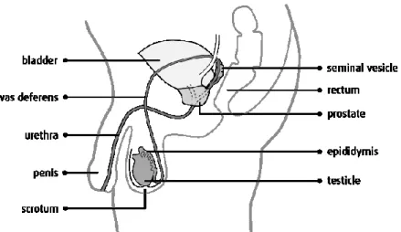
Figure 1. Male urogenital anatomy with permission from Canadian Cancer Society. ... 2
Figure 2. Diagnosed prostate cancer and related death cases in Canada 2017. ... 3
Figure 3. Age-standardized incidence rates (ASIR) for selected cancers, males, Canada, 1988–2017. ... 5
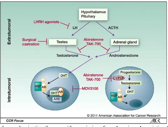
Figure 4. Androgen deprivation therapy with permissions from American Association for Cancer Research. ... 6
Figure 5. Representative chemical structures of gonadorelin and LHRH agonists that are used in prostate cancer treatment. ... 8
Figure 6. Representative chemical structure of anti-androgen agents. ... 9
Figure 7. Representative chemical structure of GnRH antagonists. ... 10
Figure 8. Representative chemical structure of taxanes and estramustine phosphate. ... 12
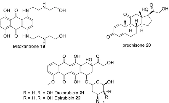
Figure 9. Representative chemical structures of anthracycline antibiotics and prednisone as a corticosteroid. ... 13
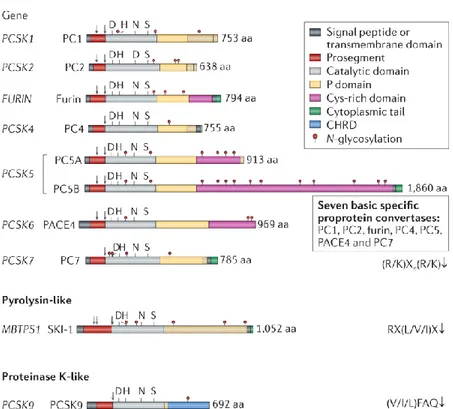
Figure 10. Schematic representation of sequences of human proprotein convertase family members. Reproduced with permission from Springer Nature publishing group. ... 18
Figure 11. Subcellular trafficking of proprotein convertases. Reproduced with permission from Springer Nature publishing group. ... 19
Figure 12. Influence of proproteins processed by furin in cancer development. Reproduced with permission from John Wiley and Sons. ... 20
Figure 13. Expression of PACE4 in prostate tissue and its silencing in tumor xenograft. A) Comparison of PACE4 with other PCs in normal and tumorous prostate. B) Representation of LNCaP tumor xenografts with different PCs silenced mRNA. CD34 staining of C) non-treated (arrows show the lumen of micro vessels) and D) PACE4-silenced tumor. PACE4 mRNA in E) Normal, and F) tumorous prostate tissues (purple indicates the presence of PACE4 mRNA). Figure adapted with permissions from Elsevier. ... 22
Figure 14. Mechanism of proteolysis catalyzed by serine proteases ... 23
Figure 15. The relaxed-eyes stereo views of crystal structure of mouse furin. a) The 3D structure of furin with inhibitor 22 in the active site shown as ribbon b) The solid surface representation of furin with inhibitor represented as ball-and-sticks c) The stick model of active site residues interacting with inhibitor 22. Reproduced with permission from Springer Nature publishing group. ... 25
xvi
Figure 16. Fluorometric assay for determination of inhibitory constant ... 26 Figure 17. Structure of 2,5-dideoxystreptamine scaffold and its derivatives as potent furin inhibitors. 29 Figure 18. Representative chemical structure and affinity of peptidomimetic furin inhibitors ... 30 Figure 19. Schematic representation of inhibitor-induced conformation alteration in furin crystal structures. Figure reproduced from Dahms et al. ... 31 Figure 20. a) The inhibition of PACE4 and furin by PC prodomains and b) the active site interacting residues. Reproduced with permission from the American Chemical Society. ... 31 Figure 21. Optimizing the number of Leu residues in the tail of the RVKR warhead leading to the discovery of Multi-Leu peptide. Reproduced with permission from the American Chemical Society. ... 32 Figure 22. Conversion of MTT to formazan in mitochondria of metabolically active cells ... 32 Figure 23. Dose-response curves in MTT assay for ML and C23 compounds on prostate cancer cell lines. Reproduced with permission from Levesque et al. ... 33 Figure 24. Chemical structures, enzymatic and cellular potencies of ML and C23... 34 Figure 25. Tumor progression following 2 mg/kg/day treatment with C23 compound. Reproduced with permission from Levesque et al. ... 35 Figure 26. The dose-dependent effect of PACE4 inhibition by C23 on cell proliferation, cell quiescence, apoptosis and pro-GDF-15 processing in vivo. Reproduced with permission from American Association for Cancer Research. ... 35 Figure 27. General structure of NOTA-labeled peptides ... 36 Figure 28. Structure of modified amino acids to be inserted at P3 of ML. ... 42 Figure 29. Structure and binding affinities of peptide inhibitors 5–16 for PACE4 and furin and their selectivity profile toward PACE4. All experiments were repeated at least twice, and data are shown as Ki ± SD. [a] 2-amino-4- guanidinobutyryl. [b] 2,3-diaminobutyryl. [c]
2-amino-3-guanidinopropionyl. [d] 2,3-diaminopropionyl. [e] 2S,3S-diaminobutyryl. [f] 2S,3R-diaminobutyryl. [g] 2S-amino-3S-guanidinobutyryl. ... 42 Figure 30. Docking of inhibitors 14 and 15 in PACE4. S-i-Dab from 14 (left) and R-i-Dab from 15 (right) in the S3 pocket of a PACE4 homology model. ... 45 Figure 31.MD simulation (1 ns) of Ac-RVKR-NH2 and Ac-RAKR-NH2 docked in a PACE4 homology model. a) Ramachandran plots for the P3 residues. The F and Y angles for Val correspond to an antiparallel β-sheet, whereas those for Ala correspond to a PPII helix. b) Variations of the hydrogen
xvii
bond angles a1 and a2 (∠N-H···O) between Gly158 of PACE4 and the P3 residues of the inhibitor
backbones. ... 46 Figure 32. Structure of control PACE4 inhibitors ... 57 Figure 33. The stereo representation of PACE4 P1–P4 active site with the Ac-RVKR-NH2 (orange)
inhibitor. The Ca2+ cation (green sphere), located deep inside the S1 subsite, is essential for its
stability. ... 58 Figure 34. Structure of P1 arginine mimetics used for PACE4 inhibitors with general structure of
Ac-DLeu-Leu-Leu-Leu-Arg-Val-Lys-NHR apart from 1. a with Leu at position P8 instead of DLeu. The
inhibition of PACE4 and furin are represented as Ki ± standard deviation, and antiproliferative
activity on PCa cell lines as IC50 ± SEM. Data adapted from Ref.166 (bData adapted from Ref.168; cNot calculable, indicates that the curve did not converged to 50% with doses up to 150 µm; dNot
determined, because of solubility/precipitation problems. ... 60 Figure 35. Energy-minimized (DFT) side chain conformers of arginine (a) and arginine mimetics (b,e−g) and estimated pKa values of relevant functional groups. Torsion angles between amidine and aromatic planes are shown (θ). ... 62 Figure 36. Superimposed induced fit docking pose of Ac-RVKX corresponding to the P5−P1 region of compounds 1 (orange) and 13 (green) in the PACE4 homology model active site. Enzyme’s side chain C atoms colored the same as corresponding ligand for clarity. H, N and O atoms are in white, blue and red color, respectively. The H bonds represented as yellow dashes. ... 63 Figure 37. Cell permeability comparison of FITC-labeled analogues of compounds 2 and 13. ... 64 Figure 38. Structure of lead compound 2 (C23). ... 85 Figure 39. a) The PACE4 affinity and antiproliferative activity of compounds 28−33. Ki and IC50 values
are means of at least two independent experiments. Errors reported as SEM for Ki and IC50. For more
details on the concentration of inhibitors in the MTT antiproliferative assay see experimental section. b) The IC50 values of selected compounds for DU145 and LNCaP PCa cell lines. ... 89
Figure 40. a) Linear relation of IC50 values for MTT antiproliferative assay on DU145 PCa cell line with
Wimley-White bilayer scale for hydrophobicity of P1' residues and b) weak correlation to PACE4 affinity of compounds 3−21. ... 90 Figure 41. Quantitative cell penetration assessment of FITC-labeled compounds 35−38 compared to control compound 34. ... 91
xviii
Figure 42. Stability of compounds 28, 31 and 32 in human plasma comparing to lead compounds 1 and
2. ... 93
Figure 43. The toxicity profile of compound 32. a) The dose-response curve for DU145 cellular toxicity was performed using PI as a staining reagent. The errors are reported as SEM. For more details on concentration of inhibitors in this assay see experimental section. b) Table showing IC50(PI) and MTD values of compound 32 in healthy CD1 mice. Data are representative of at least two independent experiments. ... 94
Figure 44. Reducing the total charge by replacing P2-Lys with Trp ... 109
Figure 45. Combining modifications at P1' and P3 of lead inhibitor 32 (Chapter 3). ... 109
LIST OF SCHEMES Scheme 1. Synthesis of Fmoc-protected residues 20a and 20b……….43
Scheme 2. Synthesis of peptide inhibitors 14–16 with modified residues………...44
Scheme 3. Synthesis of P1 arginine mimetics for inhibitors 5, 9 and 10………65
Scheme 4. Synthesis of P1 arginine mimetics for inhibitors 7, 8, 12 and 13………67
Scheme 5. Synthesis of P1 residue of compound 30, and P1-P1' adduct of compounds 32 and 33………95
Scheme 6. Synthesis of compounds 30−33………...…96
1
INTRODUCTION
The roots of word “cancer” come from a Greek “karkinos”, as described by the Greek physician, Hippocrates (460–370 B.C). However, the earliest recorded evidence of human cancer was found in mummies in ancient Egypt about 1600 B.C.1 The ancient Egyptians were treating surface tumors by
surgically removing them with similar methods that are still under practice. According to Statistic Canada, cancer is the first cause of death in Canada followed by cardiovascular diseases.
In medicine, cancer is the accepted name for a group of diseases associated with abnormal growth of eukaryotic cells. Although many types of cancer cause the growth of solid tumors, some others such as blood carcinomas are not intrinsically solid tumors. Cancer could initiate in any organ and then spread to others by translocation of cancerous cells through the blood or lymph system. Unlike benign tumors, malignant cancerous tumors are associated with high risk of fatality usually after cells migrate to foreign tissues. Cancer cells have similarities with stem cells (non-differentiated cells) and they can begin a new tumor wherever they migrate, which is termed as metastasis. For instance, a breast cancer cell could migrate to the liver and grow a new tumor, but will still be called breast cancer.2
In general, cancerous cells develop from normal cells with a damaged DNA. Two main groups of genes which are crucial in cancer development are oncogenes and tumor suppressor genes. Oncogenes are formed by the mutation of genes of normal cells called protooncogenes. Protooncogenes control cell division and growth, hence their malfunction inevitably leads to a non-controlled cell division and growth.2
Eukaryotic cells have several mechanisms to repair DNA mismatches after division. Abnormal cells undergo a programmed cell death called apoptosis.3 Apoptosis is initiated by certain intra- and
extracellular biochemical signals. A cascade of events and reactions then results in distinctive morphological changes in the cell which are terminated by cell death. On the other hand, tumor suppressor genes control the cell division, DNA repair and regulation of apoptosis. An impairment in tumor suppressor genes function can produce a cancerous cell. Many factors such as chemicals including foods, radiation, air pollution or even certain viruses are associated with mutations or damages of tumor suppressor genes or oncogenes.2
2
Historically, the first practical treatment for cancer has been the surgical removal of tumor, but even ancient physicians had noticed the post-surgery recurrence of tumors. With the development of surgical techniques, radiotherapy emerged as a complementary therapy for cancer treatment in the early 20th
century.4 Subsequently, nitrogen mustards were discovered accidentally to serve as the first cancer
chemotherapeutic agents. Nitrogen mustards were used as warfare during World War II.5 Since then, a
large number of chemotherapeutic agents has been developed. Parallel to these achievements, hormone therapy was also established for treating gender related organs tumors such as breast, ovarian and prostate cancer.
I.1. Prostate cancer
I.1.1. Prostate anatomy
The prostate is a walnut-sized gland, the main function of which is to secrete prostate fluid, a semen component (one-third of the total volume of semen). This fluid provides nutrients for sperms and facilitates their movement. During ejaculation, the muscles of the prostate gland squeeze this fluid into the urethra. The prostate gland weighs about 20 grams and is placed just beneath the bladder.
Figure 1. Male urogenital anatomy with permission from Canadian Cancer Society.6
Semen is a viscous gel at the time of ejaculation and eventually liquefies 20-30 min after ejaculation, enabling the sperms to swim.7 A group of structural proteins, including semenogelin I and II that are
3
present in the prostatic fluid, is considered to be responsible for this transformation.8 The prostatic fluid
contains high levels of citrate anions which are electrochemically balanced by high concentrations of cations such as Zn2+, Ca2+, Mg2+, K+ and Na+.9 Zinc inhibits PSA proteolytic activity with tight binding
kinetics; it is itself regulated by citrate levels in the healthy prostate.10
I.1.2. Statistics
Prostate cancer (PCa) is the most common cancer in men responsible for about 1 in 4 cases of male cancers. 11 About 15 % of Canadian men will be diagnosed with PCa during their lifetime. PCa was
attributed as the cause of death of 4100 Canadian men in 2017 and another 21300 men were diagnosed with PCa tumors. Responsible for 21% of all diagnosed cancer cases in Canadian men and 10% mortality rate in 2017, PCa has emerged as a serious healthcare issue which necessitates development of new treatments and therapeutics (Figure 2). 10
Figure 2. Diagnosed prostate cancer and related death cases in Canada 2017.10
I.1.3. Prostate cancer at cellular level
The prostate gland is comprised of a variety of cell populations: an organised epithelial lineage, a fibro-muscular stromal network, an endothelial vasculature and immune cells.12 About 95% of all prostate
cancer cases are adenocarcinoma which is a type of neoplasia of the epithelial tissue of glandular organs.13
The remaining types of prostate tumors are urothelial carcinoma (also called transitional cell carcinoma), sarcoma, small cell carcinoma, carcinoid tumours, and squamous cell carcinoma.
4
Several in-vitro cellular models have been developed for PCa cellular investigations. The gold standard model contains three cell lines: DU145, LNCaP and PC3.14 All of these cell lines are derived from
metastatic prostate cancers. The DU145 and PC3 cells are hormone independent while LNCaP cells are androgen responsive and express androgen receptors and PSA.14
I.1.4. Risk factors
The principal risk factors for prostate cancer as well as many types of cancer is age. The number of reported cases under the age of 40 is low and increases thereafter.15 However, the mortality rate for
non-metastasized prostate cancer decreases in senior men, due to greater risk of other competing causes of death.16 Genetics is the second major factor. Certain gene mutations are observed in 5-10% of people
who have PCa. For instance, BRCA1/2 mutations have been reported as risk factor in 2% PCa cases.17
Men with a first-degree relative diagnosed with PCa are two to three times more likely to develop the disease. This factor increases if patient’s relatives have diagnosed with PCa before the age of 60. Race is being identified as another prognostic factor for PCa survival. African Americans are at higher risk of diagnosis with PCa while indigenous Australians display lower rates compared to Caucasian people.18
The lifestyle also affects the PCa incidence. Cigarette smoking might increase the risk by changing the hormone levels or through increased exposure to carcinogens.19 A vegan diet was associated with a lower
risk of prostate cancer, while diets rich in animal fats were favorable for PCa, probably because of the usually high-bound obesity.20
I.1.5. Symptoms and diagnosis
Most prostate tumors are diagnosed without symptoms. However, there are some unspecific symptoms patients experience including: difficulty in urination, observation of blood in urine and pain in the genital areas. The current PCa diagnosis employs screening tests for the PSA levels. Although false positive results raise some concerns, the PSA test remains the front-line examination of the PCa. This test is a non-invasive and affordable examination which has been used routinely for several years.21 In addition, digital rectal examination (DRE) is used as a diagnostic tool complementary to the PSA test for
evaluation of PCa. The DRE test evaluates the size of the prostate by examining it through the rectum using a gloved finger. Ultimately, these tests are not sufficient, and a prostate biopsy is often necessary for a confirmatory PCa diagnosis. After biopsy, the prostate tissue samples are evaluated under the microscope, and the aggressiveness of the tumor is recorded using the Gleason scoring system.22 The
5
progression to metastatic disease, and survival chance.23 A grade 1 PCa (Gleason score (GS) 6 or lower)
is considered as low risk prostate tumor. The cells are well differentiated (not like stem cells) and rather look like normal prostate cells. Tumors with GS 7 (grade 2 or 3) are moderately differentiated which indicates an intermediate risk. Highest risk PCa tumors are allocated to grade 4 or 5 with GS 8-10 which are usually metastasized, and the cells are poorly differentiated. A gradual increase in the PSA levels with the grade of PCa is often observed.
Globally, randomized screening of a large population with the PSA test is used as a common practice. The aim of such screening is to increase the survival rate of patients by early diagnosis of the prostate tumor. Early diagnosis enables disease treatment before it advances to the unstoppable metastatic stages. However, recent findings brought some doubt about the efficacy of the PSA test on reducing the mortality of PCa patients.24-25 High levels of PSA could be due to other non-malignant prostate diseases which will
result in overdiagnosis and unnecessary biopsy and further inconvenience for false-positive cases. As represented in Figure 3, the peaks in 1992 and 2001 are due to large national screenings which were associated with over-diagnosis of PCa.
Figure 3. Age-standardized incidence rates (ASIR) for selected cancers, males, Canada, 1988–2017.10
I.1.6. Other common prostatic conditions
Prostatitis is a common prostatic condition which is sometimes caused by bacteria and is associated with
inflammation and swelling of the prostate gland. Mostly, it comes with painful and difficult urination. Three main versions of this disease are acute bacterial prostatitis, chronic bacterial prostatitis and chronic
6
non-bacterial prostatitis/chronic pelvic pain syndrome (CP/CPPS) amongst which the former is the most common.
Benign prostatic hyperplasia or prostate enlargement is caused by an overgrowth of cells which put
pressure on the urethra and discomforts urination. The condition is considered as benign and doesn’t increase the prostate cancer risk. According to Canadian Cancer Society, it is not a health issue until it reflects other potential symptoms.
I.2. Current prostate cancer treatments
Choosing a treatment for prostate carcinoma is highly dependent on the risk level of PCa, patient age, preferences and overall health. The tumor can grow at different rates based on the malignancy and stage of cancer. For less invasive tumors, doctors may prescribe active surveillance, which means patients would be closely monitored periodically using diagnostic tools. For more malignant cases, the treatments could be initiated with more radical treatments, i.e. surgical removal of the prostate gland (prostatectomy) and radiation therapy. Surgery can be followed by hormone therapy then chemotherapy as second and third lines of treatment.
Figure 4. Androgen deprivation therapy with permissions from American Association for Cancer Research.26
7 I.2.1. Hormone therapy
Hormones are among the first approved medications for prostate and breast cancers (see section I.2.3). Androgen hormones such as testosterone and its active metabolite, dihydrotestosterone (DHT), are responsible for the male characteristics viz. muscle growth, facial hair and sexual behaviour including prostate cell function and growth.27 DHT has 10-fold higher affinity for the androgen receptor compared
to testosterone, making it the main androgen hormone in the prostate gland.27 In the early stages of PCa,
the cell proliferation and regulation (apoptosis) are usually androgen dependent. Thus, androgen
deprivation therapy (ADT) is used as a tool for combating prostate tumors for early diagnosed patients.
Small molecules are utilized to lower the levels of testosterone in the whole body as well as tumorous cells to reduce tumor growth and proliferation. ADT is effective in extending the life of patients and slowing down the progress of tumor but cannot cure PCa. Tumors normally adapt to androgen deprivation and this leads to the gradual ineffectiveness of the hormone therapy.26 Several approaches in different
levels of androgen homeostasis have been used to achieve ADT including orchidectomy (surgical remove of testicles) and medications such as luteinizing hormone-releasing hormone (LHRH) agonists, anti-androgens, estrogens and more recently gonadotrophin-releasing hormone antagonists (Figure 4).28 LHRH Agonists. Luteinizing hormone-releasing hormone (LHRH, 1) or Gonadotropin-releasing hormone
(GnRH) is a hormone released by the hypothalamus which trigers the secretion of follicle-stimulating hormone (FSH) and luteinizing hormone (LH) from the anterior pituitary. After initial doses of LHRH agonists, the testosterone levels rise slightly in a condition known as flare.29 Continuing the LHRH
agonist treatment, the outcome is the reduction in the number (downregulation) of LHRH receptors. This results in reduction of circulating FSH and LH levels, along with downregulation of LHRH receptors, which cause a sharp decrease in gonadal testosterone production termed as chemical castration.30 The
structures of LHRH agonists in clinic are presented in Figure 5; leuprolide 2, goserelin 3, triptorelin 4 and histrelin 5.31 These compounds were developed by structure activity relationship (SAR) studies of
the gonadorelin hormone. Chemical castration is associated with the shrinkage of testicles. Most patients prefer medications over orchiectomy due to emotional and psychological effects of removing testicles.32
8
Figure 5. Representative chemical structures of gonadorelin and LHRH agonists that are used in prostate cancer treatment.
Androgen receptor antagonists. The anti-androgen drugs block the androgen receptor (AR) to prevent
endogenous androgens such as testosterone and dihydrotestosterone from exhibiting their biological effects. The AR antagonists are mainly used as co-therapy with LHRH agonists to reduce the flare effect (Figure 6). Steroidal anti-androgens like cyproterone acetate (10) are structurally related to testosterone and DHT. Cyproterone acetate is an antagonist for AR and suppresses the gonadal androgen production.33
The main class of AR antagonists are nonsteroidal and have a substituted anilide as core of the structure. The earlier nonsteroidal AR antagonists such as flutamide (6) and nilutamide (7) have been replaced with
9
newer agents like bicalutamide (8) and enzalutamide (9) in clinic. The hepatotoxicity of compounds 6 and 10, and the interstitial pneumonitis associated with nilutamide (7) were the main side effects that led to their replacements.34-35
Figure 6. Representative chemical structure of anti-androgen agents.
Androgen biosynthesis inhibitors. Patients with a castration-resistant prostate cancer (CRPC) are still able
to produce androgens in the adrenal gland and within the tumor. They do not respond to first line LHRH agonists and AR antagonists. The recently approved abiraterone acetate (11) is a CYP17 cytochrome P450 inhibitor interfering with the biosynthesis pathway of testosterone and DHT from cholesterol.36
Abiraterone acetate (11) is also useful in the treatment of metastatic high-risk castration-sensitive prostate cancer.37
10
Figure 7. Representative chemical structure of GnRH antagonists.
GnRH antagonists. GnRH antagonists mediate their biological effects by binding to the GnRH receptors
in the pituitary gland avoiding natural gonadorelin to activate the receptor. Unlike LHRH agonists, the antagonism action results in immediate suppression of LH release and consequently reduces the androgen release from testicles.38 Due to their immediate onset of action, GnRH antagonists are valuable
11
GnRH antagonists, readily forms gels in aqueous solutions preventing its formulation as a drug (Figure 7).39 The other member of this class, abarelix (13), has been discontinued due to severe histamine release
and allergic side effects.40-41 Further inclusion of urea and carbamoyl functionalities led to the discovery
of degarelix (14) which is currently available as the only GnRH antagonist on the market.42 Degarelix
(14) has a long duration of action compared to former agents like acyline (12) and abarelix (13). I.2.2. Chemotherapy
Chemotherapy is the practice of using chemicals to treat any disease; however, in cancer terminology, it is used for administration of certain cytotoxic chemicals that block the growth and division of malignant cells. Different classes of chemotherapeutic agents have been introduced to treat cancer but only the ones which are administered particularly for prostate cancer are discussed herein.
Taxanes. Taxanes are a class of natural products originally obtained from pacific Taxus (yews) and
discovered during a U.S. National Cancer Institute-funded screening program.43 Paclitaxel (15) and its
semisynthetic derivatives docetaxel (16) and carbazitaxel (17), are the most common taxanes used in PCa chemotherapy, all sharing a taxadiene core (Figure 8). Taxanes interfere with the normal function of microtubules during the process of cell division; thus, they are considered as mitotic inhibitors. Several groups have reported total syntheses of members of the taxane family,44 but all the commercial routes
employ semi-synthesis from yew extracts.45
Nitrogen mustards. The first developed chemotherapeutic agents for cancer were the nitrogen mustards
which share a bis-2-chloroethyl amine moiety and are still in clinical use. Their main function is to alkylate DNA twice, through intramolecular aziridinium ion intermediates and to connect covalently DNA strands together (inter-strand cross-link). The DNA double-strand needs to be opened up during cell division and as a consequence of nitrogen mustard reaction, the cell division is hampered.46 The
common nitrogen mustard in PCa treatment is estramustine phosphate (18) which has a dual hormonal (estrogen) and alkylating neoplastic (nitrogen mustard) action. Phosphate 18 is a prodrug and its active metabolites are normustine (a.k.a. bis(2-chloroethyl)carbamic acid) and estradiol.
12
Figure 8. Representative chemical structure of taxanes and estramustine phosphate.
Anthracyclines. Another important class of chemotherapeutics is the anthracycline antibiotics which
share an anthraquinone skeleton. They were first isolated from Streptomyces bacterium extracts as and examined as antibiotics; however their potent cytotoxicity led to their employment as anti-neoplastic agents.47 The main function of anthracyclines is to intercalate between DNA bases and hamper cell
division. Moreover, they are good inhibitors of topoisomerase II, a vital enzyme in DNA transcription and replication processes.48 Anthracyclines are involved in iron-mediated generation of free oxygen
radicals (quinone radicals) which cause DNA alkylation and damage.49 Doxorubicin (21) and its safer
diastereomer epirubicin (22) are widely used in PCa as well as other malignant situations (Figure 9). Another intercalating agent is mitoxantrone (19) which is also an anthraquinone and has a similar mechanism of action as the anthracyclines.
Most of the chemotherapeutic agents including taxanes and anthracyclines are administered jointly with prednisone, a corticosteroid anti-inflammatory medication, to reduce the pain and increase the quality of life of patients.50 The other beneficial effects of prednisone in PCa are the reduction of androgen levels
13
Figure 9. Representative chemical structures of anthracycline antibiotics and prednisone as a corticosteroid.
I.2.3. Targeted therapy
Cancer is a complex disease due in part to the various factors regulating human cells such as signalling, protein translocation, growth, division, protein synthesis, DNA replication and epigenetics. The undifferentiation of cancerous cells increases the possibility that each cell within the tumor has a different mutation compared to the adjacent cells. Tumors may thus adapt during treatment reducing the efficiency of medications. Cancer may vary from patient to patient and during the progression of the tumor. This huge level of complexity makes cancer one of the most challenging problems which humans have ever faced. Despite many efforts and investments, there is still no universal cure for cancer. Blocking only one pathway or target is usually not sufficient for curing tumors especially for more malignant ones, such that multiple targeted therapy has become a mainstay for treating cancer.
In the last two decades, owing to the completion of human genome map, targeted therapy received more attention from cancer scientists. Targeted cancer therapy utilizes small molecules or antibodies to block specific proteins or genes, molecular targets, that are responsible for tumor growth, progression, and cancer metastasis. The advantage of targeted therapy over current chemotherapy is due to their specificity for cancerous cells in contrast to chemotherapeutics that kill almost every rapidly-growing cell. Chemotherapeutics are cytotoxic agents. Targeted medicines are usually cytostatic and slow down or stop cell growth and proliferation rather than killing the cancerous cells. Targeted therapy along with
14
immunotherapy (modifying immune system for combating cancer) are considered as the future of cancer treatment.
Table 1.New targeted medications in clinical trials. Updated from ref. 52
Pathways Drug targets Drugs development Drug
stage AR PATHWAY AR Enzalutamide Apalutamide Darolutamide Approved Approved Phase III AR cofactors Androgen synthesis
enzymes: CYP17 Abiraterone Approved
ETS transcription
factors Transmembrane protease serine 2 PARP inhibitors: Veliparib, Talazoparib Phase II
Growth factor receptors
EGFR Afatinib, Lapatinib, Phase II
MET Cabozantinib, Tivantinib, Onartuzumab Phase II, III
IGFR Cixutumumab/IMC-A12, PLX3397 Phase II
FGFR Dovitinib/TKI258 Phase II
VEGFR Dovitinib/TKI258, Axitinib (AG013736),
PLX3397 Phase II
PI3K
PIK3 BEZ235 BKM120, GDC0980, GSK2636771, Phase I. II
AKT1 MK2206, GDC0068 Phase II
mTOR Temsirolimus, Everolimus, DS-3078a Phase II Other kinases SRC Dasatinib/Sprycel/ BMS-354825 Phase II
Cell Cycle CDKs Dinaciclib Phase I
Aurora A kinase MLN8237 (Alisertib) Phase II Protein Chaperons
HSP90 AT13387, STA-9090 Phase I, II
HSP27 OGX-427 Phase II
Clusterin/TRPM2 OGX-011/custirsen Phase III
Histone acetylation (transcriptional repression) HDAC (EZH2, CHD5, MLL2) Pracinostat SB939 Panobinostat
Vorinostat Phase III
DNA damage repair PARP PARP inhibitor Veliparib Phase II Angiogenesis VEGFR Dovitinib/TKI258, Axitinib (AG013736 Phase II
Angiopoietin 1, 2 AMG 386/Trebananib
Phase II Developmental
pathways: NOTCH. SHH, WNT
gamma secretase RO4929097
PTCH/SMOO Vismodegib/GDC-0449, LDE-225, itraconazole
Phase II Wnt-5a, Fzd8 OMP-54F28, Foxy-5
The available medications for PCa on the market are limited to hormonal therapy and chemotherapy for depriving the androgen levels and increasing the patient survival rates. Currently, many compounds are under development to target pathways other than the AR pathway (Table 1). DNA damage repair is
15
impaired in 90% of PCa cases, thus, inhibition of poly(ADP-ribose) polymerase 1 (PARP1) displayed benefits in the treatment of patients.53 PARP inhibition is also beneficial for tumors with a mutant ETS
gene (a protooncogene transcription factor).54 The growth factor receptors are other important targets for
prostate cancer which activate the tumor proliferation and differentiation by initiating the growth signal cascades. PI3K pathway kinases and other kinases which are involved in cell proliferation and apoptosis signaling as well as cell cycle homeostasis, are also targeted to treat PCa.55-57 Targeting histone
deacetylase, as DNA accessibility regulators for replication is under investigation in phase III clinical trials.58 Proteins need to have their unique tertiary and quaternary structures for performing their
functions. Targeting protein chaperones which assist in folding to the correct 3D structure has been pursued to decelerate the protein synthesis machinery of tumors.59-60 Tumor growth requires more
nutriments and consequently more blood vessels, leading to up-regulation of genes responsible for angiogenesis. Suppression of angiogenesis by inhibition of vascular endothelial growth factor receptor (VEGFR), and angiopoietin 1 and 2 has been found useful in patients with bone metastatic PCa.61-62
Developmental pathways cooperating with the AR pathway are heavily involved in PCa, and mutations in these pathways may lead to malignancy and castration resistance PCa.63
Among candidate therapies in clinical trials, the monoclonal antibody bevacizumab (an anti-VEGF), aflibercept (an anti-VEGFR), dasatinib (an inhibitor of Src), custirsen (antisense against clusterin) and tasquinimod (an anti-angiogenesis factor)64 are under investigation in different phases (Table 1).52
A revolutionary step towards greater involvement of targeted therapy (called precision oncology or personalized cancer treatment)65 is to study the molecular alterations that occur in neoplasms. Such
alterations include mutations responsible for tumor proliferation and are specific to the individuals. This study may ultimately lead to targeted treatment to combat these deleterious mutations.
I.3. Proprotein convertases I.3.1. Secretory pathway
The secretory pathway consists of the endoplasmic reticulum (ER), the Golgi apparatus organelles and the secretory vesicles. Considerable differences exist between the chemical environment inside (lumen) and outside (cytosol) of the secretory pathway. While the cytosol is reductive, the ER, Golgi apparatus and extracellular environment are oxidative. Therefore, certain oxidation reactions such as disulfide bond formation are favorable in the secretory pathway, and different types of protein are found in the lumen
16
and the cytosol. During the synthesis of proteins in a ribosome (translation), if the protein possesses a special 16–30 residue sequence called the signal peptide in its N-terminal region, it can be recognized by the signal recognition particle (SRP) on the surface of the ER. The ribosome is then connected to the ER and the protein is translocated into the ER through its N-terminal part. Once the protein synthesis is terminated, a signal peptidase enzyme removes the signal peptide and releases the protein inside the lumen. The translocated proteins in the secretory pathway could live all their life inside the secretory pathway or be moved to other places of the cell such as the membrane, or sometimes be secreted to the extracellular matrix. Some post translational modifications (PTM) are typically necessary for further translocation, activation, localization, turnover, or interactions with other proteins which are normally mediated by enzymes.66 These covalent modifications include protein cleavage, carboxy terminal
amidation, disulfide bond formation, addition of fatty acids, lipids, glycosides, ubiquitin or cofactors as well as the addition of phosphate, sulfate, methyl, hydroxyl or acyl groups to one or several amino acid residues or proteins. Addition of hydrophobic groups such as palmitoyl and farnesyl are crucial for membrane localization. Phosphorylation is particularly important in cell signal transduction because certain signaling kinases could be switched on or off with single phosphorylation or dephosphorylation.
I.3.2. The discovery of proprotein convertases
A significant group of proteins are synthesized as protein precursors (proproteins) and further activation is necessary for their biological functioning. Availability of inactive proproteins in large amounts allows the fast release of the active protein at short notice, which is especially important for hormones, clotting proteins, toxins (in poisonous animals), digesting enzymes (trypsin and chymotrypsin), caspases, collagen, and the like. The processing of proteins by amide bond lysis is one of the most imperative PTMs, which is normally mediated by proteases at a specific peptide bond. The subtilisin/kexin enzymes are one of the primary families of proteases in the activation of proproteins.
The discovery of proinsulin and proopiomelanocortin led to the development of what was later mentioned as the prohormone theory by the Steiner and Chretien groups.67-68 It was followed by the identification of
the yeast kexin, a protease related to bacterial subtilisin that proteolytically cleaves its substrates at the C-terminal of paired basic residues.69 Further studies pertaining to the proteolytic activity of yeast kexin
revealed its ability to process mammalian proopiomelanocortin, the precursor of ACTH and β-endorphin.70 These early findings led to the discovery of furin as the first human subtilisin/kexin related
17
was called PC1 (later named PC1/3) and was found simultaneously by different groups.72-73 Other similar
family members were discovered at the beginning of the 90s and now are known as PC2, PC4, PACE4, PC5/6 and PC7. They cleave their substrates at paired or single basic residues with a consensus sequence (R/K)-[X]0,2,4,6-(R/K)-↓-P1'-P2', where ↓ represents the cleavage site. For most PCs, Arg is preferred
over Lys as the basic residue. Their catalytic activity is highly Ca2+-dependent, as later confirmed by the
disclosure of the furin crystal structure in 2003.74
The subtilisin/kexin isozyme-1 (SKI-1, also called site-1 protease) and proprotein convertase subtilisin/kexin type 9 (PCSK9) being the last two PC enzymes to be discovered are slightly different compared to the other seven members because they cleave their substrates at non-basic residues. Unlike other members of the family, calcium is not essential to the proteolytic activity of SKI-1 and PCSK9.75
SKI-1 and PCSK9 are only homologous in their catalytic domain in comparison with other PCs.76-77 The
self-activation by proteolysis is the only reaction that the PCSK9 catalyzes. After removal of its prodomain, PCSK9 loses its proteolytic activity.
I.3.3. Structure of proprotein convertases
As illustrated schematically in Figure 10, PC enzymes share some similar domains in their structures including the ones listed below:
Signal peptide. A short sequence of residues in the N-terminal of PCs that is recognized by secretory
pathway for translocation into the ER.
Prodomain. The prodomain acts both as an intramolecular chaperone and inhibitor of enzymatic
activity.78-79 Some prodomain residues engage in direct interactions with the active site cleft and inhibit
the catalytic activity of PC enzymes. The variation of pH and Ca2+ during translocation from the ER to
the trans-Golgi network (TGN) and the secretory vesicles initiates the auto-catalytic removal of the prodomain in PC enzymes.80
Catalytic domain. The catalytic domain is responsible for catalyzing the cleavage of the peptide bond at
a consensus motif with paired basic residues. The mechanism of catalytic activity in PCs as in serine proteases relies on the catalytic triad (three residues of Asp, Ser and His). The details of the catalytic mechanism are discussed in section I.4.1.
18
P-domain. The P-domain plays a crucial structural role in the strong calcium and pH dependence of
PCs.81-82 PCs lose their catalytic activity without domains. A conserved RGD motif within the
P-domain has a critical role in the cellular and subcellular trafficking of PCs.83-85
Cys-rich domain. This domain has many Cys residues and contains some extra transmembrane or
cytoplasmic regions in some PCs (furin, PC4, PC5/6, PC7 and SKI-1).77, 83
Figure 10. Schematic representation of sequences of human proprotein convertase family members. Reproduced with permission from Springer Nature publishing group.86
The active site residues are highly conserved among PCs, except for PCSK9 and SKI-1. This high level of homology is reflected on the consensus sequence that they recognize in their substrates. Consequently, some PCs can recognize the substrates of other PCs although with lower affinity (this function is called redundancy). Among PCs, only crystal structures of furin have been solved untill now.74 The
demonstrated homology models for other PCs based on furin crystal structure revealed a high degree of homology around the active site.87 The disparities between PCs can be made distinguishable to a greater
19 I.3.4. Cellular and tissue distributions of PCs
The tissue distribution of PCs varies considerably between family members. Furin is ubiquitously found in the whole human body and plays a vital role in embryogenesis. PC1/3 and PC2 process most of the prohormones such as proopiomelanocortin, proglucagon and proinsulin and are thus located mostly in neural system, hypothalamus and endocrinal tissues.88-89 PACE4, PC5/6, PC7 and SKI-1 are widespread
and found in several tissues. On the other hand, PC4 is only expressed in reproductive tissues such as the testicles and ovaries.89 PCSK9 is largely expressed in the liver and regulates the LDL receptor recycling
and consequently the levels of fatty acids and cholesterol.90 In most human tissues, multiple PCs are
expressed, and process diverse substrates indicating their unique role at the cellular level.
Figure 11. Subcellular trafficking of proprotein convertases. Reproduced with permission from Springer Nature publishing group.86
The PCs are synthesized and folded similarly in the ER as described in section I.3.1 for secretory proteins. The differences between PCs appear when they leave the ER for the Golgi apparatus. The unique profile of each PC for processing their substrates is related to where they are active (TGN, secretory granules, cell surface, endosomes or extracellular matrix; Figure 11).86 The higher efficiency of PC1/3 and PC2 in
acidic pH allows them to process their substrate within the secretory granules of neuroendocrine cells, where the pH is 5 to 6. The endosomal pathway assists the journey of transmembrane furin and PC7 from the TGN to the cell surface and their return into the TGN.86
20
PACE4-FL (PACE4-A) and PACE4-altCT (the malignant isoform) are two alternative spliced versions of PACE4. The former is either secreted or is observed at the cell surface, while the latter is predominantly found within the Golgi apparatus and the endosomes.91 Some other spliced versions of PACE4 are also
produced, but they remain primarily in the ER as inactive zymogens.92 A similar pattern has been noted
for alternative splicing of PC5/6.93 The soluble 913-residue PC5/6A is secreted into the extracellular
matrix through endosomes, while the 1860-residue PC5/6B has a transmembrane domain. Similar to furin and PC7, PC5/6B is able to localize between the cell surface and the Golgi apparatus.
I.3.5. Proprotein convertases and cancer
A large body of evidence has confirmed the involvement of PCs in various levels of cancer such as carcinogenesis, tumor growth, angiogenesis and metastasis.94 The malignant overexpression of different
PCs has been documented for several types of cancer.94 The significance of PCs is due to the processing
of cancer-related substrates in the pathologic conditions. Numerous essential malignant proteins and enzymes are shown to be processed by PCs or at least they have the consensus motif for proteolytic cleavage by PCs. PC1/3 and PC2 are mostly present in neuroendocrinal tissues and they are entangled in tumors with the neuroendocrinal origin.95-96 The abnormal activity profile of both PCs is revealed to be
important in the metastasis of colorectal cancer to the liver.97 The high expression of 7B2 protein (a
neuroendocrine chaperone for PC1/3 and PC2) was counted as evidence of such role.98 PC1/3 and PC2
mRNA are also largely expressed in small-cell lung carcinomas.99
Figure 12. Influence of proproteins processed by furin in cancer development. Reproduced with permission fromJohn Wiley and Sons.100
PACE4 and furin are the most studied PCs in relation to cancer and have extensive links with various pathologic and malignant conditions.94 Furin is found to be overexpressed in several tumors, including
21
lung, head and neck carcinomas as well as endometrial, cervical and ovarian, breast, skin, gastro-intestinal tract, brain and central nervous system cancers.100 In some cases, furin tends to be a marker for
aggressiveness of neoplasms and cancer progression.100 The proteolytic activity of furin mediates the
processing of substrates such as Pro-ADAM(TS) and Pro-MT1-MMP, which results in increased aggression, migration and metastasis of tumors by release of proteases from ADAM (a disintegrin and metalloproteinase) and MMP (matrix-metalloproteases) families. Moreover, furin-mediated processing is necessary in the activation of some key growth factors and their receptors such as platelet-derived growth factor (PDGF), transforming growth factor beta 1 (TGF-β1), vascular endothelial growth factor-C (VEGF-factor-C) and insulin-like growth factor 1 receptor (IGF1R) (see Figure 12).101 The growth factors
stimulate the tumor progression by enhancing cell proliferation, angiogenesis and anti-apoptosis effects. Furin also processes integrin α-subunits, E-cadherin and N-cadherin (cell adhesion molecules) that are instrumental in the migration of cells through lymph nodes (a.k.a. metastasis).102-105
A growing body of reports implicate the role of PACE4 in pathologic conditions. The overexpression of PACE4 is associated with tumor growth and malignancy.106 Breast cancer MDA-MB-231 cells were
observed to be less aggressive (i.e. low expression of genes involved in cell growth, invasion and adhesion) when PACE4 mRNA was silenced, and most cell cycles were arrested in the G0/G1 phase.107
Administration of PACE4 inhibitors decreased the proliferation of estrogen-receptor-positive breast cancer cell line ZR-75-1 and tumor progression.108 PACE4 mediates the collagenase type IV activation
through the processing of pro-MMPs in non-melanoma skin cancers.109 Consequently, the increase in
MMP levels leads to more vulnerability of cells to carcinogens (e.g. benzopyran) and accelerated metastasis.109-110 PACE4 overexpression was also detected in non-small cell lung cancer and ovarian
cancer at both tumor cell lines and patient’s tissues.111-113 PACE4 also exhibits a unique role in the
development of prostate cancer. This will be described in more detail in the following section. I.3.6. Validation of PACE4 as a target in prostate cancer
The PACE4 enzyme has been proven to play a critical role in the progression and the malignancy of prostate. The PACE4 mRNA in DU145 cell lines was targeted and silenced using a special biochemical tool (i.e. on/off switch adapter-hepatitis delta virus ribozyme).114 The resulting cell line, called 4-2,
displayed slower proliferation. That study proposed that PACE4 has a high impact on the metastasis and malignancy of prostate tumor through processing of growth factors. More recently, pro-growth differentiation factor-15 (pro-GDF-15) was discovered as a specific substrate for PACE4.91 However,