HAL Id: tel-02463892
https://pastel.archives-ouvertes.fr/tel-02463892
Submitted on 2 Feb 2020HAL is a multi-disciplinary open access archive for the deposit and dissemination of sci-entific research documents, whether they are pub-lished or not. The documents may come from teaching and research institutions in France or abroad, or from public or private research centers.
L’archive ouverte pluridisciplinaire HAL, est destinée au dépôt et à la diffusion de documents scientifiques de niveau recherche, publiés ou non, émanant des établissements d’enseignement et de recherche français ou étrangers, des laboratoires publics ou privés.
Study of trm112, a unique methyltransferase activator
at the interface between ribosome synthesis and function
Nhan Tran Van
To cite this version:
Nhan Tran Van. Study of trm112, a unique methyltransferase activator at the interface between ribosome synthesis and function. Molecular biology. Université Paris Saclay (COmUE), 2017. English. �NNT : 2017SACLX052�. �tel-02463892�
Study of Trm112, a unique
methyltransferase activator, at
the interface between ribosome
synthesis and function
Thèse de doctorat de l'Université Paris-Saclay
préparée à l’Ecole Polytechnique
École doctorale n°573: Interfaces: approches
interdisciplinaires, fondements, applications et innovation
(Interfaces)
Spécialité de doctorat: BIOLOGIE
Thèse présentée et soutenue à Palaiseau, le 21 Septembre 2017, par
Nhan TRAN VAN
Composition du Jury:
M. Hannu MYLLYKALLIO
Directeur de recherche, École Polytechnique –CNRS (UMR 7645) Président
Mme. Béatrice CLOUET-D’ORVAL
Directrice de recherche, CNRS - Université Paul Sabatier (UMR 5100) Rapporteur
M. Jean ARMENGAUD
Directeur de recherche, CEA-Marcoule Rapporteur
Mme. Tamara BASTA-LEBERRE
Maître de conférences, Université Paris-XI (UMR 9198) Examinatrice
Mme. Valérie DE CRÉCY-LAGARD
Professeur, University of Florida (Department of Microbiology) Examinatrice
M. Marc GRAILLE
Directeur de recherche, École Polytechnique – CNRS (UMR 7654) Directeur de thèse
NNT : 2 0 1 7 S A CL X 0 52
ACKNOWLEDGEMENT
First and foremost, I would like to express my sincere gratitude to my supervisor, Dr. Marc GRAILLE for accepting me as his PhD student. I am very much grateful for his wonderful guidance, continuous supports, helps and kindness in both scientific and non-scientific stuffs. I greatly appreciate all and words cannot express how thankful I am to him. Thank you so much, Marc.
Besides my supervisor, I would like to thank the rest of the jury members: Dr. Béatrice Clouet-D’orval, Dr. Jean Armengaud, Dr. Tamara Basta-Leberre, Prof. Valérie De Crécy-Lagard and Dr. Hannu Myllykallio for accepting to evaluate my thesis. Their insightful comments and detailed discussions helped me a lot to improve my thesis.
When I first came to the lab, Gabrielle Bourgeois taught me methods and showed me everything in the lab. She is a very good friend and teacher. I learnt a lot from her. Thank you so much Gaby. A very special gratitude is for Dr. Roxane Lestini (LOB, Ecole Polytechnique) for guiding me on archaeal genetics. Many thanks go to my teammates, both the former (Gabrielle Bourgeois, Clément Charenton, Régis Back, Juliette Letoquart) and the current (Amlan Roychowdhury, Ditipriya Hazra, Nathalie Ulryck) for their helps, nice talks and discussion. It was fantastic and honored to have the opportunity to work with them as a team.
I would like to thank all colleagues in BIOC for their hospitality, helps and nice talks. I actually enjoy the lab activities a lot and I will for sure miss them so much when I leave France.
Our research is carried out in a collaborating way; thank Dr. Sarah Cianferani and Leslie Muller for the MS experiments; thank Dr. Valérie Heurgué-Hamard and Prof. Denis Lafontaine for helpful discussion on the Trm112 project.
I want to acknowledge Synchrotron SOLEIL for facilitating our crystal structural studies; many thanks for Ecole Polytechnique for funding my PhD study.
Last but not the least, I would like to thank my parents for their unconditional love and cares; thank my parents-in-law for their encouragement and taking care of my daughter. Very special thank goes to the most beloved people in my life: my wife (Nguyễn Thị Ngọc Ái) and my daughter (Trần Nguyễn Khả Ân), who are the main motivation for my life and my study. This thesis is particularly a special gift for my daughter’s 4th birthday.
ACKNOWLEDGEMENT TABLE OF CONTENTS LIST OF FIGURES LIST OF TABLE INTRODUCTION... 1 I. Eukaryotic translation ... 2
1. Translational factors and mechanism ... 2
1.1 The ribosome – the protein manufacturer ... 2
1.1.1 Structure ... 2
1.1.2 Biogenesis ... 5
1.2 mRNA – the translational template... 7
1.2.1 Capping at 5’ end ... 7
1.2.2 Polyadenylation at 3’ end... 8
1.2.3 Splicing ... 8
1.3 tRNA – an adaptor molecule... 10
1.3.1 tRNA structure ... 10 1.3.1.1 Primary structure ... 10 1.3.1.2 Secondary structure ... 12 1.3.1.3 Tertiary structure ... 12 1.3.2 tRNA function ... 13 1.4 Translation process ... 13 1.4.1 Translation initiation ... 15 1.4.2 Translation elongation ... 15 1.4.3 Translation termination ... 17 1.4.4 Recycling step ... 19
2. Methylation as a major post-transcriptional and translational modification of translational machinery... 20
2.1 Methylation ... 20
2.1.1 S-Adenosyl-L-methionine (SAM or AdoMet) ... 20
2.1.2 SAM-dependent methyltransferases ... 22
2.1.2.1 General mode of action ... 22
2.1.2.2 Classification ... 22
2.1.2.2.1 Class I SAM-dependent MTases ... 22
2.1.2.2.2 Class II SAM-dependent MTases ... 24
2.1.2.2.3 Class III SAM-dependent MTases ... 25
2.1.2.2.4 Class IV SAM-dependent MTases ... 25
2.1.2.2.5 Class V SAM-dependent MTases ... 26
2.2 Methylation of translational machinery ... 28
2.2.1.1 Base methylation... 28
2.2.1.2 Ribose methylation ... 30
2.2.1.3 Functions of tRNA methylations ... 31
2.2.2 rRNA modification ... 32
2.2.2.1 Base methylation ... 33
2.2.2.2 Ribose methylation ... 33
2.2.2.3 Functions of rRNA methylations ... 34
2.2.3 mRNA modification... 35
2.2.3.1 m6A ... 36
2.2.3.2 m1A ... 37
2.2.3.3 m5C ... 37
2.2.3.4 Functions of mRNA methylations ... 38
2.2.4 Translational protein methylation ... 38
II. Current knowledge about eukaryotic Trm112 network ... 40
1. Trm112 ... 40
2. Eukaryotic Trm112 interaction network ... 44
2.1 Trm9-Trm112 ... 45
2.2 Trm11-Trm112 ... 48
2.3 Mtq2-Trm112 ... 50
2.4 Bud23-Trm112 ... 52
2.4.1 The Bud23-Trm112 complex is involved in 40S maturation ... 52
2.4.2 Trm112 also influences 60S formation ... 54
2.5 Common themes in recognition and activation of these MTases partners by Trm112 ... 55
III. Archaeal Trm112-related research ... 59
OBJECTIVES OF THE PROJECT... 61
MATERIALS AND METHODS ... 64
RESULTS ... 88
Chapter I. Characterization of ScTrm112-Trm9 active site ... 89
Chapter II. Characterization of Trm112 interaction network in Archaea ... 125
Chapter III. Human METTL5-TRMT112 complex ... 162
DISCUSSION AND CONCLUSION ... 165
REFERENCES ... 186
LIST OF FIGURES
Figure 1. The eukaryotic 80S ribosome.
Figure 2. Eukaryotic 80S ribosome biogenesis. Figure 3. mRNA processing
Figure 4. mRNA splicing mechanism Figure 5. tRNA structures
Figure 6. Eukaryotic translation initiation model Figure 7. Eukaryotic translation elongation model
Figure 8. Eukaryotic translation termination and recycling models Figure 9. S-Adenosyl-L-methionine (SAM)
Figure 10. Class I SAM-dependent MTase Figure 11. Class II SAM-dependent MTase Figure 12. Class III SAM-dependent MTase Figure 13. Class IV SAM-dependent MTase Figure 14. Class V SAM-dependent MTase
Figure 15. Organization of eukaryotic Trm112 proteins Figure 16. Bacterial Trm112
Figure 17. Schematic representation of Trm112-MTase interaction network and of the substrates of these complexes
Figure 18. Crystal structure of YlTrm9-Trm112 complex
Figure 19. Ribbon representation of the crystal structure of EcuMtq2-Trm112 complex bound to SAM
Figure 20. Crystal structure of ScBud23-Trm112 complex Figure 21. Comparison of the Trm112-MTase interfaces Figure 22. Trm112 is present in the three domains of life
Figure 23. pH dependence enzymatic activity of ScTrm9-Trm112 complex Figure 24. Enzymatic activity of different ScTrm9-Trm112 mutants
Figure 25. Validation of the deletion of H. volcanii TRM112 gene by pop-in/pop-out Figure 26. Co-IP experiments
Figure 27. Trm112 interacting network in H. volcanii
Figure 28. HvoTrm112 solubilizes most of its methyltransferase partners Figure 29. Co-expression assay of HvoTrm11-His6 with HvoTrm112
Figure 30. An example of HvoTrm112-HvoMTase-His6 purification
Figure 31. Analyses of HvoTrm112-MTase complexes by SEC-MALLS Figure 32. Buffer optimization for enzymatic activity of HvoMtq2-Trm112 Figure 33. Enzymatic activities of HvoTrm112-Mtq2
Figure 34. Pop in/pop out result confirmed by PCR Figure 35. Co-IP of aRF1-Flag
Figure 36. Validation of the deletion of H. volcanii HVO_1032 (TRM9) gene by PCR Figure 37. Enzymatic assay for HvoTrm112-Trm9 complex
Figure 38. HVO_0019-Trm112 crystallization
Figure 39. Information on possible Bravais lattice of Hvo_0019-Trm112 crystal Figure 40. Statistics on reflection intensity along each axis in reciprocal space
Figure 41. Experimental electron density map calculated at 2.5Å resolution by Sulfur-SAD Figure 42. Crystal structure of HvoTrm112-Hvo_0019 complex
Figure 43. Sequence alignment of HvoTrm112 and Hvo_0019 sequences Figure 44. Reconstitution of A. fulgidus Mtq2-Trm112-aRF1-aRF3 complex Figure 45. HVO_1475-Trm112 complex
Figure 46. Human METTL5-TRMT112 complex
Figure 47. Docking model of cm5U into the active site of Y. lipolytica Trm9 Figure 48. HVO_0475-Trm112 complex purification
Figure 49. The superimposition of ScBud23-Trm112 (blue and light pink, respectively) onto HVO_0019-HvoTrm112 (green and pink, respectively)
Figure S1. Protein crystallization phase diagram from different crystallization methods Figure S2. Vapor diffusion crystallization
LIST OF TABLES
Table 1. Analysis of pH-dependence enzymatic activity of ScTrm9-Trm112 Table 2. Analysis of enzymatic activity of ScTrm9-Trm112 mutants
Table 3. Analysis of kinetics of different ScTrm112-Trm9 mutants Table 4. Oligomeric states of HvoTrm112-MTase complexes. Table 5. Data collection, phasing and refinement statistics
Table 6. Details of hydrogen bonds and salt bridges involved on HvoTrm112-HVO_0019 interaction
Table 7. List of putative HvoTrm112 MTase partners resulting from Co-IP experiments Table S1. Summary of different strains with their genotypes and purposes used in the thesis Table S2. Oligonucleotides and plasmids used for in vivo experiments in H. volcanii
Table S3. Oligonucleotides and plasmids used to over-express proteins in E. coli Table S4. Oligonucleotides used for site-directed mutagenesis and resulting plasmids Table S5. Different buffers used for different protein purifications in different organisms
I. Eukaryotic Translation
Protein synthesis is one of the most important processes in all living cells, leading to the synthesis of amino acid polymers based on messenger RNA (mRNA) templates. It is a tightly-regulated multi-step process occurring in the cytoplasm with the help of ribosome, a macromolecular machinery containing mainly ribosomal RNAs (rRNAs) and proteins (r-proteins), together with transfer RNAs (tRNAs) and other translational factors. The process begins when matured mRNAs synthesized in the nucleus by RNA polymerase II based on a DNA sequence are transported to the cytoplasm where the ribosome, made up of two subunits (60S and 40S), reads these mRNAs to produce corresponding proteins. By this way, the genetic information is systemically transferred from DNA to mRNA and finally proteins.
1. Translational factors and mechanism
1.1 The ribosome – the protein manufacturer
The ribosomes are known as ribozymes responsible for protein synthesis in all domains of life. They are all composed of two subunits, both built from RNAs and proteins (Figure 1A). Compared to bacterial ribosomes (sedimentation coefficient of 70S containing 30S and 50S subunits), the eukaryotic ribosomes are about 30 - 40% larger (sedimentation coefficient of 80S) and much more intricate (Ben-Shem et al., 2011; Jenner et al., 2012; Klinge et al., 2012). The small and large subunits are known as 40S and 60S subunits, respectively. Regarding its molecular weight, the eukaryotic 80S ribosome can range from 3.5MDa in lower eukaryotes to 4.5MDa in metazoa (Yusupova, & Yusupov, 2014).
1.1.1 Structure
To this day, several crystal and cryo-electron microscopy (cryo-EM) structures of eukaryotic ribosomes alone or in complex with tRNA, mRNA or proteins from different organisms have been determined, such as wheat 80S (Armache et al., 2010), yeast 80S (Ben-Shem et al., 2011), Tetrahymena thermophila 40S and 60S (Rabl et al., 2011; Klinge et al., 2012), human and Drosophila melanogaster 80S (Anger et al., 2013); Trypanosoma brucei 43S initiation complex (Hashem et al., 2013). These structures provide deep insights into structural landmarks as well as catalytic mechanism during protein synthesis by this huge machinery.
A.
B.
C.
Figure 1. The eukaryotic 80S ribosome. (A) The 80S ribosome with 40S small subunit colored in blue, 60S large subunit in yellow. Eukaryotic expansion segments are shown in red (Ben-Shem et al., 2010). (B-C) Structures of 40S and 60S subunits respectively viewed at the subunit interface. The 40S subunit includes a head (H), beak (Be), platform (Pt), body (Bo), right foot (RF) and left foot (LF). The 60S subunit exhibits a central protuberance (CP) and P-stalk. A-P-E correspond to A-site, P-site and E-site
(Adapted from Klinge et al., 2012)
In yeast, the small 40S subunit consists of a 18S rRNA and 33 r-proteins, whereas the large 60S is formed by three rRNAs (5S, 5.8 S and 25S) and 46 r-proteins (Ben-Shem et al., 2011). Among those r-proteins, two thirds have homologs in bacteria and archaea, while the remaining are specific for eukaryotes (Ramakrishnan, 2011). Compared to their bacterial counterparts, the eukaryotic subunits have several additional expansion segments (ES) and variable regions (VR) in rRNAs.The r-proteins also have C-terminal insertions/extensions and there are eukaryotic-specific proteins, resulting in larger and more complex ribosomes in eukaryotes (Ben-Shem et al., 2011; Klinge et al., 2012; Wilson, & Doudna Cate, 2012; Anger et al., 2013).
Structurally, the eukaryotic 40S subunit similarly to prokaryotic 40S is divided into the head, body, platform, beak and shoulder, left foot, and right foot regions (Figure 1B) (Klinge et al., 2012; Yusupova, & Yusupov, 2014). This subunit contains an mRNA binding site and three binding sites for tRNAs at the subunit interface. The A-site binds the incoming aminoacyl-tRNA, the P-site holds the peptidyl-tRNA attached to the nascent polypeptide chain, and the E-site accommodates the deacylated P-site tRNA after peptide-bond formation before its release from the ribosome (Schmeing, & Ramakrishnan, 2009; Klinge et al., 2012). One very important feature of this subunit is the presence of the universally conserved decoding center located in the interface surface and created by the head, the shoulder and the penultimate stem. This is the place where the base-pairing interaction between the mRNA codon and the tRNA anticodon occurs (Melnikov et al., 2012; Yusupova, & Yusupov, 2014).
Like the prokaryotic 50S, the eukaryotic 60S subunit has a crown-like shape and is composed of the central protuberance, the L1 stalk and the P-stalk (Figure 1C) (Klinge et al., 2012; Wilson, & Doudna Cate, 2012; Yusupova, & Yusupov, 2014). This subunit contains the peptidyl transferase center (PTC) where the peptide bond formation takes place. It also harbors the ribosomal exit tunnel adjacent to the PTC, which allows the nascent polypeptide chain to thread through and access the solvent side in which it undergoes processing and folding (Tu, & Deutsch, 2010; Klinge et al., 2012; Yusupova, & Yusupov, 2014).
1.1.2 Biogenesis
Ribosome synthesis is one of the most complex and energetically consuming processes in all organisms (Henras et al., 2008; Kressler et al., 2010; Thomson et al., 2013). In eukaryotes, this intricate procedure is a multiple-step, error-prone process requiring at least 200 protein factors, numerous small nucleolar RNAs and non-ribosomal factors, such as AAA-ATPases, ATP-dependent RNA helicases and kinases. These are involved in the synthesis, the maturation and the transport of individual ribosomal components and their assembly into ribosomal subunits (Kressler et al., 2010) (Figure 2A).
The eukaryotic ribosome assembly starts in the nucleus where RNA polymerase I transcribes rDNA to produce a large polycistronic precursor rRNA (35S pre-rRNA) which then undergoes several chemical modifications on specific nucleotides and nucleolytic cleavages to obtain mature 5.8S, 18S and 25S rRNAs (Figure 2B). The fourth rRNA (5S) is transcribed by RNA polymerase III, whereas the RNA polymerase II synthesizes the pre-mRNAs encoding r-proteins and other ribosomal assembly-related protein factors (Grandi et al., 2002; Granneman, & Baserga, 2004; Henras et al., 2008; Woolford, & Baserga, 2013). During transcription, the long 35S pre-rRNA is assembled with r-proteins, assembly factors and small nucleolar RNAs to form a large 90S pre-ribosome or SSU processome. Cleavage events at sites A0, A1, and A2 in 35S pre-rRNA yield to 20S and 27S pre-rRNAs, which are further processed to generate pre-40S and pre-60S particles, respectively (Figure 2) (Henras et al., 2008; Kressler et al., 2010; Oeffinger, 2016). In the final step, these particles are transported through the nuclear pores to the cytoplasm, where numerous maturation steps are required to yield the mature, functional ribosomes (Henras et al., 2008; Kressler et al., 2010). It is particularly noteworthy that there are several O-methylated sugars and base methylation introduced in rRNAs at early and late stages of ribosome biogenesis (Sloan et al., 2016). Details regarding this point will be addressed in the next parts of the thesis.
Figure 2. Eukaryotic 80S ribosome biogenesis. (A) Summary of 40S and 60S formation and maturation (Greber, 2016). (B) Pre-rRNA processing pathway (Woolford & Benserga, 2013).
1.2 mRNA – the translational template
Messenger RNAs (mRNA) carry genetic information transferred from DNA in the sequence of nucleotides, which are arranged into three-base codes called codons encoding corresponding amino acids. In eukaryotes, a primary precursor of mRNA (pre-mRNA) is synthesized by RNA polymerase II and requires a series of processing steps to produce the mature mRNA suitable for protein synthesis. These maturation steps include modifications of the 5 'and 3' ends as well as mRNA splicing.
1.2.1 Capping at 5’ end
The nascent mRNA is co-transcriptionally modified by adding a 7-methylguanosine (m7G) cap at the 5’ end through an unusual 5′ to 5′ triphosphate bond (Figure 3A). The m7G cap
Figure 3. mRNA processing. (A) mRNA capping mechanism; (B) mRNA polyadenylation model (Bentley, 2014)
is a well-conserved modification feature in eukaryotic mRNAs. The mechanism responsible for cap addition is well-known and consists of several enzymes catalyzing different reactions (Banerjee, 1980; Ramanathan et al., 2016): (1) RNA triphosphatase cleaves the γ-phosphate at the 5′ triphosphate end of mRNA to generate 5′ diphosphate; (2) RNA guanylyltransferase (EC 2.7.7.50) adds a GMP group from GTP to the 5′ diphosphate via a lysine-GMP covalent intermediate, loosing a pyrophosphate and forming the 5′–5′ triphosphate linkage; (3) An mRNA (guanine-N7-)-methyltransferase (EC 2.1.1.56), a S-adenosyl-L-methionine (SAM)-dependent methyltransferase (MTase), then catalyzes methylation on the N7 atom of guanosine to form the m7G cap (m7GpppN) named cap 0; (4) Optionally, another SAM-dependent MTase modifies the nucleotide N on the 2’ OH group of the ribose to generate the cap 1 (m7GpppNm). With regard to its functions, the m7G cap is known to protect mRNAs from the 5'-3' exonucleases (Hocine et al., 2010; Furuichi, 2015). It is also involved in splicing and in the regulation of mRNA export to the cytoplasm (Izaurralde et al., 1995). Another very important function of the mRNA cap is to promote the initiation step of the protein biosynthesis, acting like a signal for the ribosome recognition that then scans the mRNA to find the start codon (Hinnebusch, & Lorsch, 2012).
1.2.2 Polyadenylation at 3’ end
Like the 5’ end, the 3’ end of the pre-mRNAs also undergoes modifications during the maturation process (Figure 3B). The polyadenylation mechanism includes two main steps (K. M. Brown, & Gilmartin, 2003; Mangus et al., 2003; Davila Lopez, & Samuelsson, 2008): (1) The pre-mRNA is cleaved at a site characterized by two signals, a highly conserved upstream AAUAAA sequence and a downstream G/U-rich sequence. This is carried out by a series of protein factors including cleavage/polyadenylation specificity factor (CPSF) and the cleavage stimulation factor (CstF)…; (2) A polyalanine (poly(A)) tail ranging from 50 to 250 nucleotides is added at the 3’ end of the cleaved mRNA. Similarly to the 5’ cap, the poly(A) tail is also known to protect mRNA but from the 3’-5’ exonucleases. It also plays a role in mRNA transport to cytoplasm as well as in translation (Mangus et al., 2003; Hocine et al., 2010).
1.2.3 Splicing
Following transcription, eukaryotic pre-mRNAs contain an alternation of coding sequences (exons) and non-coding sequences (introns). In order to be ready for translation, the
pre-mRNAs are subjected to splicing steps to remove the introns and join the exons together. This is carried out by the spliceosome, a complex of small nuclear ribonucleoproteins (snRNPs), through a two-reaction process (Figure 4) (Fica et al., 2013; Scotti, & Swanson, 2016): (1) the formation of the intron lariat intermediate is catalyzed by a nucleophilic attack of the 2'OH of a specific branch-point nucleotide within the intron on the first nucleotide of the intron at the 5' splice site; (2) then the 3'OH of the released 5' exon performs a second nucleophilic attack on the nucleotide located just after the last nucleotide of the intron at the 3' splice site, thus connecting the exons and removing the intron lariat. Failure in RNA splicing as well as in its regulation can result in a growing number of human diseases ranging from retinal and developmental disorders to cancers, underlying the important roles of this mRNA maturation process (Scotti, & Swanson, 2016).
1.3 tRNA – an adaptor molecule
The tRNA plays an indispensable role as an adaptor molecule during the decoding process by physically linking the mRNA and the polypeptide chain. In eukaryotes, tRNAs are synthesized by RNA polymerase III as pre-tRNAs in the nucleus. In order to fulfill their complete functions, pre-tRNAs have to be processed by a variety of maturation steps: (1) removal of the 5’ leader sequence by RNase P; (2) removal of the 3’ trailer sequence by both endonuclease (tRNAse Z) and exonuclease (Rex1) enzymes; (3) addition of CCA; (4) removal of introns in some tRNAs by the combined action of an endonuclease and of a ligase; and (5) post-transcriptional modification of numerous tRNA nucleotides (Hopper, 2013; Wichtowska et al., 2013). Regarding those chemical modifications, it is known that tRNAs are the most heavily modified among the different RNAs and those modifications are required for tRNA folding, stability and function (Jackman, & Alfonzo, 2013; Vare et al., 2017). Details about this maturation step will be discussed later in the thesis.
1.3.1 tRNA structure
Similar to proteins, tRNAs also consist of primary, secondary and tertiary structures.
1.3.1.1 Primary structure
Eukaryotes can have tens to hundreds of different tRNAs depending on the organisms and some tRNAs can consist of the same anticodons (http://gtrnadb2009.ucsc.edu). In general, tRNAs have a sequence ranging from about 70 and up to 100 nucleotides (Figure 5A-B). As a conventional nomenclature (Sprinzl et al., 1998), tRNAs are numbered 1 for the nucleotide located at the 5' end and 76 for the nucleotide present at the 3' end. Similarly, the nucleotides forming the anticodon triplet are always numbered 34, 35 and 36.
Figure 5. tRNA structures. (A) Primary structure of tRNA; (B) Cloverleaf-like secondary structure of tRNA; (C) L-shaped tertiary structure of tRNA
A U G C T hairpin Acceptor stem CCA-tail D-hairpin V-loop AC hairpin AC triplet A. B. C.
1.3.1.2 Secondary structure
tRNAs fold into a conserved secondary structure known as a cloverleaf (Figure 5B), which is formed by a set of canonical Watson-Crick base pairs as well as the wobble pairings (G-U) (Auffinger, & Westhof, 2001). The cloverleaf structure consists of five regions with four helices and 3-4 loops: (1) The acceptor arm helix is formed by the base pairing of the 5’ and 3’ ends, with the latter containing the CCA 3’ end which is attached to the cognate amino acid by specific aminoacyl tRNA synthetases to form aa-tRNA. This region is normally formed by seven base pairs and possesses four unpaired nucleotides which sequence is –RCCA-3’OH at the 3’ end. R is always a purine and this position acts as a discriminator for the selection of tRNAs by the cognate synthetases (Auffinger, & Westhof, 2001); (2) The dihydrouridine (D) hairpin or D-arm, which often contains the modified base dihydrouracil. This region generally consists of four base pairs and a 7-11 residues variable hairpin loop; (3) The anticodon (A) hairpin contains a 5-bp helix and a 7-residues loop with the anticodon triplet (residues 34-36). The first position of the anticodon is called the wobble base 34 while the position 33 of the loop is always a uridine (U33) and the residue 37 is highly modified (Auffinger, & Westhof, 2001; Grosjean et al., 2010); (4) The thymine (T) hairpin or T-arm, similarly to the anticodon arm, is composed of a 4-5 bp stem and loop with seven residues, which contains a pseudouridine (ψ), a modified nucleotide in which the link between the base and the sugar is C(1’)-C5 instead of C(1’)-N1; (5) The last region is a variable loop located between anticodon and T loops and present only in some tRNAs. The length of this loop can vary between 4 and 21 nucleotides and depending on its size, tRNAs are divided into class I (the variable loop has 4-5 residues) or class II (with longer variable loop) (Auffinger, & Westhof, 2001).
1.3.1.3 Tertiary structure
The three-dimensional structure of the tRNAs was a challenging problem until its crystal structure was solved for the first time in 1974 (Suddath et al., 1974; Auffinger, & Westhof, 2001). Up to now, several structures of free tRNAs and tRNAs in complex with their cognate aminoacyl tRNA synthetases and with amino acids exist. The cloverleaf tRNA is folded into a compact structure adopting an L shape (Figure 5C). This structure is created by four helical regions of the cloverleaf, from which pairs of helices stack on each other coaxially to form two
main arms or domains: the acceptor arm is made by the acceptor helix and the T-arm, whereas the anticodon arm is formed by the D-arm and the anticodon arm. In this L-shaped architecture, the two extremities are the anticodon triplet and the 3’ CCA end, which are 75-80Å away (Auffinger, & Westhof, 2001). Moreover, this L-shaped structure is maintained by unusual hydrogen bonds occurring between the nucleotides located at invariant or semi-invariant positions (Auffinger, & Westhof, 2001; Oliva et al., 2006).
1.3.2 tRNA function
As discussed above, the main canonical function of the tRNAs is involved in the translation of genetic code as a linker between the mRNA and the amino acid sequence of the corresponding proteins by transferring amino acids, which will be inserted into the growing polypeptide chain. To perform this function, the 3’-CCA end of tRNAs are covalently attached with specific amino acids by their cognate aminoacyl-tRNA synthetases through a process called aminoacylation. This process is of particular importance since it affects the fidelity of the translation.
In addition to its well-known role in protein synthesis, tRNAs are known to possess additional functions. They are involved in the regulation of gene expression in both prokaryotes and eukaryotes (Raina, & Ibba, 2014). Moreover, aminoacylated tRNAs have been implicated as substrates for non-ribosomal peptide bond formation, post-translational protein labeling, bacterial cell envelope synthesis, and also antibiotic biosynthesis (Raina, & Ibba, 2014). tRNAs can act as precursors for the synthesis of other aa-tRNAs and could also have a potential catalytic role in certain reactions of peptidyl transfer, aminoacyl transfer and deacylation (Francklyn, & Minajigi, 2010). Furthermore, aa-tRNAs are known to be involved in the regulation of protein turnover via the N-terminal rule (Francklyn, & Minajigi, 2010).
1.4 Translation process
Translation consists of 4 different phases: initiation, elongation, termination and recycling of ribosome in all domains of life.
1.4.1 Translation initiation
In contrast to bacterial translation initiation, which needs the Shine–Dalgarno sequence of the mRNA to locate the start codon and require only three initiation factors (Voigts-Hoffmann et al., 2012), the eukaryotic translational initiation step is much more complex, requiring a scanning mechanism to identify the start codon and consists of at least 10 core initiation factors (eIFs) (Hussain et al., 2014; Aitken et al., 2016). The whole process can be divided into five different major steps (Jackson et al., 2010; Hinnebusch, & Lorsch, 2012) (Figure 6). In a first step, a methionylated initiator tRNA (Met-tRNA) forms a ternary complex (TC) with eIF2 (composed by α, β and γ subunits) and a GTP molecule before delivery to the ribosome. Next, this TC is associated with the 40S subunit linked to eIF1A, eIF1, eIF5 and eIF3s to create a pre-initiator complex 43S (PIC). Then, the PIC binds to mRNA via its interaction with mRNA cap-binding complex eIF4, thereby allowing the scanning of the mRNA in the 5' to 3' direction until the recognition of the start codon AUG in the P-site. The start codon recognition results in the formation of the 48S complex. This induces conformational changes leading to eIF1 displacement and to the hydrolysis of eIF2-bound GTP with the help of eIF5. Simultaneously, the eIF2-GDP, eIF1, eIF3 and eIF5 factors dissociate and the 60S subunit as well as eIF5B-GTP are recruited. Finally, the hydrolysis of the eIF5B-bound GTP is needed for dissociation of eIF1A and eIF5B-GDP, leaving the A-site empty to accommodate the first elongator tRNA. At the end of the initiation step, the initiator Met-tRNA occupies the P site of the functional 80S ribosome, which is ready for the elongation step.
1.4.2 Translation elongation
The translational elongation, a highly conserved step between eukaryotes and prokaryotes, is the stepwise polymerization of amino acids into the growing protein chain. In this step, the ribosome moves along the mRNA on a three-nucleotides basis called a codon in order to associate the corresponding aa-tRNA and thus allow the incorporation of the appropriate amino acid to the nascent polypeptide chain. This process can be divided into several steps and requires two eukaryotic elongation factors (eEFs): eEF1A/B and eEF2 (Figure 7) (Kapp, & Lorsch, 2004; Dever, & Green, 2012) (Figure 7).
Figure 7. Eukaryotic translation elongation model (Adapted from Dever and Green, 2012)
The elongation step starts with the 80S ribosome poised on an mRNA with the P-site occupied by the initiator Met-tRNA containing the anticodon base-paired with the start codon. The A-site in which the second codon of the open reading frame (ORF) of mRNA is present is empty, awaiting for the cognate aa-tRNA (Kapp, & Lorsch, 2004). The first step of this process consists in the formation of a ternary complex between eEF1A and the aa-tRNA complementary to the second codon in the presence of GTP. This complex then enters the empty A-site on the ribosome, from which the anticodon of the aa-tRNA is matched against the codon positioned in the A-site. A correct codon-anticodon match results in the GTP hydrolysis of eEF1A-GTP and then dissociation of eEF1A-GDP, enabling the aa-tRNA to be accommodated into the A-site. Next, the aa-tRNA at the A-site re-orientates to bring its 3' end closer to that of the tRNA present
in the P-site while eEF1A-GDP is recycled to eEF1A-GTP by an exchange factor eEF1B. The formation of the peptide bond occurs rapidly upon the nucleophilic attack of the aminoacyl-tRNA in the A-site on the ester carbon of the P-site aminoacyl-tRNA. This reaction is catalyzed by the peptidyl transferase center of the ribosome, which ideally positions the substrates for catalysis (Dever, & Green, 2012). It is then followed by the tRNAs movement to hybrid states with their acceptor arms in the P and E-sites while their anticodon loops still remain in the A and P-sites, respectively. The complete translocation of tRNAs from A and P sites to P and E sites, respectively requires the recruitment of eEF2-GTP and its GTP hydrolysis. eEF2-GDP is then released and the deacylated tRNA in the E-site dissociates from the ribosome (Kapp, & Lorsch, 2004; Rodnina, & Wintermeyer, 2011; Dever, & Green, 2012). The P-site then encompasses the tRNA bearing the elongated polypeptide chain whereas the A-site is empty, and ready for a new elongation cycle. This step is repeated until a stop codon (UAA, UAG or UGA) reaches the A-site, signaling the translation termination step.
1.4.3 Translation termination
The termination of protein synthesis takes place when one of the three stop codons (UAA, UAG and UGA) is detected in the ribosomal A-site. In eukaryotes, translation termination is catalyzed by two classes of release factors (RFs), class I eRF1 and class II eRF3 (Nakamura, & Ito, 2003) (Figure 8). The class I RF, eRF1 adopts a tRNA-like shape and recognizes all stop codons. It is responsible for the release of newly synthesized proteins from the tRNA at the P-site by triggering hydrolysis of the ester bond in peptidyl-tRNA. This results from the interaction between a universally conserved glycine-glycine-glutamine (GGQ) motif on eRF1 and the peptidyl transfer center (PTC) (Loh, & Song, 2010). Meanwhile, the class II RF, eRF3, an eEF1A-related translational GTPase, is associated to eRF1 and GTP and delivers eRF1 to the ribosome (Preis et al., 2014). eRF3 is required to complete the translation termination by enhancing the polypeptide release activity of eRF1 (Fan-Minogue et al., 2008; Eyler et al., 2013). It is known that eRF1 and the ribosome induce the hydrolysis of the GTP molecule bound to eRF3. This leads to dissociation of eRF3 from eRF1, further allowing conformational changes of eRF1 to project its GGQ motif into the PTC (Taylor et al., 2012; Preis et al., 2014).
Structurally, eRF1 consists of three well-defined domains: N, M and C (H. Song et al., 2000). The domain N (N-terminal) is involved in the recognition of the stop codons thanks to
several conserved motifs including GTx, NIKS and YxCxxxF (H. Song et al., 2000; Kryuchkova et al., 2013; Blanchet et al., 2015; A. Brown et al., 2015). The M domain (middle domain) containing the universally conserved GGQ motif is functionally analogous to the tRNA acceptor arm and this GGQ motif extends into the PTC to stimulate the polypeptide release (H. Song et al., 2000; Dever, & Green, 2012; Taylor et al., 2012; Wong et al., 2012). The C domain (C-terminal) is mainly responsible for the interaction with eRF3, mostly through hydrophobic interactions (Z. Cheng et al., 2009; Taylor et al., 2012). Moreover, it is particularly interesting to note that the glutamine side chain of GGQ motif is N5-methylated by a methyltransferase (Mtq2) in complex with a methyltransferase activator (Trm112). The methylation of the glutamine from the GGQ motif substantially increases the rate of peptide release on a subset of amino acids in
vitro in bacteria (Pierson et al., 2016), raising the question of the impact of release factor
methylation on the translation termination. The details about the methylation of the glutamine of GGQ motif by Mtq2-Trm112 complex will be discussed later in this thesis.
Figure 8. Eukaryotic translation termination and recycling models (Adapted from Dever and Green, 2012)
eRF3, for which its full-length structure is not yet available, has a variable N-terminal region known to be dispensable for translation termination (Kushnirov et al., 1988; Ter-Avanesyan et al., 1993) and a more conserved functional C-terminal region (Z. Cheng et al., 2009; Dever, & Green, 2012). The latter consists of a G-domain and of β-barrel domains 2 and 3, shares structural homology with elongation factors EF-Tu and eEF1A. It is directly interacting with domains M and C from eRF1 (Kong et al., 2004; Z. Cheng et al., 2009; Taylor et al., 2012). These interactions are important to stimulate eRF3 GTPase activity by eRF1 and by the ribosome, which is necessary for the translation termination (Z. Cheng et al., 2009; Taylor et al., 2012).
1.4.4 Recycling step
The recycling of the ribosome occurs once the complete polypeptide chain has been released. After the translation termination, the post-termination complex (post-TC) consisting of the 80S ribosome associated with an mRNA and to a deacylated tRNA and eRF1 in its P and A sites, respectively, needs to be dissociated (Pisarev et al., 2007; Jackson et al., 2012).
The exact mechanism of the recycling step is not fully understood to date, but there are two proposed mechanisms based on recent studies (Pisarev et al., 2007; Pisarev et al., 2010; Franckenberg et al., 2012; Jackson et al., 2012) (Figure 8). In a first model, the in vitro recycling of eukaryotic post-TCs is mediated by initiation factors eIF3, eIF1 and eIF1A through an energy-free mechanism functioning only in a narrow range of low Mg2+ concentrations (Pisarev et al., 2007; Pisarev et al., 2010). A second model has been recently proposed that requires ABCE1 or Rli1 in yeast, a highly conserved ATPase protein found in Eukaryotes and Archaea (Figure 8) (Pisarev et al., 2010; Kiosze-Becker et al., 2016). Following the dissociation of eRF3-GDP at termination step, eRF1 is available to interact with ABCE1, which is introduced between the two subunits of the ribosome. This leads to the hydrolysis of the ATP molecule bound to ABCE1 and the subsequent conformational changes of this later results in the dissociation of both ribosomal subunits (Becker et al., 2012; Kiosze-Becker et al., 2016). At this stage, the 60S subunit is free and ready for new cycles of translation while the small 40S subunit still holds the mRNA and the tRNA, which are subsequently released from 40S subunits either by eIF1/eIF1A/eIF3 or by ligatin (Jackson et al., 2012) (Figure 8).
2. Methylation as a major post-transcriptional and translational modification of translational machinery
It is known that eukaryotic protein synthesis is a tightly-regulated process requiring a variety of translational components. For their correct biosynthesis and functions, these factors are subjected to post-transcriptional and post-translational modifications (PTM). These PTM play key roles in enhancing and regulating the activity of the translational components and among different PTMs, methylation is the most prominent.
2.1 Methylation
Methylation is a chemical reaction, which catalyzes the transfer of a methyl group from a donor to an acceptor molecule in all living cells. Methylation plays an important role in several biological processes including biosynthesis, metabolism, detoxification, signal transduction, protein sorting and repair, and nucleic acid processing (Martin, & McMillan, 2002). This reaction is catalyzed by enzymes called methyltransferases (MTase) acting on a wide range of substrates such as nucleic acids, proteins, lipids, polysaccharides, and a range of small molecules (X. Cheng, & Roberts, 2001). Meanwhile, S-adenosyl-L-methionine (SAM) is considered as the most commonly used methyl donor for the methylation reaction (X. Cheng, & Roberts, 2001; Schubert et al., 2003; Fontecave et al., 2004).
2.1.1 S-Adenosyl-L-methionine (SAM or AdoMet)
SAM is a conjugate of a nucleotide adenosine and an amino acid methionine, known as a sulfonium compound (Figure 9A) (Fontecave et al., 2004; Kozbial, & Mushegian, 2005). SAM is known as the second most widely-used enzyme cofactor after ATP (Cantoni, 1975; Schubert et al., 2003). This is due to the fact that the strong electrophilic character of the SAM methyl group, which is brought into close proximity of a nucleophilic group from the substrate by SAM-dependent MTases, has highly favorable thermodynamics for SAM-SAM-dependent methyl-transfer reactions (Fontecave et al., 2004).
SAM is biosynthesized by SAM synthetase, which catalyzes the reaction between a methionine and an ATP. The newly synthesized SAM can further participate in a cycle, which is part of the general metabolism of sulfur-containing amino acid derivatives, by being converted to
S-adenosyl-homocysteine (SAH), which is subsequently hydrolyzed into adenosine and homocysteine by a SAH hydrolase. Homocysteine can be then either converted into glutathione or methylated to form methionine by methionine synthase. A new SAM cycle can start again with the resulting methionine (Figure 9B) (Fontecave et al., 2004). In addition to its role as the methyl donor for SAM-dependent MTases and its implication in sulfur-containing amino acid derivatives metabolism, SAM is known to act as precursor in the biosynthesis of the polyamines, nicotianamine and phytosiderophores, ethylene … (Kozbial, & Mushegian, 2005; Roje, 2006).
Figure 9. S-Adenosyl-L-methionine (SAM). (A) Chemical structure of SAM; (B) SAM cycle
A.
B.
2.1.2 SAM-dependent methyltransferases
In human, it is estimated that about 1% of all human genes encode for MTases (208 proteins) while this number is even higher in yeast, constituting around 1.2% of the genome. In addition, 30% of human MTases are linked to diseases such as cancers or mental disorders, stating the importance of this protein family (Petrossian, & Clarke, 2011).
2.1.2.1 General mode of action
The great majority of MTases (EC 2.1.1) are known to use SAM as a methyl donor to catalyze the methylation reactions (Martin, & McMillan, 2002; Loenen, 2006). Those MTases employ the general action mode for the methyl transfer reaction by promoting favorable orientation and bringing the SAM methyl group into close contact with nucleophile targets such as carbon, oxygen, nitrogen or sulfur, resulting in nucleophilic attack on the strong electrophilic methyl group of SAM and allowing the transfer of the methyl group to the various substrates (DNA, RNA, proteins, lipids, polysaccharides…) through an SN2 reaction (O'Hagan, &
Schmidberger, 2010).
2.1.2.2 Classification
Since the first crystal structure of the SAM-dependent C5-cytosine HhaI DNA MTase was solved in 1993 (X. Cheng et al., 1993; Schubert et al., 2003), there have been significant increases in the number of determined MTase crystal structures thanks to more advanced methods in structural biology. Although those SAM-dependent MTases can be defined by their action on a variety of substrates (DNA, RNA, proteins, lipids, polysaccharides, small molecules...) as well as on different atoms for methylation (nitrogen, oxygen, carbon, sulfur ...), they are structurally classified into at least five different classes (class I-V) based on their remarkably distinct structural features (Schubert et al., 2003). This characteristic of this protein family has been considered as an interesting example of the functional evolutionary convergence.
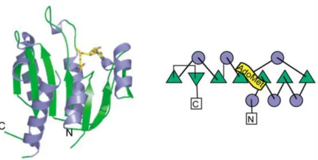
Class I MTases form the largest group with more than 60% of the total MTases in human and yeast. It consists almost all DNA MTases, some RNA as well as protein MTases but also enzymes acting on small molecules (Schubert et al., 2003; Petrossian, & Clarke, 2011; Struck et al., 2012). Despite little sequence identity, members of this class adopt a conserved Rossmann-like fold including a central seven-stranded β-sheet ending with a reversed β hairpin at the C-terminal extremity, which is surrounded by α-helices (Figure 10). The order of the β strands is ↑3↑2↑1↑4↑5↓7↑6, with the β strand 7 being anti-parallel to the other parallel strands. Helices Z, A and B are positioned on one side of the β-sheet whereas helices C, D and E are on the other side (Martin, & McMillan, 2002; Schubert et al., 2003).
Figure 10. Class I SAM-dependent MTases. An example of class I MTase tertiary structure: M.HhaI (pdb: 6MHT) (left) and its topology diagram (right) (Schubert et al., 2003)
The Rossmann-like fold is organized into two parts: the N-terminal part is responsible for SAM binding while the C-terminal part is mostly involved in substrate binding. The latter is known to be a tremendously variable region adapted to bind different kinds of substrates varying in shapes, sizes and chemistries (Martin, & McMillan, 2002). The SAM binding domain is is characterized by five highly conserved motifs (Kozbial, & Mushegian, 2005). Motif I, located in the loop between β-strand 1 and α-helix A, consists of the glycine-rich GxGxG sequence (or at least a GxG sequence), which directly interacts with the carboxypropyl moiety of SAM and is considered the hallmark of the SAM-binding site of the class I SAM-dependent MTases. Motif II
forms hydrogen bonds with the ribose hydroxyls of the SAM and is located in β-strand 2 and the adjoining turn while motif III is situated at the edge of β-strand 3 in the Rossmann-like fold and interacts with the SAM base. Motif IV interacts with the amino and sulfonium groups of the methionine moiety of SAM and encompasses β-strand 4 and the flanking loops followed by the helix corresponding to the motif V. Moreover, additional motifs such as IV, VI, VIII and/or X are known to be involved in substrate specificity whereas the motif V and VII play an important role mostly for the structural stability (Kozbial, & Mushegian, 2005).
2.1.2.2.2 Class II SAM-dependent MTases
Class II SAM-dependent MTases represent one of the smallest groups of MTases (Petrossian, & Clarke, 2011). These differ in their overall structural architecture (Figure 11) or their interaction with SAM compared to the class I MTases. This is the case of E.coli C-terminal MetH reactivation domain, which reactivates the cobalamin substrate by SAM-dependent methylation. This class II MTase has a three-dimensional structure dominated by a long central anti-parallel β-sheet flanked by groups of helices at both ends (Schubert et al., 2003). In addition, through an extended conformation, SAM binds into a shallow groove alongside the borders of the β-sheet and interacts with a conserved RxxxGY motif via hydrogen bonds (Schubert et al., 2003). In comparison to class I MTases, class II MTases undergo large conformational changes in order to position the substrate near the catalytic domain (Schubert et al., 2003).
Figure 11. Class II SAM-dependent MTases. An example of class II MTase tertiary structure: MetH (pdb: 1MSK) (left) and its topology diagram (right) (Schubert et al., 2003)
2.1.2.2.3 Class III SAM-dependent MTases
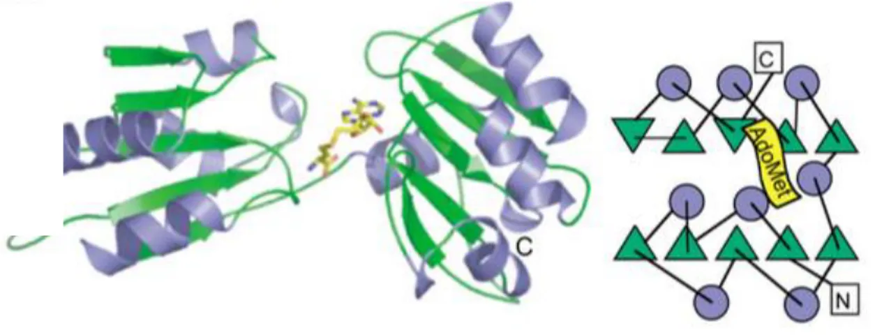
Like class II MTases, class III MTase family constitutes a very small group of MTases (Petrossian, & Clarke, 2011). This class of protein was first described in 1998 from the structure of CbiF, an MTase catalyzing SAM-dependent methylation on precorrin substrates through cobalamin biosynthesis (Schubert et al., 2003). Class III MTases contain two domains, each consisting of a five stranded β-sheet and 4 α-helices. The active site is located into a cleft at the interface between the two domains (Figure 12) (Schubert et al., 2003). Like class I MTases, members of this class exhibit a GxGxG motif at the C-terminal end of the first β-strand, but interestingly this motif is not involved in SAM binding. Meanwhile, SAM is bound to the protein between the two domains (Schubert et al., 1998; Schubert et al., 2003).
Figure 12. Class III SAM-dependent MTases. An example of class III MTase tertiary structure: CbiF (pdb: 1CBF) (left) and its topology diagram (right) (Schubert et al., 2003)
2.1.2.2.4 Class IV SAM-dependent MTases
In human and yeast, the class IV MTase family is the third largest group (Petrossian, & Clarke, 2011), known as the SPOUT MTase superfamily (shortly for SpoU-TrmD). This goup mainly consists of tRNA and rRNA MTases (Anantharaman et al., 2002; Tkaczuk et al., 2007; R. J. Liu et al., 2013) but recently some protein MTases have been shown to belong to this group (Young et al., 2012; R. J. Liu et al., 2013). This class of MTases contains a unique SPOUT domain exhibiting an unusual α/β fold with a very deep topological knot (Figure 13) (Tkaczuk et al., 2007; R. J. Liu et al., 2013). This core domain including 5 or 6 parallel β-strands sandwiched by α-helices on both sides can be separated into 2 subdomains: (1) the N-terminal subdomain
forming a Rossmann-like fold; (2) the C-terminal subdomain with a deep conserved trefoil knot known to bind SAM (Tkaczuk et al., 2007; R. J. Liu et al., 2013). The class IV MTases can be divided into two subclasses: (1) the smallest SPOUT MTases such as TrmL and RlmH containing only the SPOUT domain in which the N-terminal subdomain is involved in substrate binding (R. J. Liu et al., 2013); (2) the larger SPOUT MTases like TrmH, RlmB and RsmE showing additional domains (for instance THUMP, PUA, OB fold, or L30e….) fused at the N- or C-termini or introduced into a linker between two subdomains of the SPOUT domain, responsible for the substrate binding (Tkaczuk et al., 2007; Petrossian, & Clarke, 2009; R. J. Liu et al., 2013). Another interesting point regarding this class IV MTase family is that almost all SPOUT members except the monomeric Trm10 protein reported so far were identified as dimeric proteins (Tkaczuk et al., 2007; Oerum et al., 2017). This characteristic is known to stabilize the SAM-binding loop in the knot of one monomer through interactions with the other monomer and to play an important role in the MTase activity as the active sites are created by residues from both monomers (Tkaczuk et al., 2007; Petrossian, & Clarke, 2009; Oerum et al., 2017).
Figure 13. Class IV SAM-dependent MTases. An example of class IV MTase tertiary structure: YibK (pdb:1MXI) (left) and its topology diagram (right) (Schubert et al., 2003)
2.1.2.2.5 Class V SAM-dependent MTases
The second largest family of SAM-dependent MTases is assigned to class V, with approximately 27% and 14% of the total MTases in human and yeast, respectively (Petrossian, & Clarke, 2011). The class V MTases contain a SET domain (Suppressor of variegation, Enhancer of zeste, Trithorax), and hence are also referred as SET-domain MTase superfamily. This family
encompasses almost all proteins known to catalyze histone lysine methylations crucial for the regulation of chromatin and gene expression. In addition to histones, the SET-domain MTases also methylate some other proteins like Rubisco (Dillon et al., 2005). The SET domain consists of eight curved β strands organized into three small β-sheets (Figure 14), including the C-terminus inserted below a surface loop to generate a knot-like structure as seen in the SPOUT MTases (Dillon et al., 2005; Petrossian, & Clarke, 2009). Class V MTases have sequence similarity in their N-terminal (N-SET) and C-terminal (C-SET) domains, which harbour conserved motifs I-II and motifs III-IV, respectively. Those are known to be responsible for catalysis, SAM-binding and substrate interaction (Petrossian, & Clarke, 2009). Similarly to class III MTases, the SAM bound to the SET domain binds to a shallow groove of the protein, formed by motif I, the N-terminal part of motif III and a tyrosine in motif IV (Schubert et al., 2003; Petrossian, & Clarke, 2009). One interesting point with regard to the SAM binding mode by proteins of this family is the presence of the GxG sequence in motif I similarly to the motif I glycine-rich GxGxG sequence found in class I MTases, despite no structural similarity between the two protein classes (Petrossian, & Clarke, 2009). Moreover, some MTases have the SET-domain flanked by diverse sequences named pre- and post-SET motifs. The pre-SET motif is known to stabilize the protein structure by interacting with different surfaces of the core SET domain while the post-SET motif constitutes part of the active site, vital for the MTase activity and maybe involved in the substrate recognition and specificity (Schubert et al., 2003; Qian, & Zhou, 2006).
Figure 14. Class V SAM-dependent MTases. An example of class V MTase tertiary structure: Set7/9 (pdb:1O9S) (left) and its topology diagram (right) (Schubert et al., 2003)
2.2 Methylation of translational machinery
Methylations are by far the most frequent and important PTMs for the control and optimal efficiency of mRNA translation. This is particularly the case for tRNAs, which are heavily methylated in order to enhance their stability as well as the efficiency and accuracy of translation. rRNAs are also targeted by various post-transcriptional modifications including 2'-OH methylation, base methylation, or more complex modifications for their maturation processes and functions (Sharma, & Lafontaine, 2015). Several recently and extensively studied modified nucleotides present in mRNAs, including methylations such as N6-methyladenosine (m6A), N1-methyladenosine (m1A), and 5-(hydroxyl)methylcytosine ((h)m5C), have led to the emergence of the epitranscriptomics field (Dominissini et al., 2016; Gilbert et al., 2016). Finally, MTases also target ribosomal proteins and of translational factors (Polevoda, & Sherman, 2007).
2.2.1 tRNA methylation
According to the RNA modification database (http://mods.rna.albany.edu/mods/), 112 different modified nucleosides were found in all kinds of RNA in three domains of life to date. Among these, 93 modifications are present on tRNA molecules, with methylation being the major one. The four canonical nucleotides are the most regular substrates for tRNA MTases on either the base or the ribose moiety but modified nucleotides like pseudouridine (ψ), inosine (I) and more complex species are also subjected to methylation (Swinehart, & Jackman, 2015). Among tRNA MTases, almost all enzymes known to date belong to the class I Rossmann-like fold and class IV SPOUT-domain MTases. It is noteworthy that some recently discovered MTases could potentially form a novel class of MTase in the future (Swinehart, & Jackman, 2015).
2.2.1.1 Base methylation
The bases are the most common substrates of the tRNA MTases catalyzing modifications mainly at carbon, endocyclic nitrogen and exocyclic nitrogen atoms (Swinehart, & Jackman, 2015).
The methylation of the carbon atom at position 5 of pyrimidines (m5C and m5U) is present at multiple positions of tRNAs. These tRNA modifications are found throughout life,
with m5U identified in all 3 domains of life while m5C is present in Archaea and Eukarya (Hou, & Perona, 2010; Motorin, & Helm, 2011; Swinehart, & Jackman, 2015). Most tRNA MTases catalyzing m5C and m5U are class I MTases, with some representative members such as E.coli TrmA (yeast Trm2 and human TRMT2 homologs) catalyzing the conserved m5U54 (also called ribothymidine) in the T-loop of tRNAs and yeast Trm4 (human NSUN2 homolog) generating m5Cat different positions (Hou, & Perona, 2010; Motorin, & Helm, 2011; Towns, & Begley, 2012; Swinehart, & Jackman, 2015). The catalytic mechanism of those MTases is probably as follows. First, a conserved cysteine acts as a nucleophile to attack C6 atom from the base, which then allows the C5 atom to attack the SAM methyl group. Second, the C5 proton is abstracted by a general base in order to generate the methylated product. For TrmA, the catalytic base has been identified as a glutamate while in Trm4, an aspartate fulfills this function (Hou, & Perona, 2010; Boschi-Muller, & Motorin, 2013; Swinehart, & Jackman, 2015). Moreover, methylation events can also occur on complex modifications such as cm5U, mcm5(s2)U, ncm5U… through more complex multi-step biosynthetic reactions. This will be discussed in detail in the next part of the thesis.
Methylations on the different endocyclic nitrogen atoms such as m1A, m1G, m3G, m3C, m1ψ and m7G are found in almost all living cells except m3C and m1ψ that are absent in Archaea and Bacteria, respectively (Swinehart, & Jackman, 2015). Some examples for this methylation group include the class I MTases like yeast Trm5 (human TRMT5) forming m1G37 in Archaea and Eukarya; the yeast Trm6/Trm61 heterodimer (Trm61 is the catalytic subunit) (human TRM6) catalyzing m1A58, a highly conserved modified A58 in the T-loop of tRNAs and also the SPOUT domain MTases such as bacterial TrmD forming m1G37, yeast Trm10 (human TRMT10A) catalyzing formation of m1G9 as well as archaeal TrmY for m1ψ54 formation (Motorin, & Helm, 2011; Towns, & Begley, 2012; Swinehart, & Jackman, 2015; Hori, 2017). In term of the potential methylation mechanism, some MTases (for instance Trm5 and Trm10) use general bases like glutamate and aspartate residues, respectively, which deprotonate N1 atom. This results in the nucleophilic attack of N1 on the SAM methyl group to form the methylated targets. In contrast, for m7G class I MTases such as eukaryotic Trm8/Trm81 (human METTL1), bacterial TrmB or m3C MTases like ScTrm140, their catalytic mechanisms have not been clarified so far (Swinehart, & Jackman, 2015).
Methylation on the exocyclic nitrogen atoms like m2G, m22G, m6A, and m62A is always
catalyzed by class I MTases. The eukaryotic Trm112-Trm11 (human TRMT112-TRMT11) (where Trm11/TRMT11 are the catalytic subunit) catalyzes the formation of m2G10 while archaeal TrmG10 is responsible for the formation of both m2G10 and m22G10 (Hirata et al.,
2016). In addition, yeast Trm1 (human TRMT1) is known to form m22G26 with m2G26 as the
intermediate while archaeal Aquifex aeolicus Trm1 modifies some tRNAs to generate m2G26m2G27 and m22G26m22G27 (Awai et al., 2009). The bacterial/archaeal Trm1 proteins
contain a DPFG/DPPY conserved motif in their active site and the aspartate from this motif is likely acting as the general base to abstract a proton from N2 atom, then promoting the
nucleophilic attack on SAM methyl group for methylation to occur (Swinehart, & Jackman, 2015). Meanwhile, the m6A modification also commonly seen in mRNA, rRNA and DNA is found in tRNAs at position 37 and is catalyzed by E.coli YfiC by a still unknown mechanism (Swinehart, & Jackman, 2015).
2.2.1.2 Ribose methylation
This methylation can be found on the 2’ hydroxyl group of the ribose (2’-O-methylation) of any canonical nucleotides in all domains of life (Rana, & Ankri, 2016). A famous example for this group of MTases is bacterial TrmH (Trm3 in Eukarya), one of the founders for the SPOUT-domain MTases, generating a highly conserved 2'-O-methylguanosine at position 18 (Gm18). This protein has been well-studied and its catalytic mode is proposed to act through a conserved arginine playing the role of the general base removing a proton from the 2’-hydroxyl group and then allowing the resulting oxygen to attack the SAM methyl group for the reaction to complete (Swinehart, & Jackman, 2015; Hori, 2017). Moreover, several important 2'-O-methylations are present at positions 32 and 34 in all domains of life. Some examples include SPOUT-domain MTases like E.coli TrmJ (Cm32 or Um32), TrmL (Cm34, cmnm5Um34), aTrmJ (Cm32) but also class I MTases such as such as ScTrm7/Trm732 (Cm32) and Trm7/Trm734 (Cm34) complexes or their human orthologues containing TRMT7 (also known as FTSJ1; Cm32, Um32, Cm34, Gm34). Meanwhile, some 2'-O-methylations are specific to archaea (Cm56 catalyzed by SPOUT-domain aTrm56) or eukarya (Um44 formed by class-I ScTrm44 or human METTL19) (Hori, 2017; Marchand et al., 2017).
2.2.1.3 Functions of tRNA methylations
Although the importance of tRNA modifications has often been underestimated as the loss of most single modifications usually results in only modest or no phenotypes, the PTMs in general and the methylations in particular are crucially involved in every aspects of the tRNA, namely its structure, function, and stability (Torres et al., 2014; Swinehart, & Jackman, 2015). The effects of these methylations on tRNAs can be classified based on the positions of the modifications.
The methylations in the anti-codon loop and at positions nearby are normally related to effects on the tRNA function, namely the fidelity of mRNA decoding. This is the case for mcm5U, mcm5s2U, Cm and mnm5Um at the wobble position 34, which are directly involved in decoding through the anti-codon:codon pairing and are necessary for the translation fidelity and efficiency (Letoquart et al., 2015b; Ranjan, & Rodnina, 2016; Tuorto, & Lyko, 2016; Hori, 2017). Meanwhile, the m5C34 modification present in yeast tRNALeu(CAA) is necessary for
translation efficiency and its loss renders yeast hypersensitive to oxidative stress because of inefficient translation of UUG-containing stress response mRNAs (C. Gu et al., 2014; Swinehart, & Jackman, 2015; Ranjan, & Rodnina, 2016). Moreover, several modifications at position 37 such as a highly conserved m1G37 are responsible for enhancing the translational accuracy, whereas chemically intricate modifications like wyosine (imG) and its derivative wybutosin (yW) in eukaryotic and archaeal phenylalanine-specific tRNAPhe are known to stabilize codon:anticodon interactions by providing base-stacking interactions between the anti-codon and the A-site codon. Such modifications have a key role in preventing translational frameshifting (Ranjan, & Rodnina, 2016; Tuorto, & Lyko, 2016).
The methylations of nucleotides located in the tRNA body usually affect the folding and stability of tRNAs. Some examples are the conserved modifications m2G10 and m22G26 at the
stem of the D-loop, which both maintain the secondary and tertiary structure of tRNAs in the three domains of life (Lorenz et al., 2017; Vare et al., 2017). Meanwhile, two modifications (m1G9 and m1A9) have been identified to facilitate the correct folding of mitochondrial tRNAs (Vare et al., 2016; Oerum et al., 2017). It is also the case for m5C48 and m5C49, which are located at the junction between the variable loop and the T loop in archaeal and eukaryotic tRNAs. Furthermore, the m5U54 (forming the conserved T54, also known as ribothymidine), and
the m1A58 modifications, both located in the T loop of tRNAs, are known to stabilize a reverse
Hoogsteen base-pairing interaction between those two bases, and thus necessary for the tertiary folding of the L-shaped tRNA (Hou, & Perona, 2010). Moreover, lack of m1A58 catalyzed by Trm6/Trm61 (where Trm61 is the catalytic subunit) and m7G46 modification generated by Trm8/Trm82 (where Trm8 is the catalytic subunit) results in rapid tRNA degradation (Alexandrov et al., 2006).
Apart from direct roles in tRNA decoding and structure, tRNA methylations also encompass other additional functions. For instance the 2´-O-methylation of the tRNA anticodon loop is known to ensure effective immune response against pathogens in plants (Ramirez et al., 2015). In addition, some modifications such as 2´-O-methylations alone or in combination with methylated bases have been found crucial for temperature adaptation in thermophilic and psychrophilic organisms (Lorenz et al., 2017; Vare et al., 2017). More importantly, growing evidence suggests that defects in tRNA methylations and MTases are involved in severe human disorders such as cancers, type II diabetes, and neurological diseases. Recent extensive efforts on understanding tRNA methylations and the enzymes responsible for these modifications could lead to future discovery of novel therapeutics (Torres et al., 2014; Grosjean, 2015).
2.2.2 rRNA methylation
Like tRNAs, rRNAs are frequently methylated in all living cells, however numbers and types of methylations vary from different organisms (Przybylska et al., 2007; Piekna-Przybylska et al., 2008). E. coli has 19 base methylations and only four 2’-O-methylations. The opposite pattern is observed in Eukaryotes. Indeed, budding yeast contains 10 base methylations but 55 ribose 2’-O-methylations while in human, the numbers are 10 and around 100, respectively (Piekna-Przybylska et al., 2007; Piekna-Przybylska et al., 2008; Sharma, & Lafontaine, 2015; Krogh et al., 2016). Meanwhile, in archaea, rRNAs methylations like its eukaryotic counterparts are dominated by 2’-O-methylations (Dennis et al., 2015). These differences between three domains of life seem to be due to the presence of box C/D snoRNPs in Archaea and Eukarya (Watkins, & Bohnsack, 2012; Krogh et al., 2016). Concerning the catalytic enzymes, most 2’-O-methylations are catalyzed by small nucleolar RNPs (snoRNPs) guided by box C/D sno-RNAs, while base methylations are specifically added by conventional SAM-dependent MTases (Watkins, & Bohnsack, 2012; Sloan et al., 2016). It is also noteworthy that