Université de Montréal
Role of interleukin-1 in the pathogenesis of the infection
caused by Streptococcus suis serotype 2
par Agustina Lavagna
Département de pathologie et microbiologie Faculté de médecine vétérinaire
Mémoire présenté à la Faculté de médecine vétérinaire en vue de l’obtention du grade de Maîtrise ès sciences (M. Sc.)
en sciences vétérinaires option microbiologie
Août, 2018
ii
Résumé
Streptococcus suis sérotype 2 est un pathogène important du porc et un agent zoonotique en
émergence causant des morts subites (porcs), des chocs septiques (humains) et des méningites (chez les deux espèces), où une réaction inflammatoire sévère est caractéristique de l’infection. Une réponse rapide et efficace du système immunitaire inné contre S. suis est critique pour contrôler la croissance bactérienne et pour limiter la propagation du pathogène sans occasionner une inflammation excessive. Bien que l’interleukine (IL)-1 soit considérée comme l’un des médiateurs pro-inflammatoires les plus efficaces et produit le plus rapidement, son rôle dans la pathogénèse de l’infection par S. suis n’a pas encore été étudié.
En utilisant un modèle murin d’infection systémique bien standardisé, nous avons démontré que la souche nord-américaine de virulence intermédiaire de « sequence type » (ST) 25, la souche européenne hautement virulente ST1 ainsi que la souche épidémique chinoise ST7, induisent toutes de hauts niveaux d’IL-1 dans des organes de filtration, tels que le foie et la rate. De plus, les cellules dendritiques et les macrophages, deux types de cellules jouant un rôle central dans la pathogénèse de S. suis, sont des sources importantes de cette cytokine. Les études des mécanismes impliqués dans la production de cette cytokine ont démontré que la production d’IL-1, indépendamment de la souche bactérienne utilisée, dépendait de MyD88 et impliquait les récepteurs TLR2 et possiblement TLR7 et TLR9. Cela suggère que les composantes bactériennes responsables de l’activation cellulaire sont similaires et conservées entre les différentes souches. Cependant, seuls de très hauts niveaux de suilysine, produite par la souche ST7, provoquaient une maturation importante de proIL-1β. Cette maturation implique l’activation des inflammasomes NLRP3, NLRP1, AIM2 et NLRC4, qui est due à la formation de pores et à un efflux d’ions.
De surcroît, nous avons évalué le rôle global de cette cytokine chez des souris IL-1R-/-,
démontrant que l’IL-1 pourrait jouer un rôle bénéfique lors d’une infection systémique par S. suis en modulant l’inflammation requise pour contrôler et éliminer la charge bactérienne, ce qui favorise la survie de l’hôte. Toutefois, au-delà d’un certain seuil, l’inflammation causée par S. suis ne peut plus être contrebalancée par cette signalisation, ce qui complique la détermination exacte du rôle de l’IL-1. Une meilleure compréhension des mécanismes sous-jacents impliqués dans le contrôle de l’inflammation et de la charge bactérienne aiderait à développer de meilleures mesures de contrôle pour ce pathogène important à la fois chez le porc et chez l’Homme.
iii
Mots-clés: Streptococcus suis sérotype 2; interleukine-1; cellules dendritiques;
iv
Abstract
Streptococcus suis serotype 2 is an important porcine bacterial pathogen and an emerging zoonotic agent causing sudden death (pigs), septic shock (humans), and meningitis (both species), with exacerbated inflammation being a hallmark of the infection. A rapid, effective, and balanced innate immune response against S. suis is critical to control bacterial growth and limit the spread of the pathogen without causing excessive inflammation. Even though interleukin (IL)-1 is regarded as one of the most potent and earliest pro-inflammatory mediators produced, its role in the S. suis pathogenesis has not been studied.
Using a well-standardized mouse model of systemic infection, we showed that an intermediately pathogenic sequence type (ST) 25 North American strain, a highly pathogenic ST1 European strain, and the epidemic ST7 Chinese strain induce high levels of IL-1 in important filter organs such as liver and spleen. Moreover, dendritic cells and macrophages, which are two cell types centrally involved in the S. suis pathogenesis, are important sources of this cytokine, with the ST7 strain secreting the highest levels. The study of the underlying mechanisms involved in this production showed that, independently of the strain, IL-1β production required MyD88 and involved recognition via TLR2 and possibly TLR7 and TLR9. This suggests that recognized bacterial components are similar and conserved between S. suis strains. However, very high levels of the pore-forming toxin suilysin produced by the ST7 strain only, are required for efficient maturation of proIL-1β. Such maturation involved the activation of the NLRP3, NLRP1, AIM2, and NLRC4 inflammasomes via pore formation and ion efflux. Using IL-1R-/- mice, we demonstrated that IL-1 signaling may play a beneficial role
during S. suis systemic infection by modulating the inflammation required to control and clear bacterial burden, thus, promoting host survival. Beyond a certain threshold, however, S. suis-induced inflammation cannot be counter-balanced by this signaling, making it difficult to discriminate its role. A better understanding of the underlying mechanisms involved in the control of inflammation could help to develop control measures for this important porcine and zoonotic agent.
Keywords: Streptococcus suis serotype 2; interleukin-1; dendritic cells; inflammation;
v
Table of Contents
Résumé ... ii
Abstract ... iv
Table of Contents ... v
List of Tables ... viii
List of Figures ... ix
List of Acronyms and Abbreviations ... xiii
Acknowledgments... xvii
I- Introduction ... 1
II- Review of the Literature ... 5
1 Streptococcus suis: A growing menace ... 6
1.1 S. suis disease and transmission ... 6
1.1.1 In pigs... 6
1.1.2 In humans ... 7
1.1.2.1 S. suis human outbreaks in China ... 8
1.2 General features of S. suis ... 9
1.3 Serotype identification and geographical distribution ... 9
1.4 Virulence factors ... 12
1.4.1 The capsular polysaccharide ... 12
1.4.2 Muramidase-released protein and Extracellular factor ... 13
1.4.3 Suilysin ... 13
1.4.4 Cell wall modifications: N-deacetylation of peptidoglycan and D-alanylation of Lipoteichoic Acid... 15
1.4.5 Others ... 15
1.5 Pathogenesis ... 16
1.5.1 STEP 1: Adhesion and invasion ... 18
1.5.2 STEP 2: Survival in blood and dissemination ... 18
1.5.3 STEP 3: Central nervous system invasion: Meningitis... 19
2 Innate immune response ... 20
vi
2.1.1 Toll-like Receptors... 21
2.1.2 NOD-like Receptors... 22
2.1.3 Importance of the PRRs in S. suis infection ... 23
2.2 Inflammatory mediators ... 24
2.2.1 Mediators involved in S. suis infection: Inflammation as a hallmark of the systemic infection ... 25
3 Interleukin-1 ... 27
3.1 Main functions of IL-1 ... 27
3.2 Interleukin-1 Receptor ... 28
3.3 IL-1α ... 29
3.4 IL-1β ... 30
3.4.1 Inflammasome IL-1β-dependent and -independent maturation ... 31
3.4.1.1 NLRP3 inflammasome... 32
3.4.1.2 NLRP1 inflammasome... 33
3.4.1.3 NLRC4 inflammasome ... 33
3.4.1.4 AIM2 inflammasome ... 34
4 Role of IL-1 in the infection caused by streptococci ... 34
4.1 Streptococcus agalactiae ... 35
4.2 Streptococcus pneumoniae ... 37
4.3 Streptococcus pyogenes ... 38
4.4 What do we know about S. suis and IL-1?... 39
III- Materials, Methods, and Results ... 42
Interleukin-1 signaling induced by Streptococcus suis serotype 2 is strain-dependent and contributes to bacterial clearance and inflammation during systemic infection ... 43
Abstract ... 44
1. Introduction ... 45
2. Materials and methods ... 48
3. Results ... 54
4. Discussion ... 61
Tables and Figures ... 78
vii
1- S. suis induced high levels of IL-1 in systemic organs but not in plasma. ... 100
2- IL-1 production induced by Streptococcus suis serotype 2 is strain-dependent ... 102
3- The role of IL-1 in S. suis systemic infection depends on inflammation ... 107
V- General Conclusions and Perspectives ... 111
Conclusions ... 112
Perspectives... 113
References ... 114
Annex I: Inflammation induced by S. suis plays a major role in host survival ... xix
viii
List of Tables
Review of the Literature
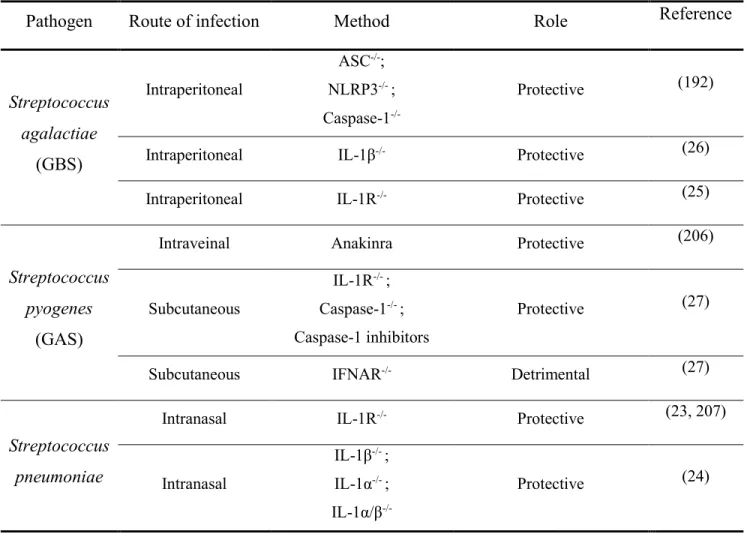
Table A. Role of IL-1 in the infection caused by streptococci ... 35
Materials, Methods, and Results
Table 1. Bacterial strains and plasmids used in this study. ... 78 Table S1.Oligonucleotide primers used in this study ... 92
ix
List of Figures
Review of the Literature
Figure A. Worldwide distribution of the most important S. suis serotype 2 sequence types isolated from both clinical pig and human cases of infection. ... 11 Figure B. Pathogenesis of S. suis infection involves 3 main steps. ... 17
Materials, Methods, and Results
Figure 1. Streptococcus suis serotype 2 induces elevated levels of IL-1β in liver and spleen but not in plasma. ... 79 Figure 2. IL-1β released from dendritic cells (DC) and macrophages (MФ) stimulated with Streptococcus suis is strain-dependent... 80 Figure 3. Role of Toll-like receptors (TLRs) and associated signaling pathways in S. suis-induced IL-1β production from dendritic cells (DCs). ... 81 Figure 4. Inflammasome implication in Streptococcus suis-induced IL-1β dendritic cell (DC) production is strain-dependent. ... 82 Figure 5. IL-1β secretion by dendritic cells (DCs) activated by S. suis is blocked by additional extracellular potassium (K+). ... 83
Figure 6. Suilysin (SLY) is involved in the maturation of Streptococcus suis-induced IL-1β by dendritic cells (DCs). ... 84 Figure 7. Co-stimulation with recombinant suilysin (rSLY) enhances Streptococcus suis-induced IL-1β production by dendritic cells (DCs), which is inhibited by cholesterol (CHOL)…… ... 85 Figure 8. Survival of wild-type (WT) and IL-1 receptor-deficient (IL-1R-/-) mice following Streptococcus suis systemic infection. ... 86 Figure 9. Plasma pro-inflammatory mediator production during Streptococcus suis systemic infection….. ... 87 Figure 10. Pro-inflammatory mediator production in spleen and liver during Streptococcus suis systemic infection ... 88
x
Figure 11. IL-1 signaling is required for control of bacterial burden in blood, liver, and spleen……….. ... 89 Figure 12. Model of the mechanisms involved in Streptococcus suis-induced IL-1β production by dendritic cells (DCs). ... 90
Figure S1. Streptococcus suis serotype 2 induces elevated levels of IL-1α in liver and spleen, but not in plasma. ... 93 Figure S2. IL-1α release from dendritic cells (DCs) and macrophages (MФ) stimulated with Streptococcus suis is strain-dependent... 94 Figure S3. Streptococcus suis-induced TNF production is inflammasome-independent. ... 95 Figure S4. Streptococcus suis-induced IL-6 and TNF secretion by dendritic cells (DCs) is independent of additional extracellular potassium (K+) concentrations. ... 96
Figure S5. IL-1β production by recombinant suilysin (rSLY) is Toll-like receptor (TLR) 4-independent….. ... 97 Figure S6. IL-1 does not modulate its own production following Streptococcus suis infection…….. ... 98
Discussion
Figure I. Role of IL-1 in the pathogenesis of the infection caused by S. suis serotype 2. ... 110
Annex I
Figure 1. Survival of wild-type (WT) and IL-1 receptor-deficient (IL-1R-/-) mice after
intraperitoneal infection with a non-lethal dose of Streptococcus suis strain SC84. ... xix Figure 2. IL-1 modulates plasma pro-inflammatory mediators during Streptococcus suis systemic infection caused by a non-lethal dose of strain SC84. ... xx Figure 3. IL-1 modulates pro-inflammatory mediators in spleen and liver during Streptococcus suis systemic infection caused by a non-lethal dose of strain SC84. ... xxi
xi
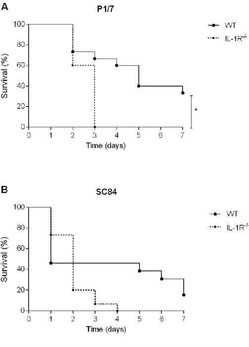
Figure 4. IL-1 is required for control of bacterial burden in blood, liver, and spleen during Streptococcus suis systemic infection caused by a non-lethal dose of strain SC84. ... xxii Figure 5. Survival of wild-type (WT) and IL-1 receptor-deficient (IL-1R-/-) mice after
intraperitoneal infection with a high dose of Streptococcus suis strain P/7. ... xxiii Figure 6. Bacterial burden in blood during Streptococcus suis systemic infection caused by a high dose of strain P1/7... xxiv
Annex II
Figure 1. S. suis strain 89-1591 induces elevated levels of IL-1 in liver and spleen, but not in plasma……. ... xxv Figure 2. IL-1 released from dendritic cells (DCs) stimulated with Streptococcus suis strain 89-1591…… ... xxvi Figure 3. Comparison of the IL-1β production by dendritic cells stimulated with S. suis serotype 2 strains 89-1591, P1/7, and SC84. ... xxvii Figure 4. Role of Toll-like receptors (TLRs) and associated adaptor proteins in S. suis-induced IL-1β production from dendritic cells. ... xxviii Figure 5. Strain 89-1591 bacterial components stimulate the production of IL-1β. ... xxix Figure 6. Comparison of the IL-1β mRNA production by dendritic cells stimulated with S. suis serotype 2 strains 89-1591, P1/7, and SC84. ... xxx Figure 7. Strain 89-1591-induced IL-1β is caspase-1, NLRP3, and AIM2 dependent. ... xxxi Figure 8. Strain 89-1591 induced IL-1β production depends on internalization. ... xxxii Figure 9. Production of IL-1β by dendritic cells stimulated with 89-1591 DNA depends on AIM2 inflammasome. ... xxxiii Figure 10. Mechanisms of IL-1β maturation by strain 89-1591. ... xxxiv Figure 11. Survival of wild-type (WT) and IL-1 receptor-deficient (IL-1R-/-) mice after
intraperitoneal infection with Streptococcus suis. ... xxxv Figure 12. Pro-inflammatory mediator production in plasma during Streptococcus suis systemic infection. ... xxxvi Figure 13. Pro-inflammatory mediator production in liver during Streptococcus suis systemic infection……... ... xxxvii
xii
Figure 14. Pro-inflammatory mediator production in spleen during Streptococcus suis systemic infection. ... xxxviii Figure 15. IL-1 is required for control of bacterial burden in blood. ... xxxix
xiii
List of Acronyms and Abbreviations
AIM2: Absent in melanoma 2ASC: Apoptosis-associated speck-like protein containing a CARD ATP: Adenosine triphosphate
BBB: Blood–brain barrier
BMEC: Brain microvascular endothelial cells CARD: Caspase activation recruitment domain CCL: Chemokine (C-C motif) ligand
CFU: Colony-forming unit CNS: Central nervous system COX: Cyclooxygenase
CPEC: Choroid plexus epithelial cell CPS: Capsule polysaccharide
CRP: C-reactive protein CSF: Cerebrospinal fluid
CXCL: Chemokine (C-X-C motif) ligand DC: Dendritic cell
DP: Dipeptidyl peptidase EF: Extracellular factor
ERK: Extracellular-signal-regulated kinase FBP: Fibronectin-fibrinogen binding protein
GAPDH: Glyceraldehyde-3-phosphatase dehydrogenase GAS: Group A Streptococcus
GBS: Group B Streptococcus GDH: Glutamate dehydrogenase
GM-CSF: Granulocyte macrophage colony-stimulating factor HPA: Hypothalamic pituitary adrenal
i.p.: Intraperitoneal
iE-DAP: γ-D-Glu-meso diaminopimelic acid IFN: Interferon
xiv IFNAR: Interferon-α/β receptor
IL: Interleukin
IRAK: Interleukin-1 receptor-associated kinase JNK: c-Jun N-terminal kinase
KC: Keratinocyte chemoattractant KO: Knock-out
LF: Lethal factor
LPS: Lipopolysaccharide LRR: Leucine-rich repeat LTA: Lipoteichoic acid
MAPK: Mitogen-activated protein kinase MCP-1: Monocyte chemoattractant protein-1 M-CSF: Macrophage colony-stimulating factor MDP: Muramyl dipeptide
MIP: Macrophage inflammatory proteins MLST: Multilocus sequence typing MOI: Multiplicity of infection MRP: Muramidase-released protein
MyD88: Myeloid differentiation primary response protein-88 Mθ: Macrophages
NCL: Novel capsular polysaccharide locus
NF-κB: Nuclear factor- kappa-light-chain-enhancer of activated B cells NK: Natural Killer cell
NLR: NOD-like receptor
NLRP1: NACHT, LRR and PYD domains-containing protein 1 NLRP3: NACHT, LRR and PYD domains-containing protein 3 NLS: Nuclear localization signal
NOD: Nucleotide-binding oligomerization domain p.i.: Post-infection
PA: Protective antigen
xv PBMC: Peripheral blood mononuclear cell PCR: Polymerase chain reaction
PG: Peptidoglycan PGE2: Prostaglandin E2 PLY: Pneumolysin PYD: Pyrin domain
RIG: Retinoic acid-inducible
RLR: Retinoic acid-inducible gene I-like receptors RTqPCR: Real-time quantitative chain reaction SLY: Suilysin
SMS: Streptococcal meningitis syndrome ST: Sequence type
STSLS: Streptococcal toxic shock-like syndrome T3SS: Type three secretion system
T4SS: Type four secretion system THA: Todd Hewitt broth agar THB: Todd Hewitt broth
THP-1: Tamm-Horsfall protein 1 TIR: Toll-interleukin 1 receptor
TIRAP: Toll-interleukin 1 receptor domain containing adaptor protein adaptor protein TLR: Toll-like receptor
TNF: Tumor necrosis factor
TRAF: TNF receptor associated factor
TRIF: TIR-domain-containing adaptor-inducing interferon-β WT: Wild-type
xvi
xvii
Acknowledgments
Firstly, I want to express my gratitude to my supervisor, Dr. Marcelo Gottschalk. Thank you for your trust and for giving me a place in your lab. Thank you for guiding me and for this project, for your support, and for always asking me to do my best. Thank you also for giving me the freedom to take the project in my own hands and to let me add as many experiments as I wanted. Last but not least, I would like to thank you for funding during my studies.
Also, a special thanks to my co-supervisor, Dr. Mariela Segura, for trusting in me as much as Marcelo and giving me a space in her lab too. Thank you for your advice and for sharing your knowledge with me. Thank you also for always adding a new perspective in every meeting and discussion.
A huge thanks to Jean-Philippe Auger, for our telepathic thoughts, for our endless discussions about this project, and for accompanying me these 2 years with all my crazy ideas.
Many thanks also to all of my colleagues for the laughs and for making it a great time in the lab. A day of working was never too long (well, maybe sometimes) or never too bad because you were there too. For the scientific and non-scientific conversations, for the help and the support: Audrey, Guillaume, Dominic, Marêva, Sonia, Agustina, Mar, and Jean-Philippe again, thank you for your friendship.
Thanks to all the members of my «comité-conseil» and jury: Dr. Marcelo Gottschalk, Dr. Mariela Segura, Dr. Levon Abrahamyan, Dr. Christopher Fernandez-Prada and Dr. Neda Barjesteh.
I am grateful to all of the institutions for the financial support GREMIP, CRIPA, NSERC – CRSNG and University of Montreal.
To all my friends, who got tired of listening how well or bad an experiment went, and were always there to listen without complaining, not even a little bit! For making me feel like home in a faraway country. Special thanks to Jasmin, Catarina and Camila for the extra-extra support.
xviii
I am also grateful to have an exceptional role model in life and in science, Florencia Albertoni-Borghese. She might not know this, but I am thankful to her for being an inspiration.
Finally, I wish to thank to all of my family in Argentina, who has always supported me to come here, and listen and advise me throughout my studies. Mama, Papa, Marce, Rama, Luli, Martin y Benja, sin ustedes no podría haberlo conseguido.
2
Streptococcus suis (S. suis) is one of the most important bacterial pathogens affecting the swine industry worldwide (2). In pigs, it has been associated with a variety of pathological processes such as meningitis, septicemia with sudden death, and endocarditis among others (3). In addition, S. suis is also an emerging zoonotic agent, responsible for meningitis and septic shock in humans (4, 5). During the last decade, the number of documented cases in humans due to this coccus has dramatically increased worldwide (4, 6). While most of them are due to close occupational contact with pigs/pork products, particularly in Western countries (7), two outbreaks were recorded in China in 1998 and 2005, with more than 200 people affected (8). In Vietnam, Thailand, and Hong-Kong, S. suis is one of the most common causes of community-acquired bacterial meningitis (9-11) . In addition, several human cases have also been reported in Canada (12).
Based on the capsular polysaccharides (CPS), 35 serotypes have been identified (5). Among these, serotype 2 is thought to be the most virulent and the most frequently isolated from diseased pigs and humans (5). The predominant S. suis serotypes isolated from clinical cases in pigs worldwide, is serotype 2, followed by serotypes 9, 3, ½, and 7, while in humans, serotype 2 is mainly followed by serotype 14 (12). Multilocus sequence typing (MLST) is a technique that allows the identification of sequence types (STs) within a serotype using genomic sequencing (13). In fact, the use of this technique in the recent years has permitted to identify the most important STs within S. suis serotype 2. Among these, the epidemic strain ST7 in China, the highly virulent ST1 in Eurasia, and the intermediate and low virulence ST25 and ST28, respectively, in North America are the four predominant sequence types (12).
Even though S. suis has been studied for several years, the pathogenesis of its infection as well as the different virulence factors involved, are still not completely understood. Similarly, the mechanisms involved in the host immune response against this pathogen remain poorly characterized. To cause disease, S. suis needs to colonize and invade the epithelial surfaces of the host. In this way, bacterial adhesins coupled with the secretion of a pore-forming toxin termed suilysin (SLY) seem to play an important role in this step (14). Once in circulation, its thick CPS will help S. suis to survive in blood and disseminate to vital organs such as liver, spleen, kidneys, and lungs (15). At this step, recognition of S. suis by host immune cells leads to the production of several pro-inflammatory mediators, which if uncontrolled, could lead to
3
the development of septic shock. If death by sepsis does not take place, and bacteremia remains high, S. suis can reach and invade the central nervous system (CNS) and cause meningitis (14).
Therefore, the severity of the outcome depends on the ability of host immune mechanisms to control bacterial growth and to limit the spread of the pathogen, without causing excessive inflammation (16). The innate immune response is initiated following bacterial recognition by specialized pattern recognition receptors (PRRs), leading to cell activation and synthesis of diverse pro-inflammatory cytokines and chemokines (17). Amongst these, interleukin (IL)-1 is regarded as one of the most potent and earliest pro-inflammatory mediators produced (18). Indeed, IL-1 is a central mediator of immunity and inflammation participating amongst others, in the induction of adhesion molecules, leukocyte recruitment and migration, production of other inflammatory factors such as lipid mediators and acute phase proteins, fever induction, HPA axis regulation, stimulation of effector functions of neutrophils and macrophages, and lymphoid cell-mediated innate and adaptive immunity (19, 20).
The term IL-1 refers to two different cytokines, IL-1α and IL-1β, which are encoded by separate genes and synthesized as precursor peptides (proIL-1α and proIL-1β). While proIL-1α is biologically active and can exert intracellular or extracellular functions, a maturation step via proteolytic cleavage is required for the activation of proIL-1β (20, 21). This activation step is mediated mainly by caspase-1, which also requires activation by multi-protein complexes: the inflammasomes.
Although IL-1 signaling plays essential roles in both immunity and sterile inflammation, uncontrolled production of this cytokine can be detrimental, as observed in cases of rheumatoid arthritis and gout (22). Likewise, during bacterial infections, synthesis of IL-1 is necessary to initiate inflammation, but disproportionate levels of this cytokine can lead to tissue damage and disease. In fact, IL-1 plays a protective role during both pneumococcal and Group B Streptococcus (GBS) infections, wherein a lack of IL-1 signaling contributes to a weak inflammatory response and higher bacterial burden (23-26). However, a recent study showed that lack of control of IL-1β production results in a lethal outcome in a mouse model of Group A Streptococcus (GAS) infection (27).
Though excessive inflammation is a hallmark of the S. suis infection, and IL-1 is a key mediator in this type of process, little is known about IL-1 involvement in the context of S. suis
4
disease. Most studies measure the cytokine as part of a group and do not focus on its mechanisms of secretion, regulation, and most importantly its overall role in the pathogenesis.
That is why, based on the observations mentioned above, we hypothesized that IL-1 plays a crucial role during the pathogenesis of the infection caused by S. suis serotype 2. While an immune activation is needed to fight the infection, a high and uncontrolled production of this cytokine significantly enhances the inflammatory reaction, fact that would be detrimental to the host.
Accordingly, the main objective of this research project was to study the production of IL-1 as part of the inflammatory response triggered by S. suis serotype 2. More precisely, its role in the pathogenesis of the infection was evaluated to identify whether it is protective and/or detrimental using a mouse model of systemic infection caused by S. suis serotype 2.
The specific objectives of this research are:
1. Characterize the production of IL-1 using a well-standardized mouse model of systemic infection (septic shock).
2. Investigate, via in vitro studies, the mechanisms implicated in IL-1 production. This objective focuses on the characterization of the different cellular sources and receptors, as well as the intracellular signaling pathways involved in the production.
3. Elucidate the role of IL-1 during in vivo infection by S. suis serotype 2 with the help of mice deficient in IL-1 receptor.
6
1 Streptococcus suis: A growing menace
S. suis is an important bacterial pathogen causing sudden death and meningitis in pigs, responsible for important economic losses to the swine industry (2). Additionally, it is also a zoonotic agent causing meningitis and septic shock in humans, and has become a public health concern, particularly in South-East Asia (12). Since being first reported in pigs in 1954, the number of cases, not only in pigs but also in humans, has increased worldwide. Therefore, the study of this pathogen is crucial to guarantee and expedite diagnosis, to develop and implement efficient treatments, and to create efficient control strategies against S. suis.
1.1 S. suis disease and transmission
1.1.1 In pigs
The natural habitat of S. suis is the upper respiratory tract of pigs, more precisely the tonsils and nasal cavities, from where it is frequently isolated (28). Moreover, S. suis can also be found in the genital and digestive tracts (29). Pigs carrying S. suis can directly transmit the pathogen to other pigs, which represents a major issue in terms of disease spread in herd. In fact, this horizontal transmission through the respiratory route essentially results from nose to nose contact and aerosols (29). In addition, vertical transmission is also possible: piglets born to sows with uterine and vaginal infections are either born infected or become infected while passing through the birth canal. Moreover, they can also acquire the bacterium after birth by close contact with the sow, her feces, and piglets from other litters, as well as from the environment (14, 30).
Several risk factors have been demonstrated to play a key role in the settlement and development of S. suis infection. These include the immunity status of the herd and the presence of other infections such as the porcine reproductive and respiratory syndrome virus (31, 32). Additionally, although no seasonal incidence has been noted in pigs, different management practices such as excessive temperature, poor ventilation, and crowding have been suggested as predisposing factors (33).
Of the various manifestations of the disease, septicemia and meningitis are by far the most striking features, but endocarditis, pneumonia, and arthritis can also be observed (14).
7
Clinical signs can differ between herds depending on the pathogenesis of the disease. Generally, pigs with peracute S. suis infection may die within hours of the onset of clinical signs, although it is not unusual for death to occur without any of them (3). The earliest sign is usually an increase in rectal temperature, accompanied by a detectable bacteremia or pronounced septicemia which, if not treated can last up to 3 weeks. During this period there is usually fluctuating fever, and variable degrees of decreased appetite, depression, and shifting lameness (29). After several days, diseased animals develop neurological signs, including opisthotonus, lateral recumbency, ataxia, incoordination, paddling, convulsions, and paralysis (3). Regarding the pathological and histopathological lesions, the most frequently observed in S. suis infected pigs are characterized by neutrophilic infiltrates and congestion of the meninges, lymph nodes, and lungs (31).
1.1.2 In humans
The first reported case of S. suis in humans was in Denmark in 1968 (34). Since then, over 1600 human cases of infection have been reported worldwide, with probably many more never diagnosed or misdiagnosed (12). S. suis infections in humans are most often reported from countries where pig-rearing is common, with the main route of entry being thought to be through contact of cutaneous lesions, most usually on the hands and arms with contaminated animals, carcasses or raw meat (7, 35). However, this situation seems to be different in some Asian countries where the oral route has been proposed, since many infections have been reported after ingestion of contaminated raw pork products (36).
Purulent meningitis is the most frequent manifestation of S. suis in humans (37). After an initial incubation period, which ranges from a few hours to days, developed symptoms are, generally, similar to those observed in the case of bacterial pyogenic meningitis which include fever, headache, vomiting, and meningeal signs (36, 38, 39). Interestingly, hearing loss is the most common sequela after recovery from purulent meningitis (35). In addition, although less frequently, S. suis can also induce other types of infections including septic shock with multiple organ failure, endocarditis, pneumonia, arthritis, and peritonitis (40-44). Septic shock symptoms include high fever, chills, headache, vomiting, and abdominal pain. Additionally, hypotension, tachycardia, multiple organs dysfunction, subcutaneous hemorrhage, disseminated intravascular coagulation, and death may occur (5).
8
Current diagnostic procedures are based on the isolation of the bacterium from blood and/or cerebrospinal fluid (CSF) by standard microbiological techniques. However, S. suis can be misidentified with other bacterial species such as Enterococcus faecalis, Aerococcus viridans, or Streptococcus pneumoniae (S. pneumoniae) (35). Moreover, bacterial cultures can result in a false-negative result as a consequence of, for example, the use of antibiotics. Nowadays, the implementation of molecular techniques such as the polymerase chain reaction (PCR) has improved the detection of S. suis (35).
1.1.2.1 S. suis human outbreaks in China
In 1998, in the province of Jiangsu, China, 25 cases of what first was believed to be food poisoning were registered. Later, those cases were categorized as the first outbreak caused by S. suis with two main clinical outcomes, streptococcal toxic shock-like syndrome (STSLS) which includes hypotension and multiorgan involvement, and streptococcal meningitis syndrome (SMS) (45). Seven years later, in 2005, another human outbreak was recorded in the province of Sichuan, China, this time with 215 cases: 28% diagnosed as STSLS, 24% sepsis and 48 % meningitis (8). This episode resulted in 39 deaths. Patients presented a sudden onset of disease with high fever, diarrhea, hypotension, petechia, disseminated intravascular coagulation, and dysfunction of multiple organs, such lungs, kidneys, liver, and heart (8, 46).
During the outbreaks, all infections occurred in backyard farmers who were directly exposed to the bacterium during the slaughtering process of pigs that had died of unknown causes or had been killed for food because they were ill. Therefore, the oral route of infection by eating raw pig could not be ruled out (5).
The strains isolated from both outbreaks were characterized as S. suis serotype 2 ST7 (47). Evidence indicated that the virulence of S. suis ST7 was high, but the particular mechanism of infection was unknown at the time and is still not completely understood. A study comparing the strain SC84, isolated from a patient with STSLS during the 2005 outbreak, and a typical highly pathogenic strain isolated from a diseased pig, 31533, showed that the strains display some differences in cytokine production. Indeed, strain ST7 showed a stronger capacity to stimulate T cells, naïve T cells, and peripheral blood mononuclear cell proliferation than strain 31533, which might help explain the high virulence of SC84 (48). Moreover, the presence of a
9
pathogenicity island probably played an important role in the rapid adaptation and increased virulence of SC84 strain (49). In addition, the production of a pore-forming toxin, suilysin (SLY) could also have contributed during S. suis invasive infections (50).
1.2 General features of S. suis
S. suis is an encapsulated Gram-positive coccus that occurs singly, frequently in pairs or occasionally, in short chain. Though it grows well in aerobic conditions, it is considered a facultative anaerobic. All strains are α-hemolytic when grown on sheep blood agar plates and some are β-hemolytic on plates containing horse blood (3). Biochemically, S. suis is characterized by absence of growth in 6.5% NaCl agar, a negative Voges-Proskauer test, production of acid in trehalose and salicin broth, and production of amylase (51).
1.3 Serotype identification and geographical distribution
To identify the different serotypes of S. suis, serological and molecular techniques can be used. Tests such as co-agglutination, capillary precipitation, or Neufeld's capsular reaction can be performed using reference antisera (52). In addition, PCR, which directly targets genes of the capsular polysaccharide (CPS), is frequently used for its simplicity and effectiveness (53). Nevertheless, despite the numerous techniques, certain strains are difficult to serotype possibly due to a novel capsular type or an acapsular phenotype. To overcome this problem, novel cps loci (NCL) have been recently under study (54, 55).
Consequently, based on the CPS, 35 serotypes have been described for the moment (types 1–34 and 1/
2) (12, 56). However, within the last years, and with the appearance of new
techniques, 6 of these serotypes were suggested to belong to species other than S. suis. As an example, serotypes 32 and 34 have been reclassified as Streptococcus orisratti (57). Yet, the classification of these strains is still controversial and there is no actual consensus. More consideration is needed on the subject since a proper classification is central to diagnostic, treatment and control of the disease (12).
Among all capsular types, serotype 2 is the most commonly associated with disease in pigs and humans. As an example, in cases of infections in pigs, serotype 2 is the most predominant in Asia (44.2%) and in North America (24.3%) (58). In Europe, however, serotype
10
2 takes the second place while serotype 9 prevails, being responsible for 61% of the cases. Concerning human cases, serotype 2 is responsible for 74,7 % of them worldwide. Since it is considered the most virulent serotype and the main cause of economic loses to the swine industry, serotype 2 became the subject of numerous studies.
Additional phylogenetic studies on S. suis can be carried out with a technique termed multilocus sequence typing (MLST). Based on the genetic diversity, this technique can further classify S. suis according to allelic types or sequence types (STs). MLST is based on the nucleotide sequencing of fragments of 6 to 7 well-conserved housekeeping genes within the bacterial genome. Allelic variation at each locus is then compared with isolate profiles in an international database. In 2002, King et al. (13) established a model of MLST for S. suis using seven different house-keeping genes: cpn60, dpr, recA, aroA, thrA, gki, and mutS).
More than 100 different STs of SS2 have been described up to date. As shown in the representative map in Figure A, there is a distinct geographic distribution of 6 predominant STs in different regions of the world. The ST1, associated with disease in pigs and humans, is mainly found in Asia, Europe, and South America (12). In North America, the STs most frequently isolated are ST25 and ST28, which can be also found in Thailand and Japan, respectively (59). In Europe, particularly in the Netherlands, ST20 strains have been isolated as the cause of disease in pigs and humans. Remarkably, ST7, responsible for the two human outbreaks in China, is endemic to this region (60). In the case of ST104, this ST is endemic to Thailand and appears to be more and more commonly isolated from human cases (12).
11
Figure A. Worldwide distribution of the most important S. suis serotype 2 sequence types isolated from both clinical pig and human cases of infection. Adapted from (10).
Different studies have tried to associate differences in genomic regions found in MLST to the virulence of the strain. Using a well-standardized mice model of systemic infection, Lachance et al. studied the difference in virulence among prevalent STs: the intermediately pathogenic ST25 North American strain (89-1591), a highly pathogenic ST1 European strain (P1/7), and the epidemic ST7 Chinese (SC84) strain (16). Mice infected with the North American 89-1591 strain showed no difference in survival with mock-infected mice, meanwhile survival curves of mice infected with the European P1/7 strain or Chinese SC84 strain differed greatly from mock-infected. Survival rates at the end of the experiment (60 h) were 90%, 40%, and 20% for 89-1591, P1/7 and, SC84, respectively. These results suggested thatST7 is more virulent than a ST1 European strain, but that they are both more than the ST25 North American strain. Interestingly, this study also showed that cytokines levels, but not bacterial burden, correlated with the degrees of virulence of ST7, ST1 and ST25 strains, suggesting a difference in these strain to activate the innate immune system (61). In the same line of studies, Auger et al. evaluated the virulence of three different STs (ST1, ST25, and ST28) from different geographical (Americas and Eurasia) and host (porcine and human) origins. However, results showed that prediction of virulence based on ST and geographical origin is difficult since strains
12
belonging to the same ST presented significant differences in virulence, which did not always correlate with a given geographical origin (62).
1.4 Virulence factors
While virulence between S. suis strains varies substantially, several bacterial components have been proposed as potential virulence factors, and many of these, have been proposed to be crucial. However, the identification of these factors has suffered from the lack of a clear definition of the term ‘virulence’ (29). Over the years, different criteria have been used to define a strain as virulent, from the clinical condition of the animal from which the strain was isolated, to diverse in vitro and in vivo studies. The differences in the model chosen, in addition to the variation in the experimental design, make it challenging to determine what is truly a virulence factor. Moreover, it is known that the presence of a certain virulence factor cannot define a strain as being virulent, while its absence cannot define it as avirulent (63). Additionally, it is important to remark that almost all of the studies on virulence and pathogenesis have used certain STs within the serotype 2, mainly ST1 and ST7 strains (15). Consequently, there is a lack of information concerning possible virulence factors of other serotypes.
1.4.1 The capsular polysaccharide
The S. suis serotype 2 CPS is a large extracellular structure made up of five different sugars: galactose (Gal), glucose (Glc), N-acetyl glucosamine (GlcNAc), rhamnose (Rha) and N-acetyl neuraminic (sialic) acid (NeuNAc) (64), and is still considered the only proven critical virulence factor. Several steps in the pathogenesis can be influenced by the presence of a thick CPS. For example, the adhesion of S. suis to host cells, which is mediated by cell wall components, seems to be reduced in the presence of the CPS, suggesting that the adhesin(s) involved in the process are partially masked by the capsule. Moreover, survival and dissemination after reaching the bloodstream were also shown to depend on the production of CPS since it confers protection against immune recognition and clearance (63). In fact, several studies with non-encapsulated mutants showed that the CPS protects S. suis from neutrophil and monocyte/macrophage–mediated phagocytosis and killing, and helps rapid clearance of the bacterium from the circulation (65-68). However, resistance to phagocytosis and clearance
13
should be considered multifactorial since encapsulated avirulent strains can be eliminated from blood within 48 h, suggesting that there are others factors involved in this process (69, 70).
In addition to its antiphagocytic properties, the S. suis CPS also plays a role in the modulation of the immune response. Several studies showed that activation of immune cells such as macrophages and DCs, by non-encapsulated mutants leads to an increased pro-inflammatory cytokines and chemokines production in comparison with the wild-type strain (67, 71-74). This suggests, that the CPS reduces the immune response by hiding cell wall immunostimulating components.
1.4.2 Muramidase-released protein and Extracellular factor
Two of the first virulence factors described for S. suis are the muramidase-released protein (MRP) and the extracellular factor (EF) (75). MRP is a 136-kDa protein, anchored to the cell wall peptidoglycan (PG) by sortase A and also released into the culture supernatant during bacterial growth (76, 77) On the other hand, EF is a 110-kDa protein only present in the culture supernatants (76).
Association of MRP and EF with virulence was observed in serotype 2 strains of S. suis and it seemed to be associated with certain countries. While strains with the phenotype MRP+EF+ have been isolated from acute cases of septicemia and/or meningitis (from either pig or human origin) in Europe and Asia, strains isolated from North America were phenotype MRP-EF- (78, 79). However, studies using isogenic mutants lacking both of these proteins appeared to be as virulent as the wild-type strain after experimental infection of new-born germfree pigs and similar results were also obtained with isogenic MRP-EF- mutants of S. suis serotype 1(75). Unfortunately, as the specific roles of MRP and EF in the pathogenesis of S. suis have not been fully clarified, they should be considered as virulence-associated markers.
1.4.3 Suilysin
Hemolysins have often been implicated as virulence factors in infections caused by various bacterial species. In fact, the pneumolysin, listeriolysin O, and streptolysin O, produced by Streptococcus pneumoniae, Listeria monocytogenes, and Streptococcus pyogenes, respectively, have been shown to contribute to the pathogenesis of disease caused by each
14
pathogen (80, 81). S. suis produces a 54-kDa thiol-activated hemolysin termed suilysin (SLY), that has the capacity to form transmembrane pores (82). SLY shares several characteristics with the previously mentioned toxins, including loss of activity upon oxidation, reactivation upon reduction, inhibition by small amounts of cholesterol, formation of transmembrane pores, and a multi-hit mechanism of action (83).
Interestingly, the gene coding this protein (sly) is highly conserved among S. suis. A study showed a 99.5% of homology between North America SC332 and the European 31533 (84). The high homology makes SLY a candidate for a vaccine against all SLY+ strains. Moreover, SLY has been showed to be very immunogenic since mouse immunization with the protein protects against a lethal dose (85). However, the role of this toxin as a virulence factor has not yet been confirmed.
In vivo studies using isogenic mutants lacking suilysin expression, showed that this protein plays a critical role in an intra-peritoneal mouse model of infection (50, 84). On the other hand, an intra-nasal mouse model using sublethal doses showed no differences in upper respiratory tract colonization between the wild-type strain and the suilysin knock-out mutant (86, 87). Concerning pig experimental models of infection, at either high or low dose of incubation, and independently of the infection route the mutant strain induced disease similarly to the WT strain, revealing that suilysin was not required for virulence. Still, SLY could play a role in the evolution of the disease by activating the host innate immune system and leading the induction of cytokines (88, 89).
In vitro studies have shown that suilysin plays important roles in the interactions with different host cells, in the induction of cell death, and in the inflammatory response. In fact, SLY positive strains were shown to be cytotoxic to endothelial and epithelial, as well as monocytes and macrophages (14, 65, 90-92). In addition, native purified SLY has been shown to induce the release of several pro-inflammatory cytokines by human and porcine BMEC (93-96), porcine peripheral blood cells (17), and porcine alveolar macrophages (88).
Finally, the presence of the gene coding SLY, similarly to MRP and EF, is correlated with high virulence of serotype 2 strains: ST1 strains are frequently MRP+EF+SLY+.
Moreover, whereas SLY is present in most Asian and European S. suis serotype 2 strains, it is only present in a limited number of North American serotype 2 strains (15).
15
1.4.4 Cell wall modifications: N-deacetylation of peptidoglycan and
D-alanylation of Lipoteichoic Acid
PG is the main component of the cell wall and it provides stress resistance and shape-determining properties (97). Since this structure can be recognized by the host, bacteria have developed different mechanisms to avoid immune recognition and killing. As it was demonstrated for Streptococcus pneumoniae, one of these mechanisms is the modification of the PG by means of N-deacetylation. (98). In the case of. S. suis, this modification is carried out by a deacetylase encoded by the pgdA gene. A study showed that S. suis enhance PG N-deacetylation in the context of an infection since expression of the pgdA gene was increased upon interaction of the bacteria with neutrophils. Moreover, in the same study, using an isogenic pgdA mutant, an attenuation in virulence in mouse and pig models of infection was demonstrated, suggesting again, an importance of this modification for S. suis infection.
Another strategy used by S. suis is the D-alanylation of its lipoteichoic acid (LTA), since it allows to modulate the surface charge and offers cationic antimicrobial peptides resistance. Similarly to N-acetylation, D-alanylation also plays a role in survival of this pathogen as demonstrated in mouse and pig models (99), where bacteria unable to produce this changes had a decreased ability to escape immune clearance a lower capacity to activate the inflammatory cascade and an impaired competence to across host barriers.
1.4.5 Others
There are many more virulence factors proposed, all summarized in Fittipaldi, et al (63) though little is known about which one in particular, can lead to the inflammatory activation and consequently to septic shock or meningitis. Among these:
• Bacterial adhesins are necessary for the attachment to the host cells. Examples are the enolase, the fibronectin-fibrinogen binding protein (FBP), and the glyceraldehyde-3-phosphatase dehydrogenase (GAPDH).
• Bacterial proteases play a critical role in the colonization and evasion of host immune defenses, acquisition of nutrients for growth and proliferation, and tissue damage during infection. Several proteases have been described for S. suis, including an
arg-16
aminopeptidase, a dipeptidyl peptidase (DPP) IV, a chymotrypsin-like, a caseinase, a phospholipase C, and a hyaluronate lyase.
• Sortases are membrane-associated transpeptidase responsible for the anchoring of surface proteins to the cell wall.
• Proteins such as Sao and glutamate dehydrogenase (GDH) have also been described to play a role in the pathogenesis of S. suis infection.
1.5 Pathogenesis
The study of the mechanisms involved in S. suis pathogenesis is complex since the development of the disease is influenced by environmental factors, immunity status of the host, and bacterial virulence factors. In fact, despite increasing research in the last years, the pathogenesis of infections due to S. suis is still not completely understood. In addition, most of the studies on this subject have been carried out using only strains from serotype 2 and concern only the development of meningitis (14, 100).
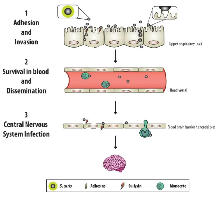
As represented in Figure B, S. suis pathogenesis can be simplified to 3 steps. Firstly, bacteria adhere and invade the epithelial cell layer of the upper respiratory tract (colonization), penetrate mucosal barriers, and gain access to blood circulation. Once in the bloodstream, S. suis can travel as free bacteria or associated to monocytes (bound and/or intracellular) and disseminate to organs such as spleen, liver, kidney, lung, and heart. If bacteremia is not controlled, it can lead to septicemia and/or septic shock. Finally, if host death by sepsis does not take place and bacteremia remains high, S. suis can also reach the central nervous system (CNS) and cross the barrier made of the brain microvascular endothelial cells and/or epithelial cells of the choroid plexus, causing meningitis (14). These 3 steps will be further explained in the next sections.
17
18
1.5.1 STEP 1: Adhesion and invasion
As mentioned before, S. suis enters the natural host (pigs) via the upper respiratory tract and resides within the palatine or pharyngeal tonsils. Once colonized, some animals may remain healthy carriers, whereas others will develop bacteremia, septicemia, and/or meningitis. For these events to happen, the bacteria need to cross the epithelial barrier in order to reach the bloodstream (14). Regarding human infections, the bacterium enters mainly through skin wounds, thus having direct access to blood circulation. However, in cases of oral route of infection bacteria may interact with the mucosal epithelial cells in the intestine (100).
Despite many studies, it is still not completely understood how S. suis is able to breach the mucosal epithelia in the upper respiratory tract. S. suis possess multiple adhesins that allow interaction with the host. Generally, adhesion to host cells is mediated via hydrophobic interactions, cation-bridging, and receptor–ligand binding. Moreover, interaction with components of the extracellular matrix such as fibronectin, laminin and collagen can be used by bacteria to favorize interactions with cells (100). However, few studies have investigated the interactions between S. suis and epithelial cells and results suggest very low levels of adhesion and invasion (91, 101).
Norton et al. suggested that suilysin-positive S. suis strains can use invasion and cell lysis as a mechanism to breach the mucosal epithelium (92). Nevertheless, strains not producing this hemolytic toxin are also able to reach the circulation and disseminate (102), suggesting that other mechanisms could also be in play.
1.5.2 STEP 2: Survival in blood and dissemination
To disseminate throughout the host and cause bacteremia and septicemia, bacteria must be able to survive in blood. As mentioned before, the CPS is a key factor for the bacteria since its presence protects bacterium against the actions of the host immune innate system.
There are several hypotheses about how bacteria travel in the bloodstream. The one proposed by Williams and Blakemore in 1990 was named the “Trojan horse theory” (103). This model describes the uptake of bacteria by monocytes (in the absence of specific antibodies) followed by intracellular survival and invasion of organs or the CNS. Though the capsule might protect against phagocytosis, it could still allow an association of S. suis with these cells. Indeed,
19
a relatively high level of adhesion (without phagocytosis) of S. suis to phagocytic cells has recently been observed (104). Thus, traveling and dissemination to target organs by exploitation of host cells as “vehicles” was postulated, the so called “modified Trojan horse theory”. Yet, is it also possible that extracellular S. suis bacteria travel free in circulation (14).
The interaction of S. suis with phagocytic cells in the bloodstream, and/or in organs is crucial for the development of the inflammatory response. It has been demonstrated that S. suis is capable of up-regulating important adhesion molecules and also several pro-inflammatory cytokines and chemokines (e.g. tumor necrosis factor alpha [TNF], IL-6, IL-1, IL-8, and monocyte chemotactic protein-1 [MCP-1]) in vivo and in vitro (71-73, 105, 106). This may result in multiple systemic effects, including contributing to the recruitment of leukocytes to the site of infection, increasing hematopoiesis and inducing fever. However, although activation of the immune system and synthesis of cytokines and chemokines contribute to the anti-infective process, their uncontrolled production can have adverse consequences for the host, leading to multi-organ failure and septic shock like syndrome.
1.5.3 STEP 3: Central nervous system invasion: Meningitis
To develop meningitis, S. suis needs to reach the CNS, and most importantly, cross the blood-brain barrier (BBB). The BBB is an anatomical and functional barrier that separates the brain from the intravascular compartment and maintains the homeostasis of the CNS. If the theory of the Trojan horse or the modified Trojan horse are correct, S. suis would arrive to the BBB inside or associated with monocytes. As the CNS is considered to be an immune privileged organ, normal circulation of monocytes into the CNS is still controversial. However the permeability to some immune cells could be modified as an adaption to the specific local microenvironment (14).
Because of the particular characteristics of the BBB, it is generally accepted that bacterial interactions with brain microvascular endothelial cells (BMEC) are mainly characterized by specific bacterial attachment with consequent invasion, toxicity and/or increase permeability. Therefore, in the case that the bacteria reach the CNS freely, it must adhere to the brain epithelium and invade it. Studies with human BMEC demonstrated that S. suis could adhere to but not invade this type of cells (90). It is possible that after the adherence of S. suis
20
to BMEC, the bacteria secrete toxic factors which would affect the endothelial cells. Such factors would increase BBB permeability, which could lead to the development of cerebral edema, increased intracranial pressure, and cerebral blood flow blockage characteristic of bacterial meningitis. Suilysin has been reported to be toxic to the BMEC, which could contribute to increased BBB permeability (90, 95). In fact, purified SLY has been shown to induce the release of several pro-inflammatory cytokines by human and porcine BMEC (93, 95). On the other hand, is it possible that suilysin-negative strains adhere to BMEC and result in cytokine production that would also lead to alteration in its permeability (93, 94). It has been shown that S. suis induces the release of pro-inflammatory cytokines and chemokines by human and porcine BMEC, murine microglia, and astrocytes (94, 106-108). However, the specific bacterial components responsible for exaggerated inflammatory reactions are not accurately known. In addition to cell wall components, bacterial CPS induces human macrophages to secrete prostaglandin E2 (PGE2) and matrix metalloproteinase 9, which may also be involved in disruption of the BBB (109).
Another model of entry of S. suis to the CNS has been suggested to be through the blood-cerebrospinal fluid barrier. In this model, S. suis invades the choroid plexus epithelial cells (CPEC), then transported within membrane-bound endocytic vacuoles to the apical side and finally exit by exocytosis onto the apical membrane of the blood–CSF barrier (110). In fact, a study with CPECs showed that S. suis induces necrosis and apoptosis (110). In addition, the translocation across the blood–CSF barrier activates neutrophils and affects the barrier´s function and integrity, further facilitating trafficking of bacteria and leukocytes (111).
2 Innate immune response
The vertebrate immune system comprises the innate and the adaptive immune system. While adaptive immunity is involved in the elimination of pathogens in the later phase of infection, and in the generation of immunological memory, the innate immunity is rapidly activated and represents the first line of defense against microorganisms. Therefore, it plays a crucial role in the early recognition of microbes and subsequent triggering of a pro-inflammatory response (112, 113). This system is composed by physical and chemical barriers, including the epidermis, the ciliated respiratory epithelium, the vascular endothelium, and mucosal surfaces with antimicrobial secretions. Additionally, among the cellular components, the innate system
21
includes macrophages, granulocytes, cytotoxic natural killer cells (NK), dendritic cells (DCs) and γδ T lymphocytes (114).
Conserved microbial motifs known as pathogen-associated molecular patterns (PAMPs) are recognized by specialized innate immune receptors, also known as pathogen recognition receptors (PRRs). The activation of the PRRs by pathogens leads to activation of the complement and coagulation cascades, opsonization and phagocytosis. Additionally, it will also activate diverse signaling pathways such as nuclear factor–kappa B (NF-κB) and mitogen-activated protein kinases (MAPKs), which promote the synthesis of a broad range of molecules, including cytokines, chemokines, cell adhesion molecules, and immunoreceptors, which together orchestrate the early antimicrobial host response (112-117).
2.1 Pattern recognition receptors
Currently, five different families of PRRs have been identified. These families include transmembrane proteins such as the Toll-like receptors (TLRs), C-type lectin receptors (CLRs), and the receptor kinases, as well as cytoplasmic proteins such as the retinoic acid-inducible gene (RIG)-I-like receptors (RLRs) and the nucleotide-binding oligomerization domain (NOD)-like receptors (NLRs). Among these, the TLRs and the NLRs are the most studied in the context of bacterial infections and will be described below.
2.1.1 Toll-like Receptors
The TLRs are a family of type I transmembrane proteins conserved from insects to humans (118). The cytosolic domain is termed the Toll/IL-1 receptor (TIR) domain and is the defining motif of the superfamily. Additionally, the extracellular portion formed by leucine-rich repeats (LRRs) is responsible for the recognition of PAMPs. Several TLRs have been reported in humans and mice (TLRs 1-13). These receptors are not only expressed in immune cells, but also in a variety of other types, including vascular endothelial cells, adipocytes, cardiac myocytes, and intestinal epithelial cells. Moreover, their expression can be modulated in response to a variety of stimuli (119).
Based on their cellular location and their ligands, TLRs are classically defined into two subgroups. TLR1, TLR2, TLR4, TLR5, TLR6, and TLR11 compose the first group. They are
22
expressed on the cellular surface and recognize main components of the microbial membrane like lipids, lipoproteins, and proteins. The other group is formed by TLR3, TLR7, TLR8 and TLR9 which are expressed in intracellular vesicles, like the endoplasmic reticulum, endosomes, and lysosomes, and recognize mainly nucleic acid from pathogens (17).
As mentioned before, after PAMP recognition, TLRs activate different intracellular signalling pathways that initiate and orchestrate the immune responses. These pathways begin with the recruitment of different adaptor molecules inside the cell. If the myeloid differentiation primary response 88 (MyD88)-dependent pathway is engaged, the cytosolic TIR domains of TLR1, TLR2, and TLR6 assemble with two proteins: MyD88 and the interleukin-1 receptor-associated kinase 4 (IRAK4). Consequently, IRAK4 undergoes (auto) phosphorylation and activates kinases IRAK1 and IRAK2. This step is followed by the recruitment of TNF receptor-associated factor 6 (TRAF 6) and the consequently activation of one of two possible signaling pathways: NF-κB activation or mitogen-activated protein (MAP) kinases activation (120) (121) which include the p38, c-Jun N-terminal kinase (JNK), and extracellular-signal-regulated kinase (ERK) pathways.
On the other side, TLR3 and TLR4, use the TIR-domain-containing adapter-inducing interferon-β (TRIF) and the TIR domain-containing adaptor protein (TIRAP) as adaptor molecule instead of MyD88. This would lead to an alternative activation of NF-κB leading to the induction of inflammatory cytokines. TLR2 and TLR4 can also use TIRAP as an adaptor protein supplementary for the recruitment of MyD88 (120, 122).
2.1.2 NOD-like Receptors
The nucleotide-binding oligomerization domain (NOD)-like receptors (NLRs) are a specialized group of intracellular receptors that, like the TLRs, represent a key component of the host innate immune system. This family of proteins is defined by a tripartite structure consisting of: (a) a variable N-terminal protein-protein interaction domain, defined by the caspase recruitment domain (CARD), pyrin domain (PYD), acidic transactivating domain, or baculovirus inhibitor repeat (BIR); (b) a central NOD domain, which mediates self-oligomerization during activation (4); and (c) a C-terminal LRR that detects PAMPs (123). Similarly to TLRs, NLRs are expressed in immune cells, including both lymphocytes and
23
antigen-presenting cells (APCs) such as macrophages and dendritic cells, but also in non-immune cells, including epithelial and mesothelial cells (123).
Likewise, stimulation of the NLRs activates signaling pathways leading to production of pro-inflammatory mediators. The MAPK and NF-κB signaling pathways are two of the main targets of the NLRs, although not through the same adaptor proteins. Additionally, there is a third target that was not seen with the TLRs, which is activation of caspase-1 leading to IL-1β and IL-18 maturation. The complexes in charge of this activation are called inflammasomes and will be explained in detail in section 3: Interleukin 1 (123). In summary, they have the ability to regulate NF-κB and MAPKs signaling indicating that they can have an important role in the pathogenesis of a variety of inflammatory human diseases (123).
NOD1 and NOD2 are the best characterized intracellular receptors in the NLRs family. NOD1 is ubiquitously expressed, while NOD2 expression is restricted to monocytes, macrophages, dendritic cells, and intestinal Paneth cells (124). They recognize PG moieties found in bacteria. NOD1 recognizes d-γ-glutamyl-meso-diaminopimelic acid (iE-DAP) (125, 126) found in the structures of all Gram-negative and some Gram-positive bacteria like B. subtilis and L. monocytogenes. On the other side, NOD2 recognizes muramyl dipeptide (MDP) a component of the PG present in all the Gram-positive and Gram-negative bacteria (127, 128).
2.1.3 Importance of the PRRs in S. suis infection
Studies involving PRR activation after S. suis infection have demonstrated that mainly TLRs, particularly TLR2, TLR4, and TLR9, are implicated in its recognition. TLR2 is a versatile receptor, as it recognizes a variety of components, including lipoproteins, PG and LTA from Gram-positive bacteria. Using TLR2 deficient mice, Lachance et al. demonstrated an increased survival rate and a significant decrease of pro-inflammatory mediators after S. suis high virulent ST1 infection that could not be associated with a lower bacterial burden. These results suggested that there might be other receptors involved. Contrarily, when TLR2 deficient mice were challenged with epidemic high virulent ST7 strain no significant difference in survival and production of mediators in comparison with wild-type mice was observed. Together these results showed that infection of mice by highly pathogenic strains of S. suis may follow TLR2-dependent or inTLR2-dependent pathways depending on the strain (129).
24
In vitro studies with murine macrophages and dendritic cells deficient in TLR2 showed a reduction in the secretion of pro-inflammatory products in response to encapsulated S. suis (105, 130). In this case, the response was totally abrogated in MyD88-deficient cells suggesting that a second pathway including other TLRs may play a role and might also participate directly or indirectly in the activation of the inflammatory cascade (105, 130). In addition, recent studies on peripheral blood mononuclear cells (PBMCs) showed an implication of the TLR2, TLR6, and TLR9 but not of TLR1 in response to S. suis ST1 and ST7 (131). The interactions with these receptors resulted in the release of several pro-inflammatory mediators, such as IL-6 and TNF, with the use of blocking antibodies for the TLRs reducing their concentrations (131). Moreover, a recent study with S. suis strains ST1, ST7 and ST25 showed the implication of TLR7 and TLR9 in the production of type-I interferon after recognition of genetic material (132).
Previous studies have demonstrated that TLR4 recognizes not only LPS from Gram-negative bacteria, but also certain toxins such as pneumolysin (133), listeriolysin O (134), and more recently S. suis-produced SLY (135). However, the capacity of S. suis and/or SLY to activate TLR4 remains controversial. In a study with whole live bacteria, the role of TLR4 in cytokine production was minimal since the production of only CXCL1 by DCs deficient in TLR4 was affected. On the other hand, a study with purified native SLY observed TLR4-dependent TNF production by peritoneal macrophages (135), suggesting that conditions tested, including concentration of toxin, could influence the results.
In the case of NOD receptors, their importance in the immunopathogenesis of infection was demonstrated for streptococci such as S. pneumoniae and S. pyogenes (136, 137). Unfortunately, there are not many studies about their role in the infection with S. suis. Lecours et al. studied dendritic cells deficient in NOD2 and demonstrated that only the release of IL-23 and CXCL1 was partially dependent on this receptor (105). In summary, the host response to S. suis is not limited to a specific receptor but requires a multimodal recognition system.
2.2 Inflammatory mediators
The secretion of cytokines and chemokines is one of the first steps necessary for the initiation of the innate immune response. They bind to specific receptors located in target