HAL Id: hal-01230394
https://hal.archives-ouvertes.fr/hal-01230394
Submitted on 19 Jul 2018
HAL is a multi-disciplinary open access
archive for the deposit and dissemination of sci-entific research documents, whether they are pub-lished or not. The documents may come from teaching and research institutions in France or abroad, or from public or private research centers.
L’archive ouverte pluridisciplinaire HAL, est destinée au dépôt et à la diffusion de documents scientifiques de niveau recherche, publiés ou non, émanant des établissements d’enseignement et de recherche français ou étrangers, des laboratoires publics ou privés.
Synthesis, Cytotoxicity, and COMPARE Analysis of
Ferrocene and [3]Ferrocenophane Tetrasubstituted
Olefin Derivatives against Human Cancer Cells
Meral Goermen, Pascal Pigeon, Siden Top, Elisabeth A. Hillard, Michel
Huché, Christian G. Hartinger, Frédéric Montigny, Marie-Aude Plamont,
Anne Vessières, Gérard Jaouen
To cite this version:
Meral Goermen, Pascal Pigeon, Siden Top, Elisabeth A. Hillard, Michel Huché, et al.. Synthesis, Cytotoxicity, and COMPARE Analysis of Ferrocene and [3]Ferrocenophane Tetrasubstituted Olefin Derivatives against Human Cancer Cells. ChemMedChem, Wiley-VCH Verlag, 2010, 5 (12), pp.2039-2050. �10.1002/cmdc.201000286�. �hal-01230394�
Synthesis, Cytotoxicity, and COMPARE Analysis of
Ferrocene and [3]Ferrocenophane Tetrasubstituted
Olefin Derivatives against Human Cancer Cells
Meral Görmen,[a] Pascal Pigeon,[a] Siden Top,*[a] Elizabeth A. Hillard,*[a] Michel Huché,[a] Christian G. Hartinger,[b] Frédéric de Montigny,[a] Marie-Aude Plamont,[a] Anne Vessières,[a] and Gérard Jaouen[a]
[a] M. Görmen, Dr. P. Pigeon, Dr. S. Top, Dr. E. A. Hillard, Dr. M. Huché, Dr. F. de Montigny, M.-A. Plamont, Dr. A. Vessières, Prof. G. Jaouen Chimie ParisTech (Ecole Nationale Supérieure de Chimie de Paris) Laboratoire Charles Friedel, UMR CNRS 7223 11 rue Pierre et Marie Curie, 75231 Paris Cedex 05 (France) Fax: (+33) 143-260-061 E-mail: siden-top@chimie-paristech.frelizabeth-hillard@chimie-paristech.fr
[b] Dr. C. G. Hartinger Institute of Inorganic Chemistry University of Vienna, Waehringer Str. 42, 1090 Vienna (Austria)
Keywords: bioorganometallic chemistry, cell culture, cytotoxicity, drug discovery, ferrocenes
Herein we report the antiproliferative effects of a series of 28 compounds against the MDA-MB-231 breast cancer cell line, including the synthesis of seven new [3]ferrocenophanyl and four new ferrocenyl compounds. For each p-R-phenyl substitution pattern investigated, the [3]ferrocenophanyl derivatives were more cytotoxic than the corresponding ferrocenyl derivative, with the highest activity found for compounds with protic substituents. Theoretical calculations of the HOMO-LUMO gap for the molecules in the Fe3+ oxidation state suggest a higher reactivity for the [3]ferrocenophanyl derivatives. A lead compound from each series, a [3]ferrocenophanyl and a ferrocenyl compound, possessing two phenol groups, were screened against the NCI/DTP 60-cell-line panel. The mean activity over all cell lines was better than cisplatin for both compounds, and both compounds showed subpanel selectivity for leukemia, CNS cancer, and renal cancer. Low systemic toxicity and lack of interaction with DNA (when in the reduced form), suggest that the compounds may act as prodrugs.
Introduction
The development of organometallic compounds for cancer therapeutics is one of the fastest growing areas of bioorganometallic chemistry.[1-4] These molecules can be generally categorized based on their mode of action, for example those where direct interaction of the metal with a biological target, such as DNA or proteins after ligand hydrolysis, is implicated.[5-9] Modeled after the action of cisplatin, these metal-centered organometallic compounds, notably containing ruthenium[9, 10] or titanium,[11] possess judiciously chosen ligands or substituents on the phenyl ring to optimize the pharmacokinetic properties of the molecule. Another class demonstrates the enthusiasm researchers have for grafting metallocenes and metal carbonyls to a variety of biomolecules to modify their biological effects.[12] These therapeutic bioconjugates include steroidal[13-15] and nonsteroidal[16-23] endocrine modulators, natural products,[24-27] and others.[28-34] In these cases, the often covalently grafted organometallic unit is usually inert to ligand substitution, but potentiates the activity of the biomolecule via modification of the pharmacokinetic profile or acts as a structural mimic.[35] A variety of other compounds are between these two classes, possessing labile biomolecule ligands, such as nucleobases.[33, 36-41] Currently in press, the book “Medicinal Organometallic Chemistry” demonstrates that the growth of structural and mechanistic diversity of organometallic compounds for cancer therapy is in full bloom.[42]
Among organometallic compounds based on endocrine modulators, the most active and well-studied are the ferrocenyl derivatives of tamoxifen (with or without the side chain present).[43] We have shown that the reversible ferrocene/ferricenium redox couple can catalyze the intramolecular formation of quinones when the ferrocenyl and phenol groups are linked by a conjugated system.[44] This mechanism is not limited to phenolic compounds: ferrocenyl moieties conjugated to an aniline or acetanilide group also show high activity against cancer cells, with IC50 values of the order of 10-7M.[45] The structural requirements for
activity seem to be 1) the presence of a ferrocene group, 2) a conjugated linker, 3) aromatic para substitution by a protic function, and 4) an ethyl group residing on the same carbon as the ferrocene group.
As a part of our studies of organometallic compounds with this motif, [3]ferrocenophanes have recently emerged as a class of notable antiproliferative agents. The [3]ferrocenophane
structure consists of a ferrocene, where the two cyclopentadienyl rings are linked by a three atom bridge, in this case carbon atoms. Molecules possessing this motif have been discussed in reviews,[46, 47] and have been studied in the context of their structures,[48-57] catalytic properties,[56, 58-63] and conjugation to biomolecules,[64, 65] inter alia. For the [3]ferrocenophane compounds based on the tamoxifen skeleton, it is supposed that their biological activity is due to a mechanism of activation similar to that of their ferrocenyl analogues. Among organometallic compounds studied so far, the highest in vitro cancer activity is exhibited by those based on the 1-(diphenylmethylidene)-[3]ferrocenophane skeleton, with para substitution on both phenyl rings[66-68] (Table 1).
Table 1. Compounds based on the 1-(diphenylmethylidene)-[3]ferrocenophane skeleton
that show high cytotoxicity against hormone-independent breast and prostate cancer cells.
R1 R2 IC50 [µM] MDA-MB-231 PC3 OH OH 0.09 ± 0.01 0.14 ± 0.01 NH2 OH 0.06 ± 0.01 0.03 ± 0.01 NHAc OH 0.09 ± 0.02 0.02 ± 0.00 NH2 NH2 0.05 ± 0.01 0.05 ± 0.00
For compounds possessing a phenol, an aniline, and/or an acetanilide, with all other parameters being equal, the presence of a [3]ferrocenophane in lieu of an n-propylferrocene yields more toxic molecules. As this phenomenon has been studied with a limited number of compounds, herein we report the results from a 28-molecule structure-activity study, which involved the synthesis and biological evaluation of 11 new compounds. These molecules are all based on the 1-(diphenylmethylidene)-[3]ferrocenophane or the
1-(diphenylmethylidene)ethylferrocene skeleton and were tested on hormone-independent MDA-MB-231 cells. A lead compound (with two hydroxy substituents) from each series was further evaluated by the National Cancer Institute Developmental Therapeutics Program (NCI/DTP) against their panel of 60 human tumor cell lines, and a COMPARE analysis was performed on these results. Cytotoxicity results have also been interpreted in light of theoretical calculations on the HOMO-LUMO gap of the oxidized molecule (Fc+) and the interaction with DNA of a reduced molecule.
Results
Synthesis
A simple and key step to obtain the desired tetrasubstituted olefins involves a McMurry cross-coupling reaction between the appropriate benzophenone and [3]ferrocenophan-1-one or propionyl ferrocene to generate the [3]ferrocenophanes (1a-17a) and the corresponding ferrocenes (1b-15b), respectively (Scheme 1, Table 2). The reaction time depends on the substituents and can vary from 90 min to three days. One caveat of the McMurry reaction is that there is a competition between the formation of the desired cross-coupled product and the two homo-coupled compounds, and this is reflected in the variety of the reaction yields. In most cases, the reactions with [3]ferrocenophan-1-one were more efficient than those using propionyl ferrocene. This can be attributed to the great avidity propionyl ferrocene shows for itself, resulting in a relatively high proportion of homo-coupled diferrocenyl species under McMurry conditions.
Table 2. Reaction conditions, yields, and isomer ratios of selected compounds.[a]
Compd R1 R2 TiCl4 [equiv] Zn[equiv] treaction [h] Yield [%] Isomer ratio Ref.
1a H H 4 6 2 93 n.a. [68]
1b H H 3 6 2 25 n.a. [84]
(E+Z)-2a OH H 4 6 2 74 81:19 [67] (E+Z)-2b OH H 3 6 2 26 50:50 [22] (E+Z)-3a OAc H Acetylation of 2a 66 82:18
(E+Z)-3b OAc H Acetylation of 2b 47 54:46
(E+Z)-4a NH2 H 5 7 48 54 87:13 [67]
(E+Z)-4b NH2 H 3 6 4 51 55:45 [45]
(E+Z)-5a NHAc H Acetylation of 4a 75 80:20 [67] (E+Z)-5b NHAc H Acetylation of 4b 91 60:40 [45]
6a OH OH 4 6 2 55 n.a. [66]
6b OH OH 4 6 2 53 n.a. [85]
(E+Z)-7a NH2 OH 4 7 24 21 58:42 [68]
(E+Z)-7b NH2 OH 13 24 4 62 50:50
(E+Z)-8a NHAc OH Acetylation of 7a 63 75:25 [68] (E+Z)-8b NHAc OH Acetylation of 7b 86 50:50
9a NH2 NH2 Deprotection of 11a 78 n.a. [68]
9b NH2 NH2 various various various 0 n.a.
10a NHAc NHAc 5 7 72 44 n.a [68]
10b NHAc NHAc various various various 0 n.a. (E+Z)-11a OAc OH Protection of 6a 46 54:46 (E+Z)-11b OAc OH Protection of 6b 65 50:50
12a OAc OAc Protection of 6a 33 n.a.
12b OAc OAc Protection of 6b 90 n.a. [86] (E+Z)-13a Br H 4 6 3.5 89 62:38
(E+Z)-13b Br H 3 6 4 13 64:36 [72]
14a Br Br 4 6 3.5 81 n.a.
14b Br Br various various various 0 n.a.
15a CN H Cyanation of 13a 74 61:39
15b CN H Cyanation of 13b 62 67:33 [72]
16a CN CN Cyanation of 14a 44 n.a.
17a CN Br Cyanation of 14a 15 44:56 [a] McMurry reaction, if not otherwise indicated; n.a.=not applicable.
In some cases, the coupling reaction did not yield the desired product. For example, the reaction between propionyl ferrocene and 4,4’-diaminobenzophenone did not lead to any trace of the expected 2-ferrocenyl-1,1-bis(4-aminophenyl)but-1-ene, 9b. Attempts to similarly obtain the NHAc-substituted compound 10b were likewise unsuccessful. However, when
propionyl ferrocene was replaced by [3]ferrocenophan-1-one, the amido compound 10a was obtained in moderate yield, although only after a reaction time of 72 h. On the other hand, the reaction between [3]ferrocenophan-1-one and 4,4’-diaminoacetophenone did not yield any desired product and therefore 9a was instead obtained by deacetylation of 10a. The McMurry reaction between propionyl ferrocene and the dibromobenzophenone also failed to yield the desired product 14b. This is surprising, as the reaction between [3]ferrocenophan-1-one and dibromobenzophenone gave the desired 14a in high yield. It should be mentioned that the failure of the McMurry reaction was only observed for the disubstituted compounds; the monosubstituted NH2 (4), NHAc (5) and Br (13) derivatives were obtained in both the
ferrocenophane and ferrocene series without difficulty.
We routinely used an excess of TiCl4 for the McMurry reaction, between three and five
equivalents, and a 1:1 proportion of the ferrocenyl ketone and benzophenone. In the case of
4a and 10a, five equivalents of TiCl4 were used, because our experience shows that the
nitrogen-containing benzophenones are less reactive than the corresponding hydroxy benzophenones. This is also reflected in the long reaction time necessary to obtain these compounds. In some cases amino-substituted compounds were obtained by an in situ reduction of nitro-substituted benzophenones. We compared the reactions using such reagents for the synthesis of 4a and 4b. For 4a, 4-aminobenzophenone gave a yield of 54% after two days of reaction and 4-nitrobenzophenone gave a yield of 21 %. For 4b, the trend was reversed, with yields of 26% and 51%, using 4-amino and 4-nitrobenzophenone, respectively. In the case of 7a and 7b, 4-nitro-4-hydroxybenzophenone, formed via a Friedel-Crafts reaction, was used directly.[68] In this case, a large excess of Zn was used to reduce the NO2 to
Scheme 2. Cyanation of monosubstituted and disubstituted bromo derivatives.
Nitriles 15a, 15b, and 16a were obtained by treating the corresponding bromo derivatives with CuCN (Scheme 2). Compound 16b was inaccessible, as its precursor 14b was not obtained via the McMurry reaction as previously discussed. In our hands the cyanation of 14a was incomplete, giving access to the mixed CN/Br derivative 17a in 15% yield with a 44:56 isomer ratio (this compound was not tested for its antiproliferative effects).
X-ray crystallography of 7b
X-ray quality crystals of E-7b were obtained from a mixture of E and Z isomers in an EtOH/H2O solution by slow evaporation. Reflections were recorded using MoKα radiation at
200 K. The ORTEP diagram, with selected bond distances and angles, is shown in Figure 1 and crystallographic data are available from the Cambridge Crystallographic Data Centre (CCDC-783005).[70] The crystal structure shows almost no distortion around the ferrocene group, but a significant deviation from planarity around the ethylene center, with an average torsion angle of 13°. The phenyl rings are twisted out of the plane created by C1-C15-C21 by 52.8 and 48.1° which is typical of such compounds.[67, 71]
Figure 1. ORTEP diagram of (E)-7b. Thermal ellipsoids shown at 50% probability level, and
hydrogen atoms have been omitted for clarity. Selected bond distances (Å) and bond angles (°): Fe1–CCp=2.061(2), C1–C2=1.372(2), C18–O1=1.376(2), C24–N1=1.420(2), Fe1–
CCentroid=179.25, Cp–Cp=2.34; C21=164.1, C5-C2-C1-C15=169.8,
C3-C2-C1-C15=_13.54, C5-C2-C1-C21=-12.58.
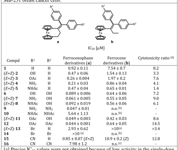
Table 3. Cytotoxicity of compounds 1–16 against hormone-independent
MDA-MB-231 breast cancer cells.
IC50 [M]
Compd R1 R2 Ferrocenophane derivatives (a) derivatives (b) Ferrocene Cytotoxicity ratio [d]
1 H H 0.92 ± 0.11 7.54 ± 0.7 8.2 (E+Z)-2 OH H 0.47 ± 0.06 1.54 ± 0.13 3.3 (E+Z)-3 OAc H 0.26 ± 0.004 1.97 ± 0.2 7.6 (E+Z)-4 NH2 H 0.21 ± 0.03 0.86 ± 0.04 4.1 (E+Z)-5 NHAc H 0.47 ± 0.04 0.65 ± 0.01 1.4 6 OH OH 0.089 ± 0.006 0.64 ± 0.06 7.2 (E+Z)-7 NH2 OH 0.061 ± 0.005 0.55 ± 0.05 9.0 (E+Z)-8 NHAc OH 0.092 ± 0.019 0.56 ± 0.06 6.1 9 NH2 NH2 0.047 ± 0.01 n.o. [b] -
10 NHAc NHAc 5.64 ± 1.13 n.o. [b] -
(E+Z)-11 OAc OH 0.049 ± 0.003 0.42 ± 0.03 8.6 12 OAc OAc 0.044 ± 0.001 0.64 ± 0.05 14.5 (E+Z)-13 Br H 2.93 ± 0.62 >10[a] >3.4 14 Br Br >10 [a] n.o. [b] - 15 CN H 0.85 ± 0.07 (E+Z) 10.9 ± 0.2 (Z) 12.8 16 CN CN 7.98 ± 1.2 n.s. [c] -
[a] Precise IC50 values were not obtained because of low activity in the single-dose
test (13b: 86% cell viability at 10-5M, 14a: 89% cell viability at 10-5M); [b]
n.o.=molecule not obtained; [c] n.s.=molecule not synthesized; [d] IC50(b)/IC50(a).
Antiproliferative effects on MDA-MB-231 cells
All new compounds, except 17a, were screened for their activity against the MDA-MB-231 human breast cancer cell line. This line is hormone independent, that is, it does not express the estrogen receptor, and therefore cell culture results are not confounded with estrogenic or antiestrogenic effects. Cells were incubated with at least three concentrations of the test compound for five days, and cell viability was determined by the staining of proteins in living cells. Cytotoxicity results (IC50 values) are given in Table 3. In all cases, the ferrocenophane
NHAc-substituted compound 5, the difference between the two compounds is small, but significant. Conversely, the diacetoxy compound 12a is almost 15 times more potent than its ferrocenyl analogue. The difference in cytotoxicity results for the nitriles 15a and 15b is even more dramatic than shown in Table 3. With an IC50 value of 60 µM, the E isomer of 15b is much
less active than its Z isomer.[72] Therefore, given a 50:50 mixture of isomers for 15b, one could predict a cytotoxicity ratio of approximately 40.
Antiproliferative effects of 6a and 6b on the NCI cell line panel
Compounds 6a and 6b were selected for further study on the human tumor cell line panel of the Developmental Therapeutics Program of the National Cancer Institute (Bethesda, MD, USA).[73] The compounds were tested on approximately 60 cell lines within nine tumor type subpanels. The cells were treated for 48 h at five concentrations ranging from 0.01 to 100 µM. Three endpoints were calculated: GI50 (the concentration where the compound inhibits cell
growth by 50%), TGI (the concentration at which the compound inhibits cell growth by 100 %), and LC50 (the concentration at which the drug decreases the original cell number by
50%), shown in Figure 2, as well as the MG-MID (full-panel mean-graph midpoint). Compounds 6a and 6b have a broad spectrum of activity, with MG-MID values (representing the average of the MG-MIDs for two experiments, using almost identical panels) lower than those for cisplatin (Table 4). The correlation between the full-panel results for the two compounds was 78% for GI50, 58% for TGI, and 58% for LC50 values.
Table 4. Full-panel mean-graph midpoint (MG-MID) GI50, TGI, LC50 values of
compounds in the NCI cell panel.[a]
Compd GI50 [µM] TGI [µM] LC50 [µM]
6a 0.18 0.03 11 1 52 3
6b 0.52 0.04 4.3 0.4 23 2 Cisplatin[b] 10.3 50.7 90.5
[a] Data from two experiments. [b] Data from NCI/DTP screening: October 2009, maximum concentration: 100 µM, after 48 h.
The GI50 results varied over approximately three orders of magnitude for 6a, and two
orders of magnitude for 6b, suggesting a more varied selectivity profile for 6a. Comparing the mean GI50 for each histological subpanel gives an indication of tumor selectivity, and a
MG-MID for all cell lines (here 0.18 µM for 6a and 0.52 µM for 6b). Table 5 shows the mean values over each subpanel for two different experiments. Compound 6a is selective for leukemia, CNS cancer, and renal cancer, and was particularly active against melanoma M14, renal cancer ACHN, and CNS cancer SF-539, with two experiments giving GI50 values less
than 20 nM. Compound 6b also showed selectivity for leukemia, CNS cancer, and renal cancer. Similar to 6a, this compound was particularly active against ACHN and SF-539, but also against UACC-62 melanoma, HL60 (TB), and SR leukemia cell lines with GI50 values
falling below 0.2 µM for two experiments. Compound 6a was more active than 6b for every subpanel of cells. The most dramatic difference in activity was noted for the renal cancer subpanel.
Table 5. Histological subpanel mean-graph midpoint (MG-MID) GI50 values of
compounds in the NCI cell panel.[a]
Cell panel 6a(exp. 1) 6a(exp. 2) 6b(exp.1) 6b(exp. 2) 6b/6a
Leukemia 0.14 0.11 0.27 0.17 1.8 NSCL Cancer 0.31 0.16 0.84 0.50 2.9 Colon Cancer 0.40 0.34 0.80 0.61 1.9 CNS Cancer 0.074 0.092 0.31 0.29 3.6 Melanoma 0.19 0.15 0.83 0.55 4.1 Ovarian Cancer 0.43 0.30 1.09 0.53 2.2 Renal Cancer 0.072 0.048 0.43 0.20 5.3 Prostate Cancer 0.41 0.048[b] 0.78 0.56 1.6 Breast Cancer 0.58 0.49 1.0 0.84 1.7 [a] Data shown are from two experiments and are the ratio of averaged values (µM); [b] data only for PC3 (excluded from averaging).
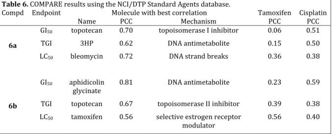
Table 6. COMPARE results using the NCI/DTP Standard Agents database.
Compd Endpoint Molecule with best correlation Tamoxifen Cisplatin
Name PCC Mechanism PCC PCC
6a
GI50 topotecan 0.70 topoisomerase I inhibitor 0.06 0.51
TGI 3HP 0.62 DNA antimetabolite 0.15 0.50 LC50 bleomycin 0.72 DNA strand breaks 0.36 0.38
6b
GI50 aphidicolin
glycinate 0.81 DNA antimetabolite 0.23 0.59 TGI topotecan 0.67 topoisomerase II inhibitor 0.39 0.38 LC50 tamoxifen 0.56 selective estrogen receptor
modulator 0.56 0.40
To gain insight into a possible mechanism of action for 6a and 6b, a COMPARE analysis[74] was carried out against the NCI/DTP Standard Agents database, a collection of
171 known molecules. The three endpoints for each seed molecule were compared with the same endpoint for the database molecules (GI50, TGI, and LC50). The molecule with the best
correlation [Pearson correlation coefficient (PCC)] and the values for tamoxifen and cisplatin are given in Table 6. The full-panel correlations with cisplatin and tamoxifen were weak (R2<0.6) for all endpoints. The best PCCs for each endpoint belong to molecules which inhibit DNA replication or cause DNA damage.
Each standard agent in the database appears with an associated mechanism: alkylating agent, antimitotic agent, DNA antimetabolite, RNA/DNA antimetabolite, topoisomerase II inhibitor, or not given/other. Averaging over the top 20 hits in each category over each endpoint, the dominant suggested mechanism for 6a is RNA and/or DNA antimetabolite (36 %), followed by an DNA alkylation (32 %), not given/other (20 %), topoisomerase II inhibition (9 %), or as a antimitotic agent (3 %). The dominant suggested mechanism for 6b was DNA alkylation (36 %), followed by acting as a RNA and/or DNA antimetabolite (22 %), topoisomerase II inhibition (18 %), not given/other (14 %), or as an antimitotic agent (9 %).
Figure 2. Mean graphs for compounds a) 6a and b) 6b from the National Cancer Institute
Developmental Therapeutic Program; data from one experiment shown.
Acute toxicity of 6a and 6b
Mice were treated by interperitoneal injection of 6a and 6b in DMSO by the NCI/DPT program.[75] One mouse was injected with 400 mg kg-1, the second with 200 mg kg-1, and the third with 100 mg kg-1 of the test compound. The mice were observed for two weeks and sacrificed if more than 20% weight loss or other signs of toxicity were noted. For 6a, the mice given doses of 400 and 200 mg kg-1 were sacrificed. For 6b, only the mouse given the highest dose of 400 mg kg-1 was sacrificed. Thus the determined maximum tolerated dose was 100 mg kg-1 for 6a and 200 mg kg-1 for 6b.
Interaction of 6b with nucleotides
Compound 6b (0.5 mM) was incubated in 10 mM NaH2PO4-Na2HPO4 buffer (pH 7.4) with
the nucleotides 5’-GMP, 5’-AMP, or 5’-CMP to obtain molar ratios of 1:2 complex/nucleotide). Analyses of the solutions were performed on a Hewlett-Packard
3D
Capillary Electrophoresis system equipped with a diode array detector at 200 nm for 10 h. As shown in Figure 3, there is no significant change in the intensity of the peaks over time, indicating that no adducts between the nucleotides and 6b were formed. Similar studies with transition metal complexes featuring leaving groups, including cisplatin and PtII analogues,[76] Ru anticancer drug candidates and developmental compounds,[77] exclusively form adducts with nucleobases, normally with highest binding preference to the N7 atoms of guanine and adenine.[78]
Calculation of HOMO-LUMO gaps for oxidized molecules
Relative differences in the HOMO-LUMO gaps of molecules can be used as a predictor of their reactivity.[79, 80] Using as a starting point our proposal that quinone methide metabolites of the test molecules are the toxic species, and that quinone methide formation involves an initial oxidation of ferrocene to ferricenium, we calculated the HOMO-LUMO gap of the oxidized (ferricenium) forms of the molecules, Table 7. The HOMO-LUMO gap of the ferrocenophanes is quite small, with an average of 1.6 eV and a range of 1.4-1.9 eV. The calculated HOMO-LUMO gaps for the oxidized ferrocene compounds are more variable, but consistently larger, with an average HOMO-LUMO gap of 6.2 eV and a range of 3.9 to 7.2 eV.
Discussion
The [3]ferrocenophane series is more active than the ferrocene series on the MDA-MB-231 cells and the 60 cell line panel overall. We previously proposed that quinone methides, formed via initial oxidation of the ferricenium moiety, could be the active metabolite in the cell, and higher cytotoxicity of the ferrocenophane molecules is consistent with this mechanism for the following reasons. Firstly, the additional electron density imparted to the ferrocene group by the bridge lowers the oxidation potential of the ferrocene, the first step in the proposed mechanism.[66] Secondly, due to the rigid conformation of the bridge, the formed quinone methide is expected to be highly strained and reactive.[67] Thirdly, Hartree-Fock
calculations suggest that the difference in activity between the ferrocenophane and ferrocene series could be due to differences in the HOMO and LUMO energy levels for the oxidized (Fc+) form of the compound. With a narrower HOMO-LUMO gap, the oxidized ferrocenophane complexes can more easily attain the first excited state and are therefore more reactive. These compounds could evolve to the quinone methide, when possible, or another reaction such as fragmentation giving rise to toxic molecules.
Indeed, the compounds that can be oxidized to a quinone methide, such as the hydroxy compounds 2 or 6, or quinone imines, such as 4 or 9, are very active. Although the best activity was provided by the diacetoxy 12a, this species should be easily transformed into 6a by hydrolysis in the cell. By contrast, comparison of the diamino compound 9a with diacetamido 10a shows a dramatic decrease in activity from 0.047 to 5.64 µM, probably because the hydrolysis of the acetamido compound is more difficult than the hydrolysis of acetoxy compound. The presence of an electron withdrawing group such as Br or CN decreases the antiproliferative activity for the ferrocene series, as previously observed,[72] and also for the [3]ferrocenophane compounds.
There are also several indications that these compounds act as prodrugs, that is, innocuous compounds which are activated in the cell to form toxic species. For example, NCI/DPT acute toxicity tests on mice for 6a and 6b showed mouse death only at high drug concentrations (200 mg kg-1 and 400 mg kg-1, respectively).[75] This suggests a selectivity for cancer cells which is consistent with our previous observation that 6b is 100x less toxic on normal cells than on cancer cells,[81] although it may also be due to a lack of bioavailability. Secondly, incubation of 6b with nucleotides did not show any direct interaction between the two, as measured by capillary zone electrophoresis (CZE). The low toxicity and lack of reaction with DNA, (though alteration of DNA after metabolism cannot be excluded), suggest that these compounds are only activated in cancer cells to form reactive species.
The COMPARE results suggest interference with DNA as the primary mechanism. These results should be regarded with caution, as mechanisms involving DNA dominate the pool of common anticancer agents. Direct attack on DNA is consistent with our proposed mechanism of quinone methide formation, as quinone methides are known to react with DNA via Michael addition.[82] Indeed, compound 6b has been shown to damage DNA, but only when incubated in living cells,[83] where it is presumably metabolized to the quinone methide or other active species. However, these results do not exclude the possibility of other targets. For example,
we have found (unpublished results) that the quinone methide forms of some of these compounds can react with glutathione and other thiols, such as those present in many proteins.
Conclusions
We have described the synthesis and antiproliferative effects for 11 new compounds based on the 1-(diphenylmethylidene)-[3]ferrocenophane or the 1-(diphenylmethylidenyl)ethylferrocene skeletons. Of the full 28-member library, compounds possessing the [3]ferrocenophane motif are, in every case, more active against the MDA-MB-231 hormone-independent breast cancer cell line than the corresponding ferrocenyl compounds. This is consistent with the smaller HOMO-LUMO gap present in the oxidized form of the [3]ferrocenophane compounds. Furthermore, those compounds which can form quinone or imine methides (with or without preliminary hydrolysis) are the most active of both the [3]ferrocenophane and ferrocene series. Results on the NCI/DPT 60 cell line panel show greater cell line selectivity for 6a than for 6b, although the qualitative subpanel selectivity patterns are similar. Possible mechanisms of action, as gleaned from a COMPARE analysis include the compounds acting as DNA alkylating agents or DNA antimetabolites. The low systemic toxicity and inertness with regard to DNA may be due to their putative behavior as oxidation-activated prodrugs.
Experimental Section
General remarks
All reactions were performed under argon using standard Schlenk techniques. Anhydrous THF was obtained by distillation from sodium/benzophenone. Thin layer chromatography was performed on silica gel 60 GF254. The preparative HPLC separations were performed on a
Shimadzu apparatus with a Nucleodur C18 column (l=25 cm, Ø=3.2 cm, particle size=10 µm)
using CH3CN as an eluent. The analytical HPLC controls were performed on a Shimadzu
apparatus with a Nucleodur C18 column (length of 15 cm, diameter of 0.45 cm, and particle
size of 5 µm) using CH3CN as an eluent. IR spectra were obtained on an IRFT BOMEM
Michelson-100 spectrometer equipped with a DTGS detector as a KBr plate. 1H and 13C NMR spectra were recorded on a 300 MHz Bruker spectrometer. Mass spectrometry was performed with a Nermag R 10-10C spectrometer. Elemental analyses were performed by the
microanalysis service of CNRS at Gif-sur-Yvette (France). HRMS measurements were performed with an LTQ-Orbitrap XL of Thermo Fischer used in positive mode with an electrospray source.
General synthesis of 7b, 13a, and 14a
Zinc powder was suspended in dry THF at room temperature, and TiCl4 was added slowly via
a syringe while stirring. The reaction mixture was held at reflux for 2 h, after which a THF solution containing the appropriate ketones was added, and reflux was continued. The reaction mixture was poured into H2O, acidified with HCl until the black precipitate
disappeared, and extracted with CH2Cl2. The organic layer was washed with H2O, dried over
magnesium sulfate, filtered, and concentrated under reduced pressure, and work-up was done as described below.
1-(4-aminophenyl)-1-(4-hydroxyphenyl)-2-ferrocenylbut-1-ene, 7b. Zinc powder (17 g,
260 mmol), TiCl4 (15.4 mL, 140 mmol), propionyl ferrocene (2.58 g, 10.67 mmol),
4-hydroxy-4’-nitrobenzophenone (2.59 g, 10.67 mmol), and reflux for 4 h. The reaction mixture was separated on a silica gel column with CH2Cl2 as the eluent. Recrystallization of the crude
product as a mixture of Z and E isomers (50:50) from CH3CN yielded 7b as an orange-red
solid (2.8 g, 62%). 1H NMR (300 MHz, [D6]DMSO): =0.99 and 1.02 (t, J=7.4 Hz, 3H,
CH3), 2.50 (q, J=7.4 Hz, 2H, CH2), 3.80 and 3.86 (t, J=1.8 Hz, 2H, C5H4), 4.07 and 4.08 (t,
J=1.8 Hz, 2H, C5H4), 4.09 and 4.11 (s, 5H, Cp), 4.99 and 5.01 (s broad, 2H, NH2), 6.42 and
6.54 (d, J=8.5 Hz, 2H, C6H4), 6.64 and 6.92 (d, J=8.5 Hz, 2H, C6H4), 6.71 and 6.73 (d, J=8.5
Hz, 2H, C6H4), 6.98 and 7.01 (d, J=8.5 Hz, 2H, C6H4), 9.25 and 9.31 (s, 1H, OH); 13C NMR
(75.4 MHz, [D6]DMSO): =15.4 (CH3), 27.1 (CH2), 67.7 (2CH C5H4), 68.6 (2CH C5H4), 68.9
(5CH Cp), 86.1 (Cip), 115.1 (2CH C6H4), 119.2 (2CH C6H4), 129.7 (2CH C6H4), 129.8 (2CH
C6H4), 134.9 (C), 136.0 (C), 137.3 (C), 139.4 (C), 149.2 (C), 155.6 (C); IR (KBr, ῠ cm-1):
3495, 3469, 3428, 3399, 3319 (OH, NH2), 3088, 2980, 2959, 2923, 2866 (CH2, CH3); MS
(CI, NH3) m/z: 424 [M+H]+; HRMS (EI, 70 eV, C26H25FeNO: [M]+) calcd: 423.1286, found:
423.1299; Anal. calcd for C26H25FeNO・0.5H2O: C 72.23, H 6.06, N 3.24, found: C 72.16, H
5.95, N 3.01.
1-[(4-bromophenyl)phenylmethylidene]-[3]ferrocenophane, 13a. Zinc powder (1.96 g, 30
mmol), TiCl4 (2.2 mL, 20 mmol), [3]ferrocenophane-1-one (1.2 g, 5 mmol),
CH3CN as eluent. Recrystallization from hexane yielded 13a as bright yellow crystals (2.08 g,
89%) as an unidentified mixture of Z and E isomers (62:38). 1H NMR (300 MHz, CDCl3):
=2.15 (s, 2H, CH2), 2.46 (s, 2H, CH2), 3.86 (s, 2H, C5H4), 3.89 (s, 2H, C5H4), 3.94 (s, 2H,
C5H4), 4.12 (s, 2H, C5H4), 6.72 (d, J=8.4 Hz, 2H, C6H4), 6.79 (d, J=8.4 Hz, 2H, C6H4),
6.88-7.33 (m, 5H, C6H5); 13C NMR (75.4 MHz, CDCl3): =28.7 (CH2), 40.6 and 40.8 (CH2), 69.1
(2CH C5H4), 69.7 (2CH C5H4), 70.4 (2CH C5H4), 70.5 (2CH C5H4), 83.3 (Cip), 86.9 (Cip),
120.1 and 120.7 (C-Br), 126.3 and 126.9 (CH C6H5), 127.5 and 128.3 (2CHarom), 129.3 and
131.0 (2CHarom), 130.5 (2CHarom), 131.4 and 132.2 (2CHarom), 135.7 (C), 139.5 and 139.6 (C),
142.0 and 142.5 (C), 142.6 and 143.0 (C); IR (KBr, ῡ cm-1): 3081, 2953, 2920, 2842 (CH2);
MS (EI, 70 eV) m/z: 468 [M]+. (79Br); Anal. calcd for C26H21BrFe: C 66.55, H 4.51, found: C
66.35, H 4.53.
1-[(di-4-bromophenyl)methylidene]-[3]ferrocenophane, 14a. Zinc powder (1.96 g, 30
mmol), TiCl4 (2.2 mL, 20 mmol), [3]ferrocenophane-1-one (1.2 g, 5 mmol),
di-p-bromobenzophenone (1.7 g, 5 mmol), and reflux for 3.5 h. Purification by HPLC with CH3CN as eluent. Recrystallization from CH2Cl2 yielded 14a as yellow crystals (2.21 g,
81%); mp: 142°C; 1H NMR (300 MHz, CDCl3): =2.33 (m, 2H, CH2), 2.64 (m, 2H, CH2), 3.99 (s, 2H, C5H4), 4.02 (s, 2H, C5H4), 4.08 (s, 2H, C5H4), 4.25 (s, 2H, C5H4), 6.90 (d, J=8.3, 2H, C6H4), 7.09 (d, J=8.3, 2H, C6H4), 7.21 (d, J=8.3, 2H, C6H4), 7.48 (d, J=8.3, 2H, C6H4); 13 C NMR (75.4 MHz, CDCl3): =28.9 (CH2), 41.0 (CH2), 69.0 (2CH C5H4), 69.6 (2CH C5H4), 70.4 (2CH C5H4), 70.6 (2CH C5H4), 83.0 (Cip), 86.7 (Cip), 120.5 (C-Br), 121.1 (C-Br), 130.7 (2CH C6H4), 131.1 (2CH C6H4), 131.6 (2CH C6H4), 132.3 (2CH C6H4), 136.7 (C), 138.6 (C), 141.6 (C), 142.0 (C); IR (KBr, ῡ cm-1): 2923, 2843 (CH2); MS (EI, 70 eV) m/z: 546
[M]+; Anal. calcd for C26H20Br2Fe: C 56.97, H 3.67, found: C 56.99, H 3.67.
General synthesis of 15a, 16a, 17a
In a Schlenk tube 13a or 14a were dissolved in anhydrous DMF. Copper(I) cyanide, dissolved in anhydrous DMF, was added dropwise and the reaction mixture was held at reflux for 12 h. The mixture was poured in 10 mL of a 10% NaCN solution and was extracted with Et2O (10
mL). The organic phase was washed with 20 mL of a 10% NaCN solution, followed by 20 mL of saturated NaCl solution. The solution was dried over magnesium sulfate, filtered, and concentrated under reduced pressure. After being passed through a small filter (filled to 3 cm with silica gel) by using a 4:1 solution of petroleum ether and CH2Cl2, the solvent was
evaporated. The desired compounds were purified with HPLC with CH3CN as eluent, and
recrystallization was performed using the appropriate solvent system as described below.
1-[(4-cyanophenyl)phenylmethylidene]-[3]ferrocenophane, 15a. 13a (0.25 g, 0.53 mmol),
anhydrous DMF (15 mL), copper(I) cyanide (0.286 g, 3.2 mmol), anhydrous DMF (12 mL), recrystallization from CH2Cl2-hexane solvent system. Orange crystals, 15a (0.162 g, 74%) as
an unidentified mixture of Z and E isomers (61:39). 1H NMR (300 MHz, [D6]acetone):
=2.40-2.46 (m, 2H, CH2), 2.70-2.73 (m, 2H, CH2), 3.96-3.98 (m, 2H, C5H4), 4.01-4.07 (m,
4H, C5H4), 4.32-4.34 (m, 2H, C5H4), 7.04-7.80 (m, 9H, C6H4 and C6H5); 13C NMR (75.4
MHz, [D6]acetone): =29.3 (CH2), 41.1 and 41.5 (CH2), 69.2 (2CH C5H4), 69.6 and 69.9
(2CH C5H4), 71.0 and 71.1 (2CH C5H4), 71.2 (2CH C5H4), 83.4 and 83.6 (Cip), 87.3 and 87.7
(Cip), 110.3 (C), 119.5 (C), 127.3 and 128.0 (CH C6H5), 128.5-129.4 (2CHarom), 130.1 and
131.1 (2CHarom), 131.2 and 132.0 (2CHarom), 132.1 and 133.0 (2CHarom), 138.2 and 139.0 (C),
140.2 and 140.3 (C), 143.3 and 143.4 (C), 149.1 and 149.4 (C); IR (KBr, ῡ cm-1): 3080, 2954, 2923, 2852 (CH2), 2226 (CN); MS (EI, 70 eV) m/z: 415 [M]+., 336, 121; Anal. calcd for
C27H21FeN: C 78.08, H 5.09, N 3.37, found: C 77.69, H 5.02, N 3.29.
1-[(di-4-cyanophenyl)methylidene]-[3]ferrocenophane, 16a and 1-[(4-bromophenyl)-(4-cyanophenyl)methylidene]-[3]ferrocenophane, 17a. 14a (1.7 g, 3.1 mmol), anhydrous
DMF (15 mL), copper(I) cyanide (3.33 g, 37.2 mmol), anhydrous DMF (12 mL). Recrystallization from CH2Cl2 gave orange crystals of 16a (0.6 g, 44%) and orange crystals of
17a (0.22 g, 15%) as an unidentified mixture of Z and E isomers (56:44).
16a; mp: 190°C. 1H NMR (300 MHz, [D6]acetone): =2.45 (m, 2H, CH2), 2.74 (m, 2H, CH2), 3.96 (t, J=1.8, 2H, C5H4), 4.05 (t, J=1.8, 2H, C5H4), 4.08 (t, J=1.8, 2H, C5H4), 4.35 (t, J=1.8, 2H, C5H4), 7.24 (d, J=8.5, 2H, C6H4), 7.52 (d, J=8.5, 4H, C6H4), 7.82 (d, J=8.5, 2H, C6H4); 13 C NMR (75.4 MHz, [D6]acetone): =29.3 (CH2), 41.3 (CH2), 69.3 (2CH C5H4), 70.0 (2CH C5H4), 71.1 (2CH C5H4), 71.3 (2CH C5H4), 82.9 (Cip), 87.4 (Cip), 110.1 (2C), 119.3 (2C), 131.3 (2CH C6H4), 132.2 (2CH C6H4), 132.3 (2CH C6H4), 133.3 (2CH C6H4), 138.7 (C), 141.1 (C), 148.2 (2C); IR (KBr, ῡ cm-1): 2948, 2922, 2848 (CH2), 2222 (CN); MS (EI, 70 eV)
m/z: 440 [M]+., 361, 278, 199, 134, 121; HRMS (ESI, C28H20FeN2: [M]+.) calcd: 440.09704,
found: 440.09657; Anal. Calcd for C28H20FeN2・0.05 H2O: C 76.22, H 4.59, N 6.35, found:
17a. 1H NMR (300 MHz, [D6]acetone): =2.40-2.45 (m, 2H, CH2), 2.68-2.76 (m, 2H, CH2),
3.96 (s, 2H, C5H4), 4.03-4.09 (m, 4H, C5H4), 4.33 (s, 2H, C5H4), 6.98 and 7.23 (d, J=8.5, 2H,
C6H4), 7.25 and 7.29 (d, J=8.5, 2H, C6H4), 7.49 and 7.50 (d, J=8.5, 2H, C6H4), 7.59 and 7.80
(d, J=8.5, 2H, C6H4); 13C NMR (75.4 MHz, [D6]acetone): =29.2 (CH2), 41.5 (CH2), 69.2
(2CH C5H4), 69.8 and 69.9 (2CH C5H4), 71.0 (2CH, C5H4), 71.2 (2CH C5H4), 83.3 (Cip), 87.3
and 87.6 (Cip), 110.5 (C), 119.3 (C), 120.9 (C), 131.2 and 131.5 (2CH C6H4), 132.1 (2CH
C6H4), 132.2 and 132.5 (2CH C6H4), 133.1 and 133.2 (2CH C6H4), 139.0 (C), 139.3 and 140.0
(C), 142.5 (C), 148.8 (C); IR (KBr, ῡ cm-1): 3066, 2955, 2917, 2902, 2842 (CH2), 2232 (CN);
HRMS (ESI, C27H20BrFeN: [M]+.) calcd: 493.01231, found: 493.01266; Anal. calcd for
C27H21FeNBr・0.26 H2O: C 65.00, H 4.15, N 2.81, found: C 64.91, H 4.44, N 2.53.
General synthesis of 11a and 12a
In a Schlenk flask, 6a (0.84 g, 2 mmol) was dissolved in anhydrous THF and NaH (0.12 g, 5 mmol, 60% suspension in oil) was added. After 10 min stirring, acetyl chloride (0.3 mL, 4.2 mmol) was added and the reaction mixture was stirred for 3 h. The mixture was poured in H2O, extracted twice with CH2Cl2, and concentrated under reduced pressure. Products were
separated by HPLC with a solution 90:10 of CH3CN/H2O as the eluent to yield the mono- and
diacetylated products. Then the products were recrystallized from EtOH to yield bright yellow crystals of 11a (0.43 g, 46%) consisting of an unidentified mixture of Z and E isomers (54:46) and bright yellow crystals of 12a (0.33 g, 33 %).
1-[(4-acetyloxyphenyl)-(4-hydroxyphenyl)methylidene]-[3]ferrocenophane, 11a. 1H NMR
(300 MHz, [D6]acetone): =2.18 and 2.27 (s, 3H, CH3), 2.30-2.40 (m, 2H, CH2), 2.62-2.74
(m, 2H, CH2), 3.94 (t, J=1.7, 2H, C5H4), 3.96-4.05 (m, 4H, C5H4), 4.27 (t, J=1.7, 2H, C5H4),
6.56 and 6.83 (d, J=8.8, 2H, C6H4), 6.86 and 7.04 (d, J=8.8, 2H, C6H4), 6.87 and 7.09 (d,
J=8.8, 2H, C6H4), 7.13 and 7.28 (d, J=8.8, 2H, C6H4), 8.18 and 8.38 (s, 1H, OH); 13C NMR
(75.4 MHz, [D6]acetone): 21.0 (CH3), 29.2 (CH2), 41.3 and 41.6 (CH2), 68.9 (2CH C5H4),
69.3 and 69.4 (2CH C5H4), 70.9 (2CH C5H4), 71.1 (2CH C5H4), 84.4 and 84.5 (Cip), 87.5 and
87.8 (Cip), 115.1 and 115.9 (2CH C6H4), 121.3 and 122.3 (2CH C6H4), 130.9 and 131.2 (2CH
C6H4), 132.1 and 132.4 (2CH C6H4), 134.8 and 135.3 (C), 135.5 (C), 140.8 and 140.9 (C),
142.0 and 142.3 (C), 150.0 and 150.6 (C), 156.7 and 157.3 (C), 169.4 and 169.6 (CO); IR (KBr, ῡ cm-1): 3434 (OH), 3077, 2915, 2853 (CH3, CH2), 1731 (CO); MS (EI, 70 eV) m/z:
464 [M]+., 422, 343; HRMS (ESI, C28H24FeO3: [M]+.) calcd: 464.1076, found: 464.10694;
Anal. Calcd for C28H24FeO3・0.54 H2O: C 70.94, H 5.33, found: C 70.66, H 5.42.
1-[(di-4-acetyloxyphenyl)methylidene]-[3]ferrocenophane, 12a; mp: 192°C (dec.) 1H
NMR (300 MHz, CDCl3): =2.23 (s, 3H, CH3), 2.31 (s, 3H, CH3), 2.36 (m, 2H, CH2), 2.68 (m, 2H, CH2), 3.97 (s, 4H, C5H4), 4.03 (s, 2H, C5H4), 4.22 (s, 2H, C5H4), 6.81 (d, J=8.7, 2H, C6H4), 7.03 (d, J=8.7, 2H, C6H4), 7.07 (d, J=8.4, 2H, C6H4), 7.24 (d, J=8.4, 2H, C6H4); 13C NMR (75.4 MHz, CDCl3): =21.3 (2CH3), 28.9 (CH2), 41.1 (CH2), 68.5 (2CH C5H4), 69.1 (2CH C5H4), 70.3 (2CH C5H4), 70.4 (2CH C5H4), 83.3 (Cip), 86.7 (Cip), 120.4 (2CH C6H4), 121.4 (2CH C6H4), 130.5 (2CH C6H4), 131.7 (2CH C6H4), 136.0 (C), 139.3 (C), 140.5 (C), 140.9 (C), 149.0 (C), 149.6 (C),169.4 (2CO); IR (KBr, ῡ cm-1): 3089, 3047, 2942, 2850 (CH3,
CH2), 1755 (CO); MS (EI, 70 eV) m/z: 506 [M]+., 464, 343; Anal. calcd for C30H26FeO4 : C
71.15, H 5.17, found: C 70.95, H 5.25.
1-(4-acetoxyphenyl)-1-(4-hydroxyphenyl)-2-ferrocenylbut-1-ene, 11b. The same
procedure as for synthesis of 11a and 12a was used with 6b (0.84 g, 2 mmol), NaH (0.12 g, 5 mmol, 60% suspension in oil), and acetyl chloride (0.2 mL, 2.4 mmol) to yield 11b (0.60 g, 65%) as an orange solid consisting of an unidentified mixture of Z and E isomers (50:50). 1H NMR (CDCl3): =0.92 (t, J=6.8 Hz, 3H, CH3), 2.20 and 2.23 (s, 3H, CH3), 2.31-2.51 (m, 2H,
CH2), 3.92 and 3.95 (s, 2H, C5H4), 4.02-4.19 (m, 7H, C5H4 + Cp), 4.72 and 4.77 (s, 1H, OH),
6.62 and 6.71 (d, J=7.9 Hz, 2H, C6H4), 6.86 (d, J=8.5 Hz, 2H, C6H4), 6.92-7.14 (m, 4H,
C6H4); 13C NMR (CDCl3): =15.4 and 15.5 (CH3), 21.2 (CH3), 27.9 and 28.1 (CH2), 68.9 and
69.0 (2CH, C5H4), 69.7 and 69.8 (2CH C5H4), 70.0 and 70.2 (5CH Cp), 86.9 (Cip), 115.1 and
115.2 (2CH C6H4), 121.1 and 121.2 (2CH C6H4), 130.4 and 130.8 (2CH C6H4), 131.1 and
131.4 (2CH C6H4), 136.6 and 136.9 (C), 136.9 and 137.4 (C), 138.5 and 138.9 (C), 142.0 and
142.3 (C), 148.9 (C), 154.0 and 154.1 (C), 169.5 and 169.6 (CO). R (KBr, ῡ cm-1): 3367 (OH), 3093, 3035, 2966, 2931, 2870 (CH2, CH3), 1728 (CO); MS (CI, CH4) m/z: 467 [M+H]+,
495 [M+C2H5]+, 401; Anal. calcd for C28H26FeO3 : C, 72.11; H, 5.61, found: C, 72.11; H,
5.67.
1-[(4-acetyloxyphenyl)(phenyl)methylidene]-[3]ferrocenophane, 3a. The same procedure
as for the synthesis of 11a and 12a was used with 2a (0.251 g, 0.62 mmol), NaH (0.05 g, 1.24 mmol, 60% suspension in oil), and acetyl chloride (0.065 mL, 0.91 mmol). After concentration under reduced pressure, the crude product was directly purified by preparative
HPLC with an CH3CN/H2O (95:5) solution as the eluent (because of decomposition on silica
gel) to give 3a (0.183 g, 66% yield) as a light yellow solid consisting of an unidentified mixture of Z and E isomers (82:18). 1H NMR (300 MHz, CDCl3): =2.15 (s, 3H, CH3),
2.16-2.26 (m, 2H, CH2), 2.47-2.59 (m, 2H, CH2), 3.95 (s broad, 2H, C5H4), 3.97 (s broad, 2H,
C5H4), 4.03 (s, 2H, C5H4), 4.20 (s, 2H, C5H4), 6.75 (d, J=8.4 Hz, 2H, C6H4), 6.99 (d, J=8.4
Hz, 2H, C6H4), 7.11-7.31 (m, 5H, C6H5); 13C NMR (75.4 MHz, CDCl3): =21.2 (CH3), 26.5
and 28.7 (CH2), 40.7 (CH2), 69.3 (2CH C5H4), 69.9 (2CH C5H4), 70.5 (2CH C5H4), 70.6 (2CH
C5H4), 83.5 (Cip), 87.0 (Cip), 120.3 and 121.2 (2CH C6H4), 126.2 and 126.8 (CH C6H5), 127.4
and 128.2 (2CHarom), 129.3 and 130.3 (2CHarom), 130.5 and 131.5 (2CHarom), 135.3 (C), 139.8
(C), 140.5 (C), 143.3 (C), 148.8 (C), 169.2 (CO); IR (KBr, ῡ cm-1): 1762 (CO), 1501, 1206; HRMS (ESI, C28H24FeO2: [M]+.): calcd: 448.11202, found: 448.11206.
1-(4-acetyloxyphenyl)-1-phenyl-2-ferrocenylbut-1-ene, 3b. The same procedure as for
synthesis of 11a and 12a was used with 2b (0.5 g, 1.23 mmol), NaH (0.098 g, 2.46 mmol, 60% suspension in oil), and acetyl chloride (0.13 mL, 1.8 mmol). After concentration under reduced pressure, the crude product was directly purified by preparative HPLC with an CH3CN/H2O (95:5) solution as the eluent (because of decomposition on silica gel) to give 3b
(0.26 g, 47% yield) a dark red oil consisting of an unidentified mixture of Z and E isomers (54:46). 1H NMR (300 MHz, CDCl3): =0.90 and 0.93 (t, J=7.2 Hz, 3H, CH3), 2.16 and 2.19
(s, 3H, CH3), 2.37 and 2.38 (q, J=7.2 Hz, 2H, CH2), 3.90 (s, 2H, C5H4), 4.08 (s, 7H,
C5H4+Cp), 6.85 and 6.95 (d, J=8.3 Hz, 2H, C6H4), 6.98 (d, J=8.3 Hz, 2H, C6H4), 7.03-7.25
(m, 5H, C6H5); 13C NMR (75.4 MHz, CDCl3): =15.4 and 15.5 (CH3), 21.2 (CH3), 27.8 and
28.1 (CH2), 68.9 (2CH C5H4), 69.7 and 69.9 (5CH Cp), 70.0 (2CH C5H4), 87.5 (Cip), 121.2
and 121.3 (2CH C6H4), 126.3 (CH C6H5), 128.2 and 128.3 (2CHarom), 129.5 and 130.0
(2CHarom), 130.3 and 140.0 (2CHarom), 137.2 (C), 137.9 (C), 141.8 and 142.0 (C), 144.1 and
144.3 (C), 149.0 (C), 169.3 and 169.4 (CO); IR (KBr, ῡ cm-1): 1757 (CO), 1496, 1193; HRMS (ESI, C28H26FeO2: [M]+.): calcd: 450.12767, found: 450.12779.
1-(4-N-acetamidophenyl)-1-(4-hydroxyphenyl)-2-ferrocenylbut-1-ene, 8b. In a flask 7b
(0.67 g, 1.59 mmol) was dissolved in 20 mL of anhydrous THF. Acetyl chloride (0.249 g, 0.23 mL, 3.2 mmol) and pyridine (0.251 g, 0.26 mL, 3.2 mmol) were added and the reaction mixture was left to stir for 3 h. The mixture was poured into H2O and extracted with CH2Cl2.
The organic phase was washed with H2O, dried over magnesium sulfate, filtered, and
petroleum ether and CH2Cl2, concentrated, and purified by HPLC (CH3CN/H2O 7:3) and
recrystallized from ether/pentane to yield 8b as orange-yellow crystals (0.628 g, 86% yield) consisting of a mixture of Z and E isomers (50:50). 1H NMR (300 MHz, [D6]acetone): =0.88
and 0.89 (t, J=7.4 Hz, 3H, CH3), 2.48 and 2.50 (q, J=7.4 Hz, 2H, CH2), 2.74 (s, 3H, CH3),
3.78 and 3.79 (t, J=1.9 Hz, 2H, C5H4), 3.93 and 3.94 (t, J=1.9 Hz, 2H, C5H4), 3.99 (s, 5H,
Cp), 6.58 and 6.69 (d, J=8.7 Hz, 2H, C6H4), 6.75 and 6.93 (d, J=8.7 Hz, 2H, C6H4), 6.83 and
7.02 (d, J=8.7 Hz, 2H, C6H4), 7.38 and 7.49 (d, J=8.7 Hz, 2H, C6H4), 8.15 and 8.18 (s, 1H,
OH), 8.99 and 9.03 (s broad, 1H, NH); 13C NMR (75.4 MHz, [D6]acetone): =16.5 and 16.6
(CH3), 24.9 (CH3), 29.0 and 29.1 (CH2), 69.4 (2CH C5H4), 70.5 (5CH Cp), 70.6 (2CH C5H4),
88.3 and 88.4 (Cip), 116.5 and 116.6 (2CH C6H4), 120.2 and 120.4 (2CH C6H4), 131.0 and
131.5 (2CH C6H4), 131.9 and 132.4 (2CH C6H4), 134.4 and 137.7 (C), 138.0 and 138.2 (C),
139.0 and 139.1 (C), 139.1 and 139.2 (C), 141.5 and 141.8 (C), 157.4 and 157.5 (C), 169.5 (CO); IR (KBr, ῡ cm-1): 3397 (OH, NH), 3097, 2967, 2930, 2871 (CH2, CH3), 1664 (CO); MS
(CI, NH3) m/z: 466 [M+H]+, 483 [M+NH4]+; HRMS (CI, CH4, C28H28FeNO2: [M+H]+) calcd:
466.1470, found: 466.1453; Anal. calcd for C28H27FeNO2.H2O: C 69.57, H 6.05, N 2.90,
found: C 69.48, H 5.75, N 2.89.
MDA-MB-231 cytotoxicity tests
Materials
Stock solutions (1 X 10⁻³ M and 1 X 10⁻² M) of the compounds to be tested were prepared in DMSO and were kept at -20°C. Under these conditions they are stable for at least two weeks. Serial dilutions in DMSO were prepared just prior to use. Dulbecco’s modified Eagle’s medium (DMEM), fetal calf serum, glutamine, and kanamycin were purchased from Invitrogen (France). Breast cancer cells MDA-MB-231 were from the American Type Culture Collection (ATCC-LGC Standards).
Culture conditions
Cells were maintained in monolayer culture in DMEM with phenol red/Glutamax I™ supplemented with 9% of decomplemented fetal bovine serum and 0.9% kanamycin, at 37°C in a 5% CO2 air-humidified incubator. For proliferation assays, cells were seeded in 24-well
sterile plates in 1 mL of DMEM without phenol red, supplemented with 9% of decomplemented and hormone-depleted on dextran charcoal fetal bovine serum, 0.9%
Glutamax ITM and 0.9% kanamycin. MDA-MB-231 were incubated 24 h at a density of 20000-30000 cells per mL. The following day (D0), 1 mL of the same medium containing the compounds to be tested, which were diluted in DMSO, was added to the plates (final volume of DMSO: 0.1%, three wells for each conditions, one plate per day). After 3 d (D3) the incubation medium was removed and 2 mL of fresh medium containing the compounds were added. At different days (D3, D4, D5 and D6), the total proteins content of each well were quantified by methylene blue staining as follows: cell monolayers were fixed and stained for 1 h in MeOH with methylene blue (2 mg mL-1) and then washed thoroughly with H2O. HCl (2
mL, 0.1m) was then added, and the plate was incubated for 1 h at 37°C. Then the absorbance of each well was measured at 655 nm with a Bio-Rad microplate reader. The results are expressed as the percentage of protein versus the DMSO control. Experiments were performed at least in duplicate.
NCI/DTP cytotoxicity and acute toxicity tests
The protocol for the determination of cytotoxicity on the 60 cell line panel can be found at http://dtp.nci.nih.gov/branches/btb/ivclsp.html; the protocol for acute toxicity testing in mice can be found at http://dtp.nci.nih.gov/branches/btb/acute_tox.html. The DTP homepage can be accessed at http://dtp.cancer.gov/.
Capillary electrophoresis
All analyses were performed on a Hewlett-Packard 3DCapillary Electrophoresis system equipped with a diode-array detector. Fused-silica capillaries (50 µm ID, total length 50 cm, effective length 42 cm) were purchased from Polymicro Technologies Inc. (Phoenix, AZ, USA). Before the first use, the capillary was flushed with 1 mM NaOH solution (HPCE grade, Fluka, Buchs, Switzerland) and H2O (HPLC grade, Roth, Karlsruhe, Germany) for 30
min each. Afterward, the capillary was conditioned for 30 min with the background electrolyte (BGE) (NaH2PO4-Na2HPO4 buffer, 50 mM, pH 7.4). Before each analysis, the
capillary was purged for 1 min with H2O, for 2 min with 0.1 mM NaOH, and for 2 min with
the BGE which was replenished after every third run. The temperature of the capillary as well as of the sample tray was kept constant at 37 °C. Injection was done by applying pressure (10 mbar) for 15 s. Voltage was maintained constant at 15 kV. The compound 6b (0.5 mM) was incubated in 10 mm NaH2PO4-Na2HPO4 buffer (pH 7.4) with 5’-GMP, 5’-AMP or 5’-CMP to
obtain molar ratios of 1:2 (complex/nucleotide). The repeated runs were recorded at 200 nm for 10 h. The experiment was repeated three times.
Theoretical calculations
Theoretical calculations were performed with Titan software [Wavefunction Inc., 18401 Von Karman Avenue, Suite 370, Irvine, CA 92612 (USA)]. A geometry optimization was performed on each molecule to find the absolute energetic minimum using the semi-empirical PM3 Hamiltonian.
Acknowledgements
We thank P. Herson for crystal structure determination, P. Schluga for nucleotide binding experiments, and the National Cancer Institute Developmental Therapeutics Program for in vitro testing. We thank the Ministère des affaires étrangères for a doctoral fellowship (M.G.), and the Agence Nationale de Recherche for financial assistance (“Mecaferol”).
[1] G. Jaouen, Bioorganometallics, Wiley-VCH, Weinheim (Germany), 2006.
[2] G. Jaouen, P. Dyson, in Comprehensive Organometallic Chemistry III, Vol. 12 (Eds.: R. H. Crabtree, D. M. P. Mingos), Elsevier Ltd., Oxford, 2007, pp. 445.
[3] C. G. Hartinger, P. J. Dyson, Chem. Soc. Rev. 2009, 38, 391.
[4] C. S. Allardyce, A. Dorcier, C. Scolaro, P. J. Dyson, Appl. Organomet. Chem. 2005, 19, 1.
[5] T. Gianferrara, I. Bratsos, E. Alessio, Dalton Trans. 2009, 7588. [6] K. Strohfeldt, M. Tacke, Chem. Soc. Rev. 2008, 37, 1174.
[7] Y. K. Yan, M. Melchart, A. Habtematiam, P. J. Sadler, Chem. Commun. 2005, 4764. [8] S. Schäfer, I. Ott, R. Gust, W. S. Sheldrick, Eur. J. Inorg. Chem. 2007, 3034.
[9] M. Melchart, P. J. Sadler, in Bioorganometallics (Ed.: G. Jaouen), Wiley-VCH, 2005, pp. 39.
[10] G. Süss-Fink, Dalton Trans. 2010, 39, 1673.
[11] U. Olszewski, J. Claffey, M. Hogan, M. Tacke, R. Zeillinger, P. Bednarski, G. Hamilton, Invest. New Drugs 2010, DOI: 10.1007/s10637-010-9395-5.
[12] N. Metzler-Nolte, Angew. Chem. 2001, 113, 1072; Angew. Chem. Int. Ed. 2001, 40, 1040.
[13] S. Top, C. Thibaudeau, A. Vessières, E. Brulé, F. L. Bideau, J.-M. Joerger, M.-A. Plamont, S. Samreth, A. Edgar, J. Marrot, P. Herson, G. Jaouen, Organometallics 2009, 28, 1414.
[14] A. Vessières, S. Top, A. A. Ismail, I. S. Butler, M. Loüer, G. Jaouen, Biochemistry
1988, 27, 6659.
[15] F. Le Bideau, E. B. Kaloun, P. Haquette, U. Kernbach, J. Marrot, E. Stéphan, S. Top, A. Vessières, G. Jaouen, Chem. Commun. 2000, 211.
[16] A. P. Ferreira, J. L. F. da Silva, M. T. Duarte, M. F. M. da Piedade, M. P. Robalo, S. G. Harjivan, C. Marzano, V. Gandin, M. M. Marques, Organometallics 2009, 28, 5412. [17] S. Top, A. Vessières, G. Leclercq, J. Quivy, J. Tang, J. Vaissermann, M. Huché, G.
Jaouen, Chem. Eur. J. 2003, 9, 5223.
[18] O. Payen, S. Top, A. Vessières, E. Brulé, M. Plamont, M. McGlinchey, H. Muller-Bunz, G. Jaouen, J. Med. Chem. 2008, 51, 1791.
[19] S. Top, J. Tang, A. Vessières, D. Carrez, C. Provot, G. Jaouen, Chem. Commun. 1996, 955.
[20] S. Top, E. B. Kaloun, A. Vessières, I. Laïos, G. Leclercq, G. Jaouen, J. Organomet. Chem. 2002, 643-644, 350.
[22] S. Top, A. Vessières, C. Cabestaing, I. Laios, G. Leclercq, C. Provot, G. Jaouen, J. Organomet. Chem. 2001, 637, 500.
[23] P. Pigeon, S. Top, A. Vessières, M. Huché, E. A. Hillard, E. Salomon, G. Jaouen, J. Med. Chem. 2005, 48, 2814.
[24] A. Arezki, E. Brulé, G. Jaouen, Organometallics 2009, 28, 1606.
[25] S. C. B. Gnoatto, A. Dassonville-Klimpt, S. Da Nascimento, P. Galéra, K. Boumediene, G. Gosmann, P. Sonnet, S. Moslemi, Eur. J. Med. Chem. 2008, 43, 1865.
[26] S. Knauer, B. Biersack, M. Zoldakova, K. Effenberger, W. Milius, R. Schobert, Anti-Cancer Drugs 2009, 20, 676.
[27] B. Long, S. Liang, D. Xin, Y. Yang, J. Xiang, Eur. J. Med. Chem. 2009, 44, 2572. [28] M. Jung, D. E. Kerr, P. D. Senter, Arch. Pharm. 1997, 330, 173.
[29] M. Schlenk, I. Ott, R. Gust, J. Med. Chem. 2008, 51, 7318.
[30] M. Patra, G. Gasser, A. Pinto, K. Merz, I. Ott, Julia E. Bandow, N. Metzler-Nolte, ChemMedChem 2009, 4, 1930.
[31] A. Houlton, R. Roberts, J. Silver, J. Organomet. Chem. 1991, 418, 107.
[32] C. W. Ong, J. Y. Jeng, S. S. Juang, C. F. Chen, Bioorg. Med. Chem. Lett. 1992, 2, 929. [33] Y. Yamamoto, K. Yamashita, M. Nakamura, Organometallics 2010, 29, 1472.
[34] M. C. Semenčić, K. Heinze, C. Foerster, V. Rapic, Eur. J. Inorg. Chem. 2010, 1089. [35] E. Meggers, Chem. Commun. 2009, 1001.
[36] A. Alama, B. Tasso, F. Novelli, F. Sparatore, Drug Discovery Today 2009, 14, 500. [37] H. Struthers, A. Hagenbach, U. Abram, R. Schibli, Inorg. Chem. 2009, 48, 5154. [38] L. Wei, J. Babich, W. C. Eckelman, J. Zubieta, Inorg. Chem. 2005, 44, 2198.
[39] D. Schlawe, A. Majdalani, J. Velcicky, E. Heßler, T. Wieder, A. Prokop, H.-G. Schmalz, Angew. Chem. 2004, 116, 1763; Angew. Chem. Int. Ed. 2004, 43, 1731.
[40] J. C. Franke, M. Plötz, A. Prokop, C. C. Geilen, H.-G. Schmalz, J. Eberle, Biochem. Pharmacol. 2010, 79, 575.
[41] P. James, J. Neudörfl, M. Eissmann, P. Jesse, A. Prokop, H.-G. Schmalz, Org. Lett.
2006, 8, 2763.
[42] Medicinal Organometallic Chemistry, 1st ed., (Eds. G. Jaouen, N. Metzler-Nolte), Springer, Berlin/Heidelberg, 2010.
[43] A. Nguyen, A. Vessières, E. A. Hillard, S. Top, P. Pigeon, G. Jaouen, Chimia 2007, 61, 716.
[44] E. A. Hillard, A. Vessières, L. Thouin, G. Jaouen, C. Amatore, Angew. Chem. 2006, 118, 291; Angew. Chem. Int. Ed. 2006, 45, 285.
[45] P. Pigeon, S. Top, O. Zekri, E. A. Hillard, A. Vessières, M. A. Plamont, O. Buriez, E. Labbe, M. Huché, S. Boutamine, C. Amatore, G. Jaouen, J. Organomet. Chem. 2009, 694, 895.
[46] G. Erker, Macromolecular Symposia 2006, 236, 1. [47] G. Erker, Polyhedron 2005, 24, 1289.
[48] A. Csámpai, A. Z. Györfi, G. I. Túrós, P. Sohár, J. Organomet. Chem. 2009, 694, 3667. [49] O. N. Kadkin, H. Han, Y. G. Galyametdinov, J. Organomet. Chem. 2007, 692, 5571. [50] L. Tebben, M. Neumann, G. Kehr, R. Fröhlich, G. Erker, S. Losi, P. Zanello, Dalton
Trans. 2006, 1715.
[51] G. Erker, G. Kehr, R. Fröhlich, J. Organomet. Chem. 2004, 689, 1402. [52] C. Arisandy, A. R. Cowley, S. Barlow, J. Organomet. Chem. 2004, 689, 775.
[53] T. J. Brunker, C. Arisandy, A. R. Cowley, L. H. Rees, S. Barlow, D. O’Hare, J. Organomet. Chem. 2004, 689, 252.
[54] P. Liptau, M. Neumann, G. Erker, G. Kehr, R. Frohlich, S. Grimme, Organometallics
2004, 23, 21.
[55] O. Kadkin, C. Näther, W. Friedrichsen, J. Organomet. Chem. 2002, 649, 161.
[56] P. Liptau, S. Knüppel, G. Kehr, O. Kataeva, R. Fröhlich, G. Erker, J. Organomet. Chem.
2001, 637-639, 621.
[57] S.-J. Jong, J.-M. Fang, Org. Lett. 2000, 2, 1947.
[58] R. Sebesta, A. Skvorcová, J. Organomet. Chem. 2009, 694, 1898.
[59] P. Liptau, L. Tebben, G. Kehr, R. Fröhlich, G. Erker, F. Hollmann, B. Rieger, Eur. J. Org. Chem. 2005, 1909.
[60] N. Faux, D. Razafimahefa, S. Picart-Goetgheluck, J. Brocard, Tetrahedron: Asymmetry
2005, 16, 1189.
[61] P. Liptau, D. Carmona, L. A. Oro, F. J. Lahoz, G. Kehr, G. Erker, Eur. J. Inorg. Chem.
2004, 4586.
[62] P. Liptau, L. Tebben, G. Kehr, B. Wibbeling, R. Fröhlich, G. Erker, Eur. J. Inorg. Chem. 2003, 3590.
[63] P. Liptau, T. Seki, G. Kehr, A. Abele, R. Frohlich, G. Erker, S. Grimme, Organometallics 2003, 22, 2226.
[64] L. Tebben, K. Bussmann, M. Hegemann, G. Kehr, R. Froehlich, G. Erker, Organometallics 2008, 27, 4269.
[65] L. Tebben, G. Kehr, R. Fröhlich, G. Erker, Eur. J. Inorg. Chem. 2008, 2654.
[66] D. Plaźuk, A. Vessières, E. A. Hillard, O. Buriez, E. Labbe, P. Pigeon, M. A. Plamont, C. Amatore, J. Zakrzewski, G. Jaouen, J. Med. Chem. 2009, 52, 4964.
[67] M. Gormen, P. Pigeon, E. A. Hillard, M. A. Plamont, D. Plaźuk, S. Top, A. Vessières, G. Jaouen, Tetrahedron Lett. 2010, 51, 118.
[68] M. Görmen, P. Pigeon, S. Top, A. Vessières, M.-A. Plamont, E. Hillard, G. Jaouen, MedChemComm 2010, 1, 149.
[69] J. E. McMurry, Chem. Rev. 1989, 89, 1513.
[70] CCDC-783005 contains the supplementary crystallographic data (excluding structure factors) for this paper. These data can be obtained free of charge from The Cambridge Crystallographic Data Centre via http://www.ccdc.cam.ac.uk.
[71] Y. L. K. Tan, P. Pigeon, E. A. Hillard, S. Top, M.-A. Plamont, A. Vessières, M. J. McGlinchey, H. Müller-Bunz, G. Jaouen, Dalton Trans. 2009, 10871.
[72] O. Zekri, E. A. Hillard, S. Top, A. Vessières, P. Pigeon, M. A. Plamont, M. Huché, S. Boutamine, M. J. McGlinchey, H. Muller-Bunz, G. Jaouen, Dalton Trans. 2009, 4318. [73] http://dtp.nci.nih.gov/branches/btb/ivclsp.html (accessed September 19, 2010).
[74] K. D. Paull, R. H. Shoemaker, L. Hodes, A. Monks, D. Scudiero, L. Rubenstein, J. Plowman, M. R. Boyd, J. Natl. Cancer Inst. 1989, 81, 1088.
[75] http://dtp.nci.nih.gov/branches/btb/acute_tox.html (accessed September 19, 2010). [76] C. G. Hartinger, P. Schluga, M. Galanski, C. Baumgartner, A. R. Timerbaev, B. K.
Keppler, Electrophoresis 2003, 24, 2038.
[77] M. Groessl, C. G. Hartinger, P. J. Dyson, B. K. Keppler, J. Inorg. Biochem. 2008, 102, 1060.
[78] A. Dorcier, C. G. Hartinger, R. Scopelliti, R. H. Fish, B. K. Keppler, P. J. Dyson, J. Inorg. Biochem. 2008, 102, 1066.
[79] N. T. Anh, Orbitales Frontières, Manuel Pratique, 2nd edition ed., CNRS éditions and EDP sciences, Paris, 2007.
[81] E. Allard, C. Passirani, E. Garcion, P. Pigeon, A. Vessières, G. Jaouen, J. P. Benoit, J. Controlled Release 2008, 130, 146.
[82] P. W. Fan, J. L. Bolton, Drug Inf. News Drug Metabol. Disp. 2001, 29, 891.
[83] I. Zanellato, J. M. Heldt, A. Vessières, G. Jaouen, D. Osella, Inorg. Chim. Acta 2009, 362, 4037.
[84] E. A. Hillard, P. Pigeon, A. Vessières, C. Amatore, G. Jaouen, Dalton Trans. 2007, 5073.
[85] A. Vessières, S. Top, P. Pigeon, E. A. Hillard, L. Boubeker, D. Spera, G. Jaouen, J. Med. Chem. 2005, 48, 3937.
[86] J. B. Heilmann, E. A. Hillard, M. A. Plamont, P. Pigeon, M. Bolte, G. Jaouen, A. Vessières, J. Organomet. Chem. 2008, 693, 1716.
![Table 1. Compounds based on the 1-(diphenylmethylidene)-[3]ferrocenophane skeleton that show high cytotoxicity against hormone-independent breast and prostate cancer cells](https://thumb-eu.123doks.com/thumbv2/123doknet/7770317.256765/4.892.105.778.505.877/compounds-diphenylmethylidene-ferrocenophane-skeleton-cytotoxicity-hormone-independent-prostate.webp)
![Table 2. Reaction conditions, yields, and isomer ratios of selected compounds. [a]](https://thumb-eu.123doks.com/thumbv2/123doknet/7770317.256765/6.892.103.754.147.921/table-reaction-conditions-yields-isomer-ratios-selected-compounds.webp)