Université de Montréal
The Implication of Cell-derived Microvesicles in Retinal
Pigment Epithelium Degeneration
Par Saeideh Shani
Département de pharmacologie et physiologie
Faculté de Médecine
Mémoire présenté à la Faculté de Médecine en vue de l’obtention du grade de Maîtrise ès sciences (M.Sc.) en Pharmacologie
Décembre 2018
Université de Montréal Faculté de Médecine
Ce mémoire intitulé :
The Implication of Cell-derived Microvesicles in Retinal
Pigment Epithelium Degeneration
L'implication des microvésicules d’origine cellulaire dans la
dégénérescence de l'épithélium pigmentaire de la rétine
Présenté par Saeideh Shani
A été évalué par un jury composé des personnes suivantes :
Hélène Girouard, Ph.D., président-rapporteur Pierre Hardy, M.D., Ph.D., directeur de recherche Gregory A. Lodygensky, M.D., Ph.D., membre du jury
Abstract
In industrialized countries, degeneration of the macula is the most common cause of irreversible vision loss after 65 years of age and is termed age-related macular degeneration (AMD). This multifactorial disease comprises two types according to the stage of progress: dry AMD (70% of AMD cases) and wet AMD (30% of AMD cases). Dry AMD causes alterations in the retinal pigment epithelium (RPE) whose role in retina homeostasis is critical. These functional and structural alterations are manifested by accumulation of metabolic and cellular waste products in the form of drusen, which appear as lipid-enriched deposits between the RPE and Bruch’s membrane. Oxidative stress is the main contributing factor, which can derive RPE cell blebbing activity and senescence. Despite extensive research; there is still no cure for dry AMD.
Our laboratory has long been focusing on the effects of microparticles originating from different cells, such as RPE-derived microparticles (RMPs). Microparticles (MPs) are membrane fragments that are released into environment upon cell activation or apoptosis and are responsible for various biological functions. microRNAs are selectively enriched in microparticles. According to our RNA sequencing profile analysis, let-7f is one of the most abundant microRNA (miRNA) in RMPs, and it may mediate oxidative stress-induced retinal cell dysfunction.
The principal objective of this work was to explore the molecular events responsible for RMP-induced RPE cell dysfunction, emphasizing the disruptive signal of let-7f. Our findings strongly indicate that RMPs released by RPE cells under oxidative stress accelerate RPE degeneration and cause subretinal deposits. Given the results regarding let-7f function, this miRNA seems to be the main suspect exerting the detrimental effect of microparticles in RPE cells.
In conclusion, verification of the role of RMPs and downstream players such as let-7f in retinal degeneration should inspire the development of therapeutic targets against AMD.
Keywords: RPE-derived microparticles (RMPs), oxidative stress, RPE cell dysfunction, Senescence, phagocytosis, miRNA let-7f, dry-AMD.
Résumé
La dégénérescence maculaire liée à l'âge (DMLA), est la cause la plus fréquente de perte de vision irréversible dans les pays industrialisés, chez les personnes de plus de 65 ans.
La DMLA est une maladie multifactorielle qui existe sous deux formes: la forme sèche et la forme humide. La forme humide qui représente 30% des cas, se caractérise par une néovascularisation choroïdienne pathologique. Des traitements efficaces existent et consiste à bloquer l’action du VEGF. La forme sèche affecte la grande majorité (70%) des patients, demeure actuellement sans traitement. Elle se caractérise par une altération de l'épithélium pigmentaire rétinien (EPR) qui joue un rôle majeur dans l’homéostasie de la rétine. Ces altérations fonctionnelles et structurales se manifestent par des accumulations dans la membrane de Bruch de déchets métaboliques et cellulaires, les drusens. Il a été démontré que le stress oxydatif est le principal facteur qui conduit à la formation de blebs et à la sénescence des cellules de l’EPR.
Notre laboratoire s'intéresse aux effets des microparticules provenant de différents types cellulaires telles que les microparticules dérivées de l’EPR (RMPs). Les microparticules (MPs) sont des fragments membranaires libérés dans l'environnement lors de l'activation cellulaire ou de l'apoptose. Ils sont responsables de diverses fonctions biologiques. Fondé sur notre profil de séquençage de l’ARN, nous avons constaté que les microparticules sont enrichies en microARNs (miARNs). Le let-7f est l'un des miARNs les plus abondants dans les RMPs, et est impliqué dans le dysfonctionnement des cellules rétiniennes induit par le stress oxydatif.
L’objectif principal de ce travail est d’étudier les mécanismes d’action des RMPs responsables du dysfonctionnement des cellules de l’EPR. Nos résultats indiquent clairement que dans des conditions de stress oxydatif les RMPs peuvent accélérer la dégénérescence de l’EPR et provoquer des dépôts sous-rétiniens. Selon nos résultats, le let-7f semble jouer un rôle majeur dans l’effet néfaste des RMPs sur les cellules de l’EPR.
En conclusion, l’étude du rôle des RMPs dans la dégénérescence rétinienne met en évidence l’intérêt de stratégie visant les miRNA tel que le Let-7f comme cibles thérapeutiques pour traiter la DMLA.
Mots-clés: Microparticules dérivées de l’EPR (RMPs), stress oxydatif, dysfonctionnement des cellules EPR, sénescence, phagocytose, miARN let-7f, DMLA sèche.
Table of content
Abstract ... i
Résumé ... ii
Table of content ...iv
List of tables ...vi
List of figures ... vii
List of acronymes ... viii
List of abreviations ...ix
Acknowledgments ... xii Introduction ... 1 1. Eye anatomy ... 1 1.1. Anterior segment ... 2 1.1.1. Cornea ... 3 1.1.2. Iris ... 3 1.1.3. Ciliary body ... 4 1.1.4. Lens ... 4 1.2. Posterior segment ... 4 1.2.1. Sclera... 5 1.2.2. Choroid ... 6 1.2.3. Retina ... 6
1.2.3.1. Retinal pigment epithelium (RPE) ... 7
1.2.4. Vitreous ... 10
2. Age-related macular degeneration (AMD)... 10
2.1. Pathophysiology of AMD ... 11
2.1.1. Atrophy age-related macular degeneration (dry type) ... 12
2.1.2. Neovascular (exudative) age-related macular degeneration (wet type) 14 2.2. Ethiology of AMD ... 15
3. Oxidative stress ... 16
3.1. Cellular senescence and aging... 19
3.1.1. Causes and characterization of senescence ... 20
3.2. RPE blebbing and microparticles (RMPs) ... 22
3.2.1. Microparticle definition ... 22
3.2.2. Microparticle formation... 24
3.2.3. Retinal pigment epithelium derived microparticles ... 26
4. Thesis Objectives ... 29 Results ... 30 Article 1 ... 31 Article 2 ... 72 Discussion ... 96 References ... 102
List of tables
List of figures
Figure 1: Schematic structure of human eye ... .2
Figure 2: Anterior segment detailed anatomy ... .3
Figure 3: Posterior segment anatomy ... .5
Figure 4: Retina’s schematic structure ... .7
Figure 5: nourishment and waste management in photoreceptors ... .8
Figure 6: The role of RPE65 in visual cycle ... .9
Figure 7: AMD interrupts detail visual recognition and central vision... 11
Figure 8: Dry AMD is recognized with drusen hallmark ... 13
Figure 9: Pathogenesis milestones in AMD progression ... 14
Figure 10: Interdependence between oxidative stress and cellular damage ... 17
Figure 11: Enzymatic antioxidant systems ... 18
Figure 12: Different types of extra cellular vesicles (EVs) ... 24
List of acronymes
~ ... Approximately % ... percente.g. ... for example i.e. ... for example µL ... microliter µg ... microgram µM ... micromolar °C ... degrees Celsius
List of abreviations
AMD... Age-related macular degeneration BrM ... Bruch's membrane
β-Gal ... Beta-galactosidase
CDKI ... Cyclin-dependent kinase inhibitors ClO- ... Hypochlorite
cm ... Centimeter CO2 ... Carbon dioxide
CTL ... Control
DDR ... DNA damage response DNA ... Deoxyribonucleic acid EVs ... Extra cellular vesicles FBS ... Fetal bovine serum FGF ... Fibroblast growth factor FITC ... Fluorescein isothiocyanate
G protein ... Guanine nucleotide-binding proteins GA ... Geographic Atrophy
H2O2 ... Hydrogen peroxide
HQ ... Hydroquinone
JMD ... Juvenile macular degeneration
LMP ... Lymphocyte-derived microparticles mCRP ... Membrane complement regulatory protein MFI ... Mean fluorescent intensity
miRNA ... MicroRNA
MMP 2 ... Matrix metalloproteinase-2 MP ... Microparticle
MPO ... Myeloperoxidase mRNA ... Messenger RNA mtDNA ... Mitochondrial DNA
μm ... Micrometer
NF-қB ... Nuclear factor kappa-light-chain-enhancer of activated B cells NIH ... National Institute for Health
nm ... Nanometer NO ... Nitric oxide
NO3- ... Peroxynitrite radical
NTA ... Nanoparticle tracking analysis O−2 ... Superoxide anion
OH ... Hydroxyl radical
PBS ... Phosphate buffered saline
PEDF ... Pigment epithelium-derived factor POS ... Photoreceptor outer segments PRs ... Photoreceptors
PSer ... Phosphatidylserine
RhoA ... Ras homolog family member A RMPs ... RPE- derived microparticles RNA ... Ribonucleic acid
ROCK I ... Receptor-operated channel I ROCKII ... Receptor-operated channel II ROI ... Reactive oxygen intermediate ROS ... Reactive oxygen species RP ... Retinitis pigmentosa RPE ... Retinal pigment epithelium
SA-β-gal ... Senescence-associated β-galactosidase SOD ... Superoxide dismutase
TCA ... Trichloroacetic acid
TNF ... Tumor necrosis factor TRAIL ... TNF-related apoptosis-inducing ligand TRAILR2 ... TRAIL receptor 2
UV ... Ultraviolet
Acknowledgments
This work was carried out at the CHU Sainte-Justine, associated with the Université de Montréal. First, I would like to thank my research director Dr. Hardy, whose throughout this work his office was always open whenever I encountered any trouble, for the support, the humanity and the wise advice that he gave me. I would also like to thank the people and the organizations that helped me and without whom this work could not have been done:
I would also like to thank Chun Yang who was full of fascinating idea and always helped me in difficult situation and Carmen Gagnon who was involved in facilitating this research project through her teaching and training on the use of the laboratory instrumentation required for my project. I also had the opportunity to benefit from my colleagues’ experience, which greatly benefited my training; as such, I would like to thank Dr. Houda Tahiri and Nasrollah Tabatabaei.
I would also like to acknowledge the responsible for graduate studies in which I was enrolled at the Université de Montréal, Dr. Réne Cardinal. I often consulted Dr. Cardinal when I came across troubles in my coursework, and he provided me with sound advice. I was always given timely notifications and reminders from kind administrators; thank you to Ms. Julie Plourde. The special thanks to go to the Hôpital Sainte-Justine Foundation and donors who supported my research and life expenses. I also thank the Canadian Institutes of Health Research for the funding to Dr. Hardy’s research program, part of which formed the basis of my master’s project and is presented in this thesis. I would thank the International office of the Université de Montréal, as well as the Department of Pharmacology and Physiology, which provided me with an additional tuition waiver.
Finally, I must express my very profound gratitude to my husband, Hamed, who tolerated difficulties, encountered over the last two years and bestowed me with the energy needed to succeed. My family abroad is also appreciated for their continuous encouragement throughout my years of study. I wish to express my gratitude to them with my sincere thanks.
Introduction
Before discussing the main subject, oxidative stress linked to degeneration of the retinal pigment epithelium (RPE) and the potential implications of the miRNA let-7f, we will briefly describe the human eye ultrastructure to facilitate understanding of the results. We will pay particular attention to the pathological effects of RPE-derived microparticles (RMPs). This part provides a general overview of the anatomy and physiology of the eye and ophthalmologic disease pathology.
1. Eye anatomy
The mammalian eye is a sense organ in which photoreceptors react to light, thus enabling visual perception. As in a camera, light reflected from an object passes through the eye, chemical signals arise, and sensitive cells within the eye are exposed to these chemicals. This stimulus, in turn, is electrically transmitted to the visual cortex of the brain and results in the perception of a processed image.
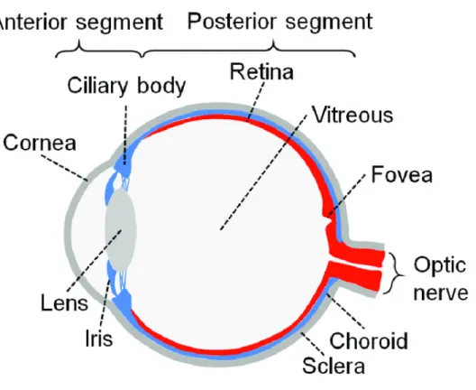
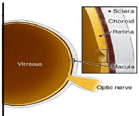
The eye globe is an approximately 2.5 cm in diameter and composed of two segments: the anterior and posterior (Figure 1).
Figure 1: Schematic structure of human eye
Human eye consist of two segments: anterior segment contains the cornea, lens, iris and ciliary body; Posterior segment contains retina, choroid, sclera and vitreous (Adapted from Chuang et al [1]).
1.1.
Anterior segment
The anterior segment usually comprises the front one third of the eye, but age and sex parameters may affect on its depth and volume. The anterior chamber in aged males diminishes more than that in females [2].
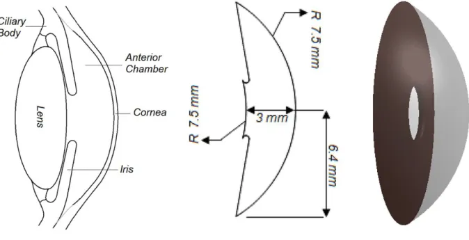
The anterior segment includes four portions, from front to back: the cornea, iris, ciliary body, and lens (Figure 2).
Figure 2: Anterior segment detailed anatomy
The anterior segment of eye includes four constituent including the cornea, iris, ciliary body, and lens (adapted from Modarreszadeh et al [3] with a little modification).
1.1.1.
Cornea
The cornea is the outermost layer of the eye globe. Despite being transparent, it protects the globe from direct contact and separates the inside structure from the outside, similarly to a window. It has two main functions: first capturing and concentrating light and second protecting the eye structure [4].
1.1.2.
Iris
The iris is a flat, colored, ring-shaped membrane behind the cornea, with an adjustable circular opening (pupil) in the center. In fact, what is called eye color is defined by that of the iris. Iris contractility can adjust the pupil diameter to allow more or less light to reach the retina [4].
1.1.3.
Ciliary body
The ciliary body is a backward continuation of the iris and is composed of muscles and processes; it is a ring-shaped thickened area that separates the posterior chamber and the vitreous body. The ciliary processes secrete a transparent gelatinous fluid in the space behind the lens called the vitreous humor, which makes up almost four-fifth of the eyeball volume. The ciliary muscle enables the shape of the lens to be changed to adjust the focus on near or far objects (a process called accommodation) [5].
1.1.4.
Lens
The lens is a transparent biconvex structure beside the cornea; it helps to concentrate light to the retina, while changing the convex angle under ciliary body contractility [5].
1.2.
Posterior segment
The posterior segment makes up the back two-thirds of the eyeball and includes the retina, choroid, sclera, and vitreous. In general, a posterior segment begins immediately after the lens and includes all optical structures behind it. These constituents are further explained below (Figure 3).
Figure 3: Posterior segment anatomy
Posterior segment contains retina, choroid, sclera and vitreous (adapted from: http://www.precisionfamilyeyecare.com/eye-encyclopedia/the-anatomy-of-the-eye-posterior/).
1.2.1.
Sclera
The sclera is the outer membrane of the eye; it is composed primarily of collagen and fewer elastic fibers, and it does not posses any vessels. This structure resembles that of connective tissue. It covers 80% of the eye and continues forward, thus forming the cornea [4].
1.2.2.
Choroid
A layer between the retina and sclera, called the choroid, is approximately 200 μm thick at birth and steadily diminishes with age [6]. The choroid contains blood vessels, melanocytes, fibroblasts, resident immunocompetent cells, and collagenous and elastic matrix. Given that the choroid is the vascular layer of the eye, it is considered one of the most highly vascularized tissues of the body. Its function has long been known to supply oxygen and nutrients to the retina. It contains five layers, which begin immediately behind the retina and include respectively, Bruch's membrane, the choroiocapillaris, Haller's and Sattler's layers (the two vascular layers), and the suprachoroidea [7].
1.2.3.
Retina
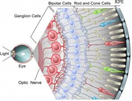
The retina has several layers. The layer containing nerve fiber is the innermost one, whereas the outermost layer is made of photoreceptors including cones and rods. The photoreceptors are specialized cells that convert light into nervous signals.
The photoreceptors lie on a cell layer called the retinal pigment epitelium (RPE), which itself receives light and performs chemical emmision, thus producing neural impulses that connect to Bruch’s membrane from the choroid. The RPE layer is further discussed below (Figure 4).
Figure 4: Retina’s schematic structure
Figure shows the arrangement of different layers: respectively, from inner to outer side: nerve fiber layer (ganglion cells), bipolar cells, photoreceptors (rods and cones) and RPE layer (adapted from: https://news.feinberg.northwestern.edu/2012/04/retina_research/) (®image courtesy of Jamie Simon, Salk Institute)
In addition, in the direction of a suppositional straight line between an object, the center of the lens, and the retina is a small area around the optical nerve of approximately 0.5 cm with a high concentration of photoreceptors, which are specialized to provide vision accuracy and detail. It is responsible for central vision, whereas the other parts of the retina process only peripheral perception. Any damage to this spot can ruin central vision.
1.2.3.1. Retinal pigment epithelium (RPE)
This pigmented layer consists of a hexagonal cell monolayer behind the photoreceptors. Retinal pigment epithelium (RPE) cells are originated from the optic
neuroepithelium. Apical side of these cells possesses multiple villi projecting forwardly to contact with the outer segments of the photoreceptor cells. Morever cells are kept contacted by tight, adherens and gap junctions in lateral sides. Eventually basal side contacts the underlying basal membrane (Bruch’s membrane) [8].
The RPE serves many supportive functions in the retina, such as cycling visual molecules, photoreceptor feeding, and removal of debris from the outer segments of the photoreceptors (Figure 5).
Figure 5: nourishment and waste management in photoreceptors
Retinal pigment epithelium (RPE) supplies the photoreceptors with nutrients and throws out the waste products from outer segment of photoreceptors (adapted from: https://www.eyecenteroftexas.com/services/eye-diseases/retina/macular-degeneration/).
Moreover, the RPE organizes a barrier between the retina and the choroid, through tight junctions (zonula occludens) in constituent cells. This arrangement results in an external blood-retinal barrier that physiologicaly prevents choroidal blood vessel penetration to the retina. The RPE has a photoprotective role, owing to the presence
of pigmented granules, including melanin and lipofuscin, which provide a protective shield while absorbing light [9].
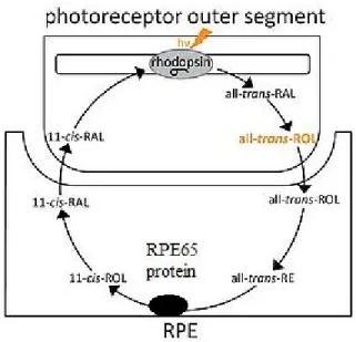
The RPE is directly involved in the maintenance of the visual system integrity via the enzyme RPE65, which transforms all-trans-retinyl ester to 11-cis-retinol, a product necessary for rhodopsin photopigment in the outer segments of photoreceptors (Figure 6) [10].
Figure 6: The role of RPE65 in visual cycle
RPE65 is critical for vitamin A metabolism in visual cycle that is responsible for converting photon to electrical pulses (adapted from: http://www.vision-research.eu/index.php?id=1180 by modification).
It also contributes to the daily renewal of the outer segments of photoreceptors [11]. Furthermore, the RPE regulates the transport of nutrients and oxygen to photoreceptors and the removal of metabolic waste from the choroid [12]. In addition,
the RPE expresses several key factors implicated in retinal development, such as fibroblast growth factor (FGF), vascular endothelial growth factor (VEGF) and pigment epithelium-derived factor (PEDF) [13-15].
1.2.4.
Vitreous
The vitreous is a clear gelatinous fluid that fills the eyeball from the lens to the retina. Its responsibility is to equilibrate the pH and to nourish the eye layers. In addition, this fluid also facilitates ocular movement [16].
2. Age-related macular degeneration (AMD)
AMD is a multifactorial, neurodegenerative disease of the retina affecting people above 65 years of age [17]. It is the leading cause of severe, irreversible vision loss in the developed world. This global disease annually affects over 8 million older people.
AMD is distinguished by a loss of central vision and resolution due to dysfunction and the death of photoreceptors (PRs) in the central part of the retina called the macula (Figure 7). AMD also detrimentally affects the retinal pigment epithelium (RPE) and Bruch’s membrane.
As the older population grows, the number of patients with currently non-treatable AMD becomes large, and AMD becomes an important health problem. According to the statistic data, 17,100 new cases of neovascular (wet) AMD and 180,000 new cases of geographic-atrophy (dry) AMD are recorded annually in Canada [18].
Figure 7: AMD interrupts detail visual recognition and central vision
The difference between a normal vision and that of AMD patient, as seen, AMD patients have problem in face recognition (adapted from Seiple et al [19]).
2.1.
Pathophysiology of AMD
AMD is categorized as depending on neovascularization changes or its progression stage [20]. Here, a well-known categorization was used, in which there are two general types of AMD: atrophy (dry type) AMDand neovascular exudative (wet type) AMD. It is possible for a person to have both dry and wet AMD at the same time in two different eyes. Moreover in the same globe it is not unusual for the severity to progress from the dry to the exudative type.
Because the onset and development of both forms of age-related macular degeneration do not follow any specific template, AMD diagnosis is too complicated to make in the earliest stages (Table I).
Table I: Clinical assortment of AMD [21].
2.1.1.
Atrophy age-related macular degeneration (dry type)
This is the first stage of AMD and the most prevalent form. Published information indicates that approximately 85% of AMD cases are of this type. In the early phase, this type is not easily recognizable by unexperienced clinicians, owing to the presence of non-tangible vision difficulties.
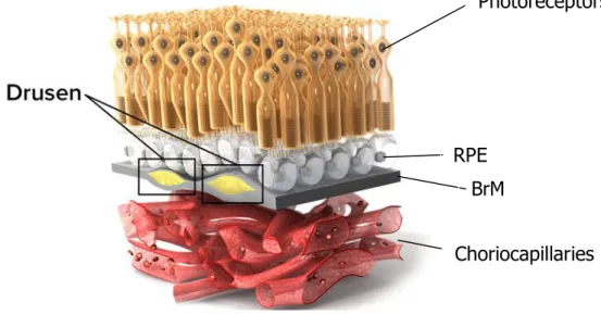
Nevertheless, the clinical hallmark of dry AMD is drusen, which are small, yellow deposits accumulating between the RPE and Bruch’s membrane (Figure 8). In fact, drusen are the compound of lipid and protein with the minor presence of trace elements. The origin of these components is not clear but according to the literatures it is likely produced by contribution of RPE and choroid. At least 40% of drusen content is lipid mostly including esterified cholesterol and phosphatidylcholine, the rest is composed of minimum 129 different proteins such as apolipoproteins [22-25].
AMD Assortment Specific hallmarks
Normal condition No drusen, No pigmented abnormality Normal aging condition Small drusen less than 63 μm
Early AMD (dry) Medium drusen (63-125 μm), No pigmentary disorder Geographic Atrophy (GA) Large drusen (125-250 μm), AMD pigmentary abnormality
These lipid accumulations slowly break down the macula and steadily demolish the retina tissue [20, 26, 27]. Although drusen are not always directly associated with AMD, they increase the risk of AMD onset. Drusen deposits vary in diameter; but only those sized between 63 and 250 μm can be characteristics of AMD [28].
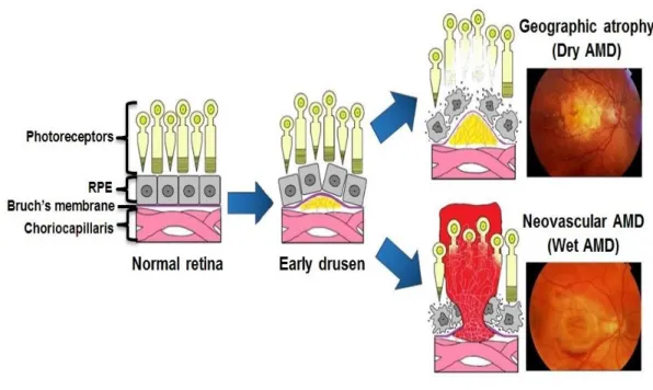
These yellow accumulations can gradually develop, and in the late stage of dry AMD may progress to geographic atrophy (GA). In advanced dry AMD or GA, the region of atrophy extends, and this is followed by the disappearance of photoreceptors over time, thus resulting in visual loss (Figure 9) [29].
Vitamin supplementation may decrease the probability of dry AMD in some cases [30] .
Figure 8: Dry AMD is recognized with drusen hallmark
Lipid spot accumulation between RPE and BrM named drusen which probably interfere with signal transfer from photoreceptors to the downstream counterparties (adapted from: http://www.scienceofamd.org/learn/)
Photoreceptors
Choriocapillaries RPE
2.1.2.
Neovascular (exudative) age-related macular
degeneration (wet type)
Patients with Atrophy age-related macular degeneration (dry AMD) are mostly susceptible to wet type (Figure 9). Uncommon blood vessels spread into the retina and over time penetrate the retina layer and may leak into the retina and induce macular swelling. This fibrovascular complex can perturb the normal RPE structure. This kind of damage is severe and can lead to legal blindness; individuals in this stage may have GA at the same time.
Figure 9: Pathogenesis milestones in AMD progression
Early stage can lead to GA or wet type as the figure shows in dry type drusen is a hallmark and in wet type, abnormal neovascular spreading into to the retina is a characteristic (adapted from: http://retina-amd.org/menu/eye-disease-amd/)
2.2.
Ethiology of AMD
AMD is a multifactorial disease that may have different risk factors, with different proportions of hereditary and environmental factors [31]. As its name indicates, age is the greatest risk factor and is inevitable. However, age is not the only cause of AMD. Some of the risk factors cannot be controlled, such as age or genetics, but several others are controllable. Below, the AMD risk factors are briefly explained:
a) Age: The risk increases after the age of 65 years old [17]. b) Sex: Females are more prone to AMD than males [32].
c) Family history: The risk of AMD is mostly associated with a family history of AMD [33].
d) Obesity: Being overweight is weakly linked to the progression of AMD, especially in late stage and advanced AMD [34].
e) Fatty diets: Studies have indicated that people who regularly consume saturated fats are predisposed to AMD, whereas consuming seafood enriched in unsaturated fats can reduce this risk [35, 36].
f) Light exposure: Studies have demonstrated that exposure to visible light and
blue light are implicated in AMD development because of their detrimental effects on photoreceptors. Most ophthalmologists recommend wearing sunglasses in the presence of ultraviolet (UV) or sun light exposure [37].
g) Smoking cigarette: The literature has confirmed an association between
smoking and AMD. Moreover the British Medical Journal has reported that public health experts from the University of Manchester believe that smoking is the most important risk factor for AMD, so smokers have a four times greater
risk of AMD than non-smokers [38]. Research has demonstrated that benzene-1,4-diol (hydroquinone), the most abundant pro-oxidant compound in cigarette smoke, can cause structural perturbations such as changes in matrix metalloproteinase-2 (MMP 2) levels and collagen IV as well as sub-RPE blebbing, all of which are related to AMD pathogenesis [39, 40].
Beyond these explanations, oxidative injury or oxidative stress is the most important underlying factor associated with triggering AMD pathogenesis through different risk factors [25]. Oxidative stress is a process of cellular damage caused by reactive oxygen intermediates (ROIs), which generally play an essential role in age-related disorders such as cancer, Alzheimer’s disease and AMD [25, 41, 42].
3. Oxidative stress
Epidemiological, clinical and experimental evidence together indicate that chronic oxidative stress is a primary contributing factor to AMD. In fact, oxidative stress is a predominance of free radicals versus antioxidant protection; it constitutes a biological attack on organisms [43] (Figure 10).
Understanding the free radical concept requires a primary knowledge of chemistry. Free radicals are defined as one or more atoms with an unpaired electron, which makes them unstable and highly reactive. To obtain stability, they must donate an electron to or gain an electron from another molecule [44, 45]. Their principal danger emerges when they attack other structural bio-macromolecules in the body. These
types of reactions are called oxidative, and over time they can lead to dysfunction or cell death.
Figure 10: Interdependence between oxidative stress and cellular damage
An imbalance between antioxidants and free radicals can threaten all biological molecules including nucleic acid, proteins and lipids (adapted from Kelly [46]).
Free radicals are reactive oxygen species (ROS), including hydroxyl radical (OH), superoxide anion (O−2), hypochlorite (ClO-), hydrogen peroxide (H2O2), peroxynitrite
radical (NO3-), and nitric oxide (NO).
The body’s defense mechanisms against ROS can be external or internal. Antioxidants are primarily external chemicals that can attenuate the deleterious effects of ROS; such chemicals may be human made, such as tocopherols, ascorbic acid, and glutathione, or can be absorbed from natural sources such as fruits. The other protection scheme includes enzymatic ROS scavengers such as catalase, peroxidase, and superoxide dismutase (SOD). These enzymes detoxify ROS through catalyzing chemical changes in their structure or reacting with lone electron pairs to
extinguish their reactivity, so that they cannot damage biological molecules [45] (Figure 11).
Figure 11 : Enzymatic antioxidant systems
Superoxide dismutase is the major constituent of enzymatic protection residing in mitochondrial matrix, this enzyme can catalyze the dismutation of the superoxide radical to oxygen or hydrogen peroxide. Glutathione peroxidase and catalase are specialized to detoxify of hydrogen peroxide (adapted from Fukai et al [47]).
The best-known role of mitochondria is to produce energy through respiration. through which the electron transport chain occurs in the inner mitochondrial membrane. This transport is mainly responsible for the production of ROS in the course of oxidative phosphorylation and has the potential for electron leakage [43]. Given the crucial function of mitochondria and their delicate structure, the presence of oxidative stress including lipid peroxidation, protein damage, and mitochondrial DNA mutation (mtDNA) is highly detrimental. Furthermore the mitochondria play a major role in apoptosis [48].
ROS are produced largely as by-products of mitochondrial respiration and subsequently can damage macromolecules. In turn, cells compromised by oxidative
stress trigger signals mediating senescence and age-related diseases [49]. Because early stages of senescence and membrane blebbing are thought to be present in the RPE, researchers have sought to explore whether these two conditions might share oxidative stress as a main cause. If so, oxidative stress may provoke RPE blebbing followed by secretion of RPE-derived microparticles, whose subretinal deposition is a sign of senescence and blebbing as a diagnostic feature of AMD [50].
3.1.
Cellular senescence and aging
One of the major effects of oxidative stress is the DNA damage response (DDR), which is followed by cellular senescence [51-58]. Senescence refers to the induction of an irreversible halting of mitosis. Recent studies have indicated that its role is a double-edged sword: on the one hand, it has a physiological function in tissue repair and development, whereas on the other hand, it is the major cause of aging and age-related disease [59].
Several lines of evidence suggest that RPE cell senescence plays a key role in the pathology of AMD. First, senescent RPE cells have been observed in primate retinas and in human cultured RPE cells; they exhibit altered morphology and reach confluency at a lower cell density [60-62]. These changes reduce their ability to form an effective barrier between the retina and the choroid [62]. Second, oxidative cell damage accelerates senescence, after which immune dysfunction can arise [63, 64]. Premature senescence has been implicated as a potentially important
pathophysiologic mediator of RPE atrophy and ultimately cell death through changing the size of the telomeres [65].
The hallmarks of senescence can be divided into three stages: primary, which is a gradual and unavoidable process throughout life and is the body’s fate; antagonistic, which involves confronting and responding to the damage; and integrative, which is the result of a cellular response and aging phenotype [66].
3.1.1.
Causes and characterization of senescence
As mentioned above, senescence is a sign of aging that can be specified by multiple cellular changes, such as decreasing telomere length, increased DNA oxidation, senescence-associated β-galactosidase (SA-β-gal) activity, increased cyclin-dependent kinase inhibitors such as p16/p15/p21, and epigenetic causes.
a) Telomere length: Telomeres, at the ends of chromosomes, are protective structures aiding in genome stability. These repetitive sequences become shorter in every DNA replication and finally lead to a constant cell cycle arrest (in G1 phase) known as replicative senescence [67]. Recent studies have reported a correlation between telomere size and age-related disease: the average telomere length in senescent cells is less than that in normal cells [68-71], in addition, telomere length is subject to change in response to stress and lifestyle [6, 69, 72, 73]. Dow and Harley (2016) reported that telomerase activator-65 significantly can add telomeric DNA in RPE chromosomal ends so it is thought to be a candidate treatment for AMD [74].
b) DNA oxidation: The oxidation of proteins and lipids may cause lipofuscin and drusen, whereas DNA oxidation may provoke cell cycle arrest [75]. Because the DNA damage repair system becomes exhausted with age, genomic inconsistencies accumulate and in turn expedite aging [76].
c) β-galactosidase activity: β-galactosidase is a lysosomal enzyme that catalyzes the hydrolysis of β-D-galactoside into basic units of sugars in the presence of water. According to Gray et al., the senescence phenotype is characterized by an increase in total β-galactosidase activity [77]. In other words, senescence-associated β-galactosidase (SA-β-Gal) is a lysosomal β-galactosidase that is a specific marker for senescent cells in acidic pH (≃ 6.0) [78]; it is a reliable proxy to detect senescence in vitro.
d) Cyclin-dependent kinase inhibitors (CDKI): CDKIs are a group of proteins inhibiting cyclin-dependent kinase. They have several activities in tumor suppressor networks, cell growth arrest at G1 phase, and eventually aging. These inhibitors include P16, P15, P21, and P57 [79].
e) Epigenetics: Epigenetics also plays an important role in the beginning of aging. It can act as an engine switch that accelerates or delays age-related disease. Among different epigenetic incidents, the major effects of miRNAs are widely known in cellular senescence. miRNAs, a group of small non-coding RNAs with a length of 21–25 nucleotides, primarily interfere in gene silencing and/or post-transcriptional regulation [80]. The multistep maturation of miRNA is orchestrated by a variety of regulatory factors; consequently, imbalance and/or dysregulation of these factors can lead to a disruption in miRNA expression and result in the
progression of various diseases [81]. In addition, they can protect against oxidative stress, through a fast and reversible response ensuring proper gene expression or regulation depending on the type of stress [82-84].
ROS-induced
RPE cell damage has a significant function in the pathogenesis of AMD.
Several studies have verified that miRNAs are implicated in the regulation of
RPE cell survival, by their ability to make mRNAs methylated following
oxidative stress [85, 86]. Taken together, we can postulate that miRNA are
likely involved somehow in AMD pathogenesis.
3.2.
RPE blebbing and microparticles (RMPs)
3.2.1.
Microparticle definition
In cell biology, a bleb is an irregular bulge in the plasma membrane of a cell, which is caused by localized decoupling of the cytoskeleton from the plasma membrane. After cell activation either by agonists or by physical or chemical stress, bleb formation results. Virtually all cell types can release membrane fragments called microparticles (MPs), generated from blebs, which represent specific characteristics of activated cells [87, 88].
MPs arise from phospholipid rich, submicron blebs (0.1 µm–1 µm) released from the plasma membranes of various types of cells in a normal state (basal level) or after apoptosis or cell activation by biological and oxidative stress (elevated level) [88-93].
They contain a variety of bioactive molecules, including proteins, biolipids, and nucleic acids such as DNAs, RNAs, and miRNAs derived from the original cells. The protein and lipid profiles of MPs may be considered a phenotypic snapshot of the cell from which they originated. Recent studies have demonstrated that miRNAs are also carried in circulating extracellular vesicles (EVs) and are probably transferred to other cells, there by altering the functions of those target cells [94, 95]. In addition, miRNAs in EVs, through the lipid bilayer, are protected from environmental stresses that may degrade miRNA. In this context, the miRNA content of vesicles can be applied to predicting disease onset and prognosis [96].
MPs are potent biological agents capable of interfering with biological signals, interacting with target cells, and eliciting both beneficial and detrimental responses. These effects are because of their ability to regulate vascular function and gene expression during inflammation and oxidative stress (Figure 12) [87, 97-102].
MPs have some similarities with apoptotic bodies and exosomes. Apoptotic cell death is a regulative process that maintains the homeostasis of a multicellular organism. Apoptosis causes cell fragmentation, after which apoptotic bodies form. These contain nuclear material surrounded by a membrane rich in phosphatidylserine (PSer) and are larger than the MPs [103] (greater than 1000 nm) (Figure 12).
Exosomes are extruded into the extracellular matrix after fusion of multivesicular endosomes with the plasma membrane. They are smaller than the MPs in diameter, varying between 50 and 100 nm. Their membranes are enriched in tetraspanines and are poor in PSer (Figure 12).
Figure 12: Different types of extra cellular vesicles (EVs)
Till now three types of EVs have been distinguished, A) exosomes are carried and secreted by intracellular vesicles; B) microvesicles (also called MPs or ectosomes) which are budding out and; C) apoptotic bodies or cell fragmentation from late stage of apoptosis [104].
3.2.2.
Microparticle formation
MPs generated by affected cells after stress cause the rearrangement of membrane phospholipids. An early event involves the reorganization of plasma membrane lipid asymmetry, alongside externalization of phosphatidylserine. Lipid rafts and their accompanying cholesterol and proteins are concentrated where the membrane buds [105], thus suggesting that MPs arise from lipid rafts or from regions with high raft content [106].
Under physiological conditions, the phospholipids are distributed asymmetrically within the lipid bilayer. Phosphatidylcholine and sphingomyelin are found on the outer layer, and phosphatidylserines and phosphatidylethanolamines are located on the inner layer of the membrane.
Any stimulus induces a calcium influx into the cytosol, thereby resulting in a reorganization of the cytoskeleton (proteolytic activity of calpain) and modulating the activity of transmembrane transporters of phospholipids including flippase, floppase, and scramblase. In the resting cell, these transporters retain the lipid asymmetry of the membrane, with early externalization of phosphatidylserine. Flippase maintains the distribution of resting phospholipids, whereas floppase directly disrupts activated cell asymmetry, and eventually scramblase externalizes phosphatidylserine during apoptosis or cell activation [87]. Other signaling proteins are also involved in MP release. The small G proteins of the RhoA family (ROCK-I and ROCK-II) are activated during budding by caspases. Caspase-activated ROCK-I promotes rearrangement of the cytoskeleton while regulating the interactions between actin and myosin. ROCK-II, via caspase 2, is also involved in the release of MPs [107, 108].
In contrast, the TNF-related apoptosis-inducing ligand (TRAIL) and respective receptor TRAIL-R2 are involved in the secretion of endothelial-derived MPs through NF-κB signaling [109]. All these complex events cause budding of the membrane and spreading of vesicles out from the origin membrane (Figure 13).
Figure 13 : Cell membrane changes in line with microvesicles formation [110].
3.2.3.
Retinal pigment epithelium derived microparticles
As demonstrated, oxidative stress induces the formation of blebs in human RPE while triggering a rearrangement response in the actin cytoskeleton [40, 111-117]. Subsequently, these membrane-derived blebs accumulate and then result in subretinal deposit formation [40, 118-121]. The excessive accumulation of any lipid-rich extracellular deposit between the RPE and Bruch’s membrane is a prominent histopathologic feature that gives rise to the earliest clinical hallmark of AMD [122, 123]. Although the proteomic profile of RPE blebs has revealed a total protein profile comprising proteins (n=314) predominantly involved in oxidative phosphorylation, cell junctions, cytoskeleton regulation, and immunogenic processes [117]. Because such
deposits have the original cell’s characteristics, they can be characterized by RPE markers such as RPE65 [124].
Our laboratory has provided the first demonstration that RPE cells take up MPs originally derived from the same cells under oxidative stress. RMP uptake is a receptor-mediated process that consequently prompts host RPE cell senescence and eventually cell death. These findings strongly suggest that RMPs function as mediators exacerbating the oxidative damage to RPE cells, and indicate a pathological role of RMPs in AMD [125]. To the best of our knowledge, RMPs contain nucleic acids, and RNA sequencing of RMPs has revealed that miRNAs are selectively enriched in RMPs.
At the National Institutes of Health (NIH), the Biomarkers and Surrogate Endpoint Working Group has formulated criteria for biomarkers of diseases as follows. Biomarkers should be: (a) an objectively measurable characteristic representing a physiologic processes, pathogenic processes or pharmacological response to a therapeutic intervention; (b) a characteristic or variable reflecting how a patient feels, functions or survives; and (c) intended to substitute for a clinical endpoint to predict either clinical benefit or harm of medicinal strategies, on the basis of epidemiological, therapeutic, pathophysiological, or other scientific evidence [126]. According to these criteria, miRNA can be considered as a biomarker.
One of the highly conserved miRNAs is the let-7 family, whose members have a key role in gene regulation and cell senescence. Zhou et al. showed that let-7 family members are expressed in retinal and choroidal endothelial cells [127] beside that AMD patients revealed 2.6 times more expression of miRNA let-7 based on serum
concentration [128]. Among members of this family, let-7f miRNA is one of the most abundant in RMPs. According to the literature, let-7f affects cell function in many other cell types [129-132]. We hypothesized that let-7f may mediate the expected effects of RMPs on RPE cell functions.
4. Thesis Objectives
Given the literature and previous laboratory findings, the roles of microvesicles such as lymphocyte-derived microparticles (LMPs) in AMD have been demonstrated [133, 134]. In this context, we sought to investigate whether RMPs contribute to RPE cell dysfunction either specifically due to miRNA let-7f or otherwise.
Our work was organized around the following objectives:
1) To investigate the role of oxidative stress in aging and RMP production. 2) To consider whether RMPs contribute to RPE cell dysfunction in vitro.
3) To assess in vivo RMP production and to examine the role of RMPs in the development of RPE cell dysfunction.
4) To explore the molecular events responsible for RMP-induced RPE cell dysfunction, emphasizing the potential let-7f signal.
Verification of the role of RMPs on the retinal degeneration and understanding of the associated mechanisms should support the development of therapies for AMD.
Results
This thesis contains two articles:
A) Extracellular microparticles accelerate oxidative damages to retinal
pigment epithelial cells
B) The implication of miRNA let-7f in the degeneration of retinal pigment
Article 1
Extracellular microparticles accelerate oxidative damages
to retinal pigment epithelial cells
Extracellular microparticles accelerate oxidative damages to retinal pigment epithelial cells
Chun Yang,1* Saeideh Shani,1* Houda Tahiri, 1 Jean-Claude Lavoie,2 Stéphane Croteau,3 and Pierre Hardy 1#
1 Departments of Pediatrics, Pharmacology and Physiology, University of Montréal,
Montréal, Québec, Canada.
2 Departments of Pediatrics and Nutrition, University of Montréal, Montréal, Québec,
Canada.
3 Department of Medicine, University of Montreal, Montreal, Quebec, Canada.
*Co-first authors, the first two authors contributed equally to this work.
Corresponding author:
#Pierre Hardy, M.D. Ph.D.
Research Center of CHU Sainte-Justine, 3175 Côte-Sainte-Catherine, Room 2714 Montréal, Québec, H3T 1C5
Phone: (514) 345-4931 (ext. 3656), Fax: (514) 345-4801 E-mail: [email protected]
Running Title: RMPs induce RPE cell dysfunction Word count: 3,493
Keywords: RPE-derived microparticles (RMPs), oxidative stress, RPE cell
Abstract
Purpose: Oxidative stress is a primary contributing factor to early dry age-related
macular degeneration (AMD). Oxidative injury to the retina may promote extracellular microparticles (RMPs) released from retinal pigmented epithelium (RPE). This study was designed to investigate whether RMPs cause RPE cell dysfunction.
Methods: RMPs were isolated from human ARPE-19 cells under oxidative stress.
The oxidative stress-induced RMPs in vitro and in vivo were characterized using Nanosight and FACS analysis. Fluorescent DiI-labelled RMPs were used to assess uptake into RPE cells. MTT assay and [3H]-thymidine incorporation assay were used
to determine cell viability and cell proliferation respectively. Expression of cell cycle genes was analyzed by quantitative RT-PCR and Western blot. The effects of RMPs on RPE cell senescence and phagocytosis were assessed.
Results: Oxidative stress induced extracellular vesicles (EVs) released from RPE
cells; RMPs constitute over 95% of the EVs. Significantly, more RMPs were released from aged RPE cells. RMPs were taken up by RPE cells in a time-dependent manner; however blockage of CD36 attenuated the uptake process. The decrease of RPE cell viability and cell proliferation by exposing with RMPs was associated with an increased expression of cyclin-dependent kinase inhibitors p15 and p21. Moreover, RMPs enhanced senescent and interrupted phagocytic activity in RPE cells.
Conclusion: This is the first study to demonstrate that oxidative-induced RMPs
produce a strong effect of inducing RPE cell degeneration. This finding leads to the postulate that RMPs exacerbate oxidative stress damage to RPE cells, which may uncover a potentially relevant process in the genesis of dry AMD.
Introduction
Age-related macular degeneration (AMD) represents the leading cause of vision loss in the elderly, which has a devastating impact on quality of life.1, 2 The most common form of AMD is dry AMD, with which 85-90 percent of AMD patients are diagnosed.3
The key features of dry AMD are the degeneration of the retinal pigment epithelium (RPE), and drusen formation.4, 5 Clinical and experimental evidence points to
oxidative stress as a primary contributing factor to AMD by promoting RPE cell dysfunction and ultimately cell death.6-8
RPE is a single layer of pigmented epithelial cells with highly organized structure and tight junctions. The structural and functional integrity of RPE is critical for maintaining photoreceptor functions and survival.9 One of the major effects of
oxidative stress is the induction of cellular senescence and apoptosis.10-18 Senescent RPE cells exhibit altered morphology and change in growth rate;19, 20 these changes could reduce their ability to form an effective barrier between the retina and the choroid.19
RPE cells are among the most actively phagocytic cells in nature. RPE cells ingest and metabolize lipid-rich photoreceptor outer segments (POS) for visual factor recycling and to prevent debris buildup.20 Impairment of RPE phagocytosis would lead to accumulation of material within the RPE and deposition of abnormal material within Bruch’s membrane.21 Drusen are extracellular lipid- and protein-containing deposits that accumulate mainly between the RPE and Bruch’s membrane, they are a hallmark of aging and early AMD.22 There is evidence suggesting that drusen are formed from extracellular microvesicles (EVs) derived from RPE cells.18, 23
In general, EVs include microparticles (MPs), exosomes, and apoptotic bodies. MPs are a heterogeneous population of shed microvesicles of 100 to 1000nm in size, formed from activated or stressed cells; exosomes are released from exocytosis of multivesicular bodies with 50 to 100 nm in diameter.24 Several studies reported the
role of RPE-derived exosomes in AMD,23, 25-27 and there have been very few reports of RPE-derived MPs (RMPs) contributing to sub-RPE deposits.18, 28, 29 However, the
effect of RMPs on RPE cell functions has not yet been established. To gain a better understanding of the pathological roles of RMPs, we isolated and characterized the RMPs from cultured RPE cells exposed to oxidative stress conditions. Most interestingly, we found that RMPs reduced RPE cell viability and impeded RPE cell functions. These findings may reveal a novel pathogenic mechanism of oxidative stress in the development of dry AMD.
Methodology Cell culture
ARPE-19, a spontaneously immortalized cell line of human retinal pigment epithelium, was purchased from American Type Culture Collection (ATCC, CRL-2302), and maintained according to standard procedures. Primary mouse RPE cells were isolated from different ages of C57BL/6 or Sod-/- knockout mice of different ages and
cultured as we described previously.30 For cell purity confirmation, confluent primary cells were fixed with 4% paraformaldehyde followed by immunohistochemistry staining with Alexa Flour 488 conjugated anti-RPE65 antibody (Novus; NB100-355AF488) (See Supplementary Fig. S1). All animal experiments were performed
according to the ARVO Statement for the Use of Animals in Ophthalmic and Vision Research and were approved by the Animal Care Committee of CHU Sainte-Justine (Montreal, QC, Canada).
Isolation and characterization of RMPs derived from ARPE-19 cells
Induction of RMPs was performed as described with modification.29 Briefly, confluent
ARPE-19 cells were cultured with medium containing sequential FBS reduction for 6 days. On day 7, cells (or primary mouse RPE cells at passage 2) were exposed to 10 microunits myeloperoxidase (MPO) for 90 minutes followed by exposure to hydrogen peroxide (H2O2) at 100 µM for 24 hours. At the end of the exposure time, isolation of
RMPs was performed as described previously.31 RMPs were then characterized with
annexin V staining32 (See Supplementary Fig. S2). To produce fluorescent lipophilic
membrane dye (DiI) labelled RMPs (DiI-RMPs), ARPE-19 cells were exposed to DiI for 12 hours before MPOexposure.
Extracellular vesicles (EVs) size analysis and quantification
To analyse EVs secreted from ARPE-19 cells with or without oxidative stress, cells were plated at 106 cells/5 cm2 and cultured without 100μM of H2O2 for 24 hours.
Differential centrifugations (i.e. 2000×g, and 12,000×g) at 4°C were performed to purify RMPs from culture supernatants. EVs profiling was analyzed by NanoSight particle-tracking system (NanoSight Ltd.) as described 33 or FACS analysis by
RMPs uptake assay
a. Uptake of DiI-RMPs by RPE cells ex vivo: RPE/choroid explants were prepared as we described previously.30 RPE/choroidal explants were incubated with DiI-RMPs and fluorescent images were taken at different incubation time points.
b. Uptake of DiI-RMPs by ARPE-19 cells in vitro: ARPE-19 cells were incubated with 10µg/ml of DiI-RMPs. At different time points, cells were collected for spectrofluorometry readings and the mean fluorescent intensity (MFI) was determined as described previously.34 To investigate whether CD36 is involved in the uptake process of RPE cells, ARPE-19 cells were pre-exposed to 15 μg/mL of CD36 antibodies (ab23680, Abcam) or isotype-matched control antibodies (anti-human IgA, Sigma) for 6 hours, followed by 24 hours DiI-RMPs incubation.
Cell viability assay and proliferation assays
ARPE-19 cells or primary RPE cells were incubated with different doses of RMPs or control vehicle. Cell viability at different time points was determined by MTT assay as described previously.35 Cell proliferation was evaluated by [3H]-thymidine
incorporation assay.36
Cell cycle analysis and quantitative RT-PCR analysis
ARPE-19 cells in log phase of growth were exposed to 10μg/mL of RMPs for 24 hours. Cells were harvested for FACS analysis as we described previously.36 Total
RNAs isolation and quantitative analysis of gene expression were performed as we described previously.36 Gene expression was normalized to 18S. PCR primers
targeting human p15, p21 and 18S were synthesized by Alpha DNA (Montreal, Quebec, Canada) based on the following sequences: p15 forward 5’-CTCTCACCCGACCGGTGCAT-3’, reverse 5’-GGTGGGTGGGGGTGGGAAAT-3’;
p21 forward 5′-GCAGACCAGCATGACAGATTT-3′, reverse
5′-GGATTAGGGCTTCCTCTTGGA-3′; 18S forward
5'-CGCAGCTAGGAATAATGGAATAGG-3'; reverse 5'-GCCTCAGTTCCGAAAACCAA-3'.
Western blot analysis
ARPE-19 cells were collected after 24 hours exposure to RMPs. Extraction of soluble proteins and fractionation by SDS-PAGE was performed as described previously.31
The anti-p15 and anti-p21 antibodies (LifeSpan BioSciences, Inc. 1:500) were used to reveal the protein levels of p15, and p21 respectively.
Senescent assay
The confluent RPE primary cells or ARPE-19 cells were exposed to 10μg/mL of RMPs for different times. Senescence-associated β-galactosidase (SABG) staining was performed as described.37 Images were taken using phase-contrast microscopy and SABG positive cells were counted.
Phagocytic activity assay
Phagocytic activity assay was performed according to a modified method.38 Briefly, photoreceptor outer segment fragments (POS) isolated from pig eyes were labeled
with 2 mg/mL of fluorescein isothiocyanate (FITC) (Sigma). RPE cells were fed with FITC-POS (10 POS/cell) for different durations. Unbound POS were removed by extensive washing with medium. The fluorescence was recorded at 485/525 nm using Spectramax GeminiXS plate reader. Phagocytosis rate was calculated by fluorescent count for each time point compared to total fluorescent count. In RMPs exposed experiments, RPE cells were pre-exposed to 10μg/mL of RMPs for 12 hours followed by incubation with FITC-POS, the rate of phagocytosis was determined after 4 hours incubation.
Animals
Hydroquinone exposed mice
C57BL/6 female mice (12-month-old) were randomly divided into two groups: mice in the control group (n=8) receiving regular drinking water, and the second group mice (n= 8) receiving drinking water containing 0.8% hydroquinone (HQ) (Fisher Scientific). Mice were kept for 45 days as described39. At the end of exposure period, eyes were immediately subjected to dissection of retina and RPE/choroid sheets.
Sod1 knock-out mice
Homozygous 129Sod1tm1Leb/j knock-out male mice and age similar 129SF1/J
wild-type male mice were purchased from Jackson Laboratory. The RPE/choroid were collected from 12-month-old sod1-/- mice and control mice respectively (n=7 mice for
In vivo RMPs characterization in mice and humans
RPE/choroid tissues were homogenized in annexin buffer followed by two steps centrifugations of 2000 rpm for 15 min and then 10,000 rpm for 20 seconds. The resulting supernatants containing MPs of RPE origin were first labeled with anti-RPE65 antibody conjugated with Alexa Flour 488 (anti-RPE65-AF488, Novus) or FITC mouse IgG2a (BD pharmingen) as isotype control, then incubated with annexin V-Cy5 (BD pharmingen). RMPs were characterized with annexin V+ and RPE65+ staining by FACS analysis and gated using 1.0-µm beads.31
Statistical analysis
Results were presented as means ± SEM from at least three independent experiments performed as duplicates or triplicates. Student’s t-test was used to determine the significant differences between two groups. One-way ANOVA followed by post-hoc Bonferroni tests was used for comparison of means from three or more groups. Statistical significance was set at P < 0.05.
Results
1. Oxidative stress promoted more extracellular microvesicles (EVs) released from ARPE-19 cells.
Human ARPE-19 cells have structural and functional properties characteristic of RPE cells in vivo40. ARPE-19 cells exposed to Hydrogen peroxide (H
2O2)is a commonly
used model to test oxidative stress susceptibility or antioxidant efficiency.41 100 μM final concentration of H2O2 has been proved to be a sub-lethal concentration to
ARPE-19 cells41-43. The nanoparticles tracking analysis of extracellular microvesicles
(EVs) revealed that EVs released from both normal culture condition and oxidative condition have a similar size distribution pattern, with diameters of 50 to over 600 nm. The average size of these EVs was approximately 200 nm and did not differ between the two groups (mean 207±3.7nm, mode 165.3±18.2nm for normal condition, mean 204.8±3.6nm, mode 150 ±11.0 nm for the oxidative condition, P = 0.94 for mean, P = 0.88 for mode). This result suggested that RPE cell derived EVs contain both exosomes (less than 100nm) and microparticles (size from 100nm~1000nm), but over 95% of EVs are microparticles (Fig. 1A). Thus, we designated these RPE derived EVs as RMPs. NanoSight analysis showed significantly more RMPs released from ARPE-19 cells exposed to H2O2 compared to control non-stimulated cells (Fig. 1B,
P<0.01).
2. RPE cells released more MPs under oxidative stress in vitro and in vivo.
To investigate whether old RPE cells are more sensitive to oxidative stress, we isolated and cultured primary RPE cells from 4-month-old or 24-month-old C57BL/6 mice. We defined age equivalence as 4-month-old C57BL/6 mouse corresponding to human age 25, and a 24-month–old mouse being equivalent to human age 69.44 After exposure with H2O2, RMPs released from RPE cells were determined by FACS
analysis measuring the amount of Annexin V+ MPs released into media. H2O2 caused
significantly more RMPs to be released from 2month-old RPE cells compared to 4-month-old RPE cells (Fig. 2A). To investigate whether the antioxidant prevented oxidative stress-induced RMPs production, RPE cells were exposed to H2O2 with or
without antioxidant (U-83836-E, inhibitor of lipid peroxidation, a derivative of vitamin E). Under oxidative stress, RMPs in the culture medium with U-83836 were significantly less than the group without U-83836. This result suggested that oxidative stress is involved in RMPs production (Fig. 2B).
Several studies have shown that exposure to HQ, a potent oxidant, induced nonlethal bleb injury and sub-RPE deposits 29, 39, 45. To investigate whether oxidants
stimulate RMPs release in vivo, we analyzed the RMPs in the RPE/choroidal tissue of the mice exposed to HQ. Consistent with other groups finding, HQ exposure induced significantly more RMPs by two folds (Fig. 2C).
Retinal dysfunction, sub-RPE deposits, and thickened Bruch’s membrane were detected in the Sod1−/− knockout mice deficient in Cu, Zn-superoxide dismutase
(Sod1).46-48 In these 12-month-old Sod1−/− mice, the average amount of RMPs released from the Sod1−/− mouse (n=7) were significantly higher than that from wild type control mice (n=7), Mean ± SEM, 655400 ± 277700 (Sod1−/−) vs. 391000 ±
376200 (control) (Fig. 2D).
3. RMPs were taken up by RPE cells in a time-dependent manner.
To assess whether RPE cells take up RMPs, we first used the primary RPE cells in RPE/choroid explants. The DiI-RMPs bound or ingested RPE cells were detected by fluorescent microscopy. The presence of DiI-RMPs along the RPE cell membrane and in intracellular compartments was identified. Fluorescence intensity was increased over time (Fig. 3A). Similar findings on primary mouse RPE cells were observed in vitro on human 19 cells. Following exposure to DiI-RMPs,
ARPE-19 cells significantly increased DiI staining detectable by spectrofluorometry, and increased in a time-dependent fashion (Fig. 3B). Nonetheless, when the membrane scavenger receptor CD36 protein on RPE cells was blocked by CD36 specific antibody, the uptake rate was significantly decreased by 47% compared to control vehicle (Fig. 3C).
4. RMPs dose-dependently reduced RPE cell viability and suppressed cell proliferation.
To investigate the effects of RMPs on RPE cell growth, we exposed ARPE-19 cells to different concentrations of RMPs at different time points. The cell viability assay showed that RMPs dose-dependently reduced ARPE-19 cell viability (Fig. 4A). The similar time-dependent reduction of cell viability with 20μg/mL RMPs was observed on primary RPE cells (see supplementary Fig. S3). Although RMPs at the dose of 10μg/mL did not significantly affect cell viability, they did remarkably suppress both ARPE-19 and primary RPE cells proliferation (Fig. 4B). Of special note, the inhibitory effects of RMPs on cell viability of both ARPE-19 and primary mouse RPE were comparable to those of RMPs derived from primary mouse RPE cells (see supplementary Fig. S4).
5. RMPs induced RPE cell cycle arrest in G0/G1 phase with an increased
expression of p15 and p21.
We further evaluated the cell cycle kinetics of RPE cells by FACS. Relative to control, RMPs-exposed cells exhibited an increase in G0/G1-phase; conversely, the proportion
of RMPs-exposed cells in S-phase markedly declined (Fig. 5A). We also performed gene expression studies to characterize specific mechanisms of this cell-cycle alteration. We found that mRNA levels of the cyclin-dependent kinase (CDK) inhibitors p15 and p21 were elevated approximately 3.1 and 4.7-fold respectively (Fig. 5B). Consistently, RMPs significantly augmented the protein levels of p15INK4b and p21Cip1, as indicated by a 1.9- and 3.2-fold induction respectively (Fig. 5C-D).
6. RMPs induced RPE cell senescence.
Along with the observation that RMPs suppressed RPE cell proliferation, RMPs significantly increased SABG activity in both primary RPE cells (Fig. 6A-B) and ARPE-19 cells (Fig. 6C-D). The senescent cells are revealed by senescence-associated β-galactosidase (SABG) stained cells in blue (Fig. 6A and 6C). The prolonged incubation of RMPs resulted in more significant induction of senescence (Fig. 6B and 6D).
7. RMPs reduced phagocytosis activity.
RPE cells are phagocytic cells, and play a vital role in phagocytosis of the tips of the photoreceptor outer segments (POS) for maintaining structural and functional integrity of the retina.49 To explore the influence of RMPs on RPE cell engulfment of POS,
confluent monolayers of RPE cells were co-cultured with FITC-labeled POS for different times. The rate of phagocytosis was increased with incubation period of time (Fig. 7A). Nonetheless, RMPs exposure significantly decreased POS phagocytic activity of RPE cells (Fig. 7B).


![Table I: Clinical assortment of AMD [21].](https://thumb-eu.123doks.com/thumbv2/123doknet/2040775.4727/27.918.132.792.163.401/table-i-clinical-assortment-of-amd.webp)