Université de Montréal
Pharmacological Interventions for the Hemodynamic
Management of Deceased Organ Donors
par Anne Julie Frenette, Pharm D., M.Sc, candidate au PhD
Département des sciences biomédicales Faculté de Médecine
Thèse présentée
en vue de l’obtention du grade de doctorat en sciences biomédicales,
option recherche expérimentale
15 Novembre 2019
Résumé
Chaque année, plusieurs milliers de patients aux prises avec une maladie chronique terminale s’ajoutent à la liste d’attente pour une transplantation d’organe, espérant ainsi prolonger leur espérance de vie. Le plus souvent, le don d’organes survient suite au décès neurologique d’un donneur, une condition qui n’est pas sans conséquence sur la qualité des organes. Les traitements pharmacologiques visant à rétablir l’homéostasie et à protéger les organes à transplanter ont été majoritairement étudiés dans des études observationnelles, au début des années 80. Depuis, très peu d’essais randomisés ont évalué l’impact d’interventions chez des donneurs sur des issues cliniques chez des receveurs. Conséquemment, le bénéfice net des traitements pharmacologiques utilisés de routine chez les donneurs d’organes après décès neurologique demeure inconnu et la rationnelle physiopathologique supportant leur utilisation est questionnable.
Cette thèse a pour visée de recenser les évidences supportant les traitements pharmacologiques employés pour la stabilité hémodynamique des donneurs d’organes après décès neurologique et de décrire le niveau d’évidence supportant leur usage. Nous visons également à identifier des cibles de recherche potentielles basées sur de nouvelles observations pathophysiologiques. Pour atteindre ces objectifs, nous avons dressé un large portrait de la prise en charge actuelle des donneurs après décès neurologique, ceci menant ensuite à l’exploration des perceptions des médecins intensivistes canadiens en regard de ces interventions. Nous avons également exploré la présentation clinique cardiaque des donneurs et nous avons identifié des barrières à la recherche clinique dans le domaine. Notre thèse a mené à 4 articles scientifiques. D’abord, nous avons démontré à l’aide d’une revue systématique des lignes directrices internationales sur la prise en charge des donneurs après décès neurologique que les recommandations actuelles sont incohérentes et que leur faible qualité méthodologique reflète la lenteur de l’émergence de la recherche dans le domaine. Ensuite, nous avons effectué un sondage national auprès de médecins des soins intensifs ayant de l’expérience dans la prise en charge des donneurs. Nous avons ainsi démontré que les perceptions de pratiques sont très variables au pays et avons attribué ces divergences d’opinions au manque de données probantes, et à la possible inexpérience relative des médecins face aux rares cas de don d’organes sur une
unité de soins intensifs. Notre troisième article a démontré que la dysfonction ventriculaire droite est fréquente après un décès neurologique, bien que la littérature actuelle ne mette l’emphase que sur la dysfonction ventriculaire gauche et ses conséquences. Nous émettons l’hypothèse que la description actuelle des conséquences hémodynamique du décès neurologique est incomplète et qu’une meilleure compréhension des mécanismes sous-jacents à la dysfonction ventriculaire droite permettrait d’identifier de nouvelles cibles thérapeutiques. Finalement, en s’appuyant sur nos observations, nous questionnons l’efficacité et la pertinence d’interventions pharmacologiques administrées de routine chez les donneurs telles que l’hormonothérapie de remplacement. Nous avons donc effectué un essai randomisé pilote visant à évaluer la faisabilité d’une étude multicentrique déterminante comparant la levothyroxine au placebo chez des donneurs potentiels. Cette étude pilote a démontré qu’une étude d’envergure était nécessaire afin d’évaluer le bénéfice de l’intervention et a permis d’identifier des barrières à la recherche spécifiques au domaine. Nous proposons que des activités de transfert de connaissances sur le niveau d’évidence supportant les interventions pharmacologiques actuelles soient implantées en préparation d’un essai randomisé contrôlé multicentrique.
Cette thèse a permis de mettre en lumière la validité questionnable du traitement pharmacologique pour la prise en charge de l’instabilité hémodynamique des donneurs d’organes tel qu’il est utilisé présentement. Nous avons fait ressortir que le traitement actuel est historiquement basé sur des données de faible évidence. Nous suggérons que l’avenir de la recherche interventionnelle chez les donneurs d’organes repose sur la capacité des cliniciens et des chercheurs à reconnaitre les zones d’incertitude dans les connaissances actuelle et à accepter des changements dans leur pratique.
Abstract
Every year, thousands of chronically ill patients are added to the transplant list, in the hope of an organ transplant that could save their life. Most frequently, organ donation occurs following neurological death of a donor, a clinical pathological condition that can jeopardize the quality and stability of organs. The body of literature on the hemodynamic consequences of neurological death and their treatment exist since the early 80’s. Since then, very few randomized trials have been performed on the neurologically deceased donor population. As a consequence, the benefit of routine pharmacological therapies for the hemodynamic management of neurologically deceased donors on recipients’ outcomes is still uncertain, and the pathological theory underlying their use remains questionable.
Consequently, this thesis aims at describing the actual body of evidence supporting the pharmacological treatment for the hemodynamic management of neurologically deceased donors and the theoretical rationale for their use. We also aimed at adding to the actual knowledge of brain death physiological hemodynamic consequences. To achieve this goal, we drew a broad portrait of the actual management of hemodynamic instability in organ donors, leading to the exploration of perceptions on these interventions. We then explored physiological consequences of neurological death at the heart level and evaluated the feasibility of conducting a multicentre trial on a pharmacological intervention in donors. Our thesis let to four research articles.
First, we demonstrated through a systematic review of international guidelines for the management of neurologically deceased donors that the existing recommendations are inconsistent and that their poor methodological quality reflects the slow emergence of clinical research in the field. Then, in a national survey of intensive care physicians with experience in organ donor clinical management, we identified varying perceptions of practices in the country. We attributed this difference in opinions to the paucity of research in the field and to the possible relative inexperience of some physicians when managing deceased donors, a relatively rare condition in the intensive care unit. Our third article suggested that right ventricular dysfunction is frequent after neurological death, although existing literature focus mainly on the occurrence and consequences of left ventricular dysfunction. We postulate that the actual description of
hemodynamic consequences of neurological death is incomplete and that a better understanding of the mechanisms underlying right ventricular dysfunction would permit to identify new therapeutic targets. Finally, based on our previous conclusions, we questioned the relevance and efficacy of levothyroxine routine administration in donors and designed a pilot randomized controlled trial to evaluate the feasibility of a multicenter definitive trial. This pilot trial permitted to identify important barriers to interventional research including neurologically deceased donors. We propose that knowledge translation activities on the actual level of evidence supporting routine interventions be implemented in the preparation of a future randomized trial.
This thesis permits to question the validity of the actual pharmacological management of neurologically deceased donors highlighting the paucity of high-evidence literature in the field and the penetrance of historical interventions and concepts. We suggest that the future of research in the field lies on the ability to recognize areas of uncertainties and the acceptance of practice change.
Table of content
Résumé ... 1 Abstract ... 3 Table of content ... 5 List of Tables ... 8 List of Figures ... 9 List of acronyms ... 10 List of abbreviations ... 11 Remerciements ... 13Chapter 1: Introduction and theoretical background ... 14
1. General introduction ... 15
1.1 Definition of neurological death in the context of organ donation ... 15
1.1.1 Historical perspectives on the concept of death ... 16
1.1.2 The actual determination of brain death ... 17
1.2 History of organ donation after neurological death ... 18
1.3 Epidemiology of organ donation ... 20
1.4 Pathophysiology of brain death ... 21
1.4.1 Anatomy of the autonomic storm and hormone depletion theory ... 22
1.3.2 Inflammation, cytokine storm and brain death ... 28
1.4 Hemodynamic consequences of brain death ... 30
1.4.1 Brain death induced cardiomyopathy ... 31
1.5 Organ donor pharmacological management ... 38
1.5.1 Pharmacological treatment of the autonomic storm ... 38
1.5.2 Pharmacological treatment of hemodynamic instability ... 39
1.5.3 Hormone replacement therapy ... 41
1.6 Professional knowledge in organ donor management ... 50
1.6.1 Organ donation organizations and coordinators ... 51
1.7 Research on organ donor care ... 52
1.7.1 Ethical challenges and outcomes in organ donation research ... 53
2. Current gaps in the literature ... 57
3. Objectives and hypotheses ... 57
Articles 1 and 2 ... 58
Article 3 ... 59
Article 4 ... 59
Chapter II: Methodology and results ... 60
1. Overview of the methodology ... 60
1.1 Worldwide management of donors after neurological death: a systematic review of guidelines ... 60
1.2 A Canadian survey of critical care physician’s hemodynamic management of neurologically deceased organ donors ... 62
1.3 Right ventricular dysfunction in neurologically deceased organ donors: an observational study in a tertiary-care organ donor referral centre ... 64
1.4 A pilot randomized controlled trial comparing levothyroxine to placebo in neurologically deceased donors ... 64
2. Results ... 66
Article 1: Worldwide Management of Donors After Neurological Death: a Systematic Review of Guidelines ... 67
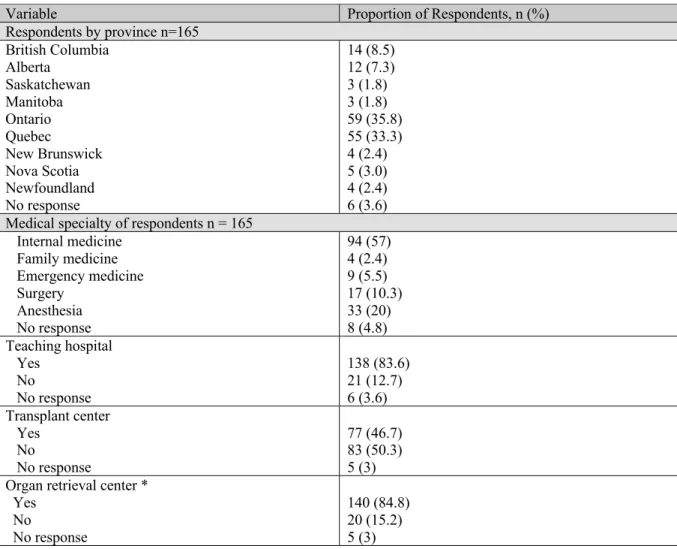
Article 2: A Canadian Survey of Critical Care Physicians’ Hemodynamic Management of Deceased Organ Donors ... 112
Article 3: Right ventricular dysfunction in neurologically deceased organ donors: an observational study in a tertiary-care organ donor referral centre. ... 147
Article 4: A Pilot Randomized Controlled Trial Comparing Levothyroxine to Placebo in Neurologically Deceased donors ... 164
Chapter III: General Discussion ... 184
Overall study conclusions ... 184
Is the theory still holding? ... 191
If organ donation is improving anyway, is research still needed? ... 195
How can we improve research in organ donor care? ... 196
Strengths of the studies presented in this thesis ... 201
Limits ... 202
Future research priorities ... 205
What is the clinical presentation of brain death in potential donors? ... 206
What is the frequency of RV dysfunction in donors and can it be prevented? ... 206
Should levothyroxine (or any of the hormone therapy component) still be used? ... 207
Conclusion ... 207
Appendix ... 211
List of Tables
Chapter 1: Introduction
Table 1. Epidemiology of cardiac dysfunction in neurologically deceased donors ... 33
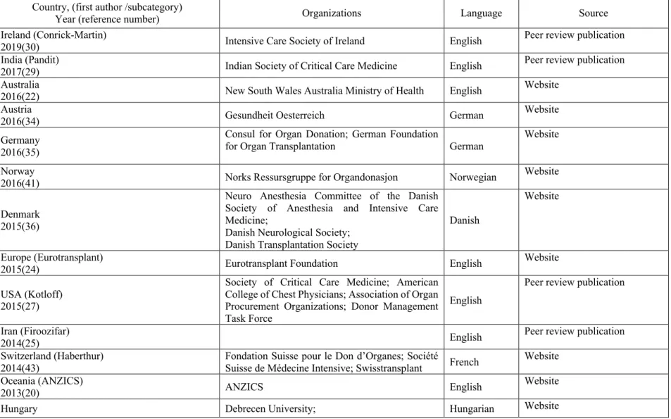
Article 1: Worldwide Management of Donors after Neurological Death: a Systematic Review of Guidelines Table 1. Description of the included guidelines ... 74
Table 2. Quality assessment with the AGREE-II instrument ... 78
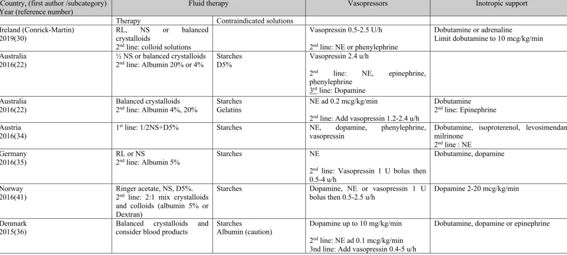
Table 3. Hemodynamic therapies ... 85
Supplemental Digital Content 2. Hemodynamic targets ... 97
Supplemental Digital Content 3. Hormone therapies ... 101
Supplemental Digital Content 4. Ventilation strategies ... 106
Article 2: A Canadian Survey of Critical Care Physician’s Hemodynamic Management of Neurologically Deceased Organ Donors Table 1. 2006 Canadian Guidelines recommendations according to survey domains ... 116
Table 3. Sources of guidance for the medical management of neurologically deceased donors ... 123
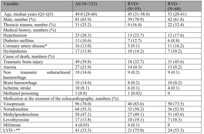
Article 3: Right Ventricular Dysfunction in Neurologically Deceased Organ Donors: An Observational Study in a Tertiary-care Organ Donor Referral Centre Table 1. Characteristics of the Study Sample ... 155
Table 2. Characteristics of subjects with isolated RV dysfunction ... 156
Table 3. Multivariate logistic regression models of variables associated with RV dysfunction ... 158
Article 4: A Pilot Randomized Controlled Trial comparing Levothyroxine to Placebo in Neurologically Deceased Donors Table 1. Characteristics of the Study Sample ... 175
Table 2. Organs Retrieved and Transplanted by Thyroid or Placebo Drug Used During Donor Management ... 176
Supplemental Table 1. Detailed reasons for exclusion by study period* ... 183
Interrater reliability of the AGREE-II instrument on the 26 included guidelines of the systematic review ... 212
List of Figures
Chapter II: Methodology and results
Article 1: Worldwide Management of Donors after Neurological Death: a Systematic Review of Guidelines
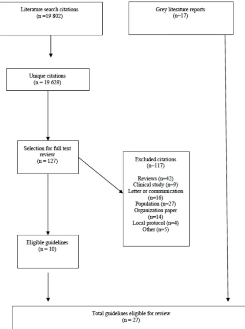
Figure 1. Guidelines selection ... 73 Figure 2. Donor management strategies, displayed as a) hemodynamic therapies, b) hormone therapy components and c) general ICU care. ... 81 Article 2: A Canadian Survey of Critical Care Physician’s Hemodynamic Management of Neurologically Deceased Organ Donors
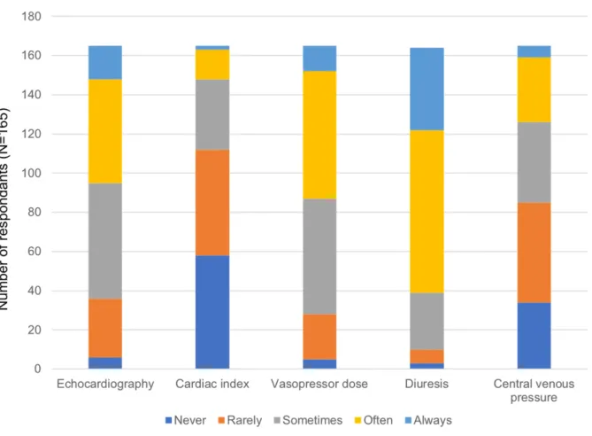
Figure 1. Medication prescribed in the context of an autonomic storm ... 126 Figure 2. Triggers for fluid administration and fluid responsiveness ... 129 Article 3: Right Ventricular Dysfunction in Neurologically Deceased Organ Donors: An Observational Study in a Tertiary-care Organ Donor Referral Centre
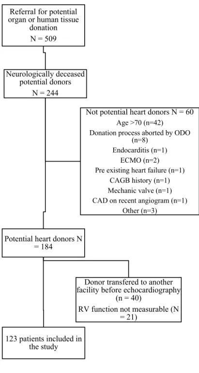
Figure 1. Inclusion of the study subjects ... 154 Article 4: A Pilot Randomized Controlled Trial comparing Levothyroxine to Placebo in Neurologically Deceased Donors
List of acronyms
CONSORT: Consolidated Standards of Reporting Trials CNTRP: Canadian National Transplant Research Program KDPI: Kidney Donor Profile Index
IRODat: International Registry on Organ Donation and Transplant OPTN: Organ Procurement and Transplantation Network
List of abbreviations
ACTH: Adrenocorticotrophic Hormone ADH: Antidiuretic Hormone
ATP: Adenosine Triphosphate CK: Creatine Kinase
CVP: Central Venous Pressure ECG: Electrocardiogram EEG: Electroencephalogram CPP: Cerebral Perfusion Pressure
DCD: Donation after Cardiocirculatory Death FSH: Follicle Stimulating Hormone
GIK: Glucose-Insulin-Potassium ICP: Intracranial Pressure
KDPI: Kidney Donor Profile Index LDH: Lactate Dehydrogenase LH: Luteinizing Hormone LV: Left Ventricle
LVEF: Left Ventricular Ejection Fraction MAP: Mean arterial pressure
NDD: Neurological Determination of Death NF-KB: Nuclear Factor-kappa B
ODO: Organ Donation Organization
PCWP: Pulmonary Capillary Wedge Pressure PVR: Pulmonary Vascular Resistance
RCT: Randomized Controlled Trial RV: Right Ventricle
SERCA: Sarco/endoplasmic reticulum Ca2+-ATPase TSH: Thyrotropin Hormone (Thyroid Stimulating Hormone) T3: Triiodothyronine
«
A ma famille et mes amis qui inspirent chacun de ces pas
Remerciements
J’aimerais remercier mes co-directeurs, Dr Emmanuel Charbonney et Dr David Williamson pour leur support, leur enthousiasme et leur implication. Vous avez eu une idée folle et vous y avez cru avec moi, merci. Merci également aux membres de l’équipe de gestion du département de pharmacie de l’Hôpital du Sacré-Cœur de Montréal ainsi qu’à mes collègues des soins intensifs pour leur ouverture d’esprit et leur flexibilité. Merci d’avoir pris le temps de vous informer de ma progression et d’avoir rassuré mes inquiétudes. Je souhaite remercier sincèrement ma famille qui m’a supporté tout au long de ces années d’étude et de travail, leurs encouragements m’ont permis de me rendre jusqu’au bout de cette aventure. Merci à mes précieux amis, nièce et collègues pour leur support moral lors de ma défense; Véro, MC, Nath, Max, Ema, Marc, Nancy, Lyne vous avez fait une différence qui fait chaud au coeur.
Un petit merci tout spécial aux extraordinaires filles de mon équipe de bateau dragon, Vous avez grandement contribué à mon équilibre de vie et à mon bonheur durant les heures et les heures de rédaction. Nos victoires et dépassements, notre esprit d’équipe et votre amitié ont nourri chacun de mes efforts. J’y ajoute un merci particulier à ma grande amie Nathalie. Merci Nath pour les fous rire, les confidences, les soupers et les encouragements, pour ta zénitude, ta sagesse, ta sensibilité et ton sourire contagieux.
I would finally like to thank my mentor Dr Maureen Meade who has been a wonderful teacher. Your always very encouraging and respectful comments permitted me to grow, and I could not have made it without you. Finally, every one of the collaborators to the articles of this thesis deserves a special thank you. Matthew, Karim, Ian, Frederick, Bram, Dave, you have all been wonderful colleagues and dear friends.
1. General introduction
For patients with end organ dysfunction, and with the exception of chronic dialysis for terminal kidney disease, organ donation is often the only option to prolong survival. During the last decades, modern medicine has made progress in the organ donation field, permitting to save thousands of lives.1 Despite these successes, the disparity between the number of organs
available for transplant and the number of patients on the waiting list is growing, and the organ shortage remains a preoccupying social issue.2 The availability of suitable organs for
transplantation rely on a continuum of careful actions along a journey that starts with potential donor identification and ends with organ-recovery surgery.3 Throughout the process, several
limiting factors may impair the ability to successfully transplant organs and contribute to organ wastage. These limiting factors include the inability to identify potential donors, the challenges related to brain death diagnosis, the absence of family or patient consent to donation, the hemodynamic instability of potential donors, and the subjective or objective low quality of retrieved organs.2-4 Each of these steps warrants improvement through research and education,
and should be addressed separately. With this in mind, we have focused on the hemodynamic instability of deceased donors and its pathophysiology, consequences and possible treatments. Specifically, this thesis aims to characterize the actual pharmacological treatment of the hemodynamic instability in potential deceased donors, to add to the actual knowledge on the pathophysiology of brain death-induced cardiomyopathy, and to determine the feasibility of assessing pharmacological interventions for the management of deceased donors’ hemodynamic instability in randomized controlled trials.
1.1 Definition of neurological death in the context of organ
donation
This thesis focuses on the pharmacological management of organ donors after determination of neurological death. Nowadays, the concept of brain death is largely accepted worldwide and, although specific criteria still vary, institutional protocols for its diagnosis are implemented in the majority of high-income countries.5,6 However, the lack of recognition of
the cessation of all brain functions as a valid death definition has limited progress in the field for decades.7 Today, although no religion formally prohibits organ donation, individuals
pertaining to specific groups may still have some personal objections due to religious motives, and organ donation knowledge evolves heterogeneously in the world. 8-10 Still, for the majority,
the concept of neurological death is well accepted and the debate has mainly moved towards defining specific criteria. The inclusion of broader criteria in the definition of neurological death is now under discussion, and philosophical and religious arguments criticize all avenues, contributing to the ethical debate.6 In this context, the review of pharmacological interventions,
the exploration of brain death pathological consequences and the implementation of research protocols in neurologically deceased donors necessitate a comprehension of the historical views of organ donation. In the next section and as a preamble to the review physiological concepts, we will present the historical perspectives on the concept of brain death as well as the historical hallmarks of organ donation.
1.1.1 Historical perspectives on the concept of death
The concept of breath as a source of life and of vital energy (Qi or prana in oriental cultures) is common to many cultures and times, and is found in ancient texts such as the Bible, the Dao De Jing and the Koran.6 Traditionally, and as far as ancient Egypt and Greece, death
was defined as the irreversible cessation of respiration and heartbeat, but until mid 1800’s some advocated that rigor mortis was the only certain criterion of death.6,11,12 As soon as the Mid-Age
period, the Rabbi Moses Maimonides observed and declared that decapitated but still moving victims were indeed dead, but until the late 19th century, the cessation of the heart was still
considered as the only admissible diagnosis of death.11,13,14 Between 1890 and 1905 several
observations of the cessation of respiration preceding that of the heart in brain injured patients with elevated intracranial pressure (ICP) led to a progressive shift in paradigm.15-20
From these years until the end of the 1950’s, several angiographic reports described comatose patients without any passage of contrast in their brain circulation, thus reinforcing a previously evoked idea of a mandatory cerebral blood flow in the maintenance of cerebral function.21 With the advent of advanced resuscitation and mechanical ventilation in the 1950’s,
raised.11,22 Comatose patients suffering from severe brain injuries were kept alive on artificial
respirators, but their prognosis was more than uncertain.11 Questions were raised and balanced
between the unethical withholding of life-sustaining manoeuvres and the not less unethical extraordinary measures applied to unconscious patients.11 In 1959, the concept of an
“irreversible” death of the nervous system was introduced, and further life supporting manoeuvres were declared futile in patients presenting this condition.6,23,24 Avoiding the word
“dead” to describe this clinical presentation, a physician named Dr Mollaret introduced the historical concept of “coma dépassé”, adding to the debate.25 This ultimate form of coma was
defined as the absence of consciousness concomitant to a complete loss of vegetative functions. Although the definition of coma dépassé does not clearly evoke brain death, the authors recognized the occurrence of death automatically following the cessation of cardiorespiratory support.25
In 1968, the Journal of the American Medical Association published a definition for “irreversible coma” that was then considered by the Harvard ad hoc committee as the new criterion for death.22,26 The Uniform Determination of Death Act resulting from the Harvard
committee integrated the whole-brain death in the accepted definition of death and importantly, legally defined the whole-brain death criterion as : “the irreversible cessation of all functions of the brain, including the brain stem”.27 Four conditions were needed at this time to meet the
irreversible coma definition: 1) unreceptivity and unresponsiveness 2) absence of movement or breathing 3) no reflexes 4) confirmatory flat electroencephalogram (EEG).26 Today, although
still not uniform in its diagnosis, the concept of neurological death is globally accepted around the world and the vast majority of organ donors are neurologically deceased.6
1.1.2 The actual determination of brain death
Although the concept of brain death is broadly accepted, two specific definitions of brain death actually exist: whole brain death and brain stem death.28 The criteria provided by the 1981
Uniform Determination of Death Act refer to the whole brain death.29 The whole brain death
implies the loss of all brain functions, including and not limited to those of the brain stem.28
This definition of brain death is the actual standard for the determination of death in Canada, US and a majority of European countries.6 The diagnosis of brain stem death refers to the loss
of brain stem functions, but does not require the irreversible cessation of all brain functions.28
However, an irreversible loss of consciousness is still required. The brain stem death definition prevails in the UK.
The clinical determination of brain death lacks sensitivity to differentiate the two definitions, since the patient is declared neurologically deceased on the basis of an irreversible loss of consciousness combined with a clinical loss of all brain stem functions.28 The clinical
exam is not sensitive enough to identify preserved perfusion to specific brain areas and particular functions such as the neuroendocrine response. Over the past years, the use of ancillary tests in the diagnosis of neurological death has gained popularity among the medical community, particularly in cases where confounding factors interfere with the clinical evaluation.30 Ancillary
tests include the assessment of brain function, perfusion or cerebral blood flow, the latter being the preferred one.30 However, when using ancillary tests, preserved intracranial blood flow or
cortical electrical activity in the context of an irreversible loss of consciousness would comply to the definition of brain stem death, but not of whole brain death.13,31 Moreover, the
identification of isolated cell activity of the neuroendocrine axis could be explained by extracranial blood supply.13 On the opposite, the sensibility of brain stem death diagnosis is
criticized in the sense that it cannot exclude a certain level of awareness.13 In summary, despite
a consensus that recognizes brain death as the ultimate end of life, with the advent of sensitive ancillary tests, the debate on the specific definition of brain death was relaunched.32,33 This
particular question of the use of ancillary tests in the diagnosis of neurological death is beyond the scope of this thesis and will not be discussed further.
1.2 History of organ donation after neurological death
The first kidney transplant attempt from a human donor in the modern era was performed by a Russian surgeon, Dr Yu Yu Voronoy, in 1936.7,14 The donor had died (cessation of heart
beat) 6 hours before the transplantation surgery and the recipient survived only 2 days to the surgery, probably due to a blood mismatch.7,14 Further attempts at kidney transplantations from
donors after cardiac death were tried in the following years. In a time before the advent of immunosuppressive drugs, successes were mitigated.7,14 In 1954, Dr Joseph Murray
During the following years, transplant centres established themselves throughout the United States and Europe, and kidney, liver, lung, and pancreas transplants were performed one after the other with success.7 However, all those transplants were from neurologically deceased
donors, only after confirming their cardio circulatory arrest.7,14,34
The first organ transplantation from a declared neurologically deceased and heart-beating donor was performed in 1963 by the Belgian surgeon Guy Alexandre, 5 years before the Harvard committee criteria were released.7,34 Alexandre proposed a definition of brain death
very similar to the one that would later be adopted by the Harvard committee, but adding the precondition of severe brain injury. His definition incorporated the following: 1) bilateral mydriasis 2) absence of reflexes 3) absence of spontaneous respiration, 5 minutes after mechanical ventilation has been stopped 4) hypotension necessitating vasoactive drugs 5) flat EEG.7,14 Although the transplant was successful, the concept of organ donation from a
neurologically deceased patient was not unanimously accepted by the medical community. In this context of controversy around the definition of death, in 1964, a single kidney was recovered from a mechanically ventilated comatose Swedish patient for the purpose of transplantation.7,19 The patient could not be declared neurologically deceased, since no law
existed to frame brain death diagnosis.7 The donor officially died from a cardiac arrest 2 days
after the transplant surgery.7 A strong debate divided the Swedish medical community for years
after this case although a new concept of death called “cerebral death” was submitted, based on this experience.7,19 Even after Alexandre’s successful kidney transplant, neurologically
deceased donors were still declared cardiorespiratory dead before donation.7 Since no brain
death law existed, transplant surgeons were reluctant to use neurologically deceased donors and were concerned of the potential adverse publicity it could do to further donation surgeries.7
Despite all doubts and debates, in 1967, Christiaan Barnard performed the world’s first successful human heart transplant in a 54-year-old patient in South Africa.35 The young female
donor and the male recipient were brought to two adjacent theaters, the recipient being prepared while the inevitable death of the donor would occur.35 Transplantation was performed only after
the donor’s electrocardiogram (ECG) shown no activity for 5 minutes concomitantly with the absence of spontaneous breathing and reflexes.35 Already 2 weeks later, Dr Barnard performed
unsustainable subarachnoid haemorrhage was declared brain-dead after a mind struggling decision taken by the attending intensive care physician.37 Still, cardiovascular death was also
confirmed before the surgery.36
1.3 Epidemiology of organ donation
The majority of organs are transplanted from donors after neurological determination (NDD) of death, but the number of organs transplanted from donors after cardio-circulatory determination of death (DCD) is rapidly rising, and currently reaches around 15% of total deceased donations.1,38,39 The actual donor rate in Canada is around 15.5-20 donors per million
population, and this number has been steadily increasing over the past 20 years.38,40 In 2016,
Canada ranked 19th among 68 countries for deceased donor rate.40 The highest donor rate in the
world is shown by Spain, with 33-46 donors per million population.1,41 However, statistics on
donor and transplant rates need to be interpreted carefully when employed for comparisons, as an inferior donor rate may simply reflect a lower rate of injuries, better healthcare services, or heterogenous definitions of donor rate.40,42 For example, some countries include in their donor
rate donors that were identified, but in whom no organs were recovered or transplanted.40 In
comparison, in Canada, donor rate represents the number of actual donors, referring to donors from whom at least one organ per million population was successfully transplanted.40 Apart
from aforementioned and from differences in organ donor care, disparities in organ procurement statistics between countries could also be attributed to the use of different models of consent to donation, referring here to the existing opt-out system (presumed consent) versus the opt-in system (explicit consent).43 However, although the opt-out system was meant to result in
increased donor rates, the difference between the performance of the two systems is not obvious, and the impact of these systems on donation rates is still under debate.43,44 In Canada, an opt-in
system in employed for consent to donation, except in Nova Scotia, a province that has recently adopted the opt-out system.44
Theoretically, potential donors include all members of the general population.40 However, not
every patient will be considered for organ donation upon its death. In the context of organ donation after NDD, the potential donor pool is limited to identified individuals that fulfill specific diagnosis criteria.40 Then, every accepted donor has the potential to give up to 8 organs.
However, organ recovery criteria are strict, and organs from donors that do not fulfill standard transplant criteria are often rejected.45 Over the last 10 years, the number of neurological deaths
secondary to traumatic brain injury has constantly decreased, thus changing the image of the typical organ donor.45,46 Patients suffering from fatal strokes and anoxic brain injuries now
represent the majority of NDD donors, the consequence being the aging of the potential donor pool.46 In response to this demographic change, potential donors that were historically
excluded from donation are now considered, and donation outcomes from expanded criteria donors are presently being studied.42,47,48 Expanded inclusion criteria for donation typically
include an age over 70 years, significant medical history in donors younger than 60 years, high-risk social behaviours or a history of viral exposure.45,49
According to the Organ Procurement and Transplantation Network (OPTN), the unified transplantation network in the USA based in Richmond Virginia, the number of organs recovered per donor has stabilized since 2000, with an average of 3.5 organs/donor for standard criteria donors and around 1.8 organ/donor for expanded criteria donors.39,50 However, numbers
vary according to geographical area, donor type (NDD or DCD) and donor’s age or characteristics (standard criteria donor or expanded criteria donor).39,50 Organ donation
performance also varies according to the target organ. Although the number of kidneys recovered per donor is high (1.54-2.00), the performance for heart (0 – 0.43) and lung (0.05-0.77) donation remains low.50
1.4 Pathophysiology of brain death
Following catastrophic brain injury, an increase in ICP that is not counteracted by a proportional increase in cerebral perfusion pressure (CPP) can lead to neurological death.51-53 First, the
compression of cerebral arteries produces brain tissue infarction.54 The ischemic pattern then
gradually progresses in a rostro-caudal fashion, from mid-brain to pons, to medulla, ultimately reaching the spinal cord. 55,56 Resulting from the injury, mass effect progressively produces
venous engorgement.54 Brain swelling compresses the brain stem which is then forced through
located in the reticular formation of the brain stem, respiratory functions are compromised, and death ensues.57
1.4.1 Anatomy of the autonomic storm and hormone depletion theory
Following brain injury, and secondary to increased ICP, ischemia within the pons breeds the Cushing reflex, a phenomenon characterized by hypertension, bradycardia and irregular breathing.18,58,59 The first description of this phenomenon was attributed to the work of Harvey
Cushing, in 1901.18 In a lecture presented before the College of Physicians of Philadelphia in
1902, he described the result of his research on an animal model in which he studied the cerebral vascular adaptation to a generalized increase in ICP.18 In response to increased ICP and in an
attempt to optimize CPP to the ischemic region, the sympathetic system is activated, causing systemic hypertension.18 Baroreceptors then activate the parasympathetic system to balance the
sympathetic system, causing bradycardia.57 With the increase in systemic blood pressure,
perfusion to the ischemic pons is re-established and ICP decreases.18 However, if the primary
cerebral insult is not alleviated, rise in ICP will instore ischemia in the pons again, to a point where the Cushing reflex is no longer sufficient to compensate.18 While ischemia reaches the
most distal midbrain, the vagal motor nucleus is destroyed, and the parasympathetic activity is abolished.58 With the progression of ischemia to the lower medulla oblongata, an unopposed
sympathetic stimulation is responsible for a systemic catecholamine surge.60 This “autonomic
storm” also called “sympathetic storm” or “catecholamine storm” is characterized by hypertension, tachycardia and vasocontriction.56,61 Ultimately, spinal cord ischemia follows,
and hypotension and cardiovascular collapse result from the loss of all sympathetic tone.57,60
The acuteness in ICP increment, the speed of neurological death and the extent of the primary injury are probable predictors of the importance of the catecholamine release, and consequently, of the severity and the duration of the autonomic storm.54,60,62,63 With slower
damage progression or with a less severe primary insult, adaptation mechanisms help the brain to autoregulate.60 However, with an abrupt increase in ICP, autoregulation fails and the
adaptation of brain circulation becomes insufficient.60 Although some authors advocate the
opposite, variability between patients’ hemodynamic parameters possibly depends on the anatomical structures involved during the brain death process.60,64 Some viable tissue may
remain despite the global cessation of brain perfusion, and this might explain the variability in hemodynamic and hormonal responses to brain death, thus contributing to the modern controversy around the determination of brain death presented earlier.54
In the occurrence of brain death, when ischemia progresses to the medulla, blood supply to the hypothalamus is impaired, causing the gland to cease its production in antidiuretic hormone. As the hypothalamus is responsible for its synthesis, the posterior pituitary lobe, irrigated by the inferior hypophyseal artery, is responsible for the release of anti-diuretic hormone.57,65 In contrast to the hypothalamus, the pituitary gland, and particularly its posterior
lobe, is anatomically protected from the brain-swelling-induced ischemia by the turcic stella.65
Also, blood supply to the posterior lobe is not as affected by ICP as the hypothalamus’ because its perfusion comes from the cavernous portion of the internal carotid artery and its branches, which are extradural arteries.65 The destruction of the hypothalamus, and not of the posterior
lobe of the pituitary gland, would be the most probable cause for low antidiuretic hormone levels, and related diabetes insipidus in brain-dead patients.65
As the posterior lobe of the pituitary gland remains sometimes completely intact, the anterior lobe of the pituitary gland, on the other hand, often suffers incomplete but important damages.66 The reason for this difference between the two pituitary gland lobes may be
anatomic, since the 2 different lobes are formed from distinct embryologic tissues; anterior (oral ectoderm) and posterior (neuro-ectoderm).57 In contrast to the posterior pituitary lobe, the
anterior pituitary lobe produces hormones (prolactin, adrenocorticotrophic hormone (ACTH), thyrotropin hormone (TSH), follicle stimulating hormone (FSH), and luteinizing hormone (LH)), and their secretion is regulated by hypothalamic hormones. The anterior lobe of the pituitary gland is irrigated by a portal venous system and by the superior and inferior hypophyseal artery. 57,66 The latter originates from the medial side of the carotid siphon, at the
level of the ophthalmic artery.66 Most of its course is intradural and therefore, necrosis of the
anterior lobe of the pituitary gland is expected during the brain death process.66 As a
consequence, levels of hormones produced by the anterior lobe should rapidly decrease after brain death, with an expected half-life of 5-34 minutes in normal renal and hepatic function conditions.66,67 However, the outer layer of the anterior lobe is irrigated by the capsular artery,
peripheral plasma after brain death, and necrosis of the anterior lobe can be incomplete, with preserved peripheral cells.65,68 These small areas of preserved cells in the pituitary gland and
sometimes even in the hypothalamus may be responsible, at least partially, for the blood detection of hormones after brain death diagnosis, and more importantly for the persistence of response to stimulation tests for some days after brain death in some individuals.67
In summary, persistence of blood hormone levels and variability in hormonal stimulation tests results in brain-dead patients theoretically depend on the mechanism of the injury, the rapidity of the brain death process, and the timing after brain death, and consequently to the extent of remaining blood supply to the pituitary gland and hypothalamus.62,69-71 However, the
pathophysiology of brain death, the theory of hormone depletion, and consequently, the choice of pharmacological agents employed for donor care mostly rely on animal models, in which such a variability is not evocated. More than 50 years after the first heart transplant from a neurologically deceased donor, the mechanisms responsible for the observed hemodynamic instability after brain death still needed elucidation. The next section will summarize findings on hormone depletion following brain death in both animal models and human studies.
Animal models
In 1984, a group of researcher conducted an experimental Chacma baboon model with the objective of understanding the effects of brain death on the heart and on the circulation.72
This article would later become one of the most cited in the organ donation field, as it set the basis for ensuing research and, consequently it led to contemporary organ donor pharmacological management.64,72-78 The experiment consisted in inducing brain death in 10
Chacma baboons by inflating a balloon in the subdural space. The study outcomes were various hemodynamic parameters (e.g., ECG, arterial pressure, pulmonary artery wedge pressure, right atrial pressure, stroke volume, cardiac output, systemic vascular resistance) and hormone blood levels (e.g., circulating catecholamines, thyroid hormones, TSH, cortisol, insulin, antidiuretic hormone (ADH) and glucagon).72 The investigators also recorded markers of hypoperfusion and
of heart damage (e.g., lactate dehydrogenase (LDH), creatine kinase (CK), glycogen, lactates).72
adrenalectomy and 2 received propranolol. Brain death occurred within 20 minutes and the animals were followed for a maximal period of 24 hours.72 Following brain death, the
investigators observed abnormal ECG, described as ventricular arrhythmias, inverted T waves or ST segment elevation. They also described a rapid increase in MAP, PCWP (pulmonary capillary wedge pressure), PVR (pulmonary vascular pressure) and CVP (central venous pressure) followed by a fall in all the observed parameters following brain death.72 The
catecholamine levels variation patterns were similar to that of hemodynamic parameters. Markers of heart ischemia (LDH and CK) rose in some animals, and a group of baboons that were not reanimated with fluids during the experiment also presented an increase in serum lactates and glycogen. Furthermore, the histological analysis of cardiac myocytes revealed the appearance of contraction bands, focal myocardial cell necrosis and interstitial edema. Bilateral vagotomy and propranolol prevented the hemodynamic and ECG changes.72 Based on these
observations, the investigators proposed that the catecholamine surge following brain death was responsible for the hemodynamic changes and for the myocardial damages, and that this phenomenon could be prevented by the administration of beta-blockers.72,74 The authors also
measured pituitary and hypothalamus hormones serum levels. They observed that thyroid hormones (triiodothyronine (T3) and levothyroxine (T4)) levels were decreased to 50% of the baseline values until being undetectable, but that TSH levels remained unchanged.72 Cortisol
levels followed a similar pathway to the thyroid hormones, and insulin levels rapidly declined in the first 5 minutes of the experiment, also reaching undetectable values. Finally, ADH disappeared from the circulating plasma within 6 hours and the animals featured clinical signs of diabetes insipidus.72
Although this study was conducted in a limited number of animals, suffered from a lack of control group and presented no statistical plan, the novelty of its findings generated enthusiasm in the scientific community.
Following this first study, the same group of investigators pursued their research with animal experiments in baboons, dogs, pigs and rats. They concluded to 4 major findings: 1) brain death is accompanied by an autonomic storm implying a surge in serum catecholamines 2) due to pituitary gland ischemia, hormone levels (anterior and posterior) are reduced following brain death 3) histologic changes in organs that translate in organ dysfunction occur following brain
death, and these changes are caused by catecholamine toxicity and hormone insufficiency 4) the administration of a hormone cocktail can prevent or treat organ dysfunction.53,64,76,79,80
Following these first experiments, other authors have corroborated these conclusions using various animal models (e.g, dogs, rats, pigs): a Cushing reflex occurred immediately after an abrupt increase in ICP, and it was predictably followed by an autonomic storm described as an increase in blood pressure and in vascular resistance. However, some variability was already observed in the presentation and duration of the autonomic storm, with some animals featuring an unexplained biphasic or tardive increase in blood pressure.71,81,82 The majority of animals
presented clinical features of diabetes insipidus, and ADH levels were often decreased to undetectable levels.63 81-83 Following the induction of brain death, thyroid hormones (T3 and
T4) levels were also decreased, reaching undetectable levels in several animal models, but in some animals thyroid hormone levels (T3, T4 and TSH) remained in the normal range.53,81-84
The pharmacokinetics of cortisol levels was not consistent between studies.82 In response to
increased ICP, some investigators observed an increase in cortisol levels in the first minutes of the experiment followed by a decline, sometimes reaching undetectable levels.53,71,81 Similarly,
insulin levels were described as decreased or normal. 53,8471,81,82
Human studies Although the pathophysiology of the autonomic storm in the period surrounding brain death
seems well acknowledged, its epidemiology and diagnostic criteria in potential donors are not reported. Animal models suggest that the autonomic storm is a milestone in the brain death process, and this theory has translated in the common description of human neurological death pathophysiology.56,64,85 However, the frequency of clinically recognized autonomic
storms in potential organ donors could be situated around 60%, depending on clinical definition.58,86 Also, some potential donors may experience not only one, but repetitive
hemodynamic changes corresponding to an autonomic storm in the hours following brain death.86
The difference between bench and beside studies may lie in different injury mechanisms leading to brain death. Two main models have been used in animal studies. In the first one, used more
commonly, ICP is rapidly increased with the injection of fluids in the subdural space, through a catheter placed via a trepanation hole in the skull.53,55,81,87 In this model, brain death predictably
occurs in minutes following the intervention (around 20 minutes).53 The acute increase in ICP
and the speed of neurological death are thought to be important predictors of the autonomic storm, thus potentially explaining the reproducibility of the animal study results.62 In humans
suffering from various causes of brain injuries, including not only traumas but also anoxia, strokes or subarachnoid haemorrhages, neurological death rarely occurs in such a predictable and rapid manner. In the second model used in animal studies, brain death is obtained by the ligation of cerebral arteries (carotid and vertebral).88 The ensuing brain death in animals implies
a complete absence of blood flow to the whole brain, which again may not always be the case in humans, where residual flow to specific area, including portions of the pituitary and hypothalamus glands, sometimes remain.22,27
A recent narrative review using a systematic search listed 32 studies that evaluated the occurrence of diabetes insipidus as a marker of posterior pituitary failure in brain-dead patients, reporting a mean frequency around 50%.32 Together, these studies included 1878 adult and
pediatric neurologically deceased patients.32 Thus, not all patients meeting the clinical definition
of brain death present with posterior pituitary failure and diabetes insipidus as the animal models suggested. As soon as the first publication on coma dépassé, Mollaret et al. observed that some cases, but not all, featured diabetes insipidus criteria.25 Since then, studies have reported diabetes
insipidus frequencies varying from 9% to 100% in humans declared brain dead.65,89-92 Individual
studies reporting a 100% frequency of diabetes insipidus in their sample all have in common small sample sizes and relatively homogeneous mechanisms of traumatic neurological death.65,91-93 Also, in these studies, describing the epidemiology of diabetes insipidus in brain
dead patient was not the primary objective.65,91-93
The 2016 narrative review cites 3 studies reporting peripheral levels of hypophysiotropic hormones (luteinizing-releasing hormone, corticotropin-releasing hormone, and growth hormone-releasing hormone) in brain-dead patients.32 These studies observed detectable levels
of these hormones in a majority of patients, but samples were small and showed interindividual variability in results.32,66,67,70 The anterior pituitary hormones levels were maintained within
used stimulation tests to assess the anterior pituitary function, and the response to insulin-induced hypoglycaemia, thyrotropin-releasing hormone and luteinizing hormone-releasing hormone have been reported as completely blunt or preserved.65-67,94-96 In the studies reporting
negative response to the stimulation test, authors attributed the preserved blood concentration of anterior pituitary hormones to extracranial sources.94 However, a persistent active secretion
could not always be ruled out, since some subjects still had positive stimulation test responses even after brain death declaration.67
Following brain death, ACTH levels remained generally in the normal range although random cortisol levels were highly variable and reported as low, normal or high.32,67,70,97-101
However, in general, healthy humans cortisol reference values have been applied to brain dead patients.102 No association was observed between hypotension and low random
cortisol levels.70ACTH stimulation test yielded no increase in cortisol in one study, but did
provoke an increase in cortisol in another more recent study, although an attenuated response was commonly observed.67,102
Although not universal, but consistent with findings in animal studies, low T3 levels and normal to low T4 levels were observed after brain death.67,70,97-101,103 However, normal TSH and
normal or high reverse T3 levels were interpreted as a euthyroid sick syndrome rather than as a pituitary failure.32,70,98,99,103 Following a catastrophic cerebral lesion leading to neurological
death, inflammatory markers are released, leading to increased intracellular nuclear factor-kappa B (NF-KB).104 Following its translocation into the cell nucleus, NF-KB induces a
reduction in deionidase-1 enzyme expression.104 A reduction in T4 to T3 conversion ensues, a
feature common among various critical care patients.99 This mechanism is probably adaptive
and constitutes a protein saving strategy.103 Also, the reduction of serum T3 are inversely
correlated with plasma levels of norepinephrine and epinephrine, which are increased in brain dead patients, particularly in those presenting an autonomic storm.103
1.3.2 Inflammation, cytokine storm and brain death
Following brain injury, a systemic inflammatory response associated with the release of inflammation mediators, the synthesis of radical oxygen species and the recruitment of
leukocytes contribute to vascular permeability and organ damage.105 A loss of
blood-brain-barrier integrity also occurs with local inflammation, contributing to cerebral edema, vasospasm and secondary injury.105 When local inflammation, edema and ICP lead to an irreversible loss
of brain functions, systemic inflammation further increases.106 In the donor’s serum, the marked
increase in epinephrine, norepinephrine and dopamine secondary to the autonomic storm surrounding brain death provokes intracellular calcium overload, which then leads to the activation of lipase, proteinase, endonuclease, and nitric oxide synthase, and to the disruption of adenosine triphosphate (ATP) synthesis.107 As a consequence of the rise in catecholamine
levels, a shift towards preferential anaerobic metabolism occurs, causing the activation of NF-KB induced apoptosis, shear stress on the endothelial wall, and gut ischemia with associated bacterial translocation.106,108 With the following destruction of vagal centres, the
anti-inflammatory response normally activated by the parasympathetic nervous system at the cholinergic receptors level is also blunted, resulting in unopposed inflammation.109
Independently of the occurrence of an autonomic storm, a central release of inflammatory mediators also occurs following brain death, causing a systemic inflammatory response, metabolic changes and a neuropeptide release from the nervous system.106 More specifically, an
increase in serum type 1 cytokines IL-1beta and TNF-alpha is observed in the hour after brain death.109 In animal models, the rise in type 1 cytokines was shown to be proportional to
the abruptness of brain death and could also be influenced by the mechanism of brain injury. 109
Although type 2 cytokines are probably not involved in post brain death inflammation, type 17 pro-inflammatory cytokine IL-6 is implicated. IL-6 serum levels increase from brain death until organ retrieval, and high concentrations of IL-6 are recovered in multiple organs.109 Specifically,
the production of IL-6 in donors’ hearts could be responsible for the induction of nitric oxide synthase in cardiac cells, and explain the early-after-brain-death cardiac failure and hemodynamic instability.109
When compared to recipients from living donors, those from brain-dead donors are more prone to allograft dysfunction.110 For example, liver and kidney recipients from donors featuring high
levels of inflammation are repeatedly reported as having worst outcomes.110 Mainly supported
by animal models, research has also shown that increased levels of inflammatory mediators are associated with donors’ myocardial dysfunction and with poorer recipients’ outcomes.106,111,112
High inflammation, demonstrated by increased levels of pro-inflammatory cytokines (TNF-alpha, IL-1beta, IL-6), major histocompatibility complex class II and adhesion molecules (ICAM-1, VCAM-1, E-selectin and P-selectin) are observed not only in donors after brain death, but also in recipients after transplant.107,112 These mediators could facilitate the ability of the
graft vasculature to present antigens to circulating T cells and contribute to graft rejection.110
Both antigen-dependant and antigen-independent immune responses influence allografts outcomes in recipients. Occurring before transplant, antigen-independent immune response depends not only on the previously described brain death induced inflammation, but also on ischemia during organ recovery and on reperfusion injury caused by the restoration of blood during organ transfer.110 The coupling of the two latter mechanisms is defined as the
ischemia-reperfusion injury.110 Ischemia-reperfusion injury thus contributes to inflammation and have
known deleterious effects on recipients’ allografts.109 However, recipients of organs from living
donors are less prone to rejection episodes and primary graft failure, and have longer survival rates that recipients from brain dead donors, and these differences appear to be independent from ischemic time.108 In a rat model, heart recipients from living donors only exhibit
ischemia-reperfusion injury characteristics, as recipients from brain dead donors featured higher levels of histological inflammation, suggesting an additive interaction between brain death and ischemia-reperfusion injury.109,112 Complement activation plays a central role in the ischemia-reperfusion
injury, and a similar pattern of C3a increase mediated by IgM was also observed following brain death.108,110 This suggests a common pathophysiology between neurological death induced
organ injury and ischemia-reperfusion injury, and the impact of brain death and of typical ischemia-reperfusion injury secondary to organ retrieval and transplant could become additive at the recipient level.109
1.4 Hemodynamic consequences of brain death
As brain death is associated with hemodynamic, inflammatory, metabolic, and potentially endocrine changes, organ dysfunction in potential donors is frequent and has direct
consequences on transplant outcomes. Donors’ organ dysfunction, not only jeopardizes the possibility of organ recovery, but also threatens recipients’ prognosis. After brain death, potential donors can present with hypotension (80%), coagulopathy (29-55%), renal failure (20-35%) and acute respiratory distress syndrome or pulmonary edema (13-18%) and importantly, with heart failure (10-56%).56,113-115 Since recipients of organs from deceased donors have worst
prognosis than recipients from living donors, independently from HLA matching, donors’ age, race or cold-ischemia time, consequences of brain death on organ function is obviously concerning.116
1.4.1 Brain death induced cardiomyopathy
In the first animal studies, changes in cardiac cells were observed and linked to the hemodynamic instability occurring after brain-death.72 Histologic changes in cardiac cells have
been described on various occasions since then, and comprise contraction bands, apoptosis, myocytolysis, necrosis and massive calcium release.117-119 When present, contraction bands are
most frequently described in subepicardic and sub-endocardic zones.119 Following brain stem
death, increased endogenous catecholamines lead to vasoconstriction, cardiac workload, and myocardial oxygen consumption.118 In the myocardium, not only inflammation, but also local
catecholamine release appears to have direct consequences on cardiomyocytes.106,112 An
observational study evaluated blood levels of endogenous catecholamines in 40 brain-dead donors in which the heart was rejected for transplant because of age, weight or recipient incompatibility.119 Catecholamines levels were drawn before brain death and up to 4
hours post brain death.119 Epinephrine and norepinephrine peak levels were 2.36 times and 8.56
times higher than normal, respectively.119 Local release of noradrenaline from myocardial
sympathetic nerve endings could lead to direct cellular cardio toxicity and cause ventricular dysfunction.53,59,120 Also, an impairment of beta-adrenergic receptor coupling and changes in
G-protein function result in a disruption of intracellular signalisation.117,121 Both mechanisms of
receptor desensitization are thought to be adaptative, in response to deleterious beta-adrenergic stimulation, but they ultimately contribute to heart dysfunction.121
Although observed consistently in animal models, cardiac lesions are not present in every human donor.119,122 Around 80% of donors show histologic signs of cardiac damage,
either necrotic, apoptotic or both, at some point between brain death and organ recovery.119 The
difference between animal models and clinical observations may be explained by the difference in beta-adrenergic receptor density in apical myocardia.118 Experiments often involved dogs, an
animal in which the receptor density is greater than in human, suggesting a higher susceptibility to catecholamines toxicity.118
Epidemiology of brain death induced cardiomyopathy
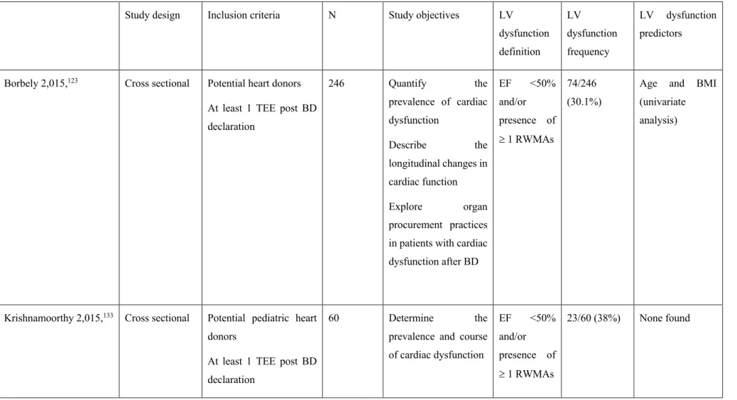
In human potential donors, observational studies report that 10 to 56% of brain-dead donors will present clinical cardiac dysfunction following neurological death.123-135 However, the definition
of cardiac dysfunction, the included populations, the timing of the evaluation, the measurement methods and the donors’ treatments at the moment of evaluation vary between studies (Table 1). The actual knowledge on the risk factors of reduced left ventricular ejection fraction (LVEF) is scarce, and only age and body mass index have been associated with impaired left heart function in univariate analyses.123 Longer time from brain death to echocardiography was also identified
as a predictor of LVEF improvement, and repeated echocardiography assessment suggests that cardiac dysfunction may be reversible, at least in some donors.135 One study evaluating the
potential impact of beta-adrenergic receptors polymorphism on the occurrence of left ventricular (LV) dysfunction suggested that genetic factors may predispose some individuals.130 Although
several reports have described LV dysfunction in brain-dead donors, right ventricular (RV) function has not been studied as extensively. In an animal model where brain death was induced in 60 mongrel dogs, high pulmonary and right ventricular pressures were observed during the autonomic storm period.136 Then, RV function decreased during the next several hours. Brain
death had a direct impact on the recipient’s RV function as well, even in the presence of basal normal pulmonary pressures.136 It was proposed that RV dysfunction may be induced by
inflammation mediators and apoptosis, similarly to LV dysfunction.137 In a mechanistic study
comparting heart donors with and without LV dysfunction, marked decrease in RV maximum contractile response to calcium was observed in heart donors featuring LV dysfunction. Response to beta-stimulation (isoproterenol) was also considerably reduced although no changes in the B1 or B2-adrenergic receptor density was observed.121
Table 1. Epidemiology of cardiac dysfunction in neurologically deceased donors
Study design Inclusion criteria N Study objectives LV
dysfunction definition LV dysfunction frequency LV dysfunction predictors Borbely 2,015,123 Cross sectional Potential heart donors
At least 1 TEE post BD declaration 246 Quantify the prevalence of cardiac dysfunction Describe the longitudinal changes in cardiac function Explore organ procurement practices in patients with cardiac dysfunction after BD EF <50% and/or presence of ³ 1 RWMAs 74/246 (30.1%)
Age and BMI (univariate analysis)
Krishnamoorthy 2,015,133 Cross sectional Potential pediatric heart
donors
At least 1 TEE post BD declaration
60 Determine the prevalence and course of cardiac dysfunction EF <50% and/or presence of ³ 1 RWMAs 23/60 (38%) None found
Examine organ procurement practices Kush 2,012,130 Retrospective
cohort
Consent to donation Stored DNA samples
1407 Describe donors’ LV ejection fraction according to genetic polymorphism
Describe RWMA and dopamine requirement according to genetic polymorphism EF <50% 10% B2Ar46 SNP Mohamedali 2,012,131 Retrospective (?) case series
Organ donors 34 Describe LV dysfunction
N/A 11/34 (32%) N/A Godino 2,010,128 Retrospective
observational
Potential heart donors Age <50 years
100 Identify and quantify the causes for exclusion of potential heart donors
Define risk factors for LV dysfunction EF < 40% 16/97 (16.5%) (3 patients missing) None Boudaa 2,003,126 Retrospective cohort
Potential heart donors 56 Identify selection criteria for heart procurement
Paul 2,003,127 Retrospective
observational
Potential pediatric heart donors
Echocardiographic screening before organ donation
23 Define the spectrum of LV dysfunction in pediatric donors EF <50% or LV shortening fraction <28% 13/23 (56.5%) N/A Hutteman 2,002,129 Retrospective observational
Patients with brain injury leading to BD
51 Evaluate the impact of TEE on patient management
Describe the incidence of LV dysfunction FAC <50% 7/51 (13.7%) N/A Dujardin 2001124 Retrospective cohort BD patient At least one echocardiogram 66 Describe the prevalence and characteristics of myocardial dysfunction N/A 28/66 (42%) N/A Kono 1,999,132 Prospective observational
Brain dead patients from non-cardiac cause
30 Describe the course of brain-death induced myocardial
dysfunction
Assess the ability of dobutamine stress echography to predict LV shortening fraction <30% 7/30 (23.3%) Positive stress echocardiography is predictor of LV function normalization
reversibility of LV dysfunction
Gilbert 1,998,125 Prospective (?)
observational
Potential heart donors 74 Evaluate cardiac function in potential heart donors Abnormal echography results leading to donor exclusion 9/74 (12.2%) N/A
TEE: Transthoracic echocardiogram; BD: brain death; LV = left ventricle; EF = ejection fraction; RWMAs = Regional wall motion abnormalities; FAC = fractional area change