UNIVERSITE DE LILLE
FACULTE DE MEDECINE HENRI WAREMBOURG
Année : 2019T H E S E P O U R L E D I P L O M E D ’ E T A T
D E D O C T E U R E N M E D E C I N E
ETUDE RETROSPECTIVE DES PATIENTS ATTEINTS D’UN CANCER
BRONCHIQUE NON A PETITES CELLULES
AVEC MUTATION BRAF V600E
Présentée et soutenue publiquement le 27/05/2019 à 18h
au Pôle Recherche
Par Geoffrey Pavaut
_____________
JURY
Président :
Monsieur le Professeur Arnaud Scherpereel
Assesseurs :
Monsieur le Professeur Laurent Mortier
Madame le Docteur Fabienne Escande
Directeur de Thèse :
AVERTISSEMENT
La Faculté n'entend donner aucune approbation aux opinions émises dans les thèses : celles-ci sont propres à leurs auteurs.
Table des matières
LISTE DES ABREVATIONS ... 1
I. INTRODUCTION ... 2 II. ARTICLE ... 12 INTRODUCTION ... 13 METHODS ... 15 RESULTS ... 17 DISCUSSION ... 26 III. DISCUSSION ... 41 IV. CONCLUSION ... 46 BIBLIOGRAPHIE ... 47
LISTE DES ABREVATIONS
ALK : Anaplastic Lymphoma Kinase
CBNPC : Carcinome Bronchique Non à Petites Cellules DCR : Disease Control Rate
EGFR : Epidermal Growth Factor Receptor INCA : Institut National du Cancer
NSCLC : Non Small Cell Lung Cancer ORR : Overall Response Rate
OS : Overall Survival
PD-L1 : Programmed Death – Ligand 1 PFS : Progression Free Survival
Cette thèse se présentera sous la forme d’une introduction suivie d’un article qui fera l’objet d’une soumission. Elle se terminera par un complément de discussion.
I.
INTRODUCTION
Prise en charge du cancer des poumons
Le cancer du poumon est une maladie fréquente, grave, avec un pronostic péjoratif à court terme. En France, en 2017, 49109 nouveaux cas ont été diagnostiqués, pour 30991 cas de décès estimés, ce qui en fait le 4e cancer par ordre de fréquence et la 1ère cause de mortalité par cancer. Le taux de survie standardisé à 5 ans entre 2005 et 2010 était de 17% (1). La mortalité par cancer du poumon est actuellement en augmentation dans la population féminine, avec 26% de décès dans cette population en 2012 (2,3). Les facteurs de risque bien connus sont le tabac, dans 80-85% des cas, également les expositions professionnelles, et notamment l’exposition à l’amiante.
Le cancer du poumon est principalement divisé en 2 types histologiques, le carcinome bronchique non à petites cellules (CBNPC), représentant environ 85% des cas, et le carcinome bronchique à petites cellules, dont l’incidence est en baisse, qui touche environ 10 à 15% des patients. Concernant les CBNPC, ceux-ci sont majoritairement représentés par les carcinomes non épidermoïdes (adénocarcinomes, carcinome à grandes cellules) dans environ 70% des cas, contre 30% pour les carcinomes épidermoïdes.
Le traitement recommandé des CBNPC au stade localisé (stade I ou II) est un traitement curatif, par chirurgie, ou par radiothérapie en cas de non-opérabilité (4,5). Pour les stades localement avancés (stade III), le traitement recommandé est une
radio-chimiothérapie concomitante, suivie d’une immunothérapie de maintenance au décours par durvalumab si la tumeur exprime PDL1 (6).
Cependant, pour la plupart des patients, environ 60%, (3,7) la maladie est diagnostiquée à un stade avancé, métastatique, pour laquelle la prise en charge repose sur un traitement systémique. Historiquement, celui-ci était représenté par la chimiothérapie, associant une molécule de 3e génération (gemcitabine, taxanes, pemetrexed, vinorelbine), à un sel de platine. L’ajout d’un traitement anti-angiogénique (bevacizumab) est discutée au cas par cas, en l’absence de contre-indication, principalement vasculaires (5). La médiane de survie sans progression était inférieure à 12 mois pour une survie globale d’environ 3,8% à 5 ans (8,9).
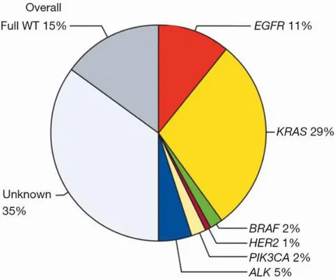
Plus récemment, le traitement en première ligne a été redéfini, avec la découverte des mutations activatrices survenant sur certains oncogènes jouant un rôle clé dans le processus de carcinogenèse. En effet, celles-ci ont permis le développement de thérapies ciblant spécifiquement les protéines mutantes issues de ces oncogènes mutés. La première décrite fut la mutation de l’Epidermal Growth Factor Receptor (EGFR), retrouvé dans 11% des cas (10) (figure 1). Celle-ci est une glycoprotéine transmembranaire constituée d’un récepteur extra-membranaire et d’un domaine tyrosine-kinase intracellulaire. L’activation de l’EGFR par la fixation de son ligand entraîne l’auto-phosphorylation du domaine tyrosine-kinase intracellulaire, conduisant à une prolifération cellulaire par la voie des MAP kinase (mitogen activated protein kinase), et la survie cellulaire par la voie Pi3K-AKT, par inhibition de l’apoptose et favorisant l’angiogénèse. En cas de mutation activatrice, le domaine tyrosine-kinase est activé sans nécessité de fixation du ligand. Elle est principalement retrouvée dans une population asiatique, féminine, sans antécédent tabagique, associée à une plus
forte proportion d’adénocarcinomes (11). Cela a permis le développement d’inhibiteurs de tyrosine kinase (TKI) ciblant spécifiquement ce domaine intra-cellulaire. La première étude évaluant un TKI inhibiteur de l’EGFR avait retrouvé un taux de réponse supérieur à la chimiothérapie, plus particulièrement dans le sous-groupe de patients porteurs de cette mutation, de 71,2% contre 47,3%, une médiane de survie sans progression de 9,5 mois contre 6,3 mois. Il n’y avait en revanche pas de différence sur la survie globale (12). Des études ultérieures ont retrouvé des résultats similaires (13,14).De plus, ces traitements sont globalement mieux tolérés par les patients (12). De même concernant le réarrangement ALK (Anaplastic Lymphoma Kinase), présent dans environ 5% des CBNPC, qui a pour conséquence une activation constante d’un domaine tyrosine kinase impliqué dans la prolifération cellulaire. Les études évaluant le crizotinib, ou plus récemment l’alectinib, ciblant spécifiquement cette protéine mutante, ont montré des taux de réponse et des médianes de survie sans progression supérieures à la chimiothérapie (15,16). On retrouve une efficacité similaire avec la mutation ROS1 et le traitement par crizotinib (17). En présence d’une de ces mutations, le traitement de première intention repose donc sur une thérapie ciblée. Ainsi, on estime que 49,5% des carcinomes pulmonaires seraient porteurs d’une mutation, et ainsi potentiellement accessibles à un traitement spécifique (10). Cependant, ce chiffre comprend la mutation KRAS, pour laquelle il n’existe, à l’heure actuelle, pas de traitement spécifique.
Parallèlement à la découverte de nouvelles mutations activatrices, le développement des plateformes de recherche en biologie moléculaire a permis de détecter un plus grand nombre de mutations lors du diagnostic de la maladie. Initialement, des tests « single-gene » étaient employés, par exemple la technique de Sanger, via des réactions en chaîne par polymérase (PCR), avec initialement une polymérisation de
l’ADN à l’aide de nucléotide pour le séquencage, puis une séparation des fragments par électrophorèse. La détection des fragments synthétisés se faisait grâce à un traceur incorporé dans l’ADN synthétisé. Actuellement, la technique majoritairement employée est celle du séquençage nouvelle génération (NGS), permettant de séquencer plus rapidement l’ADN et l’ARN (18). Cette technique permet de séquencer de nombreux nucléotides, avec la possibilité de réaliser plusieurs réactions en parallèles sans nécessité de réalisation de clones. Celle-ci permet de séquencer majoritairement une région d’intérêt, avec la réalisation de synthèse de librairie (hybridation par ARN par exemple), amplification par poymerase-chain reaction (PCR) puis le séquencage. Son développement a été primordial en oncologie, devant la possibilité de réaliser de nombreux tests plus rapidement, à un moindre coût, d’être plus exhaustif, et permettant également d’avoir une meilleure rentabilité en cas d’épuisement du matériel tumoral disponible.
En l’absence de mutation, la prise en charge est basée selon le « programmed death-ligand 1 (PDL1) », conduisant à un traitement par immunothérapie en première ligne (pembrolizumab), en cas de taux de PDL1 supérieur à 50% (19). En l’absence de mutation activatrice et en cas de taux de PDL1 inférieur à 50%, le traitement recommandé est une chimiothérapie à base d’un doublet de platine et d’une molécule de 3e génération. En deuxième ligne, les patients ayant été initialement traités par chimiothérapie pourront recevoir une immunothérapie (nivolumab, pembzolizumab) (20).
Figure 1 : Répartition du statut mutationnel dans le CBNPC, Barlesi et Al, Lancet 2016
Les mutations BRAF dans le CBNPC
Au même titre que celles précédemment décrites, la mutation BRAF, fait partie des mutations activatrices dans le CBNPC.
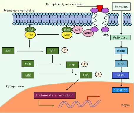
Sur le plan physiopathologique, BRAF appartient à la famille des sérine-thréonine kinases, et est un élément de la voie de signalisation des MAP (Mitogen Activated
Protein) kinases : Ras-Raf-Mek-Erk (figure 2 et 4). Elle est codée par un gène situé
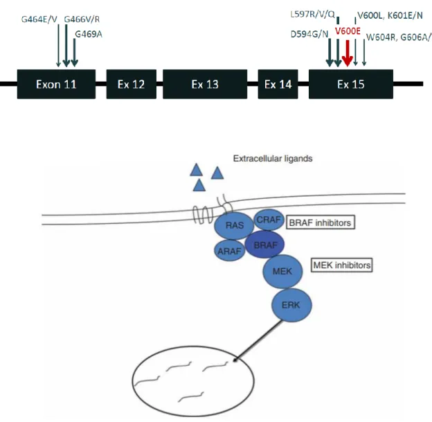
sur le chromosome 7, et est située en aval de nombreux récepteurs, notamment ceux de l’EGFR. Après plusieurs phosphorylations successives, cette voie entraîne la synthèse de facteurs de transcription impliqués dans la prolifération cellulaire et l’angiogénèse (21,22). Plusieurs types de mutations ont été décrites, la principale étant un changement d’acide aminé en position 600, avec un remplacement d’une valine (V) par un acide glutamique (E), soit la mutation V600E (classe I). Son activité kinase est
augmentée d’environ 500 fois comparativement aux non-mutés (23). D’autres mutations ont également une activité transformante in vitro : G469A, L597V, contrairement aux mutations G466V et Y472C, qui possèdent une activité kinase réduite(24).
La mutation V600E entraine une activation de la protéine BRAF, indépendamment de la protéine RAS située en amont, et active de façon continue la protéine ERK en aval, sans sensibilité au rétrocontrole négatif de cette dernière, favorisant ainsi la prolifération cellulaire (25,26).
Figure 3 : Mutations BRAF selon les exons
Figure 4 : Voie de signalisation des MAP Kinase avec sites d’action des inhibiteurs de BRAF et de MEK, Dossett et Al, 2015
Devant cet effet pro-oncogène, il a ainsi été developpé des traitements ayant pour objectif d’inhiber de façon spécifique cette protéine (sorafenib, vemurafenib, dabrafenib). Les essais (27) ont permis de découvrir un effet pro-oncogène inverse de celui attendu, avec l’apparition notamment de kérato-acanthomes et de carcinome épidermoïde cutané. Celui-ci est connu sous le nom de phénomène d’activation
paradoxale de la voie des MAP kinase en cas d’inhibition de RAF. Des modèles in vivo ont en effet montré que la fixation de l’inhibiteur de RAF sur les BRAF dits « sauvages », non-mutés, pouvait entraîner une activation de MEK via CRAF. Celle-ci s’observe en cas de concentration plasmatique basse de traitement. L’ajout d’un inhibiteur de MEK permet cependant de contrôler cet effet (28–31).
Sur le plan épidémiologique, les mutations BRAF ont été principalement décrites dans le mélanome, où les données font état d’une prévalence allant de 40 à 60% (24,32). Elle est également retrouvée dans les carcinomes papillaires de la thyroïde (30-70%), certains cancers colorectaux (5-20%) (33). Dans le cadre du cancer colo-rectal, elle représente un facteur pronostique péjoratif, de par un profil de patients plus agés, avec une maladie diagnostiquée à un stade tardif métastatique, et un profil d’instabilité micro-satellitaire élevé (34,35). Dans le mélanome, elle était associée à un indice de Breslow plus élevé, une augmentation du nombre de mitoses, un âge jeune et également un stade avancé de la maladie (36,37).
Dans le carcinome bronchique non à petites cellules, sa prévalence est de 1,7 à 5% (10,24,38) (figure 1), principalement présente dans un contexte tabagique (21,39–41). Les études antérieures font état d’une proportion d’environ 50% de mutation V600E, initialement plutôt retrouvée dans une série chirurgicale, dans une population féminine, et au contraire, sans antécédent tabagique (42,43). Cependant, d’autres études plus récentes n’ont pas retrouvé de différences concernant les caractéristiques cliniques entre les patients mutés V600E, non-V600 et les non-mutés (39,44,45). Au même titre que dans le mélanome et le cancer colo-rectal, le pronostic de ces patients serait également plus sombre, avec une moins bonne réponse à la chimiothérapie dite « classique », comparativement aux patients non-mutés (10). Concernant plus spécifiquement la mutation V600E, sous chimiothérapie, les survies sans progression
et globale seraient inférieures aux autres sous-types de mutations BRAF, sans significativité statistique (45,46). Une étude de vie réelle plus récente retrouvait cependant une médiane de survie globale plus élevée que décrite dans la littérature concernant les patients atteints de mutation V600, de 27,2 mois dans une population de mutations V600 (47).
Avant 2011, le taux de survie à 5 ans d’un mélanome métastatique non-opérable était inférieur à 10%, avec une médiane de survie inférieure à un an. Sous thérapies ciblées, les études ont retrouvé un taux de réponse de 48% à 59% pour le vemurafenib (48). Dans un essai de phase III comparant le dabrafenib à la dacarbazine, le taux de réponse était de 50% dans le groupe dabrafenib, et la médiane de survie sans progression significativement plus élevée dans ce groupe, de 5,1 contre 2,7 mois (p<0,001) (49). Une étude de phase III avait comparé le dabrafenib en monothérapie contre l’association dabrafenib-trametinib, avec une différence significative du taux de réponse, de 67% pour l’association contre 51% pour le dabrafenib en monothérapie (p<0,001). La médiane de survie sans progression était également plus longue dans le groupe combinaison, avec moins de toxicités (9,3 mois contre 8,8 mois) (50). De ce fait, la mutation BRAF représente une nouvelle cible thérapeutique en oncologie thoracique. Une première étude avait évalué le vemurafenib en monothérapie dans une cohorte de 20 patients dont 18 porteurs de la mutation V600E, retrouvant un taux de réponse de 42,0%, une médiane de survie sans progression de 7,3 mois, pour une survie globale à 12 mois de 66% (51). Dans une cohorte française de 101 patients avec mutation V600E, ayant reçu du vemurafenib après une première ligne de traitement, le taux de réponse était de 44,9%, la médiane de survie sans progression de 5,2 mois, pour une médiane de survie globale de 9,3 mois (52). Concernant le dabrafenib, autre inhibiteur de BRAF, également dans une cohorte française de 84
patients, il a été retrouvé un taux de réponse de 33%, une médiane de survie sans progression de 5,5 mois et de survie globale de 12,7 mois (53). L’association dabrafenib-trametinib a également été évaluée par ces mêmes auteurs. En 2ème ligne ou plus, le taux de réponse était de 63,2%, pour une médiane de survie sans progression de 9,7 mois (54). En 1ère ligne, sur 36 patients, le taux de réponse était de 64%, la médiane de survie sans progression de 10,9 mois pour une médiane de survie globale de 24,6 mois (55).
Ces résultats sont donc encourageants au sein de cette population, et ont permis d’obtenir l’autorisation de mise sur le marché de l’assocation dabrafenib-trametinib en mars 2017.
A l’heure actuelle, les caractéristiques cliniques de ces patients restent peu définies. De même, peu de données permettent de confirmer l’efficacité de l’association des inhibiteurs de BRAF et MEK et ce traitement n’a, à l’heure actuelle, jamais été comparé aux traitements classiques, et plus particulièrement la chimiothérapie. De même il n’existe que peu de données concernant l’utilisation de ces traitements, et leur efficacité en pratique courante, hors-essai.
Ainsi, les objectifs de notre étude sont de décrire les caractéristiques clinico-pathologiques des patients atteints d’un CBNPC porteurs de la mutation BRAF V600E, d’étudier les différents traitements reçus par ces patients en condition de vie réelle, de décrire les taux de réponse, les survies globales et sans progression, ainsi que les effets indésirables sous thérapies ciblées.
II. ARTICLE
OUTCOME OF BRAF V600E LUNG ADENOCARCINOMA : A RETROSPECTIVE STUDY
Pavaut G.1, Escande F.2, Copin, M-C.3, Gey T.4, Kedziora L.5, Amourette J.6, Scherpereel A.1, Descarpentries C.2, Cortot AB1
1 CHU Lille, Department of Pulmonary Diseases and Thoracic Oncology, Univ. Lille, F-59000, Lille, France
2 CHU Lille, Department of Molecular Biology, F-59000, Lille France 3 CHU Lille, Department of Pathology, Univ. Lille, F-59000, Lille, France
4 Clinique Teissier, Department of Pulmonary Diseases, Valenciennes, France 5 CH Denain, Department of Pulmonary Diseases, Denain, France
6 CH Arras, Department of Pulmonary Diseases, Arras, France
ABSTRACT
INTRODUCTION : BRAF mutation stands for 1.7 to 5% of lung adenocarcinomas, and V600E mutation is the most frequent type of BRAF mutations. However real-life data from patients treatment and outcomes are limited, especially in those treated with BRAF inhibitors with or without MEK inhibitors.
METHODS : We conducted a retrospective multicenter study between 2011 and 2018, including patients with non-small cell lung cancer (NSCLC) harboring BRAF V600E mutation centrally detected in Lille University Hospital. Clinical and tumor characteristics, treatments and patients outcomes were reviewed.
RESULTS : 59 patients were included. Most had adenocarcinoma (94.9%), were men (61%) and had a smoking history (57%). Disease was mainly diagnosed at stage IV (79.7%). Targeted therapies (including RAF inhibitors with or without MEK inhibitors) yieldied an overall response rate (ORR) of 52.9% and a median progression free-survival (PFS) of 6 months [2-22]. Chemotherapy yielded an ORR of 25% and a median PFS of 4 months [2-9]. Patients who did not received any targeted therapy had a median overall survival (OS) of 17.0 months [1.5-118.0] whereas those who received
at least one targeted therapy had a median OS that was not reached at the end of the study (95% CI [4.4-NR]). Median PFS on targeted therapies was improved in never-smokers compared to ever-never-smokers (9.25 months [0.5-NR] versus 2.6 [0.7–4], p=0.03).
CONCLUSION : BRAF V600E mutations in NSCLC frequently occurs in smokers. In this real-life study, targeted therapies were associated with prolonged overall survival and appeared to be more active in never smokers. Prospective and comparative studies are needed to confirm these data.
INTRODUCTION
Lung cancer is a common disease and the leading cause of cancer death in Europe. Incidence is increasing in women (3). Five-year survival rate in 2010 was about 17%, which reflects the high incidence of stage IV disease at diagnosis (1). Smoking remains the major risk factor for lung cancer.
Discovery of activating molecular alterations of key oncogenes in Non Small Cell Lung Cancer (NSCLC) (10) such as Epidermal Growth Factor Receptor (EGFR) mutations and ALK rearrangements has led to the development of targeted therapies (21,22).
EGFR and ALK tyrosine kinase inhibitors (TKI) have improved response rate and progression-free survival compared to chemotherapy, with a better tolerance profile, in EGFR-mutated and ALK-rearranged NSCLC, respectively (12,13). Targeted therapies have also demonstrated activity in NSCLC with ROS1, NTRK, RET rearrangements and MET exon 14 skipping mutations (15,16,56,57).
BRAF is a serine-threonine protein kinase in the RAS-RAF-MEK-ERK signaling pathway, which controls cell proliferation. BRAF mutations have been described in various solid tumors including melanoma, NSCLC, colorectal cancer, hairy-cell leukemia or thyroid cancer (24,32,33). BRAF mutants are usually divided into V600 mutants and non-V600 mutants. V600 mutants occur within the kinase domain of
BRAF and result in high kinase activity of BRAF which acts as a monomer, independently from RAS activation. These mutations are found in 50% of melanoma and in 1.7 to 5% of lung adenocarcinomas (10,24,39,58). V600 mutations lead to an excessive BRAF activation and thus induce cell growth and survival. Non-V600 mutants are found in many cancers and are more prevalent than V600 mutations in certain tumor types. They account for 50% of all BRAF mutations in NSCLC and 22-30% of all BRAF mutations in colorectal cancer.
In NSCLC, BRAF V600E mutation is the most frequent one and has been associated with female gender and never-smoking status (42). However, later studies have not confirmed these clinical characteristics associated with BRAF V600E mutations (38,44). BRAF V600E mutation may be associated with a lower ORR to platinum-based chemotherapy (10), and shorter overall survival compared to wild-type in a surgical series (42). Response to chemotherapy may also be inferior in V600E than non-V600E NSCLC patients (45). In vitro, BRAF V600E mutated cells are known to be sensitive to both RAF and MEK inhibitors, used either alone or in combination (23,59,60). In V600E mutant melanoma, RAF inhibitors such as dabrafenib or vemurafenib have demonstrated superiority over chemotherapy in terms of ORR, PFS and OS (61,62). For example, with vemurafenib, ORR was 48% (48). Median-PFS of dabrafenib, compared to dacarbazine in a phase III trial, was significantly higher (5.1 vs 2.7 months) with an ORR of 50% (49). To improve durability of BRAF inhibition, MEK inhibitors have been developed, in addition of the mitogen-activated protein kinase (MAPK) pathway at MEK. Interestingly, combination of RAF and MEK inhibitors increased efficacy in a phase III trial, with an ORR of 67% for dabrafenib-trametinib association, versus 51% for dabrafenib alone. Median-PFS in this study was also
higher : 9.3 months vs 8.8 months, and rates of adverse event were similar, but incidence of squamous cell carcinoma, skin papilloma and basal cell carcinoma was higher in monotherapy-arm (50). In NSCLC, RAF inhibitors alone have demonstrated some activity in V600 NSCLC with an ORR of 42.0%, a median-PFS of 7.3 months and one-year OS of 66% (51). After first-line therapy, another study has found an ORR of 44.9%, median-PFS of 5.2 months, and median-OS of 9.3 months (52). With dabrafenib, ORR was 33%, median-PFS was 5.5 months, and median-OS was 12.7 months (53). However, as for melanoma, combination of dabrafenib and trametinib was shown to be more effective and less toxic than dabrafenib alone. After first-line therapy, authors found an ORR of 63.2%, median-PFS of 9.7 months (54). In first-line therapy in previously untreated patients, ORR was 64%, median-PFS was 10.9 months and median-OS 24.6 months (55). Subsequently, the combination of dabrafenib and trametinib has been recommended as the best first-line treatment option for V600E BRAF mutated NSCLC patients by several scientific guidelines (5,63). However, the lack of a randomized trial and real-life data from V600E BRAF mutated NSCLC patients impairs the implementation of such recommendations.
Here, we conducted a retrospective study aiming at describing demographics and characteristics of real-life patients with NSCLC harboring BRAF V600E mutation, along with response to treatment, including dabrafenib-trametinib combination, and patients outcomes.
METHODS
Patients
This is a retrospective multicenter study. We included all patients diagnosed with NSCLC and harboring a BRAF V600E mutation centrally detected by the molecular
biology laboratory of Lille University Hospital. Mutations were detected by single-gene sequencing until 2015. Since 2015, next generation sequencing (NGS) Ion AmpliSeq Colon and Lung Research Panel V2 is used for sequencing. In accordance with the French legislation, the study was declared to the National Information Science and Liberties Comission (CNIL) and all patients agreed to participate to this study.
Data collected
Clinical characteristics were recorded, including age, gender, smoking history, disease stage at initial diagnosis using the 8th TNM classification, metastatic sites at diagnosis. Stage IV treatment history was collected and treatments were classified into three categories : chemotherapy (including non-RAF or MEK targeted therapies), targeted therapy against RAF (dabrafenib, combined or not with trametinib, vemurafenib) and immunotherapy. Patients received RAF or MEK inhibitors as part of a clinical trial, or off-label or under marketing authorization from March 2017. We recorded the treatment responses as assessed by the referent physician for each line of treatment until third-line treatment and for all third-lines combined. Progression-free survival (PFS) was measured frome the date of the stage IV diagnosis until progression or death, whichever occurs first. Overall survival (OS) was measured frome the date of the stage IV diagnosis until death. Objective response rate at first assessment was measured using the Response Evaluation Criteria In Solid Tumors (RECIST) version 1.1, and Immune RECIST (iRECIST) v2017 for treatment with immune checkpoint inhibitors. Briefly, response was classified into progression disease (PD), stable disease (SD), partial response (PR) and complete response (CR). Adverse events were retrospectively collected for patients treated with targeted therapies using the common terminology criteria for adverse events (CTCAE) v5.0.
Statistical analysis
Continuous variables are expressed as means (standard deviation, SD) in the case of normal distribution or medians [interquartile range] otherwise. Categorical variables are expressed as numbers (percentage). Normality of distributions was assessed using histograms and the Shapiro Wilk test. Overall survival in patients with a systemic treatment were estimated using Kaplan-Meier method.
We assessed the association of exposure to targeted therapies with overall survival at 25 months by using Logrank test. We assessed the associations of type of treatments in early lines with progression-free survival by using Logrank test. We assessed the association of smoking history with progression-free survival at the end of targeted therapies by using Logrank test.
Statistical testing was conducted at the two-tailed α-level of 0.05. Data were analyzed using the SAS software version 9.4 (SAS Institute, Cary, NC). Statistical analyzes were performed by the Methodology Biostatistics Unit of the CHU in Lille.
RESULTS
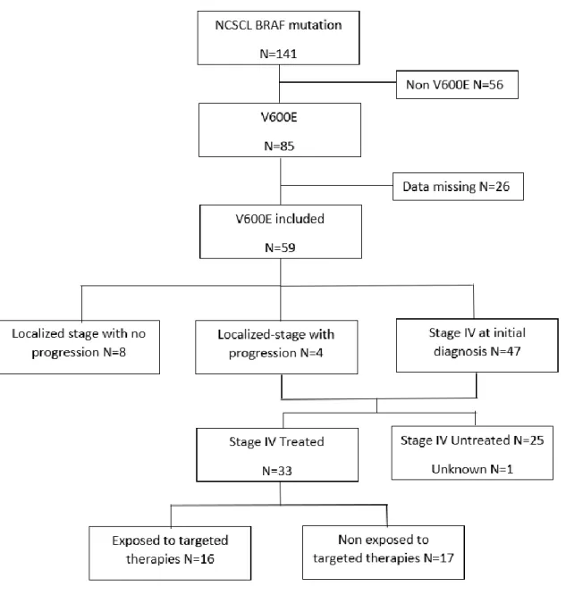
Between may 2011 and august 2018, 144 patients with lung cancer harboring a BRAF mutation were identified. 85 V600E mutations were detected, among those, 53 V600E mutations were identified by single-gene sequencing and 32 (36.4%) by NGS. 56 (63.6%) non-V600E were also detected by NGS (suppl A fig 1).
Overall, 85 V600E mutations were detected and 59 patients (69%) were eventually included in this study. The reasons for not including 26 patients were lack of data in most of the cases. Clinical characteristics of patients included in the study were not
different from those who were not included in terms of age, sex, and type of BRAF mutation.
Demographics and clinicopathological characteristics
At initial diagnosis, median age was 66.8 years (± 11.9). We found 36 men (61.0%) and 23 (39.0%) women. 32 patients (57.1%) were former or current smokers, and average number of pack years was 35.1 (± 20.9). Patients were predominantly diagnosed at stage IV (79.7%). OMS status diagnosis was 0-2 in the vast majority of patients (79.7%).
94.9 % of NSCLC were adenocarcinomas (n=56). One patient had squamous cell carcinoma, another had large cell carcinoma, and one was classified as NSCLC N.O.S. Regarding the molecular alterations, one patient was found to harbor a concurrent SMAD4 mutation, one had a KRAS andAKT1 mutation, and one KRAS and PIK3CA. Among the 47 patients with initial stage IV disease, 41 patients had lymph nodes metastasis (87.2%), 32 (68.1%) had pleura metastasis and 24 (51.1%) had lung metastasis. Other metastatic sites were bone (31.9%), adrerenal glands (14.9%), brain (12.8%) and muscles (8.5%).(table 1)
Table 1 :
Demographics and clinical characteristics
Total Patients n (%)
N=59
¨%
Age at diagnosis, years 66.8 ± 11.9
Gender Male 36 61.0 Female 23 39.0 Smoking Status Never smoker 24 42.9 Former smoker 21 37.5 Current smoker 11 19.6 Unknown 3 Pack-Year(average) in ever smokers 35.1 ± 20.9
Disease Stage at diagnosis
I-II 8 13.6
III 4 6.8
IV (metastatic) 47 79.7
OMS Status at diagnosis
0 16 27.1 1 23 39.0 2 8 13.6 3 10 16.9 4 2 3 .4 Tumor Histology Adenocarcinoma 56 94.9 Other 3 5.1
Sites of Metastases at initial
diagnosis (in stage IV) N=47
Lymphatic nodes 41/47 87.2 Pleura 32/47 68.1 Lung 24/47 51.1 Bone 15/47 31.9 Adrerenal glands 7/47 14.9 Brain 6/47 12.8 Muscles 4/47 8.5
Treatments received
Following initial diagnosis, among all 59 patients, 16 patients (27,6%) received no treatment at all. Compared to patients who received at least one line of treatment, or a local treatment, those who did not received any treatment were significantly older (73.3 ± 10.7 vs 64.4 ± 11.7, p=0.011), had higher OMS status (p<0,001) and had more pleural metastasis (80,0% vs 46% p=0.025). (suppl.B table1)
12 patients (21.1%) received local treatment including 7 patients who underwent surgery and 5 patients who received radiation therapy. Among patients with stage IV disease or with recurrent disease following local treatment (n=51), 33 (65%) received at least one systemic treatment (figure 1). First-line treatment consisted in chemotherapy for 19 (57.6%) patients, immunotherapy for 4 patients (12.1%) and targeted therapy for 10 patients (30.3%). Fourteen patients received a second-line therapy : 4 (28%) patients were treated with chemotherapy, 5 (36%) with targeted therapies and 5 (36%) with immunotherapy. Among the 5 patients who received third-line therapy, 2 patients were treated with chemotherapy, 2 with targeted therapy and one with immunotherapy. Three patients received a fourth-line treatment and one patient received a treatment until sixth line. (suppl.B table 2)
Targeted therapies consisted mainly in the combination of dabrafenib and trametinib (n=11 (65%)) (table 2). Four patients (23%) received dabrafenib alone and two (12%) were treated by vemurafenib alone.
Table 2 :
Details of patients treated with targeted therapies
First-line N=10 Second-line N=5 Third-line N=2 dabrafenib 3 1 0 dabrafenib-trametinib 6 3 2 vemurafenib 1 1 0 Efficacy
Regarding chemotherapy, overall response rate (ORR) in first-line was 35.3% (n=6), disease control rate (DCR) was 61.1% (n=11), and median progression-free survival (PFS) was 4.0 months (95% CI 1.4-11.0). In second and third-line, no patient had an objective response, three patients (50.0%) had a disease control. Median-PFS was 5.7 months (95% CI 1.0-9.0). All lines combined, ORR was 25.0% (n=6), DCR was 56.5% (n=13) and median-PFS was 4.0 months (95% CI 2.0-9.0).
Regarding targeted therapies, ORR in first-line was 50.0% (n=5), DCR was 70.0% (n=7), and median-PFS was 5.0 months (95% CI 1.0-22.0). In second and third-line, ORR was 57.1% (n=4), DCR was 71.4% (n=5), and median PFS was 6.0 months (95% CI 0.5-12.5). All lines combined, ORR was 52.9% (n=9), DCR was 70.6% (n=12) and
median-PFS was 6.0 months (95% CI 2.0-22.0). For dabrafenib-trametinib combination specifically, we found a 72.7% ORR (n=8/11).
Regarding immunotherapy, all lines combined, one patient (14.3%) had a partial response, 3 patients had control disease (42.9%), and median-PFS was 4.4 months (95% IC 1.5-6.5). (table 3) (suppl C table 1-2 ; fig 1-6)
In order to determine which patients are likely to benefit from targeted therapies, we performed subgroups analysis. We found that PFS to targeted therapies was higher in never smokers compared to smokers (median PFS 9.25 months (0.5-not reached) vs. 2.6 months (95% CI 0.7-4.0) p=0,0288) (figure 1). ORR was also higher in never smokers compared to smokers (70% vs 17%).
At data cut-off, 17 patients (28,8%) were alive. Median overall survival for stage IV patients who received any systemic therapy was 17.0 months (95% CI 5.0-43.6) (Suppl
D fig 1). It was 17.0 months (95% CI 1.5-118.0) for patients who never received any
targeted therapy and median OS was not reached for those who received at least one line of targeted therapy (95% CI 4.4-NR) (figure 2). For patients who received dabrafenib-trametinib (n=11), median-OS was not reached. It was 6.0 months (3.5-not reached) for those who received a RAF inhibitor as monotherapy (n=5). (suppl D fig 2)
Toxicity of targeted therapies
Ten adverse event were reported in patients treated with targeted therapies, 6 (60%) grade 1-2 and 4 (40%) grade 3-4. Among grade 1-2, most frequent were skin toxicities (n=3, 50%). We also found one gastro-intestinal disorder, one leukopenia and one
thrombocytopenia. All grade 3-4 were skin toxicities. 9 (90%) adverse event s occured with the combination, and only 1 (10%) with a RAF inhibitor as monotherapy. (Suppl E
table 1). Reduction dose occured in 4 patients (3 grade 3 events included one skin
adverse event), and in the aftermath of thrombocytopenia).
Table 3 :
Antitumor Activity in First, Second and Third-Line Combined
Chemotherapy N=25 Targeted Therapy N=17 Immunotherapy N=10 p Unknown 1 0 3 Best Response, n (%) PD (Progression Disease) 10 (41.7) 5 (29.4) 4 (57.1) SD (Stable Disease) 8 (33.3) 3 (17.6) 2 (28.6) PR (Partial Response) 6 (25.0) 8 (47.1) 1 (14.3) CR (Complete Response) 0 (0.0) 1 (5.9) 0 (0.0) Objective Response (CR+PR), n (%) 6 (25.0) 9 (52.9) 1 (14.3)
Disease Control Rate
(CR+PR+SD), n (%) 13 (54.2) 12 (70.6) 3 (42.9)
Progression-Free Survival,
median (95% CI), months 4.0 (2.0-9.0) 6.0 (2.0-22.0) 4.4 (1.5-6.5) 0.597
Figure 1 : Progression-free Survival to Targeted Therapies defined by Smoking status
0 = Never smoker, Median PFS = 9.25 months (0.5 - not reached)
1 = Current or former smoker, Median PFS = 2.60 months (0.7 – 4.0) p=0.03
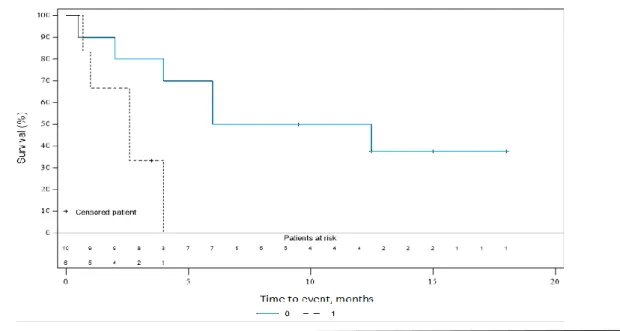
Figure 2 : Overall Survival (OS), by Targeted Therapy Status
0 = Did not receive Targeted Therapy : Median OS = 17.0 months (1.5-118.0) 1 = Receive Targeted Therapy : Median OS = Not reached (4.4-NR)
DISCUSSION
We conducted a retrospective study on real-life V600E BRAF mutants NSCLC. We found that targeted therapies yielded the best efficacy, with an ORR of 52.9% and a median PFS of 6 months. Targeted therapies were significantly more active in never-smokers.
In our study, we found a 39% proportion of women among V600E BRAF mutant NSCLC patients. Some studies have found that V600E mutations were associated with female gender. However, these prior studies were smaller or conducted in patients with early stage NSCLC (42). Based on our results, the proportion of women appears to be higher than in unselected NSCLC but lower than in EGFR-mutated or ALK-rearranged NSCLC. Regarding smoking history, 57.1% of V600E BRAF mutant NSCLC patients were current or former smokers in our series. Some authors described that BRAF mutations occur more often in current or former smokers than in never smokers, compared to wild-type (21,39,45). On the contrary, BRAF V600 mutations have been reported to be more frequently detected in never smokers (42). Our results suggest that the proportion of smokers appears to be higher than in EGFR-mutated NSCLC, but lower than in unselected NSCLC. Regarding radiological characteristics, predominant sites of metastasis were lymph nodes, lung and pleura. Brain metastasis were found to be rather rare compared to other NSCLC with oncogene addiction (15). Overall, these results draw the typical characteristics of BRAF-mutant NSCLC patients, with a high proportion of women and smokers, and a high frequency of thoracic metastasis.
We found that patients who received exclusive palliative care (27.5%) were significantly older and their OMS status was higher than those who received a treatment. Pleural metastasis may also be associated with absence of treatment, although the association we found may be due to the small sample size. This no-treatment rate was similar to that reported in Barlesi et al (10). With development of targeted therapies and their favorable tolerance profile, we could expect that the rate of patients who received exclusive palliative care would be lower. Only 16 patients received at least one targeted therapy. This may be explained because of the low prevalence of BRAF mutations. Moreover, efficacy of BRAF and MEK inhibitors have been demonstrated recently and might be initially unknown by physicians. Physicians may also lack experience of its use, which may have refrain them. Finally, Another difficulty for these patients to access to these treatment might also be that access to these targeted therapies was not easy : they were available as part of clinical trials ; however, most of the patients can not be included in clinical trials. Since march 2017, marketing authorization have been granted, but the actual clinical benefit is insufficient to justify public funding cover, and not approved for non-hospital pharmacy reimbursement or for hospital treatment.
Focusing on treatment outcomes, we found a 52.9% ORR on targeted therapy (72.7% for dabrafenib-trametinib specifically) and median PFS was 6.0 months. Median OS was not reached for patients who received at least one targeted therapy, particularly dabrafenib-trametinib combination, instead of 17.0 months for unexposed patients. Surprinsingly, no patient responded to dabrafenib alone. However we need to interprete these results with caution, because of small sample size. In metastatic melanoma, RAF inhibitors were shown to be more efficient than chemotherapy (49),
and MEK/BRAF combination improved median-PFS versus RAF inhibitor in monotherapy (50). However, there is no randomized data in NSCLC. As mentioned earlier, efficacy of dabrafenib-trametinib combination has been shown in a phase II trial, with an ORR of 64% and a median PFS of 10.9 months in first-line (54,55). ORR and PFS were not as good in our series. This may be because we performed a real-life study, and patients enrolled and treated with targeted therapies were not selected. As an illustration, 33,9% of patients had a performance status of 2 or more. Moreover, the high rate of smokers in our series may have impaired efficacy of targeted therapies.
Overall, our results, compared to those obtained with chemotherapy and immunotherapy, suggest that targeted therapies may be the best therapeutic option for BRAF V600E mutant NSCLC patients.
Coming back to smoking status, we found that median PFS to targeted therapies was higher in never smokers, compared to smokers (9.25 months (0.5-NR) vs 2.60 (0.7-4) months, p=0,03), which was also found for EGFR-TKI-therapy (11). This may be due to additional molecular alterations associated with BRAF mutations in smokers, decreased blood concentration of the drugs due to activation of liver cytochromes, or production of reactive oxygen species induced by tobacco, known to be a mechanism of resistance to TKIs (64). This data is important because of the high proportion of smokers in our study. This may need to be taken into account when making a treatment decision in this population, between chemotherapy and targeted therapy.
Results regarding efficacy of immunotherapy in our study need to be interpreted cautiously given the low number of patients. Recently, studies have shown low response rate to checkpoint inhibitor therapies, in NSCLC with EGFR or cMET mutations, and ALK rearrangements (65–67). Another study found that in BRAF mutant NSCLC, ORR with immunotherapy was 28,1% and median OS was 13.6 months (68), which sounds encouraging. This might be explained because of the high proportion of smokers in BRAF mutated NSCLC, and it is known that PD-1 inhibitors are more active in smokers than chemotherapy (69). A case-report described a great benefit to sequential treatment (targeted therapy and immunotherapy) in a NSCLC patient, with BRAF V600E and PD-L1 positive status (90%), surviving for over 7 years (70). These data suggest that immune checkpoint inhibitors might be a therapeutic option in BRAF V600E NSCLC, especially in smokers.
We found that 85 patients had a NSCLC harboring BRAF V600E mutation, among 141 BRAF mutations detected. However, we need to compare percentages of mutations detected by Next Generation Sequencing, since non-V600 mutations were not screened with the single gene sequencing. With NGS, we found 32 (36,4%) V600E and 56 (63,6%) non-V600E mutations. The proportion of V600E in our study was lower than what is found in the literature, ranging from 50% to 58% (10,39,42). However, in these prior studies, sample sizes were small (n=18-21), inclusion time was shorter (39) and mutations were not detected by NGS. For example, in Paik et al study, only G469A and D594G mutations were detected whereas we found 18 different subtypes of non-V600 mutations. Indeed, NGS makes it possible to detect a larger number of BRAF mutations, especially non-V600 mutations, which could explain why we found a higher proportion of non-V600 mutations.
Non-V600 mutants have been shown to signal as RAF dimers rather than monomers (found in class I), making them theoretically resistant to BRAF monomer inhibitors. However, pre-clinical study and case reports of response to MEK-inhibitors of non V600-BRAF patients have been published (71,72). A study evaluating trametinib in this population is ongoing in melanoma (NCT02296112). However, MEK inhibitors may have limited efficacy when given as single agent in non-V600 BRAF mutations. These mutants could be targeted by the simultaneous inhibition of RAS and downstream MAPK signaling (73). Further studies are needed in these BRAF-subtypes, especially in NSCLC.
Our study has some limitations, mainly due to its retrospective design. All BRAF-mutated NSCLC patients could not be included and some data are lacking. However, we were able to include 59 patients harboring a V600E mutation, which represents one of the largest studies published so far, and we found no difference in in terms of age and sex between patients included and those not included in this study. As this is not a randomized study, the benefit in OS observed in patients exposed to targeted therapy may rely solely on patients selection with favorable pronostic factors such as OMS status. However, the high ORR and prolonged PFS suggest a direct impact of targeted therapies on OS. We did not perform central review of radiological assessment, but RECIST criteria were used by radiologists or referent physicians most of the times, and radiological assessments reflect real-life practice, which is the final application of clinical studies.
In conclusion, in this real-life study of BRAF V600E mutated NSCLC patients, we found a high proportion of smokers and women, as well as frequent thoracic metastatic sites including lymph nodes, pleura and lung metastasis. We showed that targeted therapies are active and may be associated with prolonged survival, although they may be less effective in smokers. Additional prospective, randomized studies are needed to confirm our results.
APPENDIX
APPENDIX A : App A - Figure 1 :Genomic mutations in BRAF gene, detected by NGS
G469A 8% G596R 3% K601E 3% D594G 6% G466V 8% D594N 5% S581T 1% D594Y 1% G469V 10% G615R 1% S467L 1% G466L 1% G466A 2% N581S 3% G466E 2% G464V 3% M620I 1% S602C-S607F 1% V600E 39%
APPENDIX B :
App B - Table 1 :
Demographics and baseline clinical characteristics of treated patients versus untreated patients at initial diagnosis
Treatmend received Yes
N=42 (%)
No
N=16 (%) p
Age at diagnosis, years
64.4 ± 11.7 73.3 ± 10.7 0.011 Gender Male 25 (59.5) 11 (68.8) 0.31 Female 17 (40.5) 5 (31.2) Smoking Status Never smoker 16 (40.0) 7 (46.7) 0.66 Former or current smoker 24 (60.0) 8 (53.3)
Missing 2 1
Disease Stage at initial diagnosis
I 8 (19.0) 0 (0.0) NA II 4 (9.5) 0 (0.0)
III-IV 30 (71.4) 16 (100.0)
OMS Status at initial diagnosis
0 15 (35.7) 0 (0.0) <0.001
1 21 (50.0) 2 (12.5) 2 3 (7.1) 5 (31.3) 3-4 3 (7.1) 9 (56.2)
Sites of Metastases at initial diagnosis Lung 16 (39.0) 8 (50.0) 0.45 Brain 4 (9.8) 2 (16.7) NA Pleura 19 (46.3) 12 (80.0) 0.025 Adrerenal 4 (9.8) 3 (20.0) NA Bone 11 (26.8) 4 (28.6) 1.00 Lymphatic node 34 (81.0) 14 (93.3) 0.42 Muscular 3 (7.1) 1 (6.7) 0.42
App B - Table 2 :
Details of Treatment Received
Chemotherapy Immunotherapy Targeted
Therapy First-line (n=33, 100%) 19 (57.6%) 4 (12.1%) 10 (30.3%) Second-line (n=14, 100%) 4 (28.6%) 5 (35.7%) 5 (35.7%) Third-line (n=5, 100%) 2 (40.0%) 1 (20.0%) 2 (40.0%) Fourth-line (n=3, 100%) 3 (100.0%) 0 (0.0%) 0 (0.0%) Fifth-line (n=1, 100%) 1 (100.0%) 0 (0.0%) 0 (0.0%) Sixth-line (n=1, 100%) 0 (0.0%) 1 (100.0%) 0 (0.0%) APPENDIX C : App C – Table 1 :
Antitumor Activity in First-line
Chemotherapy N=19 Targeted Therapy N=10 Immunotherapy N=4 p Unknown 1 0 1 Best Response, n (%) PD (Progression Disease) 7 (38.9) 3 (30.0) 1 (33.3) SD (Stable Disease) 5 (23.5) 2 (20.0) 1 (33.3) PR (Partial Response) 6 (33.3) 4 (40.0) 1 (33.3) CR) (Complete Response) 0 (0.0) 1 (10.0) 0 (0.0) Objective Response (CR+PR), n (%) 6 (35,3) 5 (50.0) 1 (33.3)
Disease Control Rate
(CR+PR+SD), n (%) 11 (61.1) 7 (70.0) 2 (66.7)
Progression-Free Survival,
median (95% CI), months 4.0 (1.4-11.0) 5.0 (1.0-22 .0) 4.4 (2.0-NR) 0,79
App C – Table 2 :
Antitumor Activity in Second and Third-Line
Chemotherapy N=6 Targeted Therapy N=7 Immunotherapy N=6 p Unknown 0 0 2 Best Response, n (%) PD (Progression Disease) 3 (50.0) 2 (28.6) 3 (75.0) SD (Stable Disease) 3 (50.0) 1 (14.3) 1 (25.0) PR (Partial Response) 0 (0.0) 4 (57.1) 0 (0.0) CR (Complete Response) 0 (0.0) 0 (0.0) 0 (0.0) Objective Response (CR+PR), n (%) 0 (0.0) 4 (57.1) 0 (0.0)
Disease Control Rate
(CR+PR+SD), n (%) 3 (50.0) 5 (71.4) 1 (25.0)
Progression-Free Survival,
median (95% CI), months 5.75 (1.0-9.0) 6.0 (0.5-12.5) 3.65 (1.5-6.5) 0.57
App C – Figure 1 : Percentage of Antitumor Activity in First-line
App C – Figure 2 : Percentage of Antitumor Activity in Second and Third-Line Combined 0 10 20 30 40 50 60 70 80
Chemotherapy n=18 Targeted Therapy n=10 Immunotherapy n=3
Antitumor Activity in First-Line
Overall Response Rate Disease Control Rate
0 10 20 30 40 50 60 70 80
Chemotherapy n=6 Targeted Therapy n=7 Immunotherapy n=4
Antitumor Activity in Second and Third-Line Combined
App C – Figure 3 : Percentage of Antitumor Activity in First, Second and Third Line Combined
App C - Figure 4 : Progression Free-Survival in First-Line Therapy 0 10 20 30 40 50 60 70 80
Chemotherapy n=24 Targeted Therapy n=17 Immunotherapy n=7
Global Antitumor Activity
App C – Figure 5 : Progression Free-Survival in Second and Third-Line
Type 1 = Chemotherapy, 2 = Targeted Therapy, 3 = Immunotherapy
App C - Figure 6 : Progression Free-Survival in First, Second and Third-Line Combined
APPENDIX D :
App D - Figure 2 : Overall Survival (OS) to targeted therapies defined by combination or monotherapy
0 = RAFi in monotherapy, Median PFS = 6.0 months (3.5 – NR) 1 = Current or former smoker, Median PFS = not reached (4.4 – NR)
APPENDIX E : App E - Table 1 :
Targeted Therapies Toxicities
Grade 1 Grade 2 Grade 3 Grade 4
Dabrafenib-Trametinib (n=11) Asthenia 0 (0.0) 0 (0.0) 2 (18.2%) 0 (0.0) Cutaneous 2 (18.2%) 0 (0.0) 2 (18.2%) 0 (0.0) Gastro-intestinal 1 (9.1%) 0 (0.0) 0 (0.0) 0 (0.0) Leukopenia 1 (9.1%) 0 (0.0) 0 (0.0) 0 (0.0) Thrombocytopenia 1 (9.1%) 0 (0.0) 0 (0.0) 0 (0.0) Dabrafenib (n=4) Cutaneous 1 (25.0%) 0 (0.0) 0 (0.0) 0 (0.0) Vemurafenib (n=2) 0 (0.0) 0 (0.0) 0 (0.0) 0 (0.0)
III. DISCUSSION
Nous complétons notre discussion, avec des données non abordées précédemment,
DETECTION DES MUTATIONS EN ROUTINE
En 2019, 28 plateformes de génétique sont retenues et soutenues par l’InCA.
En présence d’un carcinome non épidermoide, ou d’un carcinome épidermoide chez un non-fumeur de stade avancé, les référentiels recommandent de rechercher de façon systématique les mutations suivantes : EGFR, KRAS, HER2, cMET, BRAF et les translocations ALK et ROS1 (4,74). Les réarrangements de RET, et amplification de MET peuvent être recherchées dans le cadre d’essais cliniques.
Selon les recommandations du Groupe Francais de Cytogénétique Oncologique (GFCO), il est également possible de rechercher de l’ADN tumoral circulant plasmatique, relargué par les cellules néoplasiques, à effectuer en cas d’échec des tests habituels, par manque de matériel, ou en cas de difficultés à la réalisation de nouveaux prélèvements. Ce type de prélèvement est également utilisé pour rechercher les mutations de résistance de l’EGFR (T790M sur exon 20) (75). Il s’agit d’un prélèvement moins invasif, important à interpréter avec prudence en cas de négativité, du fait d’une sensibilité modérée (60-70%), à renouveler si nécessaire. Dans le cadre de la mutation BRAF, on retrouve dans le cancer colo-rectal métastatique le développement de techniques de recherche de cette mutation (Intplex test), avec une spécificité et une sensitivité de 100%. Ces résultats sont cependant à interpréter avec précaution devant le faible taux de mutation de cette étude (n=5) (76). La réalisation de biopsie liquide est également employée dans le mélanome
métastatique, avec une concordance de plus de 80% comparativement aux prélèvements issus des tissus tumoraux (77). Ainsi cette technique est également utilisée dans le CBNPC avec des résultats satisfaisants (78–80), cependant sur de faibles effectifs.
Toujours dans le cadre de la détection de la mutation BRAF, un anticorps spécifique monoclonal, a été développé, VE1, en immunohistochimie (81). Celui-ci a été validé dans le cadre de la recherche de la mutation V600E, avec une sensibilité de 90,5% et une spécifité de 100%, dans une population caucasienne de 40 patients (82).
L’utilisation du séquencage haut débit reste cependant probablement la meilleure technique de détectinon des mutations en oncologie thoracique.
RESISTANCE AUX EGFR
La présence d’un autre driver oncogénique (BRAF par exemple) avec une mutation de l’EGFR, retrouvée certes de façon assez rare, est associée à un pronostic plus sombre (83). Une étude a retrouvé l’apparition d’une mutation BRAF V600E concomittante de la mutation de résistance T790M (84).
Des cas ont également été décrits d’apparition de mutation V600E comme mécanisme de résistance aux EGFR-TKI de 3e génération, notamment à l’Osimertinib (85,86). Cependant, dans notre étude, aucune mutation de ce type n’a été retrouvée.
THERAPIES CIBLEES DANS LE MELANOME
L’association dabrafenib-trametinib a montré son efficacité dans le mélanome, précédemment décrite dans l’introduction.
D’autres associations, vemurafenib-cobimetinib ont montré des résultats encourageants, de façon similaire aux données précédentes, un taux de réponse de 68% et une médiane de survie sans progression de 9,9 mois, significativement supérieurs au vemurafib en monothérapie (87).
Plus récemment, un essai a étudié la combinaison encorafenib-binimetinib contre le vemurafenib seul, avec encore des résultats supérieurs concernant l’association, une médiane de survie sans progression de 14,9 mois contre 7,3 mois (88). L’intérêt de développer de nouvelles molécules, comme l’encorafenib, est d’améliorer la tolérance et d’augmenter l’effet anti-cancéreux. Celui-ci est en effet actif sur plusieurs types de mutation V600 (E, D et K), avec une demi-vie plus longue (supérieur à 30 heures) que le dabrafenib ou encore le vemurafenib. De plus, le phénomène d’activation paradoxale serait également moindre avec celui-ci (89,90).
Concernant l’encorafenib dans le CBNPC, un essai de phase II est actuellement en cours (NCT02109653), de même qu’un autre évaluant l’association avec le binimetinib
(NCT03915951) devant débuter prochainement.
Une monothérapie par inhibiteur de MEK (trametinib) a également été étudié dans le mélanome, avec une médiane de survie sans progression significativement supérieure à la chimiothérapie, de 4,8 mois contre 1,5 mois (62). Cependant ces chiffres restent inférieurs à une association BRAFi/MEKi.
TOXICITES DES THERAPIES CIBLEES
Outre les effets du phénomène d’activation paradoxale décrits en introduction, en cas de traitement par inhibiteur de RAF et la survenue de carcinomes cutanés, d’autres effets secondaires de ces traitements ont été reportés.
Dans notre étude, les principaux effets secondaires étaient d’origine cutanée. De même que les toxicités les plus sévères (grade 3-4).
D’après Chalmers et al, dans une étude décrivant les toxicités concernant l’association Dabrafenib-Trametinib, les principaux effets secondaires décrits sont l’hyperthemie, l’asthénie, les nausées et la sécheresse cutanée. L’hyperthermie est l’effet le plus commun (44%), liée majoritairement au dabrafenib, de survenue assez précoce, sans corrélation avec la réponse au traitement. Son mécanisme n’est cependant pas connu, potentiellement lié à un métabolite du dabrafenib. Une interruption de la thérapie est parfois nécessaire selon le degré des symptômes, malgré la prise d’un traitement symptomatique (corticothérapie, anti-inflammatoires). Sur le plan cutané, les effets les plus fréquents sont les éruptions à type de rash, irritations, hyperkératoses mais également le développement de carcinomes cutanés (basocellulaire, keratoacanthome, épidermoide), dont la survenue est liée au phénomène d’activation paradoxale, diminué en cas d’ajout d’un inhibiteur de MEK.
Des toxicités oculaires, des atteintes cardiaques à type de dysfonction ventriculaire gauche, des complications hémorragiques ont également été décrites.
Ainsi les auteurs ont mis en place des recommandations de prise en charge (91).
ACCES AUX TRAITEMENTS EN FRANCE
A l’heure actuelle en 2019, l’association Dabrafenib-Trametinib est reconnue et remboursée dans le traitement du mélanome. Cependant, dans la prise en charge des carcinomes bronchiques non à petites cellules, considérant la faible incidence de la mutation V600, l’absence de donnée robuste comparative, de donnée sur la qualité de vie et sur l’absence d’impact attendu sur l’organisation du système de soins, la Commission de Transparence du médicament a jugé insuffisant le service médical
rendu pour justifier d’une prise en charge par la solidarité nationale, et a donné ainsi un avis défavorable à l’inscription de ce traitement sur la liste des traitements remboursables (92).
Celle-ci considère également la présence d’alternatives thérapeutiques, que sont la chimiothérapie et l’immunothérapie.
Cette position serait révaluée en cas de nouvelles données, notamment en termes de survie globale.
IV. CONCLUSION
Dans le cancer bronchique non à petites cellules, la mutation BRAF V600E a été retrouvé au sein de notre population chez des patients ayant un antécédent tabagique, et dans une proportion importante de population féminine. Son diagnostic correspond le plus souvent à un stade avancé de la maladie. Les patients pris en charge par soins de support exclusifs avaient un état général plus altéré.
Sur le plan thérapeutique, il semble que l’association inhibiteurs de BRAF et de MEK soit la meilleure option pour ces patients, permettant une survie prolongée. Les patients non-tabagiques ont un plus grand bénéfice de ce traitement.
Des études comparatives prospectives sont nécessaires, afin de permettre un accès à ces traitements.
BIBLIOGRAPHIE
1. Les cancers en France, Les Données, INCa, édition 2015.
2. North CM, Christiani DC. Women and Lung Cancer: What is New? Semin Thorac Cardiovasc Surg. 2013;25(2):87‑94.
3. Locher C, Debieuvre D, Coëtmeur D, Goupil F, Molinier O, Collon T, et al. Major changes in lung cancer over the last ten years in France: the KBP-CPHG studies. Lung Cancer Amst Neth. 2013;81(1):32‑8.
4. Couraud DS, Toffart, Anne Claire, Souquet P-J. Référentiels AURA en oncologie thoracique. :57.
5. Ettinger DS, Aisner DL, Wood DE, Akerley W, Bauman J, Chang JY, et al. NCCN Guidelines Insights: Non–Small Cell Lung Cancer, Version 5.2018. J Natl Compr Canc Netw. 2018;16(7):807‑21.
6. Antonia SJ, Villegas A, Daniel D, Vicente D, Murakami S, Hui R, et al. Durvalumab after Chemoradiotherapy in Stage III Non–Small-Cell Lung Cancer. N Engl J Med. ;377(20):1919‑29.
7. Debieuvre D, Locher C, Neidhardt A-C, Goupil F, Lemaire B, Blanchet-Legens A-S, et al. Évolution en 10ans du cancer bronchique non à petites cellules en fonction du sexe. Résultats de l’étude KBP-2010-CPHG du Collège des pneumologues des hôpitaux généraux. Rev Mal Respir. 2014;31(9):805‑16.
8. Recommandations professionnelles Cancer du poumon non à petites cellules - Formes localisées non opérables, localement avancées et métastatiques. 2010. (InCA).
9. Bossard N, Velten M, Remontet L, Belot A, Maarouf N, Bouvier AM, et al. Survival of cancer patients in France: a population-based study from The Association of the French Cancer Registries (FRANCIM). Eur J Cancer Oxf Engl 1990. 2007;43(1):149‑60. 10. Barlesi F, Mazières J, Merlio J-P, Debieuvre D, Mosser J, Léna H, et al. Routine molecular
profiling of cancer: results of a one-year nationwide program of the French Cooperative Thoracic Intergroup (IFCT) for advanced non-small cell lung cancer (NSCLC) patients. Lancet. 2016;287(10026):1415‑26.
11. Zhang Y-L, Yuan J-Q, Wang K-F, Fu X-H, Han X-R, Threapleton D, et al. The prevalence of EGFR mutation in patients with non-small cell lung cancer: a systematic review and meta-analysis. Oncotarget. 2016;7(48):78985‑93.
12. Mok TS, Wu Y-L, Thongprasert S, Yang C-H, Chu D-T, Saijo N, et al. Gefitinib or carboplatin-paclitaxel in pulmonary adenocarcinoma. N Engl J Med.t 2009;361(10):947‑57.
13. Rosell R, Carcereny E, Gervais R, Vergnenegre A, Massuti B, Felip E, et al. Erlotinib versus standard chemotherapy as first-line treatment for European patients with advanced EGFR mutation-positive non-small-cell lung cancer (EURTAC): a multicentre, open-label, randomised phase 3 trial. Lancet Oncol. mars 2012;13(3):239‑46.
14. Maemondo M, Inoue A, Kobayashi K, Sugawara S, Oizumi S, Isobe H, et al. Gefitinib or chemotherapy for non-small-cell lung cancer with mutated EGFR. N Engl J Med. 2010;362(25):2380‑8.
15. Shaw AT, Kim D-W, Nakagawa K, Seto T, Crinó L, Ahn M-J, et al. Crizotinib versus chemotherapy in advanced ALK-positive lung cancer. N Engl J Med. 2013;368(25):2385‑94.
16. Hida T, Nokihara H, Kondo M, Kim YH, Azuma K, Seto T, et al. Alectinib versus crizotinib in patients with ALK-positive non-small-cell lung cancer (J-ALEX): an open-label, randomised phase 3 trial. Lancet Lond Engl. 2017;390(10089):29‑39.
17. Shaw AT, Ou S-HI, Bang Y-J, Camidge DR, Solomon BJ, Salgia R, et al. Crizotinib in ROS1-rearranged non-small-cell lung cancer. N Engl J Med. 2014;371(21):1963‑71. 18. Behjati S, Tarpey PS. What is next generation sequencing? Arch Dis Child - Educ Pract.
2013;98(6):236‑8.
19. Herbst RS, Baas P, Kim D-W, Felip E, Pérez-Gracia JL, Han J-Y, et al. Pembrolizumab versus docetaxel for previously treated, PD-L1-positive, advanced non-small-cell lung cancer (KEYNOTE-010): a randomised controlled trial. Lancet Lond Engl. 2016;387(10027):1540‑50.
20. Horn L, Spigel DR, Vokes EE, Holgado E, Ready N, Steins M, et al. Nivolumab Versus Docetaxel in Previously Treated Patients With Advanced Non-Small-Cell Lung Cancer: Two-Year Outcomes From Two Randomized, Open-Label, Phase III Trials (CheckMate 017 and CheckMate 057). J Clin Oncol Off J Am Soc Clin Oncol. 2017;35(35):3924‑33. 21. Villaruz LC, Socinski MA, Abberbock S, Berry LD, Johnson BE, Kwiatkowski DJ, et al.
Clinicopathologic features and outcomes of patients with lung adenocarcinomas harboring BRAF mutations in the Lung Cancer Mutation Consortium. Cancer. 2015;121(3):448‑56. 22. Caparica R, de Castro G, Gil-Bazo I, Caglevic C, Calogero R, Giallombardo M, et al.
BRAF mutations in non-small cell lung cancer: has finally Janus opened the door? Crit Rev Oncol Hematol. 2016;101:32‑9.
23. Wan PTC, Garnett MJ, Roe SM, Lee S, Niculescu-Duvaz D, Good VM, et al. Mechanism of activation of the RAF-ERK signaling pathway by oncogenic mutations of B-RAF. Cell. 2004;116(6):855‑67.
24. Davies H, Bignell GR, Cox C, Stephens P, Edkins S, Clegg S, et al. Mutations of the BRAF gene in human cancer. Nature. 2002;417(6892):949‑54.
25. Yao Z, Torres NM, Tao A, Gao Y, Luo L, Li Q, et al. BRAF mutants evade ERK dependent feedback by different mechanisms that determine their sensitivity to pharmacologic inhibition. Cancer Cell. 2015;28(3):370‑83.
26. Pratilas CA, Taylor BS, Ye Q, Viale A, Sander C, Solit DB, et al. (V600E)BRAF is associated with disabled feedback inhibition of RAF-MEK signaling and elevated transcriptional output of the pathway. Proc Natl Acad Sci U S A. 2009;106(11):4519‑24. 27. Ascierto PA, Minor D, Ribas A, Lebbe C, O’Hagan A, Arya N, et al. Phase II trial
(BREAK-2) of the BRAF inhibitor dabrafenib (GSK2118436) in patients with metastatic melanoma. J Clin Oncol Off J Am Soc Clin Oncol. 2013;31(26):3205‑11.
28. Robert C, Arnault J-P, Mateus C. RAF inhibition and induction of cutaneous squamous cell carcinoma. Curr Opin Oncol. 2011;23(2):177‑82.
29. Heidorn SJ, Milagre C, Whittaker S, Nourry A, Niculescu-Duvas I, Dhomen N, et al. Kinase-dead BRAF and oncogenic RAS cooperate to drive tumor progression through CRAF. Cell. 2010;140(2):209‑21.
30. Poulikakos PI, Zhang C, Bollag G, Shokat KM, Rosen N. RAF inhibitors transactivate RAF dimers and ERK signalling in cells with wild-type BRAF. Nature. 2010;464(7287):427‑30.
31. Dankort D, Filenova E, Collado M, Serrano M, Jones K, McMahon M. A new mouse model to explore the initiation, progression, and therapy of BRAFV600E-induced lung tumors. Genes Dev. 2007;21(4):379‑84.
32. Curtin JA, Fridlyand J, Kageshita T, Patel HN, Busam KJ, Kutzner H, et al. Distinct sets of genetic alterations in melanoma. N Engl J Med. 2005;353(20):2135‑47.
33. Sanz-Garcia E, Argiles G, Elez E, Tabernero J. BRAF mutant colorectal cancer: prognosis, treatment, and new perspectives. Ann Oncol Off J Eur Soc Med Oncol. 2017;28(11):2648‑57.
34. Richman SD, Seymour MT, Chambers P, Elliott F, Daly CL, Meade AM, et al. KRAS and BRAF mutations in advanced colorectal cancer are associated with poor prognosis but do not preclude benefit from oxaliplatin or irinotecan: results from the MRC FOCUS trial. J Clin Oncol Off J Am Soc Clin Oncol. 2009;27(35):5931‑7.
35. Thiel A, Ristimäki A. Toward a Molecular Classification of Colorectal Cancer: The Role of BRAF. Front Oncol [Internet]. 2013 [cité 1 mai 2019];3. Disponible sur: https://www.ncbi.nlm.nih.gov/pmc/articles/PMC3828559/
36. Ponti G, Manfredini M, Greco S, Pellacani G, Depenni R, Tomasi A, et al. BRAF, NRAS and C-KIT Advanced Melanoma: Clinico-pathological Features, Targeted-Therapy Strategies and Survival. Anticancer Res. 2017;37(12):7043‑8.
37. Kim SY, Kim SN, Hahn HJ, Lee YW, Choe YB, Ahn KJ. Metaanalysis of BRAF mutations and clinicopathologic characteristics in primary melanoma. J Am Acad Dermatol. 2015;72(6):1036-1046.e2.
38. Baik CS, Myall NJ, Wakelee HA. Targeting BRAF-Mutant Non-Small Cell Lung Cancer: From Molecular Profiling to Rationally Designed Therapy. The Oncologist. 2017;22(7):786‑96.