i Université de Montréal
Lipotoxicity in diabetic cardiomyopathy
par
Taha Haffar
Programme des Sciences biomédicales Département de Médecine
Faculté de Médecine
Thèse présentée à la Faculté des études supérieures en vue de l’obtention du grade de
Doctorat philosophie (Ph. D.) en Sciences biomédicales
Juillet 2018 © Taha Haffar, 2018
ii Université de Montréal
Faculté des études supérieures
Cette thèse intitulée:
Lipotoxicity in diabetic cardiomyopathy
présentée par:
Taha Haffar
a été évaluée par un jury composé des personnes suivantes:
Dr. Yahye Merhi, président-rapporteur Dr. Nicolas Bousette, directeur de recherche
Dr. Guy Rousseau, membre du jury Dr. Paul Poirier, examinateur externe
iii
Résumé
Le diabète est en croissance à un rythme alarmant. Sa fatalité la plus importante provient de ses effets sur le système cardiovasculaire. Effectivement, le diabète est un facteur de risque majeur pour la maladie coronarienne et l’hypertension artérielle. En plus, les diabétiques courent le risque de développer une cardiomyopathie diabétique (DCM) qui est présente indépendamment de l’athérosclérose et de l’hypertension. Les causes exactes de cette maladie cardiaque ne sont pas encore définies complètement mais une anomalie du métabolisme lipidique a émergé comme étant un facteur contributeur clé. Il est intéressant de noter qu’une caractéristique commune de la DCM est l’accumulation des lipides intracellulaires ce qui est connu comme la stéatose cardiaque. Cette dernière est probablement causée par un déséquilibre entre l’absorption et la clairance cellulaires des lipides. En tant que tel, nous nous sommes concentré nos études sur l’élucidation des mécanismes de lipotoxicité dans le contexte de la DCM. Nous avons donc investigué le rôle du métabolisme lipidique sur des voies de signalisation reliées à la DCM telles que le stress du réticulum endoplasmique (ERS), l’activité du Récepteur activé par les proliférateurs de peroxysomes (PPAR), l’inflammation et la dysfonction mitochondriale avec un intérêt spécifique sur l’oxydation des acides gras.
Nous avons caractérisé l’ERS et l’apoptose induits par les lipides; nous avons confirmé que le palmitate, un acide gras lipotoxique, induit l’ERS et la mort des cardiomyocytes primaires. Ensuite, on a démontré que la lipotoxicité médiée par cet acide gras est associée à un degré d’inflammation significatif qui peut être dû à une régulation à la baisse des récepteurs de PPAR.
iv La stéatose cardiaque et la lipotoxicité peuvent altérer des voies de signalisation ce qui conduit à la dysfonction mitochondriale. On a découvert que la toxicité du palmitate est associée à une déficience de l’oxydation complète des acides gras (FAO). Spécifiquement, le palmitate atténue la β-oxydation et le cycle de l’acide citrique sans toucher à l’activité de Cpt1b qui est l’étape cinétiquement déterminante dans la FAO. Il est intéressant de noter que l’augmentation de la FAO atténue les effets toxiques du palmitate, alors que l’atténuation de la FAO de l’oléate, qui est normalement un acide gras non toxique, induit la mort cellulaire. Ces résultats suggèrent que l’augmentation de la FAO peut avoir des effets cliniques utiles comme être une cible pour le traitement de la DCM.
Nous avons également cherché à savoir si la déficience en FAO contribue à la DCM in vivo. Des études précédentes ont démontré que la FAO augmente chez les souris diabétiques. Cependant, les souris diabétiques montrent une absorption élevée des acides gras ce qui pourrait contribuer significativement à l’augmentation de la FAO dans leurs tissus cardiaques. Par la suite, nous avons tenté d’évaluer la FAO directement des mitochondries au lieu des tissus cardiaques complets afin d’éviter le facteur de confusion, qui est l’augmentation de l’absorption des acides gras. Nous avons démontré alors que les souris atteintes de diabète chronique présentent une déficience en FAO accompagnée d’une dysfonction cardiaque. En revanche, les souris atteintes de diabète aigu possédaient des fonctions cardiaques normales associées de taux normaux de FAO.
v Ensemble, ces études ont amélioré notre compréhension des mécanismes de lipotoxicité associée à la DCM et ils soulignent l’importance de l’ERS, de l’inflammation et de la dysfonction mitochondriale comme des facteurs clefs dans la promotion de la lipotoxicité dans la DCM.
Mots-clés : acides gras, cardiomyopathie diabétique, lipotoxicité, oxydation des acides gras, stress du réticulum endoplasmique, diabète type 2, stéatose, cardiomyocytes.
vi
Abstract
Diabetes is growing at an alarming rate in North America. The biggest killer of patients with diabetes is heart disease. Indeed, diabetes is a major risk factor for both coronary artery disease and hypertension. However, patients with diabetes are also at risk for developing diabetic cardiomyopathy (DCM), which is characterized as heart disease in the absence of atherosclerosis and hypertension. The exact causes of this diabetic heart disease has not been completely elucidated but abnormal lipid metabolism has emerged as a key contributing factor. Interestingly, a common characteristic of DCM is the accumulation of intra cellular lipids otherwise known as cardiac steatosis. Cardiac steatosis is likely caused by a mismatch between lipid uptake and lipid clearance from the cells. As such, we focused on elucidating the mechanisms of lipotoxicity in the context of diabetic cardiomyopathy. To this end, we investigated the role of lipid metabolism on key pathways related to diabetic cardiomyopathy including Endoplasmic Reticulum (ER) stress, Peroxisome proliferator-activated receptors (PPARs) activity, inflammation, and mitochondrial dysfunction with a specific focus on fatty acid oxidation.
We characterized lipid induced ER stress and apoptosis in vitro, using primary neonatal cardiomyocytes. We were able to confirm that palmitate, a lipotoxic fatty acid, induces ER stress and cell death in primary cardiomyocytes. We also demonstrated that palmitate mediated lipotoxicity is associated with a significant degree of inflammation which may be due to down-regulation of PPAR receptors.
vii Cardiac steatosis and lipotoxicity can alter metabolic signaling pathways leading to mitochondrial dysfunction. We discovered that palmitate toxicity is associated with an impairment of complete fatty acid oxidation (FAO). Specifically, palmitate impairs β-oxidation and citric acid cycle simultaneously with no effect on Cpt1b activity, the rate limiting step in FAO. Interestingly enhancing FAO attenuated the toxic effects of palmitate while inhibiting FAO caused oleate, which is normally non-toxic, to induce cell death. These results suggest that enhancing FAO might have some clinical utility as a therapeutic target for the treatment of diabetic cardiomyopathy.
As such, we have investigated whether impaired fatty acid oxidation might contribute to DCM pathology in vivo. Previous studies have shown that FAO is actually increased in diabetic mice. However, diabetic mice also exhibit elevated fatty acid uptake which could significantly contribute to the elevated FAO rates in these hearts. Therefore, we aimed to assess FAO rates directly from isolated mitochondria instead of whole hearts to exclude the potentially confounding factor of enhanced fatty acid uptake. We found that older (chronic) diabetic mice exhibited impaired fatty acid oxidation rates and this was associated with ER stress and impaired cardiac function. In contrast, acutely diabetic mice had normal cardiac functions which was associated with normal FAO rates.
Taken together, these studies have furthered our understanding of lipotoxic mechanisms in the context of diabetic cardiomyopathy and they accentuated the importance of ER stress,
viii inflammation, and impaired mitochondrial function as key factors promoting lipotoxicity in diabetic cardiomyopathy.
Key words: Fatty acids, Diabetic cardiomyopathy, Lipotoxicity, fatty acids oxidation, Endoplasmic reticulum stress, type 2 diabetes, steatosis, cardiomyocytes.
ix
Table of contents
Résumé………...……iii Abstract………...vi Table of content………..…ix List of tables………..………xvi Supplementary tables………...……xvii List of figures………..……xviii Supplementary figures………..…...…xxii Abbreviation list………..…...…xxiii Acknowledgements………...…xxviiiChapter 1: Introduction and literature review………..…………1
1.1 The heart………1
1.1.1 Heart anatomy……….……1
1.1.2 Heart physiology……….………3
1.2 Cardiomyocytes……….………6
1.2.1 Description, histology and contraction………...…………6
1.2.2 Cardiomyocytes metabolism………...………8
1.2.3 Rat neonatal cardiomyocytes cultures………...……11
x
1.3.1 Description and classification………...………13
1.3.2 Fatty acids oxidation……….……17
1.3.2.1 β-oxidation……….………17
1.3.2.2 Citric acid cycle……….……21
1.3.2.3 Electron transport chain……….………24
1.3.3 Triacylglycerol and lipid droplets synthesis……….……27
1.3.4 Phospholipids synthesis………30
1.4 Diabetes and heart disease………...………32
1.4.1 Introduction………...………32
1.4.2 Type 1 Diabetes Mellitus………..………37
1.4.3 Type 2 Diabetes Mellitus………..………38
1.4.3.1 Insulin resistance and glucotoxicity………...…41
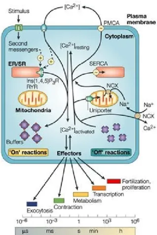
1.5 Diabetic cardiomyopathy……….……43 1.5.1 Introduction………...………43 1.5.2 Cardiac hypertrophy………..…………44 1.5.3 Diastolic dysfunction………46 1.5.4 Calcium dysregulation………..………47 1.5.5 Fibrosis………..………50 1.5.6 Steatosis………52 1.5.7 Lipotoxicity………...……54 1.5.8 Hyperglycemia………..………58
1.6. Mechanism implicated in diabetic cardiomyopathy………...…60
xi 1.6.1.1 Endoplasmic Reticulum stress and the unfolded
Protein response………60
1.6.1.2 Endoplasmic Reticulum stress in diabetic cardiomyopathy..…64
1.6.1.3 Endoplasmic Reticulum stress induced apoptosis…….………67
1.6.2 Inflammation……….…68
1.6.2.1 Peroxisome proliferator-activated receptors………..…71
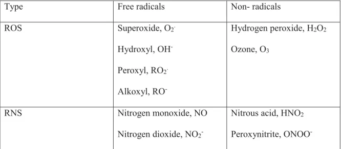
1.6.3 Oxidative stress and Reactive oxygen species….……….…72
1.6.4 Mitochondrial dysfunction………75
1.6.4.1 Fatty acids oxidation………..……75
1.7 Apoptosis……….……79
1.7.1 Extrinsic pathway………..……79
1.7.2 Intrinsic pathway………...…80
1.8 Rationale, hypothesis and objectives…………..……….…81
Chapter 2: Saturated fatty acids induce endoplasmic reticulum stress in primary Cardiomyocytes………84 2.1 Authors contribution………85 2.2 Context……….………85 2.3 Abstract………86 2.4 Introduction………..………87 2.5 Methods………89 2.6 Results………..……93 2.6.1 Palmitate induces cell death of primary NCMs, which is prevented
xii
by the addition of oleate………..………...…………...…93
2.6.2 Distinct lipid staining in palmitate treated NCMs………93
2.6.3 Palmitate induces ER stress………..………99
2.6.4 Palmitate induces apoptotic cell death………100
2.6.5 The protective effect of oleate is associated with a decrease in ER stress………103
2.6.6 Palmitate causes increased ubiquitination of Grp78………...……103
2.7 Discussion………..………104
2.8 Acknowledgements………111
2.9 Supplementary data………112
2.10 Reference……….…115
Chapter 3: Cardiomyocyte lipotoxicity is mediated by Il-6 and causes downregulation of PPARs………122 3.1 Authors contribution………..……123 3.2 Context……….………..………123 3.3 Abstract………..…124 3.4 Introduction………124 3.5 Methods………..………126 3.6 Results………129
3.6.1 Time dependent effects of palmitate on PPARs……….……129
3.6.2 Lipotoxicity induces marked cytokine expression in primary cardiomyocytes………...…131
xiii
3.7 Discussion………..………134
3.8 Acknowledgements………138
3.9 Supplementary data………138
3.10 Reference……….…140
Chapter 4: Impaired fatty acid oxidation as a cause for lipotoxicity in cardiomyocytes ……146
4.1 Authors contribution………..…147 4.2 Context………...……147 4.3 Abstract………..…148 4.4 Introduction………149 4.5 Methods………..…151 4.6 Results………154
4.6.1 Palmitate impairs fatty acid oxidation in primary cardiomyocytes ...…154
4.6.2 Impaired FAO is not due to loss of carnitine palmitoyl-transferase (Cpt1b) or loss of mitochondrial integrity………..…155
4.6.3 Enhancing fatty acid oxidation attenuates palmitate-mediated lipotoxicity………..…156
4.6.4 Impairing fatty acid oxidation causes lipotoxicity………..……157
4.7 Discussion………..………158
4.8 Acknowledgements………165
xiv Chapter 5: Lipotoxic Palmitate Impairs the Rate of β-Oxidation and Citric Acid Cycle
Flux in Rat Neonatal Cardiomyocytes………...……170
5.1 Authors contribution………..………171 5.2 Context……….………..………171 5.3 Abstract………..………172 5.4 Introduction………173 5.5 Methods………..……175 5.6 Results………180
5.6.1 Palmitate impairs complete FAO but not Cpt1b activity………180
5.6.2 Palmitate impairs β-oxidation……….……183
5.6.3 Palmitate impairs citric acid cycle flux………...…186
5.6.4 Palmitate does not impair FAO through oxidative stress…………...…187
5.6.5 Palmitate causes the accumulation of sn1,2 Diacylglycerol………...…188
5.7 Discussion………..…189
5.8 Acknowledgements………194
5.9 Reference………...…195
Chapter 6: Overexpression of Cpt1b reduces both ER stress and oxidative stress in STZ/HFD mice and in palmitate treated T293………202
6.1 Context………...…202
6.2 Introduction………202
6.3 Methods………..…204
xv 6.4.1 STZ mice exhibit hyperglycemia, hyperlipidemia, ER stress and
Oxidative stress………...………208
6.4.2 Fatty acid oxidation is impaired in chronic STZ/HFD mice models…..211
6.4.3 Cpt1b overexpression is protective in STZ/HFD mice………...…212
6.4.4 Palmitate induces ER stress and oxidative stress in T293 cells………..212
6.4.5 Overexpressing Cpt1b protects against palmitate induced lipotoxicity..214
6.5 Supplementary data………217
Chapter 7: Discussion and conclusion……….…219
7.1 Discussion………..…219
7.2 Conclusion……….…236
xvi
List of tables
Chapter 1
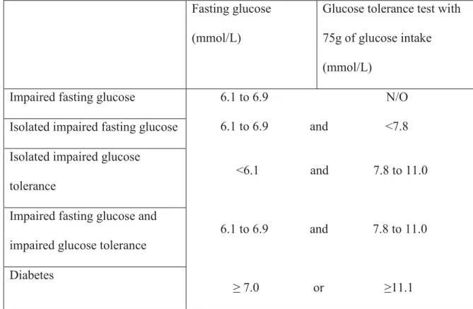
Table 1.1: Diabetes Canada diagnostic criteria for diabetes………..……34 Table 1.2: Selected examples of free reactive species………...……73
xvii
Supplementary tables
Chapter 2
xviii
List of figures
Chapter 1
Figure 1.1 Heart anatomy……….…2
Figure 1.2 Calcium-induced calcium-release model………8
Figure 1.3 Fatty acids and glucose metabolism in cardiomyocytes………...……10
Figure 1.4 Initial steps of Fatty Acids Oxidation………...……18
Figure 1.5 β-oxidation reaction of fatty acids………20
Figure 1.6 Overview of citric acid cycle………23
Figure 1.7 Calcium signaling and homeostasis………..…50
Figure 1.8 The three arms of ER stress………..…64
Figure 1.9 Inhibiting FAO is associated with lipotoxicity and mitochondrial dysfunction…...78
Figure 1.10 Extrinsic and Intrinsic pathways of apoptosis………81
Chapter 2 Figure 2.1 Palmitate causes time dependent cell death in NCMs, which is prevented by the co-administration of oleate………...………94
Figure 2.2 Marked differences in BODIPY staining in NCMs treated with oleate, palmitate, Or oleate + palmitate for 24 hours………...………97
Figure 2.3 Marked differences in BODIPY staining in NCMs treated with oleate, palmitate, Or oleate + palmitate for 8 hours………...………..98 Figure 2.4 Oleate and palmitate exhibit similar uptake rates and intracellular lipid
xix
accumulation in NCMs………..……101
Figure 2.5 Palmitate induces ER stress in NCMs………102
Figure 2.6 Palmitate induces apoptotic cell death in NCMs………107
Figure 2.7 Palmitate causes the ubiquitination of Grp78 protein in NCMs……….…108
Chapter 3 Figure 3.1 PPAR target gene expression is induced early on, and then repressed later, in palmitate treated neonatal cardiomyocytes (NCMs)………..…130
Figure 3.2 Palmitate induces the down regulation of Cpt1b and PPAR-d protein levels……132
Figure 3.3 Palmitate induces the expression of inflammatory mediators Tnf-a and Il-6 in neonatal cardiomyocytes (NCMs)……….…133
Figure 3.4 Palmitate induced cell death in neonatal cardiomyocytes (NCMs) is attenuated With treatments that reduce Il-6 expression………...………...……135
Chapter 4 Figure 4.1 Palmitate impairs fatty acid oxidation in primary rat neonatal cardiomyocytes (NCMs)………..……155
Figure 4.2 Palmitate causes a time dependent loss of mitochondrial membrane potential in NCMs………..……159
Figure 4.3 Enhancing fatty acid oxidation attenuates palmitate mediated lipotoxicity in primary NCMs………...…162
xx
Chapter 5
Figure 5.1 Palmitate impairs complete fatty acid oxidation in primary neonatal
Cardiomyocytes (NCMs)……….………..…181 Figure 5.2 Palmitate impairs ß-oxidation in primary neonatal cardiomyocytes (NCMs)……182 Figure 5.3 Palmitate impairs citric acid cycle (CAC) flux in primary neonatal
Cardiomyocytes (NCMs)……….…………..……184
Figure 5.4 300 μM oleate or palmitate for 8 hours induces increased ROS generation but Not overt oxidative stress in primary neonatal cardiomyocytes (NCMs)……..…185 Figure 5.5 Palmitate induces increased 1,2 diacylglycerol (DAG) in primary neonatal
cardiomyocytes (NCMs)………188
Chapter 6
Figure 6.1 High-fat diet feeding (HFD) leads to hyperglycemia, hyperlipidemia, ER stress And oxidative stress in STZ mice…………..………210 Figure 6.2 Chronic feeding of high-fat diet causes a significant decrease of Fatty acids
oxidation in STZ mice………211 Figure 6.3 Overexpressing Cpt1b using AAV9 Cpt1b rescued mice from STZ/HFD induced
FAO inhibition………...…213 Figure 6.4 Palmitate induces ER stress and oxidative stress in T293 cells……….…215 Figure 6.5 Enhancing fatty acid oxidation attenuates palmitate mediated lipotoxicity in T293
xxi
Chapter 7
xxii
Supplementary Figures
Chapter 2
Supplementary Figures S2.1: Palmitate induces ER stress in mouse neonatal cardiomyocytes (mNCMs)………..…113 Supplementary Figures S2.2: Palmitate induces markers of ER stress in a dose and time
dependent manner in rat neonatal cardiomyocytes (rNCMs)…114
Chapter 3
Supplementary Figures S3.1: Acadl, Acsl, and Cpt1b are legitimate PPAR target genes in rat primary cardiomyocytes (NCMs)……….…138 Supplementary Figures S3.2: Protein levels of Acadl and PPAR-α in NCMs treated with
palmitate………139
Chapter 6
Supplemental Figure S6.1: Cardiac mitochondria isolated from STZ/HFD mice are sensitive to etomoxir and carnitine……….…217 Supplemental Figure S6.2: Overexpressing Cpt1b attenuates Atf6, Chop and sXbp1 genes
xxiii
List of abbreviations and acronyms:
SA Sinoatrial Node
AV Atrioventricular Node
FAO Fatty acid oxidation
PDH Pyruvate dehydrogenase
AMPK AMP-activated protein kinase
ACC Acetyl-CoA carboxylase
RXR Retinoid x receptor
PPRE PPAR response elements
ALA Alpha linolenic acid
EPA Eicosapentaenoic acid
DHA Docosahexaenoic acids
LPL Lipoprotein lipase
CACT carnitine/acylcarnitine translocase
ACAD Acyl CoA dehydrogenase
NADH Nicotinamide adenine dinucleotide
FADH2 Flavin adenine dinucleotide
CAC Citric acid cycle
DAG Diacylglycerol
LD Lipid droplets
xxiv
PIP2 Phosphatidyl inositol bisphosphate
OGTT Oral Glucose Tolerance Test
T1DM Type 1 diabetes
T2DM Type 2 diabetes
MODY Maturity Onset Diabetes of the Young
NDM Neonatal Diabetes Mellitus
SNP Single nucleotide polymorphism
CAPN10 Calpain-10
TCF7L2 Transcription factor 7-like 2
GLUT4 Glucose transporter type 4
IRS Insulin receptor substrate
NEFA Non-esterified fatty acids
PKC Protein kinase C
JNK c-Jun N-terminal Kinase
PTEN Phosphatase and TENsin homolog deleted on chromosome ten
SHIP Src Homology 2 domain-containing Inositol 5-Phosphatase
AGE Glycation end-products
RAGE Receptor for advanced glycation end products
ROS Reactive oxygen species
DCM Diabetic cardiomyopathy
SERCA sarco/endoplasmic reticulum Ca2+-ATPase
NCX Sodium-Calcium exchanger
xxv
CaMK Ca2+/calmodulin-dependent protein kinase
PLB Phospholamban
PKA Protein kinase A
HSL Hormone sensitive lipase
apoB apolipoprotein B
VLDL Very low-density lipoproteins
LDL Low-density lipoprotein
CD36 Cluster of Differentiation 36
FATP1/6 Fatty acid transport protein 1/6
FABPpm membrane associated fatty acid binding protein
GPI Glycosylphosphatidylinositol
TZD Thiazolidinediones
STZ Streptozotocin
MAPK Mitogen-activated protein kinase
NF-κB Nuclear factor kappa B
PI3K Phosphoinositide 3-kinase
RNS Reactive nitrogen species
GFAT glutamine:fructose-6-phosphate amidotransferase
ER Endoplasmic reticulum
GRP78 Glucose-related peptide 78
UPR Unfolded Protein Response
PERK Protein kinase R (PKR)-like endoplasmic reticulum kinase
xxvi
IRE1 Inositol Requiring Enzyme 1
XBP-1 X-box binding protein 1
elF2-α Eukaryotic Initiation Factor 2 alpha
ATF4 Activating Transcription Factor 4
UTR Open Reading Frame (ORF) in their 5’ untranslated region
CHOP CAAT/Enhancer binding protein (C/EBP) homologous protein
S1P site 1 protease
S2P site 2 protease
CREB cAMP response element binding protein
CRE cAMP response element
UPRE Unfolded protein response element
ERSE ER stress response element
ERAD Endoplasmic-reticulum-associated protein degradation
SR Sarcoplasmic reticulum
PP1 Protein Phosphatase 1
p38 MAPK p38 mitogen-activated protein kinases
BI-1 Bax Inhibitor-1
IKK IκB kinase
TLR-4 Toll Like Receptor 4
PGC-1α Peroxisome proliferator-activated receptor gamma coactivator 1-alpha
LCAD Long-chain acyl-CoA dehydrogenase
CD95 Cluster of Differentiation 95
xxvii
TL1A TNF-like ligand 1A
TNFR1 TNF-receptor 1
DR-3 Death receptor 3
FADD Fas associated death domain
xxviii
Acknowledgements
First and foremost, I would like to express my sincerest appreciation to my supervisor Dr. Nicolas Bousette for giving me the opportunity to undertake my PhD degree in his laboratory. This thesis would not have been possible without his input and encouragement. I’m sincerely grateful for your patience, motivation, and helpful guidance during these last years. You have been an incredible mentor and wonderful human being, and I have learned a lot from you, be it in science or life in general. You cared so much about my work and future career goals. I consider myself extremely lucky to have you as my supervisor.
I would also like to thank my friends and colleagues Ali Akoumi and Dr. Félix-Antoine Bérubé-Simard. Your support created an amazing lab atmosphere.
1
Chapter 1: Introduction and literature review
1.1 The heart
1.1.1 Heart anatomy
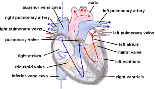
The heart is a vital organ in the human body. This hollow intrathoracic muscular organ has the role of pumping blood to the blood vessels of the body through its rhythmic contractions. The heart is made up of left and right sectors, each of which consists of two cavities: an atrium and a ventricle. The four cavities thus distinguish a “right heart” and a “left heart,” which normally do not communicate with each other (Figure 1.1). The role of the atria is to receive the blood and then redistribute it to the ventricles, while the role of the ventricles is to provide the force necessary to propel the blood towards the organs. The left and right ventricles are separated by a septum called the interventricular septum (1).
The heart has four valves, which are non-muscular elastic membranes. Two separate each atrium from the adjacent ventricle and are called atrioventricular valves. A third valve called the aortic valve separates the left ventricle from the aorta, while the fourth valve, the pulmonary valve, is located between the right ventricle and the pulmonary artery. These valves prevent the blood from going backward and thus preventing backflow (2).
2
The heart wall is made of three different layers: 1) endocardium, 2) myocardium, and 3) pericardium. The endocardium is a sheet of epithelial tissue found in the inner layer, which is in direct contact with the blood thus preventing it from clotting. The myocardium is the heart muscle and the thickest component of the heart wall. It is the only part of the heart wall that contracts, and it is made mainly from cardiac muscle cells. The myocardium is composed of identifiable thick and thin filaments organized in sarcomeres that give it striated appearance like skeletal muscle (3). Finally, the pericardium is a double-walled sack that contains the pericardial fluid whose role is to prevent friction during the heartbeat, while giving the heart sufficient freedom of movement to achieve rapid and vigorous contractions. Additionally, the heart is shaped by connective tissue made from collagen, among other cells. Since it does not conduct electric signals, it provides a boundary for the heart’s electrical conduction system.
3
Cardiomyocytes and fibroblasts represent the majority of heart cells (5). Endothelial cells, vascular smooth muscle cells, and different stem cells account for the rest. In adult rat hearts, cardiomyocytes represent around 25% of total cells and almost 80% of total heart volume (5). Cardiomyocytes contract simultaneously because, unlike other muscle cells, they are connected to each other by intercalated discs. The electrical resistance between adjacent myocytes is low because of gap junctions within the intercalated disks. These gap junctions allow ions to move freely between two cardiac cells, enabling the cells to contract simultaneously, since the electrical signal can reach all cells in few milliseconds.
1.1.2 Heart physiology
The heart pumps blood across the body through two circulations called the systemic circulation and the pulmonary circulation. The systemic circulation consists of transporting blood full of oxygen and nutrients to the peripheries before transporting blood full of CO2 and waste out of
the peripheral organs (6). The pulmonary circulation consists of transporting blood between the lungs and the heart.
Blood transport function in a cycle where the heart is at its center. The cycle starts with venous, deoxygenated blood returning to the right atrium via the superior and inferior vena cava. The blood continues to the right ventricle followed by the lungs via the pulmonary valve. The pulmonary valve is connected to the pulmonary trunk, which splits into the right and left pulmonary arteries. These two arteries transport deoxygenated blood to the right and left lungs
4
respectively. It is important to note that the term “arteries” indicates the blood vessels coming out of the heart independently of whether the blood is oxygenated or deoxygenated. The oxygenated blood returns to the heart via the left and right pulmonary veins and fills the left atrium. Once the left atrium is filled, the blood passes to the left ventricle via the mitral valve, which then ejects it into the systemic circulation through the aorta. Interestingly, heart cells are not supplied with oxygen and nutrients by either of the two circulations but by a circulation specific to them: the coronary circulation. This circulation, which is irrigated during the diastole, is formed of two coronary arteries (right and left) that are derived from the aortic artery and divide into a network bringing nutrition to the cardiac cells (7). Each day, the heart pumps the equivalent of 8,000 liters of blood, the equivalent of 100,000 heartbeats.
Each heartbeat consists of three major movements: atrial systole, ventricular systole (contraction) and ventricular diastole (relaxation). During the atrial systole, the atria pump blood to the ventricles (though almost 70% of the blood flows passively). During the ventricular systole, which is the strong contraction, the ventricles contract and eject the blood accumulated from atrial systole to different organs and peripheries through the arteries. This step is followed by a rest phase known as diastole where the pressure in the ventricles decreases, allowing the filling of the atria. The rhythm, frequency, and rate of this cycle are regulated by two nervous systems known as the sympathetic and parasympathetic nervous system. These two systems influence the conduction system of the heart.
The conduction system, which generates and distributes electrical impulses, is composed of specialized cardiac muscle cells found in the heart wall. It consists of: the sinoatrial node (SA),
5
the atrioventricular node (AV), the bundle of His, the bundle branches, and the Purkinje fibers. The SA node, or sinus node is where the heartbeat generates; it is considered the heart’s natural pacemaker (3). The cells of the SA node can spontaneously depolarize and generate an electrical impulse that reaches the left and right atria. The AV node receives the electrical signal from the SA node, then transmits the signal to the ventricles. The AV node is the only pathway by which the depolarization can pass from the atria to the ventricles. The conduction of the electrical impulse through the AV node is slow enough to prevent the atria and ventricles from contracting and filling up with blood simultaneously. The His bundle, which is an extension of the atrioventricular node, is divided into two branches, each of which is divided into a network of fibers called the Purkinje fibers. The Purkinje cells are myocytes, not nerve cells, that conduct the depolarization to all part of the ventricles. The sinus node, the bundle of His and the Purkinje network are the centers of the autorhythmicity of the cardiac fiber, each having their own intrinsic rhythm (3). They can generate their own potential action and dictate the heart rhythm. The one with the fastest rhythm dictates the rhythm of the others, since their slower rhythms cannot be imposed. The SA node is the primary center and controls the heart rate in normal conditions. The bundle of His and the Purkinje network are secondary centers that can only impose their rhythm if the SA node is failing.
Propagation of the electrical impulse from the sinus node to cardiomyocytes induces their contraction. This contraction, which is well paced and synchronized, is partially modulated by the sympathetic and parasympathetic nervous systems. The sympathetic system increases the heart rate, while the parasympathetic system decreases it.
6
Cardiac cells possess one or several electrophysiological properties, including automaticity, excitability, conductivity, contractility and refractoriness.
Automaticity is controlled by the conduction system which represents less than 1% of heart cells. However, all myocardial cells can conduct an electrical impulse. Excitability is defined by the cells ability to react to an electrical, mechanical, or chemical stimulus and convert it into a mechanical function such as contraction (3). Excitability depends on the refractory period, which is defined as the inability of cardiomyocytes to respond to any stimulus, cancelling its excitability. The refractory period in cardiomyocytes spans the depolarization phase, the plateau phase, and most of the repolarization phase (about 250 ms). Due to the refractory period, cardiac cells cannot contract until the end of systole. This protects the heart from arrhythmias and helps coordinate the heart contractions (8).
1.2 Cardiomyocytes
1.2.1 Description, histology and contraction
Cardiomyocytes are rod-shaped cells responsible for cardiac contraction. They are about 80-140μm long and 15-25μm in diameter, and usually have a single nucleus located in the center. In human hearts, binucleated, trinucleated, and tetranucleated cardiomyocytes can also be found (9). Like other striated muscle cells, cardiomyocytes consist mainly of myofibrils, wherein the contractile units of the cell are found: the sarcomeres. Myofibrils are made up of mainly three proteins: 1) actin, 2) myosin, and 3) titin. These proteins are organized into two types of
7
filaments: 1) thick and 2) thin filaments. Multiple myosin molecules are clustered together to form each thick filament while thin filaments are made from actin and other proteins. The striated appearance of cardiac muscle is caused by overlapping arrays of thick and thin filaments, which appears under a microscope as a sequence of dark and light bands. Dark bands, named A Bands, are found in the center of sarcomeres. They are flanked by two light bands named I bands. While I bands contain only thin filaments, A bands contain both thin and thick filaments. Dark Z-lines separate two adjacent sarcomeres and serve as an anchoring point for actin filaments.
Even though thin and thick filaments are juxtaposed, they can only interact in the presence of calcium. Calcium released from the sarcoplasmic reticulum binds to troponin, causing tropomyosin to modify the actin structure. As a result, modified actin molecules can interreact with myosin, causing thin filaments to slide along thick filaments. This contraction needs adenosine triphosphate (ATP) and thus depends on the activity of myosin molecules and the ability of myosin to convert chemical energy (ATP) into motion. This huge demand for ATP caused by the continuous activity of cardiomyocytes is explained by the high density of mitochondria in these cells (around 40% of cytoplasm).
In order to contract simultaneously, gap junctions within intercalated discs connect cardiomyocytes together to form one functional syncytium. This allows the wave of excitation to reach all cardiomyocytes. When an action potential reaches the cell, a depolarization occurs, followed by the entry of calcium into the cells through voltage-gated calcium channels. These channels are present on cell membranes and transport the “trigger calcium” into the cell. Trigger
8
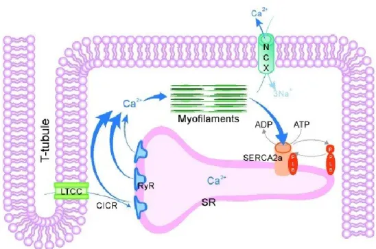
calcium binds to ryanodine receptors on the sarcoplasmic reticulum membranes and induces the release of intra-sarcoplasmic calcium. This mechanism is called calcium-induced calcium release and it increases the concentration of cytosolic free calcium (Figure 1.2). Free calcium binds to troponin and triggers a cascade of events that leads the sarcomeres to contract. Finally, calcium is sequestered back to sarcoplasmic reticulum and out of the cell, which leads to muscle relaxation.
Figure 1.2: Calcium-induced calcium-release model (10).
1.2.2 Cardiomyocytes metabolism
The heart’s need for energy is enormous; it beats more than 100,000 times per day and consumes more energy than any other organ (11). Almost 95% of ATPs synthesized in cardiomyocytes
9
come from mitochondrial oxidative phosphorylation (12), while the rest are obtained from glycolysis and GTP (via the Krebs cycle) (13, 14). Myocardial ATP pool turnover, under normal conditions, is about ten seconds, so the heart must continually produce enough ATP to maintain its contractile function (15). It is important for the heart muscle to adapt quickly to physiological changes and the availability of different substrates.
Cardiac metabolism is characterized by a large number of interdependent and highly regulated reactions that, together, orchestrate the energetic needs of the heart. Indeed, cardiomyocytes can use carbohydrates, lipids, ketone bodies, and amino acids as substrates for energy production (16). Glucose and fatty acids are the main energy substrates, while the contribution of others is minimal (Figure 1.3). Normally, in adult cardiomyocytes, fatty acids represent the main energy source, while glucose represents less than 10% of energy sources (17). However, these percentages are flexible and they depend on the physiological state of the heart (18). The alteration of substrate choice depends on substrate availability, oxygen supply and work load. For example, in a fasting state, fatty acids are the main energy source for the heart, while in a postprandial state, glucose becomes the main energy source (19). This equilibrium between glucose and fatty acids was first described by Randle et al. in 1963 (20). He demonstrates that an increase in cardiomyocytes’ use of fatty acids is reciprocated by an inhibition of glucose utilization and vice versa. This is known as the Randle cycle and was later confirmed by McGarry et al. in 1977 (21).
10
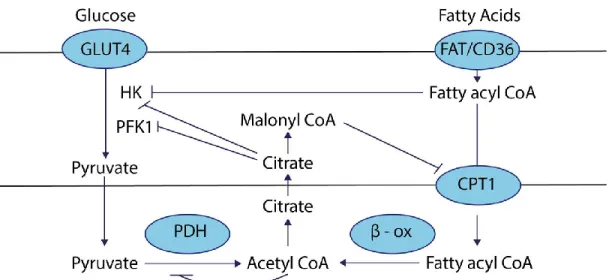
Figure 1.3: Fatty acids and glucose metabolism in cardiomyocytes (22).
Several mechanisms are involved in glucose/fatty acid counterbalance. First, fatty acids inhibit glucose oxidation through phosphofructokinase 1 (23) and pyruvate dehydrogenase (20). The mechanism involves citrate and acetyl-CoA respectively, which are both intermediates of fatty acid oxidation (FAO). Citrate inhibits phosphofructokinase 1 activity and thus inhibits glycolysis. Acetyl-CoA induces pyruvate dehydrogenase (PDH) kinase, which inhibits PDH activity and thus inhibits pyruvate oxidation.
Second, AMP-activated protein kinase (AMPK) is a metabolic sensor and plays a key role in energy homeostasis. AMPK can phosphorylate Acetyl-CoA carboxylase (ACC) and thus inhibits its activity (24) (25). ACC catalyzes malonyl-CoA synthesis, which is a potent inhibitor of Cpt1b. Since Cpt1b catalyzes the rate-limiting step of the FAO process, inducing AMPK activity leads to FAO upregulation.
11
Third, PPARs are master regulators of lipid metabolism and fatty acids are natural ligands of PPAR (26). When bound to fatty acids, PPARs translocate to the nucleus, where they heterodimerize with retinoid X receptors (RXRs) and bind PPAR response elements (PPREs). PPREs can be found in the promoters of many metabolic genes, including but not limited to: CD36 (27), Cpt1 (28), and PDK4 (29). CD36 translocates free fatty acids across the cell membrane and Cpt1 catalyzes the rate-limiting step for FAO. Therefore, PPARs are key factors of lipid metabolism in cardiomyocytes. Additionally, PDK4 phosphorylates and thus downregulates PDH activity, leading to inhibition of glucose oxidation.
Finally, insulin directly impacts FAO by modulating AMPK activity. Insulin inhibits AMPK activity, and thus inhibiting Cpt1b activity and FAO in cardiomyocytes (30). Insulin, at the same time, induces Glut-4 translocation and the transport of glucose into cytosol. In consequence, insulin augments glucose utilization and glucose oxidation (31).
1.2.3 Rat neonatal cardiomyocytes cultures
Cultures of rat neonatal cardiomyocytes formed the main model used in our studies. These cardiomyocytes were isolated directly from the ventricles of 1- to 3-day-old rats. Fibroblasts and myocytes are the main components of rat heart ventricles (5). For this reason, fibroblasts were separated from cardiomyocytes by incubating the cells’ mixture for one hour. This was sufficient for fibroblasts to attach onto the plate but not enough for cardiomyocytes to do so. Cardiomyocytes were then transferred to another plate and incubated for seven days before performing experiments. We incubated for seven days, since the hearts of newborn mammals
12
adapt from the dramatic nutrient changes that occurs after birth, becoming less dependent on carbohydrates and more dependent on fatty acids (from colostrum and milk) as energy fuel (32). Simultaneously, capacity for FAO increases significantly during post-natal development (32, 33). Our cell cultures were almost 95% pure cardiomyocytes, which was confirmed through sarcomeric staining. This method provides a mean to study intracellular mechanisms in one specific cell type: cardiac myocytes. This model is known as the “redifferentiation model,” where cardiac myocytes dedifferentiate when isolated from hearts before redifferentiating into spontaneously beating cells after incubating the cells in 2-10% fetal bovine serum for a few days (34).
We have used primary cardiomyocyte cultures, since they have many important advantages over cell line cultures. First, most cell lines come from tumor cells or transformed cells. These cells divide in an uncontrolled manner which is a behavior not found in primary cell culture. For example, H9C2 is a cell line derived from rat heart myoblasts. These cells can be passaged up to four months (35). However, primary rat cardiomyocytes do not divide at all. Second, physiological, morphological, and phenotypical traits found in primary cells are not always replicated in cell line cultures (36). This discrepancy is reflected in cellular responses and signaling pathways where primary cells isolated directly from tissue maintain most of the functions found in vivo, though this which is not the case for cell lines. A clear example is the H9C2 cell line, which is isolated from the ventricles of embryonic rats, though they exhibit many features of skeletal muscle (35).
13
It’s important to mention that neonatal and adult cardiomyocytes are not identical. For example, some ion channels are not fully developed in neonatal cardiomyocytes (37). Additionally, some proteins (isoforms) implicated in contraction are not expressed in neonatal cardiomyocytes which is not the case for adult cardiomyocytes (37). Therefore, In vitro culture of Rat adult cardiomyocytes closely resemble the in-vivo myocardium of adult rats and hence it is a better model to study rat hearts. However, there are two main advantages for using neonatal over adult cardiomyocytes. First, culturing cardiomyocytes isolated from adult rats is laborious and cost intensive when compared to neonatal cardiomyocytes. Indeed, culturing neonatal cardiomyocytes yield more cells since they are less sensitive to the calcium-containing medium, a medium through which cells are reintroduced after dissociation (38). The disparity in calcium sensitivity is likely associated with Transverse tubule, since the Transverse tubule network is not fully developed until the adult period (39). Second, neonatal cardiomyocytes start beating spontaneously after they enter the dedifferentiation-redifferentiation cycle, unlike cardiomyocytes isolated from adult rats which require pacing to trigger contraction (40).
1.3 Fatty acids
1.3.1 Description and classification
Fatty acids are essential molecules in all organisms. They are carboxylic acids (COOH) attached to a hydrophobic aliphatic chain. They can be saturated or unsaturated depending on the absence or the presence of double bonds in their aliphatic chain. Unsaturated fatty acids are referred to as monounsaturated if they possess only one double bond in their aliphatic chain; they are
14
referred to as polyunsaturated if they possess two or more double bonds. Almost all double bonds found in native fatty acids have a cis configuration. A cis configuration bends the hydrocarbon chain, which affects the shape of the molecule and confers specific physical and chemical properties. A trans configuration is usually obtained after hydrogenation of the double bond. Additional classifications are used depending on the length of the carbon chain and the position of the double bonds. Concerning the number of carbons in the aliphatic chain, fatty acids are classified into four groups: 1) short chain fatty acids (less than five carbons), 2) medium chain fatty acids (6 to 12 carbons), 3) long-chain fatty acids (13 to 21 carbons), and 4) very long-chain fatty acids (22 carbons or more).
Since the physiological role of unsaturated fatty acids is more dependent on the relative position of the double bonds to the methyl group than saturated fatty acids, an alternative classification is used. The last methyl group from the carboxyl group is called omega (ω) and it is given the number 1. Omega-3 fatty acids are polyunsaturated fatty acids where three carbons from the methyl group form the first double bond, which means it is located between carbon 3 and carbon 4. Omega-6, omega-7, and omega-9 fatty acids also exist. There are three major omega-3 fatty acids: 1) alpha linolenic acid (ALA), 2) eicosapentaenoic acid (EPA) and 3) docosahexaenoic acids (DHA). The main source of ALA is fish oil and the protective role of omega-3s against cardiovascular disease is well established (41-43). Unlike omega-3s, omega-6s are proinflammatory, prothrombotic, and hypertensives (44).
The two most common fatty acids are palmitic and oleic acids (45). They are saturated and unsaturated fatty acids respectively. Palmitate has a carboxylic group and a 15-carbon aliphatic
15
chain for a total of 16 carbons. Oleate has a total of 18 carbons and one unsaturation between carbon 9 and carbon 10. A simplified nomenclature is used where the number of carbons followed by the number of unsaturated molecules are indicated. The positions of double bonds are specified by superscript numbers preceded with Δ. For example, palmitate is abbreviated 16:0 and oleate is abbreviated 18:1 (Δ9). Additional well-known fatty acids in human diet
include: arachidonic acid 20:4 (Δ5,8,11,14), stearic acid 18:0, and linoleic acid 18:2 (Δ9,12). It is
important to note that saturated and monounsaturated fatty acids are metabolized differently in cardiomyocytes and significantly diverge in intracellular signaling (46, 47). For example, saturated fatty acids induce insulin resistance, while monounsaturated fatty acids induce insulin sensitivity in diabetic patients (48, 49). Additionally, palmitate causes mitochondrial dysfunction and increases mitochondrial reactive oxygen species, which is correlated with apoptosis in L6 skeletal muscle cells (50). Indeed, we showed that palmitate is a toxic fatty acid while oleate is neutral and even protective (51, 52).
Fatty acids are poorly soluble in water. Their hydrophobic characteristics are based on their molecular structure. Water solubility is positively correlated with the number of double bonds and negatively correlated with the length of the hydrocarbon chain. Therefore, fatty acids in organisms are rarely found free. In blood vessels, they are attached to a carrier protein: serum albumin (53). They are also conjugated to glycerol and converted to triacylglycerol then packed into lipoproteins and chylomicrons (54). Lipoproteins are spheres of decreasing diameter but with increasing density. Their center contains hydrophobic lipids such as triglycerides and cholesteryl ester and engulfed with apolipoproteins and amphiphilic lipids such as phosphatidylcholine. In the heart, lipoprotein lipase (LPL) acts on lipoproteins to release fatty
16
acids from triglycerides (55). The released fatty acids are transported into cardiomyocytes via passive diffusion (56) or via several membrane transporters, including CD36, FATP1/6, or FABPpm (57). Once inside the cells, Acyl-CoA-synthetase “activates” fatty acids by adding a CoA group. The CoA group confers hydrophilic properties onto fatty acids that permit them to further interact with hydrophilic cytosolic enzymes (58). For long-chain fatty acids, five isoforms of acyl-CoA synthetase exist in mammals (59-61). Each isoform differs in its subcellular location (62) and it has been shown that fatty acids’ intracellular fate depends on the action of specific ACSLs (61). For example, heart-specific ACSL1 knockout in mice is associated with FAO impairment and cardiac hypertrophy (63).
In cardiomyocytes, lipid droplet synthesis and phospholipid synthesis are two other possible fates, beside oxidation, for fatty acids. Lipids are the main component of cell membranes (phospholipids) and they provide energy for various metabolic process. Fatty acids are stored as energy depots in triacylglycerol form. Using triacylglycerol instead of polysaccharides as energy depots has two main advantages. First, oxidation of fatty acids yields almost twice as much ATP compared to polysaccharides oxidation (64). Second, triacylglycerols are hydrophobic, unlike polysaccharides, which are hydrated when stored as glycogen or starch. Triacylglycerols are denser than polysaccharides, since cells can store more triacylglycerol (two grams of triacylglycerol per gram of polysaccharide) (3). Indeed, obese people can store enough fats to meet their energy needs for months, while the human body cannot store more than one day worth of energy in the form of glycogen (3).
17
Fatty acids also play an indispensable role as signaling molecules (for example eicosanoids) and as cofactors (65). Additionally, fatty acids consumed through dietary, participate in cell membrane synthesis. They reduce the melting point and augment membrane fluidity (66). Thus, they can modify some receptors’ activity (67).
1.3.2 Fatty acid oxidation
1.3.2.1 β-oxidation
FAO occurs in peroxisomes and mitochondria. Even though a small fraction of fatty acids is oxidized in peroxisomes (32), mitochondria is considered the main site for β-oxidation. Small, medium, and long-chain fatty acids are oxidized in mitochondria, while the peroxisomal system is much more active on very long-chain fatty acids (68). Peroxisomal oxidation cannot degrade the fatty acid completely (69), but it shortens the fatty acid chain with a few cycles of β-oxidation. The shortened fatty acid translocates to mitochondria for further β-oxidation. Oxygen is the final electron acceptor in both mitochondria and peroxisome. However, one difference between the peroxisomal and mitochondrial oxidation pathways is that the former produces H2O2 while the latter produces H2O (70, 71). An additional difference between the two is in
ATP synthesis. ATP is generated in mitochondrial oxidation, which is not the case for peroxisomal oxidation (72).
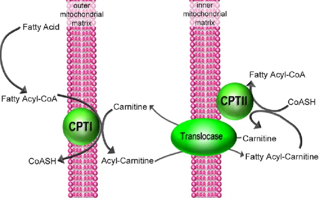
Long-chain fatty acids cannot diffuse freely through mitochondrial membranes (73). Therefore, in cardiomyocytes, the CoA group of fatty acids is exchanged with the carnitine group through
18
CPT1B (Figure 1.4). CPT1B, which stands for carnitine-palmitoyl-transferase 1b, commits fatty acids to mitochondrial oxidation. The reaction catalyzed by CPT1B is the rate-limiting step for FAO, which regulates the number of fatty acids entering mitochondria (74, 75). The newly synthesized acyl-carnitine can diffuse freely through the outer mitochondrial membrane. Once it is in the mitochondrial intermembrane chamber, it is transported into a mitochondrial matrix with the help of carnitine/acylcarnitine translocase (CACT) (76). CACT exchanges carnitine with an acylcarnitine across the inner mitochondrial membrane. Subsequently, carnitine diffuses freely to the cytosol, where it is used by Cpt1b for the following reactions. Once inside the mitochondria, acyl-carnitine is converted back to acyl-CoA by Cpt2 (77).
Figure 1.4: Initial steps of Fatty Acids Oxidation (adapted from (78)).
19
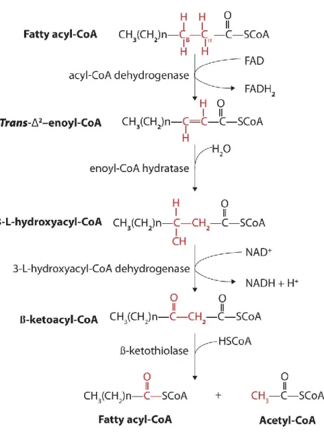
Mitochondrial acyl-CoA then enters the β-oxidation pathway. This pathway is a four-step reaction (Figure 1.5). The enzymes involved are in the following order (79): Acyl CoA dehydrogenase (ACAD), enoyl CoA hydratase, L-hydroxyacyl CoA dehydrogenase, and 3-ketoacyl CoA thiolase. ACAD catalyzes the first step of β-oxidation when it introduces a trans double bond between carbon number 2 and carbon number 3. This reaction is called dehydrogenation and it produces one molecule of FADH2 as a by-product. Several members of
the ACAD family have been found in the human genome and they are all involved in β-oxidation (80). Very long-chain acyl-CoA dehydrogenase, long-chain acyl-CoA dehydrogenase, medium chain acyl-CoA dehydrogenase, and short chain acyl-CoA dehydrogenase are responsible for the dehydrogenation of very long-chain CoA, long-chain CoA, medium chain acyl-CoA, and short chain acyl-CoA respectively. Enoyl CoA hydratase catalyzes the second step of β-oxidation. It adds one water molecule to the double bond between carbon 2 and carbon 3 and it converts 2-trans-enoyl-CoA to L-3-hydroxyacyl-CoA. L-3-hydroxyacyl CoA dehydrogenase follows enoyl CoA hydratase and it is responsible for the second dehydrogenation of the β-oxidation cycle. L-hydroxyacyl CoA dehydrogenase converts hydroxyacyl-CoA to 3-oxoacyl-CoA. In this step, NAD+ is the electron acceptor. 3-ketoacyl CoA thiolase catalyzes the fourth and last step of the β-oxidation cycle when it reacts with one molecule of free CoA in order to split the original fatty acid into acetyl-CoA and acyl-CoA.
At the end of each β-oxidation cycle, an acetyl-CoA is produced and the fatty acid chain is shortened by two carbons. One additional molecule of the reducing equivalents nicotinamide adenine dinucleotide (NADH) and flavin adenine dinucleotide (FADH2) is produced (58). These
20
phosphorylation. The β-oxidation cycle continues until the fatty acid is entirely catabolized to acetyl-CoA (58).
Figure 1.5: β-oxidation reaction of fatty acids (81).
If the fatty acid carbon chain is unsaturated, or if contains an odd number of carbons, then additional enzymes are required (58). The double bonds found in fatty acids usually have a cis
21
configuration which enoyl CoA hydratase cannot recognize. Therefore, in monounsaturated fatty acids, such as oleate, an additional enzyme is required such as enoyl CoA isomerase. Enoyl CoA isomerase converts the cis configuration in fatty acids to trans configuration. For polyunsaturated fatty acids, such as linoleic acid, two enzymes are employed: enoyl CoA isomerase and 2,4 dienoyl CoA reductase.
If the fatty acid chain contains an odd number of carbons, then the chain will be catabolized to several acetyl-CoA molecules and one propionyl-CoA molecule. Propionyl-CoA is metabolized to succinyl-CoA by a mechanism that involves three enzymes: 1) propionyl-CoA carboxylase, 2) methylmalonyl-CoA epimerase, and 3) methylmalonyl-CoA mutase. Succinyl-CoA is subsequently fed into the citric acid cycle (CAC) as an intermediate (82).
1.3.2.2 Citric acid cycle
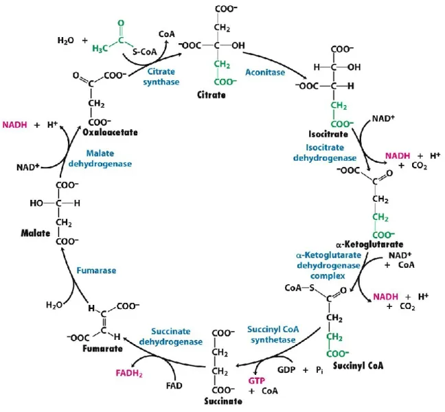
Acetyl-CoA, the final product of β-oxidation, is further oxidized by the CAC, followed by oxidative phosphorylation. The citric acid cycle (CAC) and oxidative phosphorylation are mutual for both glycolysis and β-oxidation pathways. The CAC is also named Krebs cycle after the German researcher Hans Krebs who described the steps of the cycle (83). Like β-oxidation, the CAC takes place in the mitochondrial matrix. It is an eight-step reaction catalyzed by at least eight different enzymes: 1) citrate synthase, 2) aconitase, 3) isocitrate dehydrogenase, 4) α-ketoglutarate dehydrogenase, 5) succinyl-CoA synthetase, 6) succinate dehydrogenase, 7) fumarase, and 8) L-malate dehydrogenase (Figure 1.6) (58). First, a CoA group is cleaved from acetyl-CoA. Then citrate synthase combines oxaloacetate with acetate to form citrate. Next,
22
aconitase converts citrate to isocitrate. The latter is metabolized to α-ketoglutarate by isocitrate dehydrogenase, then to succinyl-CoA by α-ketoglutarate dehydrogenase. One molecule of CO2
and two molecules of NADH are released during the last two steps. Succinyl-CoA synthetase converts succinyl-CoA to succinate and GTP. Next, succinate dehydrogenase metabolizes succinate to produced fumarate, which is then coupled with the synthesis of one FADH2
molecule. Fumarase activity converts fumarate into to malate, which is further converted to oxaloacetate by L-malate dehydrogenase. A third molecule of NADH is formed (84). At the end of each cycle, acetyl-CoA yields two molecules of CO2, three molecules of NADH, one
molecule of FADH2, and one molecule of GTP (85). CO2 is cleared out of the cell as a waste,
while NADH and FADH2 enter the electron transport chain where they are used as precursors
23
Figure 1.6: Overview of citric acid cycle (86).
In cardiomyocytes, the CAC is modulated by substrates availability and by the cell energy needs (87). For example, when the NADH/NAD+ ratio is elevated, citrate synthase (88), isocitrate
dehydrogenase (89), and α-ketoglutarate dehydrogenase (89) are downregulated. Additionally, CAC enzymes are regulated by the cycle intermediates such as oxaloacetate and acetyl-CoA.
24
CAC flux is regulated by anaplerotic and cataplerotic reactions (90). Anaplerosis is simply the regeneration of CAC intermediates while cataplerosis is the loss of CAC intermediates. Therefore, CAC intermediates can be used as precursors for cataplerotic reactions or they can be replenished by anaplerotic reactions. Some fatty acids such as palmitate do not contribute to anaplerosis (91), while glucose and pyruvate are important anaplerotic substrates (92, 93). Pyruvate carboxylation via pyruvate carboxylase or malic enzymes produces oxaloacetate and malate respectively (94). Pyruvate can be used as a substrate for α-ketoglutarate regeneration (93). Propionate and amino acids are also metabolites for anaplerotic reactions (93, 95). The extent of these reactions depends on various circumstances including substrate and oxygen supply. Indeed, the importance of anaplerosis was established when a perfused heart was supplied with ketone bodies as an energy substrate (96). A rapid deterioration in contractile function was observed and this decline was only reversed by supplementation of glucose and pyruvate (97).
1.3.2.3 Electron transport chain
NADH and FADH2 generated in the CAC and in fatty acid beta oxidation are used to donate
electrons for the mitochondrial respiratory chain. NADH and FADH2 are oxidized to NAD+ and
FADH2+ respectively and the electrons released are captured by oxygen. Oxygen is transformed
into water and this is coupled to the ATP synthesis. This process is called oxidative phosphorylation and it employs five complexes (NADH: ubiquinone oxidoreductase (or complex 1), succinate dehydrogenase (or complex 2), cytochrome bc1 complex (or complex 3), cytochrome c oxidase (or complex 4), and F0F1-type ATP synthase (or complex 5)) and two
25
electron carriers (ubiquinone and cytochrome c). The five protein complexes are bound to the mitochondrial inner membrane, unlike CAC enzymes, which are localized in the mitochondrial matrix (except for succinate dehydrogenase) (94-96). The first four complexes expulse H+ ions into the mitochondrial intermembrane chamber, creating a PH gradient. Therefore, the electron transport chain does not synthesize ATP directly. Instead, it generates energy by creating a proton gradient between mitochondrial inner and outer membranes. This proton gradient is capitalized by the last complex to generate ATP through chemiosmosis. The last complex, called F0F1 ATP synthase, returns most of the protons to the mitochondrial matrix, which enables it to synthesize ATP from ADP (98).
A small fraction of intermembrane protons “leaks” to the mitochondrial matrix through the uncoupling proteins. In cardiomyocytes, two main isoforms of uncoupling proteins exist: UCP-2 and UCP-3 (99). They transport protons back to the mitochondrial matrix without being coupled to ATP synthesis, hence their name. Instead of ATP, UCPs capitalize on the proton gradient by releasing energy in form of heat (100). UCP-1 is highly concentrated in brown adipose tissue and it plays a pivotal role in thermogenesis (101, 102).
Electrons carried by NADH are transferred to complex 1 in the electron transport chain, where flavoprotein is the electrons’ acceptor. Electron flux is transferred from one complex to the other (complex 2, 3, and 4), inducing the reduction and the oxidation of several electron transporters: ubiquinone and cytochrome B, C1, C, A, and A3. Electron transfer is coupled to proton expulsion from the mitochondrial matrix to the intermembrane space. Oxygen, the final electron
26
acceptor, obtains its electrons from cytochrome A3, then combines with two molecules of H+ to generate H2O.
Unlike NADH, FADH2 transfers its electrons directly to complex 2, leading eventually to the
synthesis of two molecules of ATP (instead of three molecules of ATP for NADH) (58).
ATP and ADP are transported through the mitochondrial membrane in opposite direction by antiport (translocase) (85). ATP exits the mitochondria to be used by the contractile units and ions pumps, while ADP enters the mitochondria to be used by oxidative phosphorylation. In healthy adult cardiomyocytes, the ATP pool is only sufficient for ten seconds (15). Therefore, the robust activity of mitochondrial oxidative phosphorylation is indispensable. For it to work flawlessly, mitochondrial oxidative phosphorylation needs a constant supply of oxygen (through the coronary circulation), as well as ADP, protons, and electrons. In prenatal conditions, rat hearts have low activity in CAC enzymes and the mitochondrial respiratory chain. However, a few days after birth, enzyme activity is upregulated severalfold (85, 103, 104).
At the end of the electron transport chain, the complete products of palmitate oxidation to carbon dioxide and water is:
27
1.3.3 TAG and lipid droplets synthesis
Triglycerides consists of two molecules attached together: glycerol and fatty acids. Glycerol is a three-carbon alcohol with a hydroxyl group for each carbon. Triglycerides can be found mainly in animal fats and vegetable oils. Most animal triglycerides are saturated triglycerides, which means they are composed of saturated fatty acids (58). These triglycerides are flexible and agglomerate together because they lack double bonds in their carbon chain. This is why lard and butter are solid at room temperature, while vegetable oils, in contrast, are liquid at room temperature since their triglyceride are unsaturated. Unsaturated triglycerides contain double bonds in their aliphatic chain. These double bonds usually have a cis configuration, causing the hydrocarbon chains to lose their flexibility and their ability to form agglomerates. In the food industry, unsaturated fatty acids found in vegetable oil are partially hydrogenated to improve their shelf life. A by-product of fatty acids hydrogenation is trans fats (105). These fats are unsaturated because their double bonds have a trans configuration. There is a clear association between trans-fat consumption and cardiovascular disease (106).
Upon entering the cell, fatty acids can be esterified and used for TAG synthesis. In cardiomyocytes, TAG is mainly synthesized from glycerol 3-phosphate. This pathway is a four-step reaction with each four-step catalyzed by a different enzyme. First, GPAT condenses acyl-CoA with glycerol 3-phosphate to form lysophosphatidic acid. This step is common for all glycerolipid synthesis and is considered the rate-limiting step in the TAG synthesis (107). Another acyl transferase, AGPAT, transfers a second acyl-CoA to lysophosphatidic acid. Phosphatase acts on phosphatidic acid (the product of the second step) and transforms it to
28
diacylglycerol (DAG) by removing a phosphate group. The final step produces TAG by condensing a third acyl-CoA with DAG. This step is catalyzed by DGAT and is the only committed step in TAG biosynthesis (108). Since DGAT is located on the ER membrane, TAG is considered a product of the ER (109). Another pathway for TAG synthesis involves the acylation of MAG. However, this pathway is not active in cardiomyocytes and is mainly found in hepatocytes and adipocytes.
TAG is a neutral lipid and its moderate presence in cardiac cytoplasm is harmless and might even be protective (110). Since DAG is associated with ER stress (111, 112), inflammation (113) and insulin resistance (114), the transformation of DAG to TAG by DAGT is considered a protective mechanism (115). Indeed, an increase in the DAG/TAG ratio is associated with increased cardiac lipotoxicity (116). Additionally, DGAT2 deficiency (DGAT2 -/-) in newborn
mice is a lethal condition associated with a severe decrease in TAG levels (117).
TAGs are stored in specific organelles called lipid droplets (LD). While the main site for the synthesis of LDs are adipocytes, LDs can also be found in almost all cells, including cardiomyocytes. LD is filled with TAG and cholesterol ester, which are engulfed with a monolayer of phospholipids (118). Additionally, a wide array of proteins is found on the outer layer of lipid droplets. Protein composition varies among cell types, which are categorized into nine groups (119): 1) PAT family, 2) lipid metabolism, 3) membrane traffic, 4) cytoskeleton, 5) chaperone, 6) ER, 7) mitochondria, 8) cell signal, and 9) miscellaneous. The PAT family, also known as perilipins, are the most abundant protein on LD membranes.
29
There are five isoforms of perilipin proteins numbered 1 to 5 (120). Perilipin 1 is dominant in white and brown adipocytes (121), while perilipin 2, 3, 4, and 5 can be found in cardiomyocytes (122). Perilipins are considered the gatekeepers of lipid droplets. They are phosphorylated by PKA and they play a major role in lipolysis by modulating the interaction between lipases (hormone sensitive lipase and adipose triglyceride lipase) and TAG in lipid droplets (123, 124). Initially, LDs were thought to play a passive role in energy and TAG. However, this role is far from the truth. LDs have been found to play a dynamic role in lipids’ metabolism, lipotoxicity, cell membrane homeostasis, signalization pathways, and protein modification (125, 126). Additionally, in leucocytes, LDs are considered as inflammatory organelles (127). Since DGAT (the final enzyme in TAG synthesis) is bound on ER membranes, ERs are considered the site for LD biogenesis (109).
The exact mechanism for LD biogenesis is still unknown and several hypotheses have been proposed. The most accepted one suggests two steps for LD synthesis. First, TAG accumulates between the hydrophobic bilayers of ER membranes, and as TAG concentration increases, a len-shaped bulge starts to appear. Second, for reasons not well understood, TAG stops accumulating and LDs bud off through a mechanism similar to dewetting (128). Cytosolic LDs can also derive from existing LDs through fission. However, this process is not dominant (129). The stored triglycerides are ultimately hydrolyzed, then oxidized to meet the energy needs of cardiomyocytes. Interestingly, cardiomyocytes accumulate lipid droplets during fasting states; however, in a fed state, lipid droplets become scarce (130).
![Figure 1.8: The three arms of ER stress [adapted from (353)].](https://thumb-eu.123doks.com/thumbv2/123doknet/2039654.4673/92.918.113.820.112.562/figure-arms-er-stress-adapted.webp)