Any correspondence concerning this service should be sent to the repository administrator:
[email protected]
O
pen
A
rchive
T
oulouse
A
rchive
O
uverte (
OATAO
)
OATAO is an open access repository that collects the work of Toulouse researchers
and makes it freely available over the web where possible.
This is a publisher-deposited version published in:
http://oatao.univ-toulouse.fr/
Eprints ID: 4815
To link to this article :
DOI:10.1039/B909035E URL : http://dx.doi.org/10.1039/B909035ETo
cite
this
version:
Tîlmaciu, Carmen and Soula, Brigitte and Galibert, Anne Marie and Lukanov, Petar and Datas, Lucien and Gonzalez, Jesus and Fernandez Barquin , Luis and Rodriguez Fernandez, Jesus and Gonzalez-Jimenez, Fernando and Jorge, Jose and Flahaut, Emmanuel Synthesis of superparamagnetic iron(III) oxide nanowires indouble-walled carbon nanotubes. (2009) Chemical Communications (n° 43). pp. 6664-6 666. ISSN
Synthesis of superparamagnetic iron(III) oxide nanowires in double-walled
carbon nanotubesw
Carmen-Mihaela Tıˆlmaciu,
aBrigitte Soula,
aAnne-Marie Galibert,
aPetar Lukanov,
aLucien Datas,
bJesu´s Gonza´lez,
cLuis Ferna´ndez Barquı´n,
dJesu´s Rodrı´guez Ferna´ndez,
dFernando Gonza´lez-Jime´nez,
eJose Jorge
eand Emmanuel Flahaut*
bReceived (in Cambridge, UK) 8th May 2009, Accepted 28th August 2009 First published as an Advance Article on the web 17th September 2009 DOI: 10.1039/b909035e
The synthesis and characterization of superparamagnetic iron(III) oxide nanowires confined within double-walled carbon nanotubes by capillary filling with a melted precursor (iron iodide) followed by thermal treatment is reported for the first time.
Carbon nanotubes (CNT) are undoubtedly one of the most striking discoveries in recent years, as these structures possess unique properties, including the ability to encapsulate foreign materials inside their cylindrical cavity, for applications in different fields.1,2One simple approach to fill nanotubes is to use capillary forces, which induce the filling with either a solution3,4or a melted material.5,6The latter was proved to
lead to a better filling rate in comparison with the wet chemistry route. In the present study we have explored the filling of double-walled CNT (DWNT) with melted iron diiodide (FeI2), as precursor for CNT-encapsulated magnetic
iron nanowires, which are planned to be used for biomedical applications, such as hyperthermia.7
DWNT with a selectivity of ca. 80% were synthesized by catalytic chemical vapour deposition (CCVD) as described in earlier work.8 The resulting DWNT-containing composite powder was then soaked in an aqueous HCl solution, in order to separate the CNT, by dissolving all the remaining oxide material, as well as unprotected metal (Co, Mo) particles. Unlike nitric acid or other oxidizing acids, HCl does not damage the DWNT (no opening of the DWNT, no oxidation of the tips and defects which may be present in the outer sidewall9). The acidic suspension was filtered on 0.45 mm pore
size cellulose nitrate membrane (Whatman) and washed with deionized water until neutrality. The resulting DWNT, denoted extracted DWNT (pristine), were then dried overnight in an oven at 80 1C.10DWNT (ca. 15 mg) with median inner diameter of 1.35 nm and median outer diameter of 2.05 nm,8 were intimately mixed in glove-box conditions with anhydrous
iron(II) iodide (ca. 500 mg), with a purity of 99.99+% (Aldrich), in an approximate molar ratio of 1 : 1.3 (volumic excess = 36). The violet–black mixture-powder was then transferred into a quartz ampoule and sealed under vacuum. It was then placed in a tubular furnace and heated at 5 1C min 1
to 690 1C (ca. 100 1C above the melting point of FeI2, 587 1C).
The sample was kept for 300 min at this temperature and melted FeI2entered within the DWNT, due to capillary forces.
It was then slowly cooled in two steps, at 0.1 1C min 1to a temperature of 567 1C (20 1C below the melting point of FeI2)
and then at 1 1C min 1 to room temperature. A black aggregate with metallic reflects was obtained, which was again ground in a mortar, until a powder was obtained. In order to eliminate the iron(II) iodide which was obviously in excess at the surface of the tubes, the resulting powder was then transferred in a small quantity of deionized water, sonicated for ca. 1 min in a sonication bath, filtered and then washed with deionized water, until the yellow colour of the filtrate could no longer be observed. The FeI2@DWNT powder was
dried overnight at 80 1C and then reduced in hydrogen at 250 1C for 5 h. The sample was then stored (in air) before observation by high-resolution transmission electron microscopy (HRTEM) and characterization by57Fe Mo¨ssbauer spectroscopy as well as magnetic measurements.
The filled DWNT were first imaged by HRTEM with a JEOL-JEM-2100 (operated at 200 kV), before and after the treatment in hydrogen.
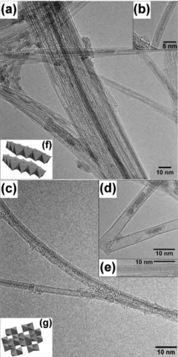
Fig. 1(a) and (b) show two typical HRTEM images of FeI2@DWNT observed before the reduction in hydrogen.
Most of the DWNT were found to be clean of deposits on the outer wall, and were filled with crystallized material. Either in the case of bundles (Fig. 1(a)) or for separated tubes (Fig. 1(b)), strong contrasts arranged in a regular fashion could be clearly detected inside the DWNT and on almost all their observable length. This regular alignment of dark spots indicates a very nice crystallization of FeI2 (structure
model of bulk FeI2 represented in Fig. 1(f)). Based on the
visual inspection of all HRTEM images, the filling yield was estimated to be higher than ca. 60%. In a previous study,4 similar DWNT were filled with a much lower yield starting from a precursor in solution (FeCl3). The presence of some
FeI2nanocrystals located outside the DWNT was observed to
a very limited extent, suggesting that the washing conditions with deionized water are efficient, but could still be optimized (longer time). EDX analysisw confirmed the concomitant presence of both iron and iodine in the sample. Besides,
aUniversite´ de Toulouse, UPS, INP, Institut Carnot Cirimat,
118 route de Narbonne, 31062 Toulouse Cedex 9, France
bCNRS, Institut Carnot Cirimat, 31062 Toulouse, France.
E-mail: fl[email protected]; Tel: +33-5 61 55 69 70
cMALTA Consolider Team, DCITIMAC, Facultad de Ciencias,
Universidad de Cantabria Santander, Spain
d
DCITIMAC, Facultad de Ciencias, Universidad de Cantabria Santander, Spain
e
Laboratorio de Magnetismo, Fisica, Ciencias, Universidad Central de Venezuela, Caracas, Venezuela
w Electronic supplementary information (ESI) available: Additional experimental data. See DOI: 10.1039/b909035e
6664 | Chem. Commun., 2009, 6664–6666 This journal is c The Royal Society of Chemistry 2009
COMMUNICATION www.rsc.org/chemcomm | ChemComm
Downloaded by INP Toulouse on 20 June 2011
Published on 17 September 2009 on http://pubs.rsc.org | doi:10.1039/B909035E
elemental analysis indicated 12.3 wt% of iron in the sample, which is a quantitative confirmation of the good filling rate evidenced by the HRTEM images.
Fig. 1(c)–(e) present HRTEM images of filled DWNT after the treatment in hydrogen. In Fig. 1(d), a discontinuous filling with apparently poorly organized material is shown, while in Fig. 1(c) and (e) the filling was achieved on all the observable length of the tubes. Although the filling yield was still estimated to be over 60%, the crystallized filling material was found
more discontinuous than before the reduction treatment, with the frequent observation of crystals only a few tens of nanometres in length (Fig. 1(c)), suggesting that some chemical modification occurred. This was confirmed by EDX analysis which indicated the absence of iodine (oxygen was also detected but could not be related to the presence of an iron oxide because the starting DWNT already contained traces of oxygen11). The XRD pattern shows the presence of low-intensity broad peaks at the angles corresponding to g-Fe2O3, although the small quantity and small size do not
allow a full analysis of the data (size of crystallites). It is however clear that no peak of bcc-Fe was present, unlike in our previous work.4
The observation of crystalline material inside the DWNT is not an evidence that the final compound obtained after reduction in hydrogen is actually iron metal. After treatment in hydrogen, the sample was analyzed by 57Fe Mo¨ssbauer spectroscopy at three different temperatures (RT, 77 K and 45 K) and the results (Fig. 2) indicated that the iron within the tubes was in the form of superparamagnetic (SPM) iron(III) oxide nanoparticles, both at RT and liquid nitrogen temperature. At 45 K, a magnetic contribution (39% area, present as a large hyperfine magnetic field distribution, with two relative maxima at 160 kG and 460 kG) was superimposed with the SPM quadrupolar doublet. The fact that at 45 K the magnetic contribution was 39% of the total area indicates that the Mo¨ssbauer blocking temperature TBMhas not been reached
(this temperature is determined when the respective areas of the SPM and magnetic contributions are equal).12
In order to confirm the superparamagnetism of the iron(III) oxide nanowires, magnetic measurements were also performed, with a Quantum Design MPMS SQUID, from 2 to 300 K. Usual zero-field-cooled (ZFC) and FC measurements were performed to observe magnetic irreversibility.
For the hysteresis loop M(H) measurements were obtained between + 10 KOe and – 10 KOe.
Fig. 3(a) shows the FC and ZFC curves for the iron oxide nanowires inside the DWNT from 2 to 300 K at different constant magnetic fields (20 r H r 1000 Oe). On the one hand, the ZFC susceptibility curves exhibit a broad peak indicating the blocking temperature TBw, as found in
super-paramagnetic compounds (TBw = 32 K for H = 20 Oe),
related to the mean particle size. The TBw decreased
monotonically with increasing the magnetic field. Such a peak in the ZFC curve generally implies a distribution in particle size and anisotropy.13 On the other hand, the FC branch Fig. 1 HRTEM images of FeI2filling within (a) a DWNT bundle; (b) a
single DWNT; (c) DWNT filled with superparamagnetic iron(III) oxide nanowires; (d) DWNT with large diameter, filled with iron(III)
oxide nanoparticles; (e) smaller diameter DWNT filled with iron(III)
oxide nanowire; (f) structure model of bulk FeI2; (g) structure model
of bulk iron(III) oxide.
Fig. 2 Mo¨ssbauer spectra of the reduced samples at RT, 77 K and 45 K. The experimental result (black dots) and the theoretical fit (continuous line) are shown.
This journal is c The Royal Society of Chemistry 2009 Chem. Commun., 2009, 6664–6666 | 6665
Downloaded by INP Toulouse on 20 June 2011
Published on 17 September 2009 on http://pubs.rsc.org | doi:10.1039/B909035E
displays a continuous increase when decreasing T, without tendency to magnetization saturation below TBw, showing a
MFC(TBw) E 0.61MFC(10 K) and an irreversibility of
MZFC(TBw) E 0.63MFC(TBw). Above TBw, the ZFC and FC
data show a perfectly reversible behaviour at all fields. The lack of FC saturation points out to the existence of a (quasi) independent relaxation and then a magnetically ideal SPM behaviour. In a similar system comprising Co nanoparticles in mesoporous (SiO2) media,14 the increasing magnetic
inter-particle (dipolar) interactions by increasing Co concentration were deduced from the FC curves. This resulted in a saturation (MFC(TBw) E MFC(10 K) and MZFC(TBw) E MFC(TBw)) of
that FC branch, visible at 14% Co in pores. Indeed, this FC feature is here providing some useful information on the arrangement of the wires in the tubes; namely, the Fe-oxide nanowires encapsulated with a high filling ratio should be then separated in discrete long portions in a chain-like arrangement, which is supported by TEM observations (Fig. 3(c) and (d)). The distance between chain wires should be some tenths of nanometers to result in a very weak dipolar interaction (p1/r3) to allow the observed ideal SPM behavior displayed in Fig. 3(a). Furthermore, to display such a behaviour, we suggest that the randomly arranged wires (possibly g-Fe2O3)
present a feeble inter-wire magnetic coupling through the carbon wall, due to the average distance around 1 nm. Indeed, nanometric sols of g-Fe2O3in PVA in various arrangements
and interparticle distances have been the subject of extensive research, from independent to highly-coupled nanoparticles and chains.13–15In the case of particles of 8 nm size, separated by an average distance of ca. 64 nm, a blocking temperature very similar to the present results was observed in the ZFC/FC curves,15 with a ZFC peak E 40 K, and a FC continuous increase with MFC(TBw) E 0.79MFC(10 K) and
MZFC(TBw)E 0.85MFC(TBw). The ideal superparamagnetism
of the iron oxides is also obvious in the M(H) hysteresis data. Between 2.2 and 40 K, a symmetrical hysteresis loop was observed, indicating the ferromagnetic nature of the iron nanosystem (either nanowires or particles) in this range of temperatures which is below the blocking temperature (TBw= 32 K).
As an example Fig. 3(b) shows the hysteresis loops at 2.5 and 10 K. In the superparamagnetic region (TBw4 32 K) the
magnetization curves exhibit no hysteresis and both remanence and coercivity are equal to zero. Fig. 3(c) shows the magnetization curves at 100 and 300 K with no hysteresis loop. From the hysteresis curves at different temperatures (Fig. 3(d)) we can obtain the values of the coercivity. As predicted by the theory of superparamagnetism13the coercivity
is equal to zero (no hysteresis) for temperatures above TBw.
In agreement with 57Fe Mo¨ssbauer spectroscopy and magnetic measurements data, the reduction of FeI2 in
hydrogen was probably directly followed by oxidation, leading to superparamagnetic iron(III) oxide crystallized nanowires (supported by HRTEM observations) forming chain-like structures inside DWNT (the exact nature of those structures, wires or particles, is under investigation).
FP6 Marie Curie RTN CARBIO (N1 35656) is acknowledged for financial support. J. G. acknowledges the MALTA-Consolider Ingenio 2010 Program as well as the Franco–Venezuelan Cooperation Program PCP, ‘‘Nanotubos de Carbono’’ for financial support.
Notes and references
1 C. Lamprecht, J. Danzberger, P. Lukanov, C.-M. Tıˆlmaciu, A.-M. Galibert, B. Soula, E. Flahaut, H. J. Gruber, P. Hinterdorfer, A. Ebner and F. Kienberger, Ultramicroscopy, 2009, 109, 899.
2 E. Heister, V. Neves, C. Tıˆlmaciu, K. Lipert, V. Sanz Beltran, H. M. Coley, S. Ravi, P. Silva and J. McFadden, Carbon, 2009, 47, 2152.
3 E. Borowiak-Palen, E. Mendoza, A. Bachmatiuk, M. H. Rummeli, T. Gemming, J. Nogues, V. Skumryev, R. J. Kalenczuk, T. Pichler and S. R. P Silva, Chem. Phys. Lett., 2006, 421, 129.
4 J. Jorge, E. Flahaut, F. Gonza´lez-Jı´menez, G. Gonza´lez, J. Gonza´lez, E. Belandria, J. M. Broto and B. Raquet, Chem. Phys. Lett., 2008, 457, 347.
5 B. Ballesteros, G. Tobias, M. A. H. Ward and M. Green, J. Phys. Chem. C, 2009, 113, 2653.
6 E. Flahaut, J. Sloan, S. Friedrichs, A. I. Kirkland, K. S. Coleman, V. C. Williams, N. Hanson, J. L. Hutchison and M. L. H. Green, Chem. Mater., 2006, 18, 2059.
7 R. Klingeler, S. Hampel and B. Bu¨chner, Int. J. Hyperthermia, 2008, 24, 496.
8 E. Flahaut, R. Bacsa, A. Peigney and C. Laurent, Chem. Commun., 2003, 1442.
9 E. Flahaut, A. Peigney, Ch. Laurent and A. Rousset, J. Mater. Chem., 2000, 10, 249.
10 E. Flahaut, A. Peigney, W. S. Bacsa, R. R. Bacsa and C. Laurent, J. Mater. Chem., 2004, 14, 646.
11 V. Datsyuk, C. Guerret-Pie´court, S. Dagre´ou, L. Billon, J. C. Dupin, E. Flahaut, A. Peigney and Ch. Laurent, Carbon, 2005, 43, 873.
12 S. Mørup, J. Dumesic and H. Topsøe, in Applications of Mo¨ssbauer Spectroscopy, ed. R. Cohen, Academic Press, New York, 1980, vol. II, p. 1.
13 P. Prene´, E. Tronc, J.-P. Jolivet, J. Livage, R. Cherkaoui, M. Nogue´s, J.-L. Dormann and D. Fiorani, IEEE Trans. Magn., 1993, 29, 2658.
14 A. F. Gross, M. R. Diehl, K. C. Beverly, E. K. Richman and S. H. Tolbert, J. Phys. Chem. B, 2003, 107, 5475.
15 J. L. Dormann, D. Fiorani and E. Tronc, Adv. Chem. Phys., 1997, 98, 283.
Fig. 3 Magnetization as a function of temperature at different magnetic fields. (a) The ZFC curve shows the blocking temperature TBwof superparamagnetic behaviour. (b) Magnetization vs. H showing
the symmetric hysteresis at two temperatures below TBw. (c) In the
superparamagnetic region the magnetization curves exhibits no hysteresis. As an example we show the magnetization at 100 and 300 K. (d) Temperature dependence of the coercivity (zero above TBw).
6666 | Chem. Commun., 2009, 6664–6666 This journal is c The Royal Society of Chemistry 2009
Downloaded by INP Toulouse on 20 June 2011
Published on 17 September 2009 on http://pubs.rsc.org | doi:10.1039/B909035E