lonic, Cellular and Molecular Mechanisms
underlying the QT Prolongation and Arrhythmïas
in Dïabetïc Cardiocomplïcatîons
par
Yiqiang Zhang
Programme des Sciences biomédicales Département de Médecine
Faculté de Médecine
Thèse présentée à la Faculté des études supérieures en vue de l’obtention du grade de
Doctorat philosophie (Ph. D.) en Sciences biomédicales
Octobre 2005 © Yiqiang Zhang, 2005
(I
-J
ç
J1
4-Direction des bibliothèques
AVIS
L’auteur a autorisé l’Université de Montréal à reproduire et diffuser, en totalité ou en partie, par quelque moyen que ce soit et sur quelque support que ce soit, et exclusivement à des fins non lucratives d’enseignement et de recherche, des copies de ce mémoire ou de cette thèse.
L’auteur et les coauteurs le cas échéant conservent la propriété du droit d’auteur et des droits moraux qui protègent ce document. Ni la thèse ou le mémoire, ni des extraits substantiels de ce document, ne doivent être imprimés ou autrement reproduits sans l’autorisation de l’auteur.
Afin de se conformer à la Loi canadienne sur la protection des
renseignements personnels, quelques formulaires secondaires, coordonnées ou signatures intégrées au texte ont pu être enlevés de ce document. Bien que cela ait pu affecter la pagination, il n’y a aucun contenu manquant.
NOTICE
The author of this thesis or dissertation has granted a nonexclusive license allowing Université de Montréat to reproduce and publish the document, in part or in whole, and in any format, solely for noncommercial educational and research purposes.
The author and co-authors if applicable retain copyright ownership and moral rights in this document. Neither the whole thesis or dissertation, flot substantial extracts from ît, may be printed or otherwise reproduced without the authot’s permission.
In compliance with the Canadian Privacy Act some supporting forms, contact information or signatures may have been removed from the document. While this may affect the document page count, t does not represent any loss of content from the document.
Faculté des études supérieures
Cette thèse intitulée:
Ionic, Cellular and Molecular Mechanisms Underlying the QT
Prolongation in Dîabetic Cardiocomplïcatïons
présentée par:
Yiqiang Zhang
a été évaluée par un jury composé des personnes suivantes:
Dr. Stanley Nattel président-rapporteur Dr. Zhiguo Wang directeur de recherche Dr. Jacques Billette membre du jury Dr. Alvin Shrier examinateur externe
Or. Rémy Sauvé représentant du doyen de la FES
Le diabète est une maladie grave qui est associée le plus souvent à des complications sévères dont les plus fréquentes sont les pathologies cardiovasculaires. Cinquante pour cent des patients diabétiques meurent suite à des cardiopathies parmis lesquels plus d’un quart des patients présentent un prolongement de l’intervalle QT, qui constitue un facteur de prédiction de la mortalité chez les patients diabétiques. Les études chez les modèles diabétiques des rats et des souris prouvent que la diminution des canaux I et I est le majeur facteur. Ce pendant, plusieurs des problèmes importants
ne sont pas expliqués. D’abord, les rôles de canaux ‘Kr et ‘Ks qui n’existent pas chez les rats et les souris adultes, ne sont pas clair, mais ils jouent un rôle important determinant l’APD chez l’homme. De plus, les profils des changements des courants ioniques trouvés chez les rats et les souris ne peuvent pas expliquer complètement la survenue de troubles de l’intervalle QT chez les patients diabétiques. Enfin, le diabète est principalement un trouble métabolique pour lequel les niveaux de glucose, de ROS (Reactive Oxygen Species), de TNf-Œ (tumor necrosis factos-Œ) et de ceramide sont élevés. A contrario, le niveau de l’insuline est réduit et la voie de signalisation de l’insuline est diminuée. Cependant, ces perturbations métaboliques comme les mécanismes potentiels pour les désordres électriques diabétiques ont été négligé dans le passé.
Cette thèse a pour objectif d’apporter de nouveaux éléments permettant d’approcher plus en détail les mécanismes cellulaires et moléculaires responsables des troubles des rythmes et de la prolongation de l’intervalle QI, chez les individus présentant un diabète. L’hypothèse principale est
que le courant ‘Kr transporté par les canaux HERG chez l’homme joue un rôle majeur dans la survenue des troubles du rythme associés au diabète , et que la réduction du courant IKr/HERG est le
résultat. d’une part d’une diminution de l’expression du canal HERG, mais aussi d’autre part, d’une régulation fonctionnelle négative liée au stress métabolique ainsi qu’aux protéines de signalisation.
Nous avons tout d’abord vérifié l’altération de l’APD et de l’intervalle QT dans les myocytes ventriculaires isolés provenant d’un modèle de lapin présentant un diabète de type-l induit par l’alloxan. Dans ce modèle, nous avons montré que parmi les principaux courants ioniques, la réduction d’IKr était un facteur majeur dans l’altération de l’APD et de l’intervalle QT. Nous avons également démontré que chez les animaux diabétiques, les perturbations métaboliques associées au diabète entraînaient d’importantes dysfonctions des canaux IKr/HERG à cause de la réduction de l’expression de la protéine HERG, ainsi que en raison de la régulation dysfonctionnelle par les ROS,
par l’oxydation protéique, par la peroxydation lipidique qui sont augmentées, et par la réserve endogène d’antioxydants qui est réduite. Ces effets sont contrés par un traitement à l’insuline qui normalise le niveau de glucose ou par l’antioxydante vitamine E, permettant de récupérer le courant IKt/HERG ainsi que l’APD et l’intervalle QT. Ces résultats sont confirmés par le fait que les lapins diabétiques traités à l’insuline ou à la vitamine E ne présentent aucuns signes d’arythmies.
Au cours d’études plus détaillées, nous avons montré que la fonction du canal HERG était a son maximum dans un contexte de normoglycémie, et que cette fonction était altérée en hypo- ou hyper-glycémie suite à la sous-production d’ATP ou la surproduction de ROS, respectivement.
Nous montrons aussi que l’activité basale de la protéine kinase B (PKB), directement corrélée
à l’action d’insuline, est essentielle à la fonction normale du canal HERG. De plus, deux autres
facteurs de signalisation impliqués dans le diabète, le TNF-a et le céramide affectent la fonction des canaux IKr/HERGvia un même mécanisme de l’augmentation de ROS.
L’ensemble de ces travaux nous permet de conclure que la modulation fonctionnelle du courant IKt!HERG joue un rôle central dans la prolongation de l’intervalle QT et l’apparition de troubles du rythme associés au diabète. Le dysfonctionnement du courant IK/HERG est le résultat d’un équilibre net entre les facteurs d’activation du courant, tel que l’insuline et la PKB, et les facteurs inhibant le courant tels que l’hyperglycémie, les RO$, le TNf-Œ et le céramide. Ainsi, l’augmentation du courant IKr/HERG par manipulation de l’expression ou par modulation fonctionnelle pourrais retarder voir même renverser les désordres électriques du myocarde associés a la pathologie diabétique.
Mots-clés: Cardiomyopathie diabétique, intervalle QT, arythmies, IKr/HERG, insuline, hyperglycémie, protéine kinase B, YNf-a, céramide, ROS.
ABSTRACT
Heart diseases account for half of ail deaths among people with diabetes. Up to one fourth of diabetic patients have prolonged QT interval. QT prolongation, primarily reflecting the lengthening of ventricular action potential duration (APD), can cause sudden cardiac death due to the occurrence of lethal ventricular arrhythmias, and has been considered as a predictor of rnortaÏity in both type I and type II diabetic patients. Previous studies found reduction of several ion currents (e.g. reduced It and I) in experimental models of diabetes. These studies contribute significantly to our current understanding of the ionic mechanisms for diabetic QT prolongation, but several important issues remained unresolved. First, previous studies were conducted nearly exclusively in rats and mice, the species in their adulthood not expressing phenotypic and physiologically significant Igj afld ‘Ks that are otherwise the major repolarizing currents determining the plateau phase and total APD in humans. Second, the profiles of changes of ion currents found in rats and mice could hardly fully explain the clinical diabetic QT prolongation. Finally, diabetes is primarily a metabolic disturbance with elevated levels of glucose, reactive oxygen species (ROS), tumor necrosis factor-a (TNf-a) and ceramide, and reduced insulin and impaired insulin signaling transduction. However, these metabolic perturbations as the potential mechanisms for the diabetic electrical disorders have been overlooked in the past.
In this thesis, we aimed at delineating the underlying ionic, cellular, molecular and signaling mechanisms for diabetic QT prolongation and the associated arrhythmias, and shedding the light on developing novel and rational therapies. We hypothesized that Ii and its pore forming Œ-subunit HERG (human ether-a-go-go related gene) is the major contributor to the diabetic QT prolongation and the impairment of IjHERG in diabetic heart is a combined effect of the down-regulation of I channel protein and of the negative functional modulation of
‘Kr’1HERGby metabolic stresses and signaling molecules.
We reproduced the prolongation of APD and heart-rate corrected QT (QTc) interval in alloxan-induced type 1 diabetic rabbits, and identified the significantly impaired ‘Kr as the major ionic mechanism for diabetic APD prolongation, while other ion currents as the minor contributors to this abnormality. We demonstrated that diabetic metabolic perturbations cause the dysfunction of Ig./HERG in diabetic hearts by significant down-regulation of HERG protein and by impairing IK/HERG function due to increased levels of ROS, oxidations of lipids and
proteins in diabetic myocardiurn and simultaneously decreased endogenous antioxidant reserve (total antioxidant status), which can be prevented by insulin or vitamin E therapy to decrease the glucose level and/or to reduce the oxidative stress, rendering normalized IKt/HERG function and APD and QTc interval, and preventing the associated arrhythmias.
Our studies further revealed that the maximum HERG function operates under normoglycernia, and depression of HERG function occurs with either hypoglycemia or hyperglycemia, due to underproduction of ATP in the former, and overproduction of ROS via oxidative phosphorylation in the latter.
We found that the activity of protein kinase B (PKB), a down-stream component of insulin signaling pathway, is essential for proper function ofIKr/HERG. Moreover, TNF-Πand
ceramide, which are known to be deleterious factors critical to the progression of diabetes, both impaired IKr/HERG function via the common pathway, the increases in ROS.
Based on these data, we conclude that IKr/HERG K channelopathy serves as the ionic basis for diabetic QT prolongation and accompanying anhythmias, and oxidative stress caused by hyperglycemia as the major metabolic mechanism for diabetic IK/HERG K channelopathy. The dysfunction of IKr/HERG is a combined effect of decrease in the enhancing factors (e.g. insulin, PKB) and increases in the suppressing factors (e.g. hyperglycemia, elevated ROS, TNF c and cerarnide). Thus, increasing IKr”HERG by manipulating HERG expression and by functional
modulation on related cellular signaling molecules such as antioxidants could retard and even reverse, at least partially, the electrical disorders in diabetic hearts.
Key words: Diabetic cardiomyopathy, QT interval, Arrhythmia, IKr/HERG, Insulin, Hyperglycemia, Protein kinase B, Tumor necrosis factor-Œ, Ceramide, Reactive oxygen species.
TABLE 0F CONTENT
RÉSUMÉ.
ABSTRACT .iii
TABLE 0F CONTENT y
LIST 0F TABLE xiii
LIST 0F FIGURES xiv
LIST 0F SIGNS AND ABBREV1ATIONS xviii
ACKNOWLEDGMENTS xxi
CONTRIBUTION 0F AUTHORS xxiii
DEDICATION xxvi
PART I. INTRODUCTION AND REVIEW 0F THE LITERATURES J
I QT PROLONGATION AND ARRHYTHMIAS IN DIABETIC
CARDIOCOMPLICATIONS 4
1.1 Dïabetes Mellitus and Diabetic Complications 4
1.1.1 Diabetes, a prevalent and costly disease 4
1.1.2 Cardiomyopathy and cardiovascular complications in diabetes 5
1.1.3 QT prolongation and arrhythmias in diabetic hearts 7
1.1.4 Animal models of diabetes mellitus 8
1.2 QT interval, APD, and Ion Channels: Physiology and Pathology 10
1.2.1 QT interval as a function ofcardiac repolarization 10
1.2.1.1 EÏectrocardiogram (ECG) 10
1.2.1.2 Ion channels and action potential 10
1.2.1.3 Long QTsyndromes (LQTSJ) 11
1.2.2 Inwardcurrents 17
1.2.2.1 Na current: ‘Na 17
1.2.2.2 Ca2 currents: ‘CaL, ‘CaT 20
1.2.3. 1 Transient outward K cztrrent; I,,. 21
1.2.3.2 Rapid delayed rectifier K currents: ‘Kr 23
1.2.3.3 Slow DeÏayed recttfier K currents: ‘Ks 28
1.2.3.4 Inward rectifier K currents: ‘K? 29
1.2.3.5 Other cardiac ion channels 3]
1.3 Current Knowledge of Ionic Mechanisms for Prolonged QT Interval in Diabetic
Hearts 32 1.3.1 Inwardcurrents 33 1.3.1. 1 Sodium currents 33 1.3.1.2 Calcium currents 33 1.3.2 Outwardcurrents 34 1.3.2.1 I,0andI5 34 1.3.2.2 ‘K? 36 1.3.2.3 Irp 36
1.4 Current Knowledge of Cellular/Molecular Mechanisms in Diabetic
Cardiocomplications 3$
1.4.1 Hypo- and hyper-glycemia 38
1.4.2 Insulin-PI3K-PKB signaling pathway 39
1 .4.3 Tumor Necrosis factor-a and sphingolipid metabolites 40
1 .4.4 Oxidative stress 41
1.5 Questions 43
1.6 Reference 44
PART II. ORIGINAL CONTRIBUTIONS 78
2 AIMS 0F THE STUDIES 79
2.1 Working Hypothesis 79
2.2 Specific Aims of the Project 79
3 CRITICAL ROLE 0F ‘KR IN DIABETIC QT PROLONGATION AND THE
ASSOCIATED ARRHYTHMIAS 81
3.2 Introduction.84
3.3 Metliods 86
3.3.1 Preparation of rabbit model of type I insulin-dependent diabetes mellitus 86 3.3.2 Implantation oftelemeters and ECG recording in conscious rabbits 86
3.3.3 Isolation cf rabbit ventricular myocytes 87
3.3.4 Whole-cell patch-clamp recording 87
3.3.5 Western blot 89
3.3.6 Data analysis 89
3.4 Results 90
3.4.1 Diabetic QI prolongation and the associated arrhythmias 90 3.4.2 Functional alterations of cardiac ion currents in diabetic hearts 91 3.4.3 Relative contributions cf various ion currents te APD lengthening in IDDM.... 92
3.4.4 Altered protein levels of various ion channel subunits 93
3.5 Discussion 94
3.5.1 Dysfunction of IKr/HERG as the major ionic mechanism for diabetic QT
prolongation and the associated arrhythmias 94
3.5.2 Potential implications of the findings 97
3.5.3 Possible limitations ofthe study 97
3.6 Acknowledgement 98
3.7 References 99
3.8 Figures and Figure Legends 107
4 EXPERIMENTAL THERAPIES 0F DIABETIC QT PROLONGATION AND
ARRHYTHMIAS 115
4.1 Summary 117
4.2 Introduction 118
4.3 Methods 119
4.3.1 Rabbit model of type I insulin-dependentdiabetes mellitus (DM) 119 4.3.2 Implantation oftelemeters and ECG recording in conscious rabbits 120
4.3.3 Surface ECG recording in anesthetized rabbits 120
4.3.5 HEK293 celI culture . 121
4.3.6 Whole-cell patch-clamp recording 121
4.3.7 Western blot 122
4.3.8 lmmunohistochemistry 123
4.3.9 lmmunocytochemistry 123
4.3.10 Measurement of intracellular reactive oxygen species (ROS) 124
4.3.1 1 Lipid peroxidation assay 124
4.3.12 Protein oxidation assay 125
4.3.13 Total endogenous antioxidant assay 125
4.3.14 Data analysis 125
4.4 Resuits 126
4.4.1 Insulin corrects diabetic QT prolongation and suppresses arrhythmias 126 4.4.2 HERG K channel dysfunction as the ionic mechanism for diabetic QT
prolongation: role of high glucose 127
4.4.3 Oxidative stress leading to impairment of HERG K channel: vitamin E
treatment 128
4.4.4 Reduced protein level and membrane density of HERG K channels 130
4.5 Discussion 132
4.5.1 lkr/HERG K channelopathy as an ionic mechanism for diabetic QT
prolongation and the associated arrhythmias 132
4.5.2 Metabolic perturbation as the mechanism for dysfunction of HERG K
channels 134
4.5.3 Insulin and vitamin E treatment of diabetic APD/QT prolongation and the
associated arrhythmias 135
4.5.4 Potential implications of the findings 136
4.5.5 Possible limitations of the study 137
4.5.6 Conclusion 138
4.6 Ackuowiedgements 139
4.7 References 140
5 IMPAIRMENT 0F HERG
W
CHANNEL FUNCTION BY HYPOGLYCEMIA AND HYPERGLYCEMIA 158 5.1 Summary 160 5.2 Introduction 161 5.3 Experimental Procedures 162 5.3.1 CelI culture 1625.3.2 Whole-celI patch-clamp recording 162
5.3.3 Pharmacological probes 162
5.3.4 Intracellular reactive oxygen species (ROS) measurement 163
5.3.5 Data analysis 163
5.4 Resuits 164
5.4.1 Effects of glucose on IHERG 164
5.4.2 Role of glycolysis and oxidative phosphorylation on glucose-induced ‘HERG
enhancement 166
5.4.3 Role cf intracellularAlP in maintaining HERG function 167
5.4.4 Role cf ROS on hyperglycemia-induced ‘HERG depression 169
5.5 Discussion 171
5.5.1 Depression cf HERG function in hypoglycemia likely resuits from
underproduction ofATP 172
5.5.2 Depression of HERG funcfion in hyperglycemia results from overproduction of
ROS 174
5.5.3 Impairment cf HERG function might contribute to Q-T prolongation caused by
hypoglycemia and hyperglycemia 176
5.6 Acknowledgements 177
5.7 References 17$
5.8 Figures and Figure Legends 181
6 NORMAL FUNCTION 0F HERG
W
CHANNELS REQUIRES BASAL PROTEINKINASE B ACTIVITY 194
6.1 Abstract 196
6.3 Materials and Methods. 198
6.3.1 Cellculture 198
6.3.2 Gene transfection 198
6.3.3 Whole-CeH Patch-Clamp Recording 199
6.3.4 Immunocytochemistry 199
6.3.5 Data analysis 200
6.4 Resuits 200
6.4.1 Effects ofwortmannin on HERG currents (IHERG) 200
6.4.2 Effects of constitutively active or dominant negative PI3K on ‘HERG 201
6.4.3 Effects of constitutively active or dominant negative PKB on ‘HERG 202
6.4.4 Imuunocytochemical analysis of active PKB 202
6.5 Discussion 203
6.6 Acknowledgements 206
6.7 References 207
6.8 Figures and Figure Legends 210
7 IMPAIRMENT 0F HERG
W
CHANNEL FUNCTION BY TUMOR NECROSISFACTOR-Œ: ROLES 0F REACTIVE OXYGEN SPECIES 215
7.1 Abstract 217
7.2 Introduction 21$
7.3 Experimental Procedures 21$
7.3.1 CeIl disposition 218
7.3.2 Whole-ceII patch-clamp recording 219
7.3.3 Western Blot 219
7.3.4 Intracellular Reactive Oxygen Species (ROS) Measurement 219
7.3.5 Data analysis 219
7.4 Resuits 220
7.4.1 Impairment of HERG function by TNF-Π220
7.5 Discussion 222
7.6 Acknowledgements 223
7.7 References 224
7.8 Figures and Figure Legends 226
8 CERAMIDE CAUSES METABOLIC PERTURBATION LEADING TO HERG K
CHANNEL DYSFUNCTION AND ABNORMAL SLOWING 0F CARDIAC
REPOLARIZATION 230
8.1 Summary 232
8.2 Introduction 233
8.3 Experimental Procedures 234
8.3.1 Cellculture 234
8.3.2 Whole-ceH patch-clamp recording 234
8.3.3 Drugs and treatments 234
8.3.4 Western blot 235
8.3.5 lmmunocytochemistry 235
8.3.6 Intracellular reactive oxygen species (ROS) measurement 235
8.3.7 Data analysis 236
8.4 Resuits 237
8.4.1 Effects of membrane permeable ceramide on ‘HERG expressed in HEK293
Ceils 237
8.4.2 Effects cf endogenous ceramide generated by sphingomyelinase on ‘HERG 237 8.4.3 Effects of inhibitors to PTK, PKA or PKC on ‘HERG modulation by Ceramide 238 8.4.4 Lack 0feffects of ceramide on HERG protein expression level 239
8.4.5 Role of reactive oxygen species (ROS) in IHERG modulation by ceramide 239
8.5 Discussion 241
8.6 Acknowledgements 244
8.7 References 244
9 GENERAL DtSCUSSION AND CONCLUSIONS.257
9.1 Summary of Novel Findings and Their Significances 257
9.1.1 ‘Kras a major contributor to the APD/QT prolongation in diabetic hearts 258
9.1.2 Metabolic perturbations cause IKr/HERG channelopathy thereby the APD/QT
prolongation in diabetic hearts 258
9.1.3 Hypoglycemia and hyperglycemia impair HERG K channel function with
different mechanisms 260
9.1.4 Basal activity of protein kinase B is essential for proper IKr/HERG functions 261 9.1.5 TNF-a and ceramide depress IKt/HERG function by generating ROS 262
9.2 Potential Limitations 265
9.2.1 Animal model used in this study 265
9.2.2 Ion current and AP recordings in diabetic cardiomyocytes 266 9.2.3 Links between insulin, TNF-a I ceramide and intracellular ROS 266
9.3 future Directions 268
9.4 Conclusions 271
9.5 References 272
10 APPENDIX xxvii
LIST 0F TABLE
Chapter I
LIST 0F FIGURES
Chapter 1
Figure 1. Diabetic Complications 6
f igure 2. Relationships between ECG, APD and ion currents 13
f igure 3. Subunits of voltage-gated sodium chamiel and sites responsible for diseases ...19
Figure 4. Comparison of cardiac delayed rectifier K currents in heterogenous
system 24
figure 5. Unusual kinetics ofIia/HERG channel 25
Figure 6. Action potentials of rat ventricles 32
Figure 7. Current knowledge ofionic disturbance in diabetic heart 37
Chapter 3
Figure 1. Electrical disorders in rabbits with insulin-dependent diabetes mellitus 107
f igure 2. Comparison of various K+ currents in ventricular cardiomyocytes behveen
healthy and diabetic rabbits 10$
f igure 3. Comparison of L-type Ca2 current (ICaL) and fast Na current (‘Na) in
ventricular myocytes between healthy and DDM rabbits 110
Figure 4. Relative contributions ofvarious ion currents to APD prolongation in
ventricular myocytes from 1DDM rabbits Iii
figure 5. Alterations ofprotein levels of outward ion channel subunits revealed by
Western blot analysis 112
Figure 6 Alterations of protein levels of Œ-subunits of‘CaLand ‘Na revealed by Western
blotanalysis 114
Chapter 4
Figure 1. Electrical disorders and the ionic mechanism in rabbits with insulin-dependent
Figure 2. Metabolic mechanisms for diabetic APD prolongation: high glucose and insulin
treatment 14$
Figure 3. Metabolic mechanisms for diabetic APD prolongation and insulin
treatment 150
Figure 4. Role of oxidative stress in diabetic QT/APD prolongation and vitamin E
treatment 152
Figure 5. Alterations of protein levels of HERG K channels 153
Figure 6. Subcellular distribution and membrane density of HERG channel protein.... 155
Figure 7. Schematic chart ofthe proposed mechanisms ofdiabetic QTc
prolongation 157
Chapter 5
Figure 1. Effects of glucose on HERGK current(1HERG) stably expressed inHEK293
ceils 181
Figure 2. Effects of glucose on‘FIERG under conditions with conected osmolarity 183
Figure 3. Effects of complete inhibition of glucose metabolism or inhibition of glycolysis
OflIHERG 124
Figure 4. Effects of inhibition ofoxidative phosphorylation on ‘HERG 186
Figure 5. Effects of depletion of intracellular ATP on‘HERG 187
Figure 6. Effects ofnon-hydrolysable ATP (AMP-PCP) and GTP on ‘HERG 188
Figure 7. Effects of antioxidants on‘KERG depression induced by hyperglycemia 189
Figure $ Effects ofthe oxidant generating system xanthine/xanthine oxidase on ‘HERG
under normoglycemia 190
Figure 9 Oxidative phosphorylation and intracellular levels ofreactive oxygen species
(ROS) measured by CM-H2DCFDA fluorescence dye 191
Figure 10 Effects of antioxidants vitamin E and superoxide dismutase mimetic MnTBAP,
on intracellular ROS levels under hyperglycemia 192
Figure 11 Effects of the superoxide generating system xanthine/xanthine oxidase on the
Chapter 6
Figure 1. Effects ofwortmannin on‘HERG stably expressedinHEK293 ceils 210
Figure 2. IHERGin celis transfected with constitutively active PI3K or dominant negative
PI3K 211
figure 3. ‘HERGin HEK293 celis transfected with constitutively activePKB or dominant
negativePKB 212
figure 4. Immunocytochemical analysis of active PKB in HEK293 ceils with anti-phospho
(S473)-PKB antibody 213
Chapter 7
Figure 1. Impairment ofHERG function by TNF-Π226
Figure 2. Mechanisms for HERG depression by TNf-Π228
Chapter 8
Figure 1. Analog data showing the effects of membrane permeable ceramide (C2) on
HERG current(IHERG) expressed in HEK293 ceils 247
Figure 2. Characterization of‘HERGwith prolonged exposure to ceramide (C2) 24$
figure 3. Characterization of‘HERGdepression caused by sphingomyelinase 249
Figure 4. Effects ofinhibitors oftyrosine protein kinases (TPKs) and atypical protein
kinase C (PKC) on‘HERGmodulation by ceramide (C2) 250
Figure 5. Effects of inhibitors of protein kinase A (PKA) or protein kinase C (PKC) on
‘HERGmodulation by ceramide (C2) 251
Figure 6. Expression level ofHERG protein determined by immunoblotting with membrane protein preparations extracted from HERG-expressing HEK293
celis 252
Figure 7. Role ofreactive oxygen species (ROS) on‘HERGmodulation by ceramide ... 254
Figure $ Effects ofvitamin E (VitE) or MnTBAP (an SOD mimic) on intracellular levels
Chapter 9
Figure 1. Schematic diagram ofpathophysiology of diabetic arrhythmias 260
Figure 2. Signaling factors in diabetic arrhythmias 263
LIST 0F SIGNS AND ABBREVIATIONS
[Ca2]1 Intracellular calcium concentration
ATP Adenosine triphosphate
AF Atrial fibrillation
AP(s) Action potential(s)
APD Action potential duration
APD50 50% depolarization ofAP
APD90 90% depolarization of AP
AT-Il Angiotensin II
AV node Atrioventricular node
Bis Bisindolylmaleimide
Ca2 Calcium (ion)
CaM Calmodulin
CRF Congestive heart failure
CR0 Chinese hamster ovary
CM-R2DFDA 5 -(and-6)-chloromethyl-2’, 7 -dichlorodihydrofluorescein diacetate
CNS Central nerve system
DAD Delayed early afterdepolarizations
1DM Diabetes mellitus
ECG Electrocardiogram
EAD Early afierdepolarizations
GAPDH Glyseraldehyde-3 -phosphate dehydrogenase
GluT4 Glucose transporterase
H202 Rydrogen peroxide
HEK Rurnan embiyonic kidney
HERG Human ether-à-go-go-related gene
Hf Reart failure
IDDM Insulin-dependent diabetes mellitus
‘Cal (or ‘Cal) ‘HERG(orJHERG) ‘K (or IK)
‘1(1 (or IKI) ‘KATP(orIKATP) ‘Kr (orIKr) ‘Ks (oriKs) ‘NCX (or iNcx) ‘to (or I) IsS IVF ‘-V JLN syndrome K KChIP K LPC LQTS MAPK minK MiRP1 MI MIS MnTBAP + Na NIDDM 02 0W PBS PIP2 PI3K
T-type Ca2 current/channel HERG K cunents/channel
Delayed rectifier K current/channel
+
inward rectifier K currentlchannel ATP-sensitive K currentlchannel
Rapidly-activated delayed rectifier K current/channel Slowly-activated delayed rectifier K current/channel
+ 2+
Na -Ca exchanger current /channel Transient outward K current/channel
Q
uasi-steady-state outward K current Idiopathic ventricular fibrillation Current-VoltageJerveli and Lang-Nielsen syndrome Potassium (ion)
K-channel interacting protein Voltage-gated potassium channel Lysophosphatidylcholine
Long QT syndrome
Mitogen-activated protein kinase Minimal potassium channel MinK related peptide I Myocardial ischemia
Myocardial ischemia syndrome
Mn (III) tetrakis (4-benzoic acid) porphyrin chloride; Sodium (ion)
Non-insulin-dependent diabetes mellitus Superoxide anion
Hydroxyl group
Phosphate-buffered saline Phosphatidyl 4,5-biphosphate Phosphoinositide 3-kinase
PKA Protein kinase A
PKB Protein kinase B
PKC Protein kinase C
Lengthening of QT interval
RMP Resting membrane potential
ROS Reactive oxygen species
RW syndrome Romano-Ward syndrome
SMase Sphingomyelinase
SOD Superoxide dismutase
SR Sarcoplasmic reticulum
STZ Streptozotocin
TdP Torsade de pointes
TNf-ΠTumor necrosis factor-a
TNF R TNF receptor
VF Ventricular fibrillation
VGKCs Voltage-gated potassium channels
VitE Vitamin E
VT Ventricular tachycardia
WMN Wortmannin
ACKNOWLEDGMENTS
This thesis is really the work of more than the person named on the titie page.
I want to express my deepest gratitude to my supervisor Dr. Zhiguo Wang for his expert support and guidance throughout these years. I thank Dr. Wang for his accepting me as a student afler rny coming to Montreal as a science visitor, and for sharing with me his rich scientific and personal experience which I will continue to learn in my future life. Without bis essential advises, bis encouragement and inspiration, my study would neyer bave been succeeded.
Dr. Stanley Nattel is gratefully acknowledged for his constructive scientific counsel and review in our collaborative projects and his nice recommendation in rny applications for a studentship and a postdoctoral fellowship.
Thanks also for creating an outstanding research environment with honesty and friendship which extends from the workplace in the Dr. Wang laboratory, in the Institut de Cardiologie de Montreal (1CM), and in the Université de Montréal. I bighly appreciate the expertise at any ‘time-point’, which I needed from any ofthe staff at the 1CM where the work for this thesis was carried out.
I would like to thank ah my collaborators from China: Dr. Baofeng Yang and bis teams, for significant contribution to our experimental and writing processes. I express my sincere gratitude to my supervisors, Professors Jiancheng Huang and Shibo Xu during my undergraduate and graduate studies, and my first employer Professor Chuangguang Zhong, for their sustained fascinating me with the passion to science, their great help in my development of scientffic and personal quality and their maximum sympathy to me under any circumstance.
I want to sincerely thank the colleagues I worked with, whose company bas been very inspiring and whose friendship will aiways stay in my heart: Dr. Jingxiong Wang, Dr. Huizhen Wang, Dr. Hong Han, Dr. Huixian Lin, Xiaofan Yang, Marc-Antoine Gilhis, Louis R. Villeneuve, Dr. Jiening Xiao, Ling Xiao, Dr. Liming Zhang, Hong Long, Dr. Stephen Zicha, Dr. Huanhuan Gao, Xiaobin Luo, Dr. Marc Pounier. Dr. Gemot Schram, Daniel Henera, Denis Chartier, Dr. Yukiomi Tsuji, Chantal Maltais, Natalie L’Heureux, Evenlyn Landiy, and people in the laboratories of Dr. Nattel, Dr. Terence E. Hébert, Dr. Céline fiset, and Dr. Bruce G. Allen, and rnany others, with whom I have been interacted and belped for various needs.
My special thanks go to classmate Jingxiong Wang for his in-time help in work and in life and for his friendship, to Huizhen Wang for her first-hand help to initiate my learning in Dr. Wang’s lab, to Xiaofang Yang for her ready-for-help daily assistance, to Louis R. Villeneuve for bis sophisticated assistance in confocal experiments, and to Marc-Antoine Gillis for bis professional aids in telemetry ECG recordings, and to Dr Ange Maguy for his kind help in working out the French version ofthe thesis summary.
I address my warmest thanks to Karine Bouthillier, Emilie Nadeau, Stephenie Blanchet, Nathalie Degrasse, Benoit Chambarron and Kristine Perez for their professional assistance in animal care, making the animal studies undergo smoothly; and to Jacqueline Loiselle, Luce Bégin, and other secretaries for their excellent secretarial and other helps. Special recognition is due as well to the administrative office and audio-vision section staff at the Centre de Recherche de l’ICM. I want to say “Merci!” to Madame Denise Varemes and the stuff in the Faculté de médecine and the Faculté de étudies supérieurs of the Université de Montréal, for their helps in my PhD studies and preparing for this thesis.
I would like to extend my gratitude to Dr. Terence E. Hébert, Dr. Céline Fiset, Dr. Bruce G. Allen, Dr. Jacques Billette, and Dr. Alvin Shrier for their insightful suggestions and constructive comments in my thesis studies.
I gratefully acknowledge financial support for this study from the Canadian Institutes of Health Research, the Canadian Diabetes Association, the Fonds de la Recherche de l’Institut de Cardiologie de Montréal, the Fonds de Recherche en Santé du Québec; and the Heart and Stroke Foundation of Canada for ftinding me the Doctoral Research Award since 2003.
I thank my dear friends Xiao Zhang, Xiaoli Tang, and Xiang Wan for their warm-hearted support and care in spite ofthe thousands kilometres distance between us.
Finally I want to say ‘Xiè Xiè!’ to my wonderful parents Qingyun and Xiuping for their endless care and love. b my loved wife Haiqing who has been supporting me facing any obstacle and sharing our joys since our first meet; and to ail others of my families. This work would not be donc without their support.
CONTRIBUTION 0F AUTHORS
The following is a statement regarding the contributions of co-authors and myseif to the six articles which have been published or subrnitted for publication, and included in this thesis.
1. Zhang Y, Wang Xiao J*, Bai Y, Wang J, Zhang H, Yang B, Wang Z. Ionic mechanisms underlying the abnormal QT prolongation and the associated arrhytbmias in diabetic rabbits: Critical role of rapid delayed rectifier K current. Arn.i PhysioÏ Submitted, December 2005 (* indicates these two authors have equal contribution).
Dr. Zhiguo Wang and I proposed the original research plan. I designed and carried out the experiments in every step and analyzed the data and prepared for the figures and manuscripts. Dr. Huizhen Wang, Dr. Jiening Xiao, and Haiqing Zhang were involved in the experiments of Western blot. Yunlong Bai and Dr. Baofeng Yang were involved in the recording ofiKs and ‘Na.
Dr. Jingxiong Wang participated in animal handling and myocyte isolation. Dr. Z. Wang had the original idea and fully supervised the work and finalized the manuscript for publication.
2. Zhang Y, Xiao J*, Wang H*, Wang J, Villeneuve LR, Zhang H, Lin H, Bai Y, Yang B, Wang Z. IK/HERG as a target for experimental therapies of diabetic QT prolongation and arrhythrnias. Arn.i Physiot Submitted December 2005 (* indicates these two authors have equal contribution).
Dr. Z. Wang and I proposed the original research plan. I designed and carried out the experiments in every step and analyzed the data and prepared for the manuscript. Drs. J. Xiao and H. Lin participated in the experiments to test the oxidative status of heart tissues. Dr. H. Wang and H. Zhang, Y. Bai and Dr. B. Yang participated in the experiments of Western blot and imrnunocytochemistry. Dr. J. Wang participated in the animal handiing, myocyte isolation, and ion current recording. Louis R. Villeneuve contributed to the cryosectioning and the observation of immuno-histochemical and immunocytochemical specimens. Dr. Z. Wang had the original idea and fully supervised the work and finalized the manuscript for publication.
3. Zhang Y, Han H, Wang J, Wang H, Yang B, Wang Z. Impairments ofHERG (Humanether
à-go-go related gene) K channel function by hypoglycemia and hyperglycemia: Sirnilar phenotype but different mechanisms JBioÏ Chem 2003;27$:10417-10426
Dr. Z. Wang and I proposed the original research plan. I designed and performed the experiments in celi culture, ion current recording, and RO$ staining, and analyzcd the data and worked out the draft of this article. Dr. Hong Han and Dr. H. Wang participated in the experiments of ROS staining, and Dr. J. Wang worked partially on the patch-clamp recording. Dr. Yang comments on the designing of experiments. Dr. Z. Wang had the original idea and flilly supervised the work and finalized the manuscript for publication.
4. Zhang Y, Wang H, Wang J, Han H, Nattel S, Wang Z. Normal function of HERG K channels expressed in HEK293 celis requires basal protein kinase B activity. FEBS Lett. 2003; 534:125-132
In this paper, Dr. Z. Wang and I proposed the original research plan. I designed and did the experiments, analyzed the data and wrote the manuscript. Dr. H. Wang and Dr. H. Han participated in the immunocytochemical studies and Dr. J. Wang participated in the experirnents of ion current recording. Dr. Stanley Nattel provided effective co-direction of this work and Dr. Z. Wang offered close instruction in the whole process, refining the proposai, clarifying the notion, and producing the final version of this article.
5. Wang J*, Wang H*, Zhang Y, Gao H, Nattel S, Wang Z. Impairment of HERG K Channel Function by Tumor Necrosis factor-Œ: Role of Reactive Oxygen Species as a Mediator. J BioÏ Chem, 2004; 279(14):132$9 13292. (* indicates both authors have equal contribution).
In this work, I designed the protocols for detecting ROS generated by TNf-u in HEK293 cells and cardiornyocytes, performed the experirnent and analyzed the data. Dr. J. Wang performed the experiment and analyzcd the data about the effects of TNF-Πon HERG current and on action potential in myocytes and involved in the experiments about ROS. Dr. H. Wang and Dr. Huanhuan Gao performed the Western blot experiments. Dr. Natte! provided effective
co-direction of this work and Dr. Z. Wang offered close instruction in the whole process, refining the proposai, ciarifying the notion, and producing the final version ofthis article.
6. Wang J, Zhang Y, Wang H, Lin H, Yang B, Wang Z. Sphingoiipid Metabolite Ceramide Causes Metabolic Perturbation Leading to HERG K Channei Dysfunction and Abnormal Siowing of Cardiac Repolarization. (in preparation)
I designed and performed the experiments to detect the ROS generated by ceramide in HEK293-HERG ceiis, and anaiyzed the data. Dr. J. Wang demonstrated the depressive effects of ceramide on HERG channei in HEK293 ceils using different pharmacological approaches. Dr. H. Wang and Dr. H. Lin participated in the Western blot experiments. Dr. Yang provided effective co-direction of this work. Dr. Z. Wang offered close instruction in the whoie process, refining the proposai, clarifying the notion, and producing the finai version ofthis article.
DEDICATION
I
wouldtike
to dedicate thistltesis to my
family,my
relatives, andmy
friends,whose
continued support and encouragement along die way
have
meant more
Wme than they
Unattended patients of diabetes mellitus (DM) are at high risk of developing cardiac and vascular diseases, such as arrhythmias and hypertension, which can be life-threatening; therefore, it is important to understand die mechanisms underlying the diabetic cardiac and vascular complications.
Previous studies on diabetic vascular and heart diseases have demonstrated the impaired cardiac function independent of vascular and other diseases, suggesting the existence of a primary myocardial defect in DM, so-called diabetic cardiomyopathy. Diabetic cardiomyopathy i s characterized by electrical remodeling with aberrant electrophysiology, metabolic remodeling with malignant biochernical processes and anatomical remodeling with progressive loss of cardiomyocytes, which resuit in impaired cardiac function and increased risk of lethal arrhythmias, with the prolongation of cardiac QT interval as a predictor. These studies have suggested the transient outward K channel (Ito) to be a contributor to the QT prolongation. However, these studies were mainly done in diabetic rat and mouse models, and it is unlikely applicable to humans. The metabolic stresses, such as increased reactive oxygen species, and the defective biochemical process including insulin insufficiency. are also considered to cause diabetic arrhythmias because ofthe resultant functional impairment of cardiac myocytes.
This dissertation focuses on the role of the rapid delayed rectifier K1 channel (IKr) that is
encoded mainly by human ether-à-go-go related gene (HERG) which is a critical player in maintaining regular cardiac rhythm, in the diabetic QI prolongation and anhythmias, and deciphers the possible mechanisms using celi and animal models. The ultimate goal is to provide novel and rational approaches for the treatment of cardiac arrhytlmiias in DM patients.
The first chapter introduces the basis of cardioelectrophysiology and provides extended review of our understanding to the diabetic QT prolongation and alThythmias, and the questions remained unanswered. The main parts of the thesis include: in Type 1 diabetic rabbits, how the cardioelectrophysiology changes and what are the underlying mechanisms (Chapters 3 and 4); how glucose level affects IKr/HERG function in an HEK293 celi model (Chapter 5); the role of protein kinase B (PKB), a downstream target of insulin signaling cascade, in sustaining proper IKr/HERG activity (Chapter 6); and how tumor necrosis factor-Œ (TNf-Œ) and ceramide, both of which are diabetic signaling molecules, modulate the flinction of IK/HERG and change the cardiac function (Chapter 7, and 8). Overali discussion of these results is followed at the end of the dissertation (Chapter 9).
J
QI PROLONGATION AND ARRHYTHMIAS IN
DIABETIC CARDIOCOMPLICATIONS
1.1
Diabetes Mellïtus and Diabetic Complications
1.1.1 Diabetes, a prevalent and costly disease
Diabetes mellitus (DM), a suent kilÏer, is a chronic metabolic disease caused by inherited and/or acquired absolute or relative deficiency in production of insulin by the pancreas, or by the ineffectiveness of the insulin produced (known as insulin resistance). $uch kind of deficiency resuits in increased concentration of glucose in blood, which in turn damages functions of many systems ofthe body, in particular the heart, blood vessels, nerves, kidneys, etc.
There are mainly two common types of DM: the insulin-dependent DM (IDDM, Type 1) and the non-insulin-dependent (NIDDM, Type 2). IDDM is characterized as a symptom that the pancreas islet fails to produce insulin, which is essential for survival. This form develops most frequently in chiidren and adolescents, but is being increasingly noted later in life. NIDDM results from the body’s inability to respond properly to the action of insulin produced by the pancreas. Type 2 diabetes is much more common and accounts for around 90% of all diabetes cases worldwide.’ It occurs most frequently in adults, but is being noted increasingly in adolescents as well.
DM is a major health problem worldwide. Now more than 2.4 million or 6.7% of Canadians,2 and 18.2 million or 6.3% of Americans have diabetes.3 Globally, there are currently almost 200 million diabetic patients. In Canada, more than 60,000 new cases of diabetes come to light eveiy year. Moreover, half of people with diabetes are unaware of their condition. In some countries this figure may rise to 80%. According to the World Health Organization (WHO)s Diabetes Program of the Division of Noncommunicable Diseases and Mental Health (NMH/DIA), recent estimates predict that if current trends continue the number of persons with diabetes will be more than double, from 175 million to 300 million in the next two decades.45
The greater proportion of the increase is likely to occur in the developing countries, the communities that can least afford the treatments.
Diabetes is a costly and even fatal disease. WHO estimates that 45% of health budgets is spent on diabetes-related illnesses. A person with diabetes incurs medical costs that are two to five times higher than those of a person without diabetes. This is due to more frequent medical visits, purchase of supplies and medication, and the higher likelihood of being admitted to a hospital. Health care costs for diabetes and its complications amount to more than $9 billion a year for the Canadian.2 It is the fourth main cause of death in most developed countries, the leading cause of blindness and visual impairment in aduits, and the most common cause of amputation which is not the result of an accident. It is a bit better that this disease is the seventh leading cause of death in Canada. Diabetic patients are two to four times more likely than people without diabetes, to develop cardiovascular disease, which is the number one cause of death in industrialized and most of the less deveÏoped countries. Increased risks and occurrence of heart attack, stroke, and high blood pressure are found in people with diabetes.
There is considerable scope however, to lessen the economic and social burden, and the patients’ torment resulted from diabetes, provided that work continues in identifying the most effective prevention and control policy.69 There is conclusive evidence that good control of blood glucose level can substantially reduce the risk of developing complications and slow their progression in all types of diabetes. The management of hypertension and hyperlipemia is equally important. In all societies, better control of these parameters would contribute to a substantial improvement of quality of life. Once diabetic complications occur, weYl monitored conditions such as arrhythmia-prevention will help to improve the health of the diabetics.
1.1.2 Cardiomyopathy and cardiovascular complications in diabetes
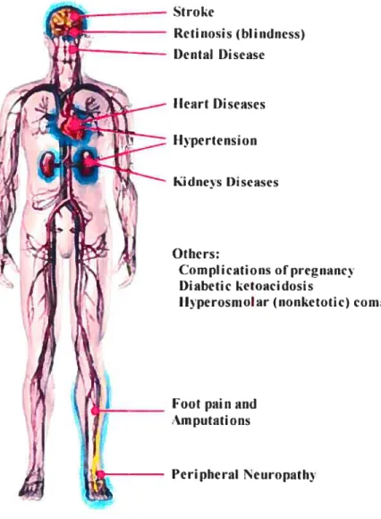
Due to the increased glucose level and insulin insufficiency and resistance, diabetes can result in a number of complications that have a major impact on the health and well-being, depending on the serious degree of the diabetic diseases. These include neuropathy or nerve problem, cardiovascular complications (e.g. hypertension, coronaiy heart disease, micro- and macrovascular complications), retinopathy or eye problem, nephropathy or kidney damage, diabetic foot diseases, etc (f igure 1).
Cardiovascular disease represents the major cause of morhidity and mortality in diabetic patients and heatt disease is the leading cause of diabetes-related deaths. Aduits with diabetes have heart disease death rates and risk for stroke, hoth about two to four times higher than aduits without diabetes. About 65% of deaths among people with diabetes are due to heart disease and stroke. Heart disease accounts for approxirnateiy 50% of ail deaths among people with diabetes.8
Stroke Retinosis (hlindiiess) Dental l)isease Ilcart Diseases Ilpertension kidiiets I)iseases Otiiers: Complications ofpregrianc i)ïabetic ketoaciclosis
Jlvperosiiioiar (nonketotic) conia
Foot painand
.m putatiOflS
Figure 1. Diabetic Complications. (Moditied from healthcentersonhin.com)
Prior to 1972, the increased cardiovascular morbidity and mortality in diabetics had been attributed to vascular diseases. In 1 972, Rubler et al first described a specific type of
cardiomyopathy related to diabetes,1° suggesting that there exist rnyocardial disease as an
independent clinical entity.’1 The terrn “diabetic cardiomyopathy” was thus proposed, describing
an early complication of diabetes that shows diastolic dysfunction and then followed by systolic dysfunction. Since then, a variety of disciplines including noninvasive and invasive cardiac methodologies, as well as epidemiologic studies, have provided information that lias changed our view on the relation of diabetes to cardiac diseases. Instead of an exclusive focus on coronary artery disease, it is now recognized that heart muscle can be independently involved in diabetic patients. In diabetics without known cardiac diseases, abnormalities of left ventricular mechanical function have been demonstrated in 4O-5O% of subjects and it is primarily a diastolic phenomenon. Lefi ventricular hypertrophy may eventually appear in the absence of hypertension. The diastolic dysfunction appears related to interstitiaÏ collagen deposition, largely attributable to diminished degradation.12 Clinical studies demonstrated that children and adolescents with type 1 diabetes have signs of subclinical cardiac dysfunction since their early disease stage; in this population comorbidity such as ischemic heart disease or hypertension can be considered as absent or minimal.’3
When presenting with other cardiovascular complications (e.g.. ischemic heart disease or hypertension), diabetic patients have a much worse prognosis than non-diabetic patients and are more prone to progress to congestive heart failure.’4’6 The underlying diabetic cardiomyopathy appears to contribute to accelerated heart failure. Diabetic cardiomyopathy in general is characterized by metabolic remodeling with malignant biochemical processes,’7”8 electrical remodeling with aberrant cardiac electrical and contractile fiinctions and arrhythmias, 11,19-21 and anatomical remodeling with progressive loss of cardiomyocytes.22 These alterations can be primary (due to loss of direct insulin action) or secondary (due to the metabolic perturbations associated with the disease). Cardiomyopathy will result in irnpaired cardiac performance and increased risk of lethal arrhythmias. Despite the obvious importance of diabetic cardiomyopathy, a better understanding of this complex and very likely multi-factorial problem at the cellular and molecular levels is lacking. Much of what is known about the pathogenesis of diabetic complications lias been gleaned from studies using animal models, including rat, mouse, rabbit, and dogs, etc.
1. 13 QT prolongation and arrhythmias in diabetic hearts
Electrocardiogram (ECG) recorded from diabetic patients show prorninent electrical disturbance with prolonged QT interval according to more than 6 long-term epidemiological
studies (including the EURODIAB, the Neuropathy Study Group of the Italian Society for the Study of Diabetes, the 3rd National Health and Nutrition Examination Survey) involving more than 20 countries and 30 centers, and 11 thousand diabetic patients.2333 It was found up to 26% diabetic patients have electrical disturbance along with QT prolongation and dispersion,34 including the diabetics of both children and adolescents.28 The QT prolongation and dispersion are associated with an increased risk of sudden cardiac death in diabetics have been considered as a predictor of mortality in diabetic patients and,35’36 due to the occurrence of lethal ventricular arrhythrnias known as Torsade des Pointes or long Q-T syndrome (LQTS).3738 These findings can also be applied to type 2 diabetes.3032’3941
Studies also demonstrated that both hyperglycemia of diabetes, and hypoglycemia in diabetes therapies, can prolong cardiac QT interval.34’4248 Indeed, QT prolongation and heterogeneity of QT prolongation (QT dispersion) have been suggested as the predictor of mortality in both IDDM and NIDDM. 26;33;3437;38;4O
Despite this serious reality, it is only in recent years that people start to understand the ionic and cellular/rnolecular basis for the diabetic QT prolongation.
1.1.4 AnimaI models of diabetes mellitus
There has been littie progress in characterizing the ionic, cellular and molecular features of diabetic cardiomyopathy in patients, much less their mechanisms. It remains unclear whether there are differences between insulin dependent and non-insulin dependent forms of diabetes mellitus with respect to the development of diabetic cardiomyopathy. Defining cardiac changes in diabetic humans is obviously extremely important in light of the fact that diabetes is a chronic disease flot well represented by animal models. However, animal models are the most used subjects to understand the mechanisms ofdiabetic complications.
Although we now have animal models for both type 1 and type 2 diabetes, the separation of these two categories is flot total, just as these two are flot completely separated in human. Most used type II diabetic animal models are: genetic, obese diabetic mouse and fatty Zucker rat, nutrition-induced diabetic sand rat and spiny mouse, rodents with decrease in f3-cell mass induced by pancreatectomy and streptozocin (STZ), and diabetic offsprings of selective inbreeding of rodents.49
Type 1 diabetic animais can be subdivided into two groups: chemical-induced, and spontaneous/ genetic modified.50’5’ Genetic models include the nonobese diabetic (NOD) mouse model ofdiabetes and also the 33 rat model,52 which will not be discussed in detail in this thesis. lnduced diabetes can be achieved by pancreatectomy or by destroying the pancreatic 13-ceil with a number of chemicals, including alloxan and STZ which are frequently used. Alloxan and SIZ, initially used as active anti-tumor reagents, are deleterious and can selectively damage islet celis, thus can be used to induce IDDM.5356 The f3-cell is uniqueiy sensitive to alloxan because its plasma membrane contains ionized sulphydryl (SH) groups related to insulin release which are not found in other tissues.57 The sensitivities to alloxan or STZ in different species are not the sarne, e.g. guinea pig and some big-size animals including dog and rabbit, are resistant to STZ, therefore it is difficuit to make diabetes in these species.5° functionally, both alloxan and $TZ interfere the glucose transport, alter the SH groups of glucokinase, and impair mitochondrial function by inducing the formation of free radicals.58 Repeated injections with low doses of STZ into genetically susceptible mice can induce diabetes similar to human IDDM, in which a pathogenic involvement of cell-mediated imrnunity and the coexistence of a marked pancreatic insulitis are encountered and apoptotic death of 3-cell is a responsibie mechanism. However, this model is different from the autoimmune type 1 diabetes seen in spontaneous animal models.58
Both chemical-induced and genetic-modified diabetic animal models have been generated to facilitate the studies in the pathology of diabetes. By far the most extensively employed animal models are the $TZ-induced diabetes mellitus in rat and mouse and alloxan induced rabbits and dogs. Rats treated with SIZ for weeks to months develop hypoinsulinernic diabetes mellitus, and mice are usually treated for a single dosage of STZ in order to develop DM. Chronic diabetes diminishes contractility and, most especially, relaxation, and prolongs the duration of contraction in rabbit hearts,59 which may due to the alteration of myosin isoenzyme composition (i.e. switch from Vi to V3 isomyosin) and are reversible with insulin.6° Effects on ‘passive” diastolic compliance have been variable. These contractile abnormalities are correlated with several alterations in excitation-contraction (E-C) coupling, ion transport and exchange, including the prolongation of AP, and the depressed sarcoplasmic reticular tSR) calcium pumping and sarcolemmal NaJCa exchange.”216’ While the STZ-diabetes mellitus rat and mouse are reliable, well-characterized models, they may flot be the most relevant model for human disease, particular in their different electrophysiological profiles.
1.2
QT interval, APD, and Ion Channels: Physiology and Pathology
1.2.1 QT interval as a function of cardiac repolarization
Heart rhythm is generated by sequential movement of inward and outward ion currents across the membrane of the cardiomyocyte; disturbance of the balance will result in aberrantly altered cardiac electrical activities, so called arrhytbmias.
1.2.1.1 Electrocardiogram (ECG)
The electrical activity ofheart can be detected using a technique called electrocardiogram (ECG) which represents the electrical current moving through the heart during heartbeat cycles. The current movement is divided into several parts, and each part is given an alphabetic designation in the ECG. Each heartbeat begins with an impulse from the heart’s pacemaker (sinus or sinoatrial node) located at the entrance to the right atrium. This impulse activates the upper chambers of the heart (atria), represented as a P wave in the ECG recording. Then the excitatoiy stimuli pass over the atrioventricular node (AV node) to the lower chambers of the heart (ventricles) and generate the QRS complex in ECG. The electrical pulse then spreads back over the ventricles wall in the opposite direction, resulting in the recovery T wave. Therefore, the QT interval which is the duration between the onset of
Q
wave and the termination of T wave represents the activity of ventricular contraction. Many kinds of cardiac abnorrnalities can be seen on an ECG. for example, the heart rhythm may be abnormal: too fast, too slow, or irregular. By reading an ECG, doctors can usually determine where in the heart the abnormal rhythm starts and what its causes may be.1.2.1.2 Ion channets andaction potentiat
The basis of ECG is the ion currents passing tbrough the protein-structured “ion channels” on cell membrane of cardiomyocytes and channels on membrane of cell organelle sucli as sarcoplasmic reticulum tSR) which is a pool of calcium storage. Cell plasma membranes are highly selective barriers forming the outer surface of eukaryote cells including cardiomyocytes. Inside the cell, membranes of many organelles such as the $R and the mitochondria also have similar structure as the plasma membranes. Assembled from lipids and
proteins in sheet like fashion, plasma membranes give the celi individuality and a certain level of control over its internai environrnent, and permit the development of concentration gradients (e.g. ions) cruciai to the function cf signaling moiecules.
An ion channel is a pore-forming protein or more typically an assembly of severai proteins on the membrane. Such Tmulti-subunit” assemblies usually involve a circular arrangement of identical or reiated proteins closely packed around a water-fflled pore through the plane of the membrane or lipid bilayer. Ion channels are generally highly selective and impermeable to most ions. By conducting and controlling the flow of ions, ionic channels help establish the small negative voltage that ail celis possess at rest, or fire the cell membrane when a certain voltage threshold reaches. Ion channels can generally be classffied according to their “gating” (the open-close of a channel): voltage-gated and ligand-gated ion channels; or based on their permeability to ions: cation, anion, or Na, Ca2, K, Cï channels, etc. Ions passing though the channel will generate either inward or outward currents.
The cardiac action potential (AP) is generated by the concerted action of depolarizing (inward) and repolarizing (outward) currents conducted by different ion channels, pumps and transporters. As the ventricular activity is the main part responsible for die QT inteiwal, it will be focused on the ventricle when discussing AP and ion chaimels. In ventricle, major inward currents include sodium current (‘Na) and calcium current (ICa). and outward currents include the
Ca2-independent transient outward K current (Ito), the rapid and the slow activating delayed
rectifier outward K currents (iKr and ‘Ks, respectively), and the inward rectifier K current (IKI) which conducts outward cunent at membrane potential between —80 and O mV.6265 A small change in the potential across the cell due to various stimuli is sensed by the Na channel protein
(‘Na), which alters its conformation from resting close state to open, allowing a large, rapid Na influx, producing the typical rapid rising upstroke phase O depolarization -- membrane potential becomes positive compared to the resting membrane potential which is between -65mV to -85 mV in different heart ceils. A rapid phase 1 repolarization then ensues in some types ofcells (e.g. atrial and ventricular myocytes). because of the outward movement of K via the transient outward K channels (Ito). Calcium channels open when the membrane depolarizes to the
threshold during phase O and phase 1. Following phase 1 is the long lasting plateau (phase 2) of the action potential, reflecting a balance between inward cunent, largely through L-type Ca2 channels (ICaL), and outward current, mainly through delayed rectifier K+ channels (IK, including
I and ‘1(s). It is ‘Kr that initiates the repolarization phase 2 in the action potential. The net outward current during Phase 3 repolarization cornes from the increasing I1(, along with the inactivation of ‘CaL. ‘Kr reach maximum at about 90% depolarization of AP (APD90). Final repolarization is accomplished by the increasing outward movement of K through inward rectifier chaimels (‘1(1) beginning from phase 3 and reach stable at cornplete repolarization. Slow response cells, for example the sinus node and the AV node have slow depolarization during phase 4 due to the activity of the pacemaker channel. In these ceils, a rapid phase 1 upstroke is absent, and initial depolarization is accomplished by the opening of L-type (and perhaps also T-type) Ca2 channels.
The burst and the decay of inward and outward cunents during an electrical pulse cycle in a ceil form different phenotypes of AP at different regions of the heart because of the different expression in ion channels. For example in ventricle, there are three distinct cell types: epicardial, rnidmyocardial (M) and endocardial celis particularly in species with larger size. APs of epimyocardial and M ceils differ frorn endocardial cells with respect to the morphology of phase
1. These cells possess a prominent‘to which mediates the notch and is responsible for the ‘spike and dome’ morphology in APs. M celis are distinguished frorn the other cell types in that they display a smaller ‘Ks, but a larger late sodium current (INaL) and sodium-calcium exchanger current (INCx).6674 These intrinsic electrical heterogeneities in ventricular myocardium underlie the longer action potential duration (APD), which is more pronounced in the presence of antianhythmic agents with class HI actions. The preferential prolongation of the M cell action potential resuits in the development of a transmural dispersion of repolarization, which can be examined from the ECG showing a prolonged QT interval and a change of the T rnorphology. Exaggeration of these heterogeneities due to inherited (i.e. gene mutation of channels and their regulatory molecules) or acquired means (e.g. drug exposure, exercise, and secondary to diseases such as ischemia, diabetes, etc.) enhances reentrant excitation, a common mechanism for many cardiac anhythmias.75’76 The acute electrophysiological response of a myocyte to exogenous stressors such as drugs, myocardial ischemia, diabetes, or autonomic activation likely reflects changes in function of individual ion channels (also called remodeling), including channels activated by specific stimuli such as ATP depletion and muscarinic stimulation. More chronic responses to such exogenous stressors may also include changes in gene expression.
CURRENTS Ix, ‘NCX — — -‘hi 200 ms
figure 2. Relationships between
al., 2002.65)
ECG, APD and ion currents. (Adapted from Roden DM, et
In theory, inward cunents depolarize (meaning the membrane potential is brought to more positive) the cardiac ccli membrane and maintain the APD, whereas outward ctirrents repolarize (meaning the membrane potential is brought back to more negative) cardiac celis and shorten the APD. Therefore, decreases in outward currents and/or increased inward currents will
ECG T
AP OI
—I
resuit in the prolongation of APD thereby the QT interval. Figure 2 describes the relationship between a typical ECG, an AP, and ion currents.
1.2.1.3 Long QTsyndromes (LQTS)
LQTS is a disorder characterized by a prolongation of the QI interval on the ECG and a propensity to ventricular tachyarrhythmia, which may lead to syncope, cardiac arrest, or sudden death. Defects in the genes conesponding to certain ion channels or impairment of ftinctions of these chaimels resuit in QT prolongation, reflecting the abnormally long delay between the electrical excitation (or depolarization) and relaxation (repolarization) of the ventricles of heart. Heart rate corrected QTc values above 0.44 seconds are generally considered abnormal in human, although age- and sex-specific abnormal QTc values have been proposed. Certain triggers such as intense physical exercise or excitement, and being suddenly startled or badly frightened, result arrhythmias, e.g. torsade de pointes (TdP), a life-threatening form of ventricular tachycardia and sudden cardiac death.
LQTS is recognized as the Romano-Ward (RW) syndrome (familial occurrence with autosomal dominant inheritance, QT prolongation, and ventricular tachyanhythmias) or the Jervell and Lang-Nielsen (JLN) syndrome (familial occurrence with autosomal recessive inheritance, congenital deafness, QT prolongation, and ventricular anhythmias). Conventionally, LQTS is divided into two catalogues: genetic or inherited, and acquired or drug-induced. Based on recent efforts in deciphering the arrhythmias in cardiac or noncardiac diseases, it is rational to classify a new class of QT prolongation which may also lead to LQTS. Many other syndromes or diseases have been correlated to and probably a reason of QT prolongation. This “pathological LQTS” may occur in ischemia and infarction, chronic heart failure, and noncardiac conditions, such as electrolyte abnormalities (e.g. hypokalemia, hypocalcemia), pulmonary embolism, CNS diseases (e.g. cerebrovascular disorders), hypothermia,77 hypothyroidism,788’ and both IDDM and NIDDM which have been discussed in Section 1.1.3. Yet, the ionic mechanisms underlying these pathological LQTS are just becoming understood by intensive studies in electrophysiology, cellular and molecular biology.
Genetic testing of LQTS is unfortunately stiil flot readily available, and it may not be possible to determine with certainty a particular patient’s genotype. However, at least one recent article daims that LQT 1, 2, and 3 can frequently be identified by the ECG pattern.82 LQT 1 has
a prolonged QT interval with a normal to high T wave amplitude, a broad based T wave, and an indistinct T wave onset, caused by dysfunction of the pore-forming Œ-subunit of I chaimel due to either lost-function mutation or drug induced or diseases-modulated channel impairment. LQT 2 accounts for about 35% of the long QT syndrome patients, resulted from the depression of‘Kr current. It is characterized by a prolonged QT interval, low amplitude and bifid T waves in over 60% of cases. LQT 3 patients have a prolonged QT interval with a delayed-onset, peaked and widened T wave, and a long, isoelectric ST segment. They have a rate dependence of the QT interval prolongation and develop TdP at slow rates. The positional candidate gene approach was used to establish that the gene responsible for this chromosome 3-linked LQTS is the cardiac sodium channel gene SCN5A.83 In 1995, Schott et al.84 reported on a large f rench kindred with a complex cardiac phenotype including sinus bradycardia, abnormal heart rate variability, defects
in cardiac repolarization (denoted on the ECG as prolonged QT interval), and sudden cardiac death. This report described the first finding of ‘type 4 long QT syndrome’ and mapped the autosomal dominant disease to human chromosome 4q25-27, a site distinct from the location of any other ion channel gene implicated in the genesis of inherited arrhythmia. LQT5 and LQT6 are diseases resulted from the dysfunction of the channel accessory subunits minK and MiRP 1, of I and ‘Kr channel, respectively. The Andersen syndrome is a rare, inherited disorder characterized by periodic paralysis, long QT interval with ventricular arrhythmias, and skeletal developmental abnormalities, and subtyped into LQT7 due to its prolonging QT interval and its distinct substrate for arrhythmia susceptibility: in reduced extracellular K, it induces delayed afterdepolarizations (DAD) and spontaneous arrhythmias. Homozygous KVLQT1 and KCNE1 mutations are associated with congenital deafness (JLN syndrome) and account for less than 1% of cases of LQT$. Approximately 200 different mutations of these genes have been found so far. Significant phenotypic variation of the ECG findings (T wave morphology), factors triggering cardiac events, and risk of cardiac events exist, depending upon which gene and which mutations ai-e involved. Because flot ah known cases of LQTS are caused by mutations of the above genes, other genes causing this disorder are expected to be identified in the future.
Table 1 lists the chromosomal locus, gene and ion currents related to these LQTS.8587 Drug-induced and pathological QT prolongations may also lead to increased risk of ventricular tachyarrhythmias (e.g., TdP) and sudden cardiac death in a similar ionic mechanism as observed
In sum, ionic basis in LQTS demonstrates that reduction of outward current (e.g. IKr, iKs)
and/or increase of inward current (e.g. ‘Na, ‘CaL), will delay the repolarization of AP, lead QT
prolongation and predispose to LQTS.
Table 1. Genetic Background oflnherited Forms ofLQTS.
LQTS DIR Chromosomal Gene Target Alteration
type Locus (channel, etc.)
D KVLQTJ(KCNQ1)
LQTI llpl5.5 IK5(Œ)
(heterozygotes)
LQT2 D 7q35-36 HERG ‘Kr
‘1’
LQT3 D 3p2l-24 $CN5A ‘Na
t
calcium signalings [Ca2j
LQT4 D 4q25-27 ankyrin-B, G NaJK-ATPase, ‘NCX transient
t
InsP3 receptor D KCNEJ
‘I,
LQT5 21q22.1-22.2 ‘Ks(f3,
minK) (heterozygotes) LQT6 D 21q22.l-22.2 KCNE2 IKr(f3,MiRP1) LQT7(Andersen D 17q23 KCNJ2 (Kir2.]) ‘KI
syndrome)
LQT8
(Timothy D l2pl3.3 C’AC’NAJc (‘C’av].2) ICaL
syndrome) KVLQT] (KCNQ]) JLN1 R Iipl5.5 IKs(Œ) (homozygotes) KCNE] JLN2 R 21q22.1-22.2 ‘Ks
(f3,
minK)) (homozygotes)D: autosomal dominant inheritance; R: autosomal recessive inheritance;