Bacterial intestinal flora associated with enterotoxaemia in Belgian Blue
calves
C. Mantecaa, G. Daubea, V. Pirsona, B. Limbourgb, A. Kaeckenbeecka, J.G. Mainila
a. Department of Infectious and Parasitic Diseases-Bacteriology, Faculty of Veterinary Medicine, University of
Liège, Liège B4000, Belgium
b. Laboratoire Provincial de Lutte contre les Maladies du Bétail, Ciney B5590, Belgium
Abstract
The enterotoxaemia syndrome in Belgian Blue calves is characterised by a high case fatality rate, sudden death, lesions of haemorrhagic enteritis of the small intestine and, quite often an absence of other clinical signs but its cause has not been yet identified. As a first step in this identification, the aerobic and anaerobic intestinal flora of a population of 78 calves, originating from farms located in southern Belgium and that died in circumstances defined as "calf enterotoxaemia" (study population) and of 64 calves that died in other circumstances (control population) were studied qualitatively and quantitatively. The colonies were identified after subcultures with appropriate API sugar sets. Anaerobically Clostridium perfringens was isolated in higher numbers (mean values of 107-107.5 colony forming units (CFU) versus 104-105 CFU per ml of intestinal content) and from more animals
(79 versus 19%) in the study population than in the control population, although individual results from both populations could overlap. Other clostridial species, i.e. mainly urease-negative C. sordellii and C. bifermentans, were isolated in high numbers (>106 CFU per ml of intestinal content) from a few animals in the study
population only. All but one of the 705 C. perfringens isolates from both populations belonged to the A toxin type and none of the urease-negative C. sordellii was toxigenic. Gram-negative anaerobes were not isolated in high numbers from any of the samples. Aerobically β-haemolytic E. coli were significantly more frequent among the study population, but were isolated from only 25% of the animals. Salmonella Typhimurium was isolated from only two animals in the study population. Less than 1% of the E. coli isolated were verotoxigenic and one-third were necrotoxigenic. At this stage only non-enterotoxigenic type A C. perfringens are thus statistically associated with the enterotoxaemia syndrome in Belgian Blue calves and fulfil the first of the Koch's postulates.
Keywords: Bovine enterotoxaemia; Cattle-bacteria; Belgian Blue cattle; Clostridium sp.; Clostridium
perfringens; Toxins
1. Introduction
Bovine enterotoxaemia is characterised by a high case fatality rate, sudden deaths, lesions of haemorrhagic enteritis of the small intestine and, quite often, an absence of other clinical signs (Popoff, 1990; Manteca and Daube, 1994; Songer, 1996). The syndrome is relatively frequent in Belgian Blue calves, since more than 10,000 of them die from it every year in Belgium (Limbourg, unpublished data). Environmental parameters, clinical signs, macroscopic and microscopic lesions were recently specified from a survey of more than 70 cases occurring in different farms in Belgium (Manteca et al., 2000). These cases show similarities, but also differences, when compared to cases occurring in other cattle breeds in other countries (Popoff, 1990; Daube, 1992; Manteca and Daube, 1994; Songer, 1996). For instance, macroscopic lesions consisted of haemorrhagic enteritis, mainly of the small intestine, but sometimes also of the large intestine. Lesions of other organs were not consistent. Microscopically, the intestinal lesion was necrosis of the top of the intestinal villi, of the intestinal epithelial cells and/or of the intestinal villus vascular axis (Manteca et al., 2000).
The aetiological agent of bovine enterotoxaemia has not been formally identified. Clostridium perfringens has been incriminated, like in other cattle breeds, although other clostridial species and/or genera can be associated with similar clinical signs and lesions (e.g. Clostridium sordellii, Salmonella sp.) (Manteca and Daube, 1994). Moreover, until now, no large scale quantitative case-control study has been performed.
The goal of the present work was to fulfil the first of the Koch's postulates by studying qualitatively and quantitatively the aerobic and anaerobic intestinal flora of a population of 78 Belgian Blue calves that died of "calf enterotoxaemia" (Manteca et al., 2000) and of a similar calf population that died of other causes.
2. Materials and methods
2.1. Animals
The 78 cattle, which had died either suddenly or very rapidly, with diffuse or localised lesions of acute haemorrhagic enteritis at necropsy, composed the study population (Manteca et al., 2000) and 64 other cattle, with a formal exclusion diagnosis of enterotoxaemia composed the control population. All control cases, as well as more than 90% of the study cases (Manteca et al., 2000) were Belgian Blue suckling calves between 2 and 4 months of age. Necropsy was performed within 24 h of death. All calves originated from farms located in southern Belgium.
2.2. Samples for bacteriological analysis
Bacteriological analyses were performed on the small intestine contents of all animals after removal of 30 cm ligated loops with the most obvious lesions (if any) and, in some animals, the contents of the rectum. For long-distance transport, the samples were placed in anaerobic jars with the BBL Gaspak Anaerobic System (Becton-Dickinson). Ligated intestinal loops were also removed above and below of the lesions in four animals with localised lesions of haemorrhagic enteritis.
2.3. Smears of intestinal contents
Smears of the intestinal contents were stained by Gram's and Ziehl-Neelsen's methods to estimate the proportions of Gram-positive flora and of sporulated bacteria, respectively. The Ziehl-Neelsen's method for spores is an adaptation from the method for acid-fast bacteria with the carbol fuchsin heated until boiling. 2.4. Aerobic bacteriological analysis
Small intestinal and rectal contents were inoculated onto Columbia Agar plates with 5% sheep blood (Becton-Dickinson) and Gassner Agar plates (Merck-Belgolabo) which are selective for Enterobacteriaceae. Lactose-fermenters (coliforms) grow as blue colonies and non lactose-Lactose-fermenters as yellow colonies. Bacteria were allowed to grow overnight at 37°C. Tetrathionate enrichment broth (Oxoid) Drongen, Belgium for Salmonella was also inoculated, incubated for 48 h at 42°C, and subsequently inoculated onto Gassner Agar plates. Identification of Enterobacteriaceae was performed using API-20E sugar sets (BioMérieux) Marcy l'Etoile, France.
2.5. Anaerobic bacteriological analysis
The 100 µl of the small intestinal and rectal contents were sampled and resuspended in 900 µl (dilution 1) of Brain Heart Infusion broth (Gibco BRL) with 0.5% thioglycollate (BHI-thioglycollate). The 10-fold dilutions (2-8) were made in 900 µl of BHI-thioglycollate. Dilutions were subsequently plated onto three different growth media for the anaerobic bacteriological analysis: Ana-blood base agar (Gibco BRL) plates with 8% cattle blood (Ana-blood plates), Ana-blood plates with polymyxin and kanamycin (S.E.R Supplement, Oxoid) to select for Gram-positive bacteria (Ana-pol plates), and Ana-blood plates with cycloserin (TSC Supplement, Oxoid) which are selective for a few clostridial species, especially C. perfringens and C. sordellii (Ana-cyclo plates).
Two methods of bacterial cell count determination were used during the course of the study. During the first phase, 100 µl of dilutions 5-8 were inoculated manually onto the three Ana agar plates. Bacteria were allowed to grow overnight at 37°C in anaerobic conditions in HP11 jars (Oxoid). During the second phase, dilutions 1-4 were inoculated using a Model D Spiral Systems plater (LED Techno) onto the three Ana agar plates, 14 cm in diameter. Bacteria were allowed to grow overnight at 37°C under anaerobic conditions (90% N2, 5% H2, 5%
C02) in a Don Whitley MK3 Anaerobic Workstation (LED Techno). Dilutions 1-4 were also heated at 75°C for
20 min in a water bath and inoculated onto Ana-pol plated for count determination of sporulated bacteria. Presumptive identification of colonies was made on the basis of their aspect and the presence of haemolysis. Final identification of anaerobes was performed using APIRapid32A sugar sets (BioMérieux) after subculture in aerobic, microaerophilic and anaerobic conditions.
2.6. Gene probes and colony hybridisation assay
The gene probes for the α, β, ε, and ι toxins and for the enterotoxin of C. perfringens were derived as previously described; positive controls were as published (Daube et al., 1996).
The gene probes for the lethal toxin (LT) and haemorrhagic toxin (HT) of C. sordellii were derived by PCR from the genes coding for the B and A toxins of C. difficile strain VIP 10463, respectively, on the basis of the
sequence homology between these genes (Barroso et al., 1990; Sauerborn and Von Eichel-Streiber, 1990; Moncrief et al., 1997). The primers were derived from the published sequences of the A and B toxin-encoding genes of C. difficile with Oligo® software (National Biosciences). The primers for A toxin-encoding genes were:
5'-ATAAGAAAATAGTAACTTCAGCA-3' and 5'-AAACTTCAAGCAGAAATAG-3' with an annealing temperature of 48.4°C; and the primers for B toxin-encoding gene were: 5'-TCCACAGAAATAGAAGCAGCAA-TA-3' and 5'-ATAGCCAATCAATCCATCCTCATA-3' with an annealing temperature of 50.7°C. The PCR reactions were performed with a DNA Thermal Cycler (Perkin-Elmer).
The PCR conditions were as already described (Daube et al., 1994). The PCR procedure was: 5 min at 94°C followed by 30 cycles consisting of 1 min at 94°C, 30 s at the annealing temperature, 3 min at 62°C. The PCR products were purified with a PCR purification kit (Qiagen) and radiolabeled as described elsewhere (Daube et al., 1996). The A/HT toxin probe was 3101 bp long and the B/LT toxin probe was 3420 bp long. The C. sordellii positive control was the toxigenic reference strain C582.
The probes for the Verotoxins 1 and 2 (VT1 and VT2) and for the Cytotoxic Necrotizing factors 1 and 2 (CNF1 and CNF2) of E. coli which can be associated with haemorrhagic enteritis in cattle were derived as previously described; positive controls were also as published (Mainil et al., 1993; Oswald et al., 1994).
The probes were radioactively labelled and the colony hybridisation assay was performed as previously described (Daube et al., 1996).
2.7. Statistical analysis
The percentage of positive animals from each population in the various tests was compared using the Fisher test. The mean value and the standard error of the bacterial counts were compared using the t-test of Welch and the Barnett test, respectively. Correlations between the different bacterial populations present in different portions of the intestinal tract were analysed using the Spearman test.
3. Results
3.1. Smears
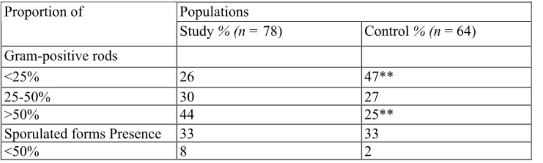
The 44% of the intestinal content smears in the study population had an estimated proportion of Gram-positive rods of more than 50% compared to 25% in the control population (P < 0.01; Fisher test) (Table 1). On the other hand no difference was observed between the two populations for the presence of sporulated forms after Ziehl-Neelsen's staining.
3.2. Aerobic growth
Even if β-haemolytic E. coli were significantly (P < 0.05; Fisher test) more frequent in the study population than in the control population, they were isolated from only 25% of the animals in the former (Table 2). Salmonella Typhimurium was isolated from two animals in the study population. On the other hand, streptococci and enterococci were isolated more frequently from the control population than from the study population (Table 2). 3.3. Anaerobic growth
Gram-positive, but not Gram-negative, anaerobes were isolated from the highest dilutions of the samples from both populations. All the Gram-positive bacteria belonged to the genus Clostridium. On the basis of the results of bacterial identification, two groups were observed: C. perfringens, and other clostridial species. Both were isolated more frequently (P < 0.01; Fisher test) in the study population (79 and 19%, respectively) than in the control population (19 and 5%, respectively) (Table 3). No significant difference was observed in the isolation rate of C. perfringens using either method of bacterial cell count determination (Table 3). On the other hand, the
isolation rate of other clostridial species in the study population was much higher using the anaerobic cabinet (Table 3).
The other clostridial species identified in samples from the study population were: urease-negative C. sordellii (n = 16, 20%), C. bifermentans (n = 15, 19%), and C. baratii, C. beijenrikii, C. botulinum, C. butyricum, C. cadaveris, C. fallax, C. glycolicum, C. limosum, C. paraputrificum, C. ramosum, C. sporogenes, C. tertium, C. tetani and C. tyrobutyricum, from only a very few samples each.
3.4. Anaerobic bacterial counts
The mean values of C. perfringens counts were significantly higher (P < 0.01; Welch's f-test) in the study population than in the control population: 107-107.5 CFU (Colony Forming Units) compared to 104-105 CFU per
ml of intestinal content. However, some individual counting results in both populations overlapped (Fig. 1). In the four calves with localised intestinal lesions, the counting results of C. perfringens were significantly higher at the site of the lesions, than above or below the lesion (Fig. 2). When present (in 20% of the animals in the study population), C. sordellii was also isolated in very high numbers (>106 CPU per ml of intestinal content).
Only C. perfringens colonies grew on Ana-pol plates after heating of the samples at 75°C for 20 min (= sporulated forms). No significant difference (P > 0.05; Fisher test) was however observed between the number of CFU in samples from both populations and the numbers of sporulated forms of C. perfringens did not correlate with the number of non-sporulated forms in either population (Spearman test).
3.5. Bacterial populations of the rectum
No difference was observed between bacterial identification and counts performed on the rectal contents from 49 animals of both populations. No significant correlation was observed between the rectal and the intestinal flora either (Spearman test).
3.6. Bacterial typing
The 523 C. perfringens isolated from the study population and 182 isolated from the control population were hybridised with the probes derived from the genes coding the α, β, ε and ι toxins and for the enterotoxin. All C. perfringens isolated were positive with the probe for the α toxin, and one, isolated from an animal in the study population, with the probe for the enterotoxin, but none with the probes for the β, ε and L toxins.
None of the C. sordellii isolates hybridised with the probes for the ι T and HT toxins, although the C. difficile and C. sordellii positive control strains did.
Of 210 E. coli isolated from animals in both populations 10 non-haemolytic isolates tested positive with the probes for the VT toxins. Fifty other isolates hybridised with the probe for CNF1 and 25 with the probe for CNF2. The CNF 1-positive isolated were mainly β-haemolytic.
Table 1 : Proportion of Gram-positive rods vs. total number of bacterial cells and sporulated forms vs. total number of Gram-positive rods in smears of intestinal contents from study and control population animals after Gram's and Ziehl-Neelsen's staining, respectively
Populations Proportion of Study % (n = 78) Control % (n = 64) Gram-positive rods <25% 26 47** 25-50% 30 27 >50% 44 25**
Sporulated forms Presence 33 33
<50% 8 2
Table 2 : Bacteria isolated in highest counts from intestinal contents of study and control population animals after growth in aerobic conditions
Populations Bacterial species Study % (n = 78) Control % (n = 64) Non-haemolytic E. coli 86 82 β-haemolytic E. coli 25 12* Salmonella sp. 2 0 Proteus sp. 9 10 Streptococcus/enterococci 33 56* No growth 3 4 * P < 0.05 (Fischer test)
Table 3 : Clostridial species isolated from intestinal contents of study and control population animals after growth in anaerobic conditions using the methodology #1 or #2a
Clostridial flora Total Methodology #1 Methodology #2
Study % Control % Study % Control % Study % Control %
(n = 78) (n = 64) (n = 50) (n = 46) (n = 28) (n = 18)
C. perfringens 62 (79) 12 (19)** 40 (80) 9 (20)** 22 (79) 3 (17)**
Other clostridia 15 (19) 3 (5)** 2(4) 1 (2) 13 (46) 2(11)**
a See Section 2.
** P < 0.01 (Fischer test).
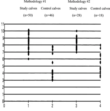
Fig. 1. Individual values (log10 of CFU) of Clostridium perfringens flora per ml of intestinal content from study
and control population animals after growth in anaerobic conditions using the methodology #1 or #2 (see Section 2).
Fig. 2. Individual values (log10 of CFU) of total and Clostridium perfringens flora per ml of intestinal content
from the four study population animals with localised lesions of haemorrhagic enteritis (above, lesion site, and below) after growth in anaerobic conditions. Estimation (score 0-4) of the haemorrhagic character of the intestinal wall and content is presented in parallel.
4. Discussion
Different clostridial species cause intestinal disorders and enterotoxaemia in various animal species, including ruminants (Popoff, 1990; Manteca and Daube, 1994; Songer, 1996). Enterotoxaemia in cattle is rarer than in small ruminants. In Belgian Blue calves, enterotoxaemia is defined as a sudden death syndrome with lesions of haemorrhagic enteritis of the small intestine at necropsy (Manteca and Daube, 1994; Manteca et al., 2000). The infectious aetiology has not been identified, although C. perfringens is often regarded as responsible, by analogy with the situation in small ruminants (Popoff, 1990; Manteca and Daube, 1994; Songer, 1996).
C. perfringens was indeed isolated in higher numbers and from more animals in the study population than in the control population (Table 3), although individual results from either population can overlap (Fig. 1). Similar results were observed in the four animals with localised lesions of haemorrhagic enteritis: C. perfringens counts were much higher at the place of the lesions than above and below. At this stage, and in the absence of any other bacterial species, or anaerobes or aerobes isolated at high frequency from the study population compared to the control population, only the bacterial species C. perfringens quantitatively fulfil the first of the Koch's postulates and may be regarded as a candidate infectious aetiological agent in enterotoxaemia of Belgian Blue calves in Belgium.
If this hypothesis is confirmed in the future and in analogy with other types of enterotoxaemia, like in small ruminants (Popoff, 1990; Daube, 1992; Songer, 1996) one may speculate that the C. perfringens present more especially in the large intestine divide anarchically and invade the small intestine in high numbers, as a consequence of some disturbance of the intestinal peristaltism (Manteca and Daube, 1994). It has indeed been observed that most clinical cases occur after a change in diet (Manteca et al., 2000). However, it is strange that the total intestinal anaerobic flora does not increase markedly. This may be due to the limits of the technique for growing anaerobic bacteria. A greater difference was indeed observed during the second phase of the study when an anaerobic incubator, which provides much better conditions for anaerobic growth than jars do, was used (Table 3). C. perfringens may also have a natural advantage compared to other anaerobes to multiply in very large numbers, in the small intestines of Belgian Blue calves. In that respect it is noteworthy that the bacterial
counts in animals from the control population are high compared to the data in the literature: 10 versus 102.5 CFU
per ml of intestinal content (Smith, 1975; Daube, 1992).
Alternatively, the total number of C. perfringens present in the intestine may not be so relevant, if some strains produce a toxin that induces the lesions and clinical signs, like the β toxin in newborn lambs and piglets, the ε toxin in pulpy kidney disease in lambs, or the enterotoxin in humans and animals (Niilo, 1978; Hatheway, 1990; Popoff, 1990; Daube, 1992; Songer, 1996; Johnson and Gerding, 1997). Determination of the toxin types of the C. perfringens isolated from both populations is however disappointing as only non-enterotoxigenic toxin type A strains were detected, confirming previous results (Daube et al., 1996) and excluding the toxins β, ε and i and the enterotoxin. If C. perfringens is the aetiological infectious agent, the toxin responsible for the intestinal lesions and for the death of the calves may be the a toxin, as suspected in other animal species, or one of the minor toxins (Hatheway, 1990; Popoff, 1990; Daube, 1992; Songer, 1996; Griffiths et al., 1998; Netherwood et al., 1998). The recently described β2 toxin (Gibert et al., 1997) which is associated with haemorrhagic intestinal lesions in piglets (Klaasen et al., 1999; Garmory et al., 2000) but which was not looked for, is another candidate. Other clostridial species were also isolated in high numbers but only from a few animals in the study population, especially during the second phase of the study, once again emphasising the importance of anaerobic growth conditions. On the other hand, Gram-negative anaerobes were not isolated in high numbers from any of the samples but they are at the same time not incriminated as a cause of enteroxaemia in cattle (Al-Mashat and Taylor, 1983a; Manteca and Daube, 1994). Of the other clostridial species, urease-negative C. sordellii and C. bifermentans were the most frequent. C. bifermentans is regarded as non-pathogenic, but C. sordelli producing the HT toxin has been incriminated in cases of enterotoxaemia, besides its known role in gas gangrene, although no experimental reproduction of the clinical syndrome was successful (Cottereau et al., 1962; Niilo et al., 1963; Arseculeratne et al., 1969; Popoff, 1990; Al-Mashat and Taylor, 1983a,b; Songer, 1996). When C. sordellii is present, high numbers of C. perfringens are also present, which makes the discussion on the potential role of the former impossible. Moreover, the urease-negative C. sordellii isolated in this study were non-toxigenic as determined by colony hybridisation, and their identity itself must be questioned (Cato et al., 1986). In general, the other clostridial species isolated can be eliminated as infectious aetiological agents in the cases of
enterotoxaemia in Belgian Blue calves in this particular study, although a role cannot be formally excluded for one or another of them in individual cases.
Similarly, a role of toxigenic E. coli and Salmonella species cannot be formally excluded in isolated cases of sudden death with haemorrhagic enteritis, as in other host species (Cherifi et al., 1990; De Rycke et al., 1999; Mainil, 1999). But on the whole, bacterial species other than C. perfringens were not significantly associated at high frequency with the clinical cases of enterotoxaemia of this study. The massive multiplication of C. perfringens, and perhaps also of other clostridia may also explain the reduction of the number of streptococci and enterococci in animals from the study population in comparison to the control population, as a outcome of bacterial competition.
In spite of these results, confirmation of bovine enterotoxaemia in calves remains a problem for routine diagnostic laboratories. The results obtained in this study show that only a quantitative determination of the C. perfringens in the intestinal contents, with a cut-off value between 106 and 107 CFU per ml, permits such a
diagnostic confirmation, as alternatives for simplifying bacterial diagnosis, such as faecal smears, faecal bacterial counts and smears of small intestinal contents, have all failed. Moreover, the absence of haemorrhagic enteritis in a small proportion of cases at necropsy (Manteca et al., 2000) and the observation of relatively high C. perfringens counts in some animals in the control population (Fig. 1) does complicate the diagnosis. The diagnosis of enterotoxaemia therefore remains a balance between a series of factors, including environmental circumstances, clinical signs, lesions and bacterial analysis. Further studies will attempt to fulfil the other classical Koch's postulates and, hopefully, molecular Koch's postulates.
Acknowledgements
The authors thank Dr. M.-P. Marot, Mrs. P. Simon, N. Collin and K. Renier for their help in the collection of the samples and in the analysis of the results. The authors would also like to thank the Provincial Laboratories, the veterinary practitioners and the farmers for their collaboration. This work was financially supported by the "Ministère de la Région wallonne" and the Belgian Federal Ministry of Agriculture (Research Projects # 5551, 5631 and 5740).
References
Al-Mashat, R.R., Taylor, D.J., 1983a. Bacteria in enteric lesions of cattle. Vet. Rec. 112, 5-10.
Al-Mashat, R.R., Taylor, D.J., 1983b. Production of enteric lesions in calves by the oral inoculation of pure cultures of Clostridium sordellii. Vet. Rec. 112, 141-146.
Arseculeratne, S.N., Panabokke, R.G., Wijeesundr, A., 1969. The toxins responsible for the lesions of Clostridium sordellii gas gangrene. J. Med. Microbiol. 2, 37-53.
Barroso, L.A., Wang, S.Z., Phelps, C.J., Jonhson, J.L., Wilkins, T.D., 1990. Nucleotide sequence of Clostridium difficile toxin B gene. Nucl. Acid Res. 18, 4004.
Cato, E.P, George, W.L., Finegold, S.M., 1986. Genus Clostridium Prazlowski. In: Sneath, P.H.A., Mair, N.S., Sharpe, M.E., Holt, J.G. (Eds.), Bergey's Manual of Systematic Bacteriology, Vol. 2. Williams & Wilkins, pp. 1141-1200.
Cherifi, A., Contrepois, M., Picard, B., Goullet, P., De Rycke, J., Fairbrother, J.M., Bamouin, J., 1990. Factors and markers of virulence in Escherichia coli from human septicaemia. FEMS Microbiol. Lett. 70, 279-284. Cottereau, P., Gilbert, H., Joubert, L., Oudar, J., Pierre, M., 1962. Deux cas d'entérotoxémie bovine à Clostridium sordellii. Rev. Méd. Vét. 113, 34-41.
Daube, G., 1992. Clostridium perfringens et pathologies digestives. Ann. Méd. Vét. 136, 5-30.
Daube, G., China, B., Simon, P., Hvala, K., Mainil, J., 1994. Typing of Clostridium perfringens by in vitro amplification of toxin genes. J. Appl. Microbiol. 77, 650-655.
Daube, G., Simon, P., Limbourg, B., Manteca, C, Mainil, J., Kaeckenbeeck, A., 1996. Hybridization of 2659 Clostridium perfringens isolates with gene probes for seven toxins (α, β, ε, τ, µ, and enterotoxin) and for sialidase. Am. J. Vet. Res. 57, 496-501.
De Rycke, J., Milon, A., Oswald, E., 1999. Necrotoxic Escherichia coli (NTEC): two emerging categories ofhuman and animal pathogens. Vet. Res. 30, 221-233.
Garmory, H.S., Chanter, N., French, N.R, Bueschel, D., Songer, J.G., Titball, R.W, 2000. Occurrence of Clostridium perfringens beta2-toxin amongst animals, determined using genotyping and subtyping PCRassays. Epidemiol. Infect. 124, 61-67.
Gibert, M., Jolivet-Renaud, C, Popoff, M.R., 1997. Beta2 toxin, a novel toxin produced by Clostridium perfingens. Gene 203, 65-73.
Griffiths, N.J., Walton, J.R., Edwards, G.B., Bennett, M., China, B., Mainil, J., Vandevenne, S., Hart, C.A., 1998. The prevalence of Clostridium perfringens in the horse. Rev. Med. Microbiol. 9, S52-S64.
Hatheway, C.L., 1990. Toxigenic clostridia. Clin. Microbiol. Rev. 3, 66-98.
Johnson, S., Gerding, D.N., 1997. Enterotoxemic infections. In: Rood, S., McClane, B.A., Songer, J.G., Titball, R.W. (Eds.), The Clostridia — Molecular Biology and Pathogenesis. Academic Press, New York, pp. 117- 140. Klaasen, H.L., Molkenboer, M.J., Bakker, J., Miserez, R., Hani, H., Frey, J., Popoff, M.R., van den Bosch, J.F, 1999. Detection of beta2 toxin gene of Clostridium perfringens in diarrhoeic piglets in The Netherlands and Switzerland. FEMS Immunol. Med. Microbiol. 24, 325-332.
Mainil, J.G., 1999. Shiga/verocytotoxins and Shiga/verotoxigenic Escherichia coli in animals. Vet. Res. 30, 235-258.
Mainil, J.G., Jacquemin, E., Kaeckenbeeck, A., Pohl, P., 1993. Association between the effacing (eae) gene andthe Shiga-like toxin-encoding genes in Escherichia coli isolates from cattle. Am. J. Vet. Res. 54, 1064-1068.
Manteca, C, Daube, G., 1994. L'entérotoxémie en Belgique. I. Introduction et contexte bibliographique. Ann. Méd. Vét. 138, 155-164.
Manteca, C, Daube, G., Jauniaux, T, Limbourg, B., Kaeckenbeeck, A., Mainil, J.G., 2000. L'entérotoxémie enBelgique: II. Epizootiologie élémentaire et pathologie descriptive. Ann. Méd. Vét. 144, 75-84.
Moncrief, J.S., Lyerly, D.M., Wilkins, T.D., 1997. Molecular biology of Clostridium difficile toxins. In: Rood, S., McClane, B.A., Songer, J.G., Titball, R.W. (Eds.), The Clostridia — Molecular Biology and Pathogenesis. Academic Press, New York, pp. 369-392.
Netherwood, T, Wood, J.L., Mumford, J.A., Chanter, N., 1998. Molecular analysis of the virulence determinants of Clostridium perfringens associated with foal diarrhoea. Vet. J. 155, 289-294.
Niilo, L., 1978. Enterotoxigenic Clostridium perfringens type A isolates from intestinal contents of cattle, sheep and chickens. Can. J. Comp. Med. 4, 357-363.
Niilo, L., Ruth, E., Moffat, E., Avery, R.J., 1963. Bovine enterotoxaemia: II. Experimental reproduction of the diseases. Can. Vet. J. 4, 288-299.
Oswald, E., Pohl, P., Jacquemin, E., Lintermans, P., Van Muylem, K., O'Brien, A.D., Mainil, J., 1994. Specific DNA probes to detect Escherichia coli strains producing cytotoxic necrotising factor type 1 and 2. J. Med. Microbiol. 40, 428-434.
Popoff, M.R., 1990. Les entérotoxémies. Rev. Méd. Vét. 1990, 479-491.
Sauerborn, M., Von Eichel-Streiber, M., 1990. Nucleotide sequence of Clostridium difficile toxin A. Nucl. Acid Res. 18, 1629-1630.
Smith, L.D.S., 1975. The Pathogenic Anaerobic Bacteria, 2nd Edition. Thomas, Springfield, IL, pp. 115-176. Songer, J.G., 1996. Clostridial diseases of domestic animals. Clin. Microbiol. Rev. 9, 216-234.