Activity and Expression of Na[K-ATPase in Thoracic
Aorta of Normal Pregnant ami Experimentai
Preeclamptic Rats
by
Chunyan LI
A thesis submitted to the Department ofBiomedical Science in
conformity with the requirement for the degree of Master of Science
Montréal University
Montréal, Québec, Canada
L4
I
I
\J J I_
de Montréal
Direction des bibliothèques
AVIS
L’auteur a autorisé l’Université de Montréal à reproduire et diffuser, en totalité ou en partie, par quelque moyen que ce soit et sur quelque support que ce soit, et exclusivement à des fins non lucratives d’enseignement et de recherche, des copies de ce mémoire ou de cette thèse.
L’auteur et les coauteurs le cas échéant conservent la propriété du droit d’auteur et des droits moraux qui protègent ce document. Ni la thèse ou le mémoire, ni des extraits substantiels de ce document, ne doivent être imprimés ou autrement reproduits sans l’autorisation de l’auteur.
Afin de se conformer à la Loi canadienne sur la protection des renseignements personnels, quelques formulaires secondaires, coordonnées ou signatures intégrées au texte ont pu être enlevés de ce document. Bien que cela ait pu affecter la pagination, il n’y a aucun contenu manquant.
NOTICE
The author of this thesis or dissertation has granted a nonexciusive license allowing Université de Montréal to reproduce and publish the document, in part or in whole, and in any format, solely for noncommercial educational and research purposes.
The author and co-authors if applicable retain copyright ownership and moral rights in this document. Neither the whole thesis or dissertation, nor substantial extracts from it, may be printed or otherwise reproduced without the author’s permission.
In comptiance with the Canadian Privacy Act some supporting forms, contact information or signatures may have been temoved from the document. While this may affect the document page count, it does flot represent any loss of content from the document.
ACTIVITÉ ET EXPRESSION DE LA NAIK-ATPASE
DANS L’AORTE DE RATE AVEC PREÉCLAMPSIE
EXPÉRIMENTALE
Présenté par
Chunyan Li
est évalué par un jury composé des personnes suivantes:
Dre. A. Chorvatova
Président Rapporteur
Dr. S. Orlov
Membre du jury
TABLE 0f CONTENTS
ABSTRACT
. ivDEDICATION
viiiACKNOWLEDGMENTS
ixLIST 0F FIGURES
xLJST 0F TABLES
xiiUST 0F ABBREVIATIONS
xiiiJ. INTRODUCTION
Ï
1.1 Pregnancy
1
1.1 .1 vascular functions in pregnancy
1
1.1.2 Possible mechanisms for pregnancy-induced decrease in
blood pressure 4
1.2 Pharmaco-mechanical coupling—Na[K-ATPase 4
1.2.1 Membrane hyperpolarization in pregnancy 4
1.2.2 Biological function ofNa’K-ATPase 5
1.2.3 Subunit composition 5
1.2.4 Distribution and physiological function 8
1.2.5 Function of Na pump in vascular issues 9
1.2.6 Specific inhibitors 10
1.2.7 Endogenous digitalis-like factor 10
1.3 Preeclampsia
14
1.3.1 Definition ofpreeclampsia 14
1.4 Experimental preeclampsia .18
1.4.1 High-sodium intake mode! 19
1.4.2 Stress inpregnancymodel
21 1.4.3 Othermodels
21
2. RESEARCH OBJECTIVES
233. MATERIALS AND METHODS
243.1 Animal
24 3.2 Materials
24 3.3 Organ Bath Assay
25
3.4 Concentration-Response Curves to KCÏ 25
3.5 Relaxation Curves to KCÏ 2$
3.6 Preparation of Membrane Protein 29
3.7 Western Blot 30 3.$DataAnalysis 31
4. RESULTS
334.1 Contractile Effect ofOuabain 33
4.2 Vasoconstrictory Response to KCI 33
4.3 Relaxation Cuiwes to KC1 3$
4.3 Western Blot Analysis ofNaJK-ATPase al and a3 isoform Expression
41
5. DISCUSSION
47 5.1 Overview 47 5.2 Ouabain-induced contraction 4$ 5.3 Vasoconstrictor activity 49 5.4 Relaxation to KCÏ 51 5.5WesternBlot 53 5.6 Summary ofExperiments 545.7 future Study
.57
6. REFERENCES
RÉSUMÉ
La gestation chez le rat s’accompagne d’une diminution des effets des vasoconstricteurs, laquelle est absente chez la rate avec prééclampsie expérimentale (PE), un désordre induit durant la gestation et caractérisé par de l’hypertension et un état augmenté de vasoconstriction. Ces manifestations sont considérées être associées aux changements de l’activité de NaJK-ATPase (jompe à sodium) dans le muscle lisse vasculaire, qui contribue elle à l’entretien d’un gradient électrochimique de Na et de K à travers la membrane cellulaires. Cependant, elle joue un rôle important dans le reglage du tonnees vasculaire. Les études précédentes ont prouvé que des ligands endogènes de la pompe à sodium sont augmentés dans le plasma et utérine pendant la gestation et encore plus en PE. Dans ce travail, nous proposons que l’activité et l’expression de la pompe à sodium sont modifiées dans le muscle lisse vasculaire durant la gestation et à la PE. Nous avons mesuré les effets inhibiteurs de la ouabaïne sur l’activité de la pompe et l’expression protéique de la sous unité et de la NaJK-ATPase dans l’aorte de rate gestante avec ou sans PE. Pour développer le modèle de preeclamptie expérimentale, nous soumettrons des rates gestantes à un supplément sodique pour 7 jours (saline 0.9% , comme breuvage), correspondant à la
dernière des 3 semaines de gestation chez cette espèce. Des anneaux d’aorte des rates sont suspendus dans des bains à organe et des réponses au KCI sont obtenues en l’absence ou en présence de ouabaïne. L’activité de la NaIK-ATPase est mesurée indirectement par la relaxation au KC1 dans une solution physiologique sans K. L’expression de la sous-unité et1 est mesurée par buvardage Western. La ouabaïne produit des contractions sur les anneaux d’aorte, cette réponse est augmentée dans une solution faible en K, de même que chez la non-gestante sous diète forte en sel. Le groupe gestant s’est montré résistant aux effets vasoconstricteurs du KC1, mais pas les rates avec prééclamptie expérimentale. La relaxation au KC1 est réduite dans l’aorte de rate gestante comparaterement à la non gestante. La ouabaïne inhibe cette relaxation, avec de façon plus importante durant la gestation. L’expression de la sous-unité cd de la Na!K-ATPase est réduite dans les aortes de rate gestante et augmentée dans la prééclampsie. L’ensemble de nos résultats suggèrent que l’activité de la NaJK-ATPase est augmentée dans les muscles lisses vasculaires durant la gestation pour favoriser les l’hyperpolarisation des cellules musculaires lisses et
diminuant ainsi leurs réponses aux vasoconstricteurs. Le PE diminue l’effet inhibiteur de l’ouabaïne, ce qui suggère que l’activité de pompe à sodium serait réduite dans cette condition. L’expression augmentée de la protéine de la sous-unité al bourrait être un effet compensatoire à cette réduction d’activité.
Mots Clés
2 Prééclampsie, NaJK-ATPase, supplément sodique, ouabaïne, aorte, musclesABSTRACT
During late gestation in rats, resistance to vascular responses to vasopressor agents is observed, but absent in rats with experimental preeclampsia (PE), a pregnancy-induced disorder characterized by high blood pressure and increased vasoconstriction. These manifestations are believed to be associated with changes in the activity of Na!K-ATPase (sodium purnp) in vascular smooth muscle, since it contributes to the maintenance of an electrochemical gradient of Na and K across ceil membrane and plays a major role in vascular tone regulation. Previous studies showed that endogenous sodium pumps ligands are increased during pregnancy and even more in PE. In the present work, we hypothesize that sodium pump activity and expression in vascular smooth muscle would change in response to pregnancy and PE. Thoracic aortas of normal pregnant and of experimental preeclamptic rats were assayed for inhibitory effects of ligands of the sodium pump, as well as for protein expression of Œ-isoform of NaJK-ATPase. Non-pregnant and pregnant rats were used; experimental preeclamptic model was developed by giving pregnant rats 0.9% NaC1 supplement to drink for the last 7 days of gestation. Responses of aortic rings to KC1 were obtained in the presence of ouabain, and inhibitor of sodium pump, in normal (5.8 mM) or low (2.0 mM) potassium medium. Relaxations to KC1 were obtained by incubating aortic rings in K-free solution and precontracting with phenylephrine (PhE) in the presence of inhibitors of the sodium pump. KC1 was gradually re-added into the bath to induce relaxation. Protein expressions of cd and ct3 isoforms of sodium pump were measured by Western blot. It was found that Ouabain produced contractions on isolated aortic rings, which was increased in low K solution, as well as in non-pregnant rats with high salt diet. Pregnant group showed refractoriness to vasoconstrictor effects of KCY, but experimental preeclamptic rats not. Relaxation to KC1 was significantly reduced in aorta from pregnant compared to non-pregnant rats. Ouabain produced concentration-dependent inhibition in this relaxation, that was significantly larger in aorta of normal pregnant than in non-pregnant rats. Expression of al subunit of sodium pump did flot change in aortas of pregnant rats, but was significantly increased in aorta of preeclamptic rats. Based on the above observations and experimental results, we concÏude that sodium pump in vascular smooth muscle is increased during pregnancy to favor vascular smooth muscle
hyperporization and thus reduce vasoconstriction. Experimental PE decreased the inhibitory effect of ouabain. which suggests sodium pump activity is decreased in pathological state of PE. Increased protein expression of al isoform may be a biological compensational effect.
Key words:
Preeclampsia, Na/K-ATPase, sodium supplement, ouabain, aorta, vascuar smooth muscle, vasoconstrictionThis thesis is dedicated to my family for their love,
support and encouragement.
ACKNOWLEGEMENT
I would like to thank my supervisor, Dr. Jean St-Louis, for the help,
guidance and enlightenment that he has provided during my researching work.
I would also like to
thank
our Research Assistant, Benoit Sicotte, for
the technical expertise, heÏp in animal handiing.
I am also very grateful to the Research Center and the foundation of
LIST 0F FIGURES
Figure 1. Increase in cardiac output from the non-pregnancy state throughout Pregnancy
2
figure 2. Blood pressure trends (sitting and lying) during pregnancy
3 Figure 3. The sodium pump
6
Figure 4. Putative three-dimensional model ofthe topological structure ofthe Na!K
ATPase
7
Figure 5. Structure ofthree cardiac glycosides
11 Figure 6. Structure ofthe ouabain analogs
12 figure 7. Trophobalst invasion into the spiral arteries in the placental bed in normal
pregnancy and in preeclampsia 16
Figure 8. Arterial pressure ofthe rats in normal diet or in diet with sodium Supplement
20
Figure 9. Organ bath assays
26
Figure 10. Experiment protocol ofKCl-induced contraction 27 Figure 11. Experiment record ofKCl-induced contraction 2$ figure 12. Experiment protocol ofKCI-induced relaxation 29 Figure 13. Experiment record of KC1-induced relaxation 30 figure 14. Contraction induced by ouabain on the aortas of non-pregnant and pregnant
rats in normal and increased sodium intake 34
Figure 15. Concentration-response curves to KCI in aortic rings from non-pregnant and pregnant rats in normal diet and on 0.9% NaC1 supplement. Rings were
incubated in normal physiological otution 37
Figure 16. Concentration-response curves to KC1 in aortic rings from non-pregnant and
pregnant rats in normal diet and on 0.9% NaCI supplement. Rings were
Figure 17. Ouabain induced a concentration- dependent (0.01, 0.03, 0.lmM) inhibition in
KC1-induced relaxation
42
Figure 18. Digoxin (0.1-1.0 .iM) and marinobufagenin (0.01- 0.1 1iM) did flot have any
inhibitory effect on KCI-induced relaxation 43
Figure 19. Expression of ai- subunit of the NaIK-ATPase by Western blot in the aortas f
non-pregnant and pregnant rats 45
Figure 20. Expression ofOEl- subunit ofthe NaJK-ATPase by Western blot in the aortas of
pregnant rats with or without sodium supplement 46
Figure 21. Immunoassay ofmarinobufagenine (MBG) and ouabain like compound (OLC)
in urine for 24 hours
55
Figure 22. Scheme of relationship among PE, ouabain inhibition and sodium pump activity
& expression
LIST 0F TABLES
Table. I Plasma levels ofmarinobufagenine (MBG) and ouabain like compound (OLC) in non-pregnant control individuals, the trimester of normal pregnant women and
patients with PE 14
Table. 2 Cocktail Protease Inhibitors
30
Table. 3 Western Blot Antibodies
32
Table. 4 Parameters of concentration-response curve to KC1 in aortic rings, bathed in
normal physiological solution, from non-pregnant and pregnant rats on normal
and increase sodium intake 36 Table. 5 Parameters of concentration-response curve to KC1 in aortic rings, bathed in low
K solution (2.0 mM), from non-pregnant and pregnant rats on normal and
increase sodium intake 39
Table. 6 Isoform protein al expression changes with pregnancy and experimental
LIST 0f ABBREVIATIONS
ANG II angiotensin II
ADP Adenosine diphosphate ANP atrial natriuetjc peptide AVP arginine vasopressin ATP Adenosine triphosphate DMSO dimethylsulfoxide
EDLF endogenous digitalis-like factor Emax maximal response
EC50 concentration producing 50% of maximal response ECL enhanced chemijumjnescence
HRP horseradish peroxidase KBS Krebs bicarbonate solution MBG marinobufagenin
NaJK-ATPase sodium pump, sodium-potassium ATPase NO nitric oxide NP non-pregnant NP-40 Nonidet P-40 PE preeclampsia Pg pregnant PhE phenylephrine
OLC ouabain-like compound
RAAS rennin-angiotensin-aldosterone system SDS sodium dodecyl sulfate
SPL sodium pump ligand
SVR systemic vascular resistance TBS tris base solution
1.1
Pregnancy
In human being, normal pregnancy causes profound physiologic changes in severai systems to adapt to a new situation with different challenges and pregnancy needs. These maternai changes concem cardiovascular, hematological, pulmonary, urinary and endocrine systems, etc. for exampie. there is a large expansion of plasma volume, an increase in heart rate, cardiac output, ail components of renin-angiotensin-aidosterone (RAAS) system, and elevated sait retention. Ail these body changes are made to accommodate the demands of gestation.
1.1.1 Vascular Functions in Pregnancy
During normal pregnancy, dramatic changes take place in cardiovascular system, such as: increased heart size (by about 12%), increased cardiac output by (30-40%), increase heart rate by about 15% beats/ mm, increase renal blood flow (by about 70%) and increase blood volume (by 50%), decrease peripheral vascular resistance and blood pressure [123]. Despite the large increases in blood volume and cardiac output (Fig.1) [37], arterial biood pressure does not rise during normal pregnancy. In contrast, the diastolic blood pressure decreases in mid-pregnancy and then graduaily increases after 26 to 2$ weeks to pre pregnancy values at term (Fig.2) [38]. The decreased blood pressure resuits from decreased systemic vascular resistance (SVR). The SVR decreases to a minimum at midpregnancy foliowed by a graduai rise until tenu [1].
Moreover, plasma renin activity, plasma angiotensin II (ANG II) and aldosterone level increase 5 to 10 times during this period. However, the normal pregnant women also have some refractoriness to the pressor effects of exogenously given ANGII. It lias been shown
that nulliparous women who later become preeclarnptic lose this refractoriness prior to developing clinical signs ofPE [2].
8
o
o 5. 90- B5- 75-70- I I I t I I I I I I I t P-P 5 8 12 16 20 24 25 32 36 60 PN G.staton (wk)Figure 1. Changes in cardiac output (upper panel), stroke volume (middle panel), and heart rate (Iower panel) throughout pregnancy in human. P-P, pre-pregnancy values; PN, values afler parturition. (From [37] Hunter S, Robson S: Br Heart J 6$: 540, 1992)
I 4 j 4 I I t 4 4 P-P 5 5 12 1 20 24 26 32 3ê 4G PN 90 80. E 7O• t I t I I I I I I I L I p-P 5 8 12 16 20 24 28 32 36 40 PN
120
Svtolic
“110
ZiaoIic2
16
20
24
28
32
36
40
Post.
natal
Weeks of prenancy
figure 2. Blood pressure profiles (sitting and lying) during pregnancy. (From [38]
1.1.2 Mechanisms for Pregnancy-Induced Decrease in Blood Pressure
As mentioned, the decrease in bÏood pressure is attributable to the fali in decreased SVR, which is believed to link to diminished constriction of blood vessels
[79].
The exact mechanisms for the fail in SVR during pregnancy are poorly understood. Several theories have been proposed for this hemodynamic adaptation, such as down-regulation of membrane receptors for vasopressor ligands (vasopressin, norepinephrine, etc.) in vascular tissues, increased liberation of an endogenous vasodilator (prostacyclin, nitric oxide, etc.) acting as physiological antagonist to vasopressor [66]. However, pregnancy associated blunted responses to vasoconstrictors has been observed in isolated blood vessels of pregnant rats in the presence and absence of endothelium, indicating that the phenomenon is not consequent to autonomous reflex or to some endothelial dysfunction [65, 76-79]. More recent hypothesis proposes smooth muscle-relaxing effects of the elevated progesterone level, the presence of the one or more circulating substances exerting a vasodilatory effect on the arterial and venous vasculature. This may involve nitric oxide (NO), prostaglandins or atrial natriuretic peptide (ANP) [1]. Attention lias focused on the production of NO in pregnancy, since it plays a role in the control of vascular resistance in human [76]. However. no single mechanism emerges as the only and major one.1.2
Pharmaco-mechanical Coupling, the NaIK-ATPase
1.2.1 Membrane Hyperpolarization in Pregnancy
A promising explanatïon for the decreased vascular resistance is membrane hyperpolarization in vascular smooth muscle celis. Electrophysiological data have shown that resting membrane potential is more negative in vascular smooth muscle celi membrane of vasculature during pregnancy in rat [80]. This pregnancy-induced
hyperpolarization is believed to relate to alteration in cellular active transport system
sodium pump). This transport system is believed to play an important role in maintaining membrane potential. Increased sodium pump activity may promote hyperpolarization in plasma membrane, and reductions of its activity can result in increase muscle contraction [67]. Studies of blood ceifs from patients with essential hypertension have generally demonstrated reductions in NaJK-ATPase activity [6$]. In the present study, we investigate the activity and protein expression of the Na/K-ATPase in aortic rings of rats to attempt to determine its contribution to pregnancy-induced decrease in blood pressure.
1.2.2 Biological Functions ofNa/K-ATPase
The sodium pump is responsible for establishing and maintaining the electrochemical gradient in animal celis. for every molecule of ATP hydrolyzed. three Na ions from the intracellular space are extruded and two K ions from the external medium are uptaken (Fig.3). Thus, the sodium pump contributes substantially to the maintenance of cellular ionic homeostasis. The electrochemical gradients are then hamessed by other membrane proteins for a variety of essential cellular functions, including electrical membrane potential changes mediated by ion channels, osmotic balance of the cdl, active uptake of molecules, like neurotransmitters, amino acids, sugars, nucleosides, and extrusion of Ca2. It is also thought to be critically involved in functions such as cellular growth and differentiation, as well as contraction ofvascular smooth muscle. Therefore the inhibition
of the Na pump produces an ionic redistribution that may promote the increase in
vascular tone [3].
1.2.3 Subunit Compositions of Na/K-ATPase
The NaIK-ATPase is a member of a large family of P-type ATPase, which hamess the energy derived from the hydrolysis of the terminal pyrophosphate bond of ATP to drive the transport of cations such as Na, K, H, Ca2. It is composed of 3 protein subunits (Œ, f3,y).
+ +. Figure i. The sodium pump. This camer protem actively pumps Na out and K into a
ceil against their electrochemical gradients. For every molecule of ATP hydrolyzed, three
Na are pumped out and two K are pumped in. The specific inhibitor ouabain and K
compete for the sarne site on the extracellular side of the pump. (From [119) Bruce Alberts et ai: Molecular Biology ofThe Ceil, Fourth Edition, Chapter 11.)
The ci subunit, which is referred to as the catalytic subunit. is responsible for most ofthe
activities. It contains also intracellular ATP hydrolytic site and the extracellular digitalis glycoside-binding site. It has a relative mass of 100-113 KDa, depending on the isoforms
cri to cr4. ci subunit crosses the membrane 10 times, forming trans-membrane domains Ml to M10; both N- and C- termini are localized on the cytosolic side (Fig.4).
3g
Kand ouabain
binding site
sodium
&ectrochemical
gradient
I
o
potassium
eIectrochemica
gradient
LNa-binding
site
CYTOSOL
fAD]
+P1
2)
The
f3
subunit is highly glycosyiated and has a relative molecular mass of about 60 KDa. The mass ofthe protein moiety of this unit is 36-3 8 KDa, depending on the isofors f31- f33. Thef3
subunit crosses the membrane only once, and the N-terminus is localized on the intracellular side of trans-membrane (fig.4). The proper roles of these proteins are sf11 not entirely clear. More recent resuits have shown that thef3
subunit makes direct contact with the a subunit [4], thereby stabilizing the a subunit and assisting in its transport from the endoplasmic reticulum to the plasma membrane [5].The third subunit of NaIK-ATPase, the ‘y subunit of 7-11 KDa, was first identffied as a
component invo Ives in the binding of t3Hl ouabain, but ‘y subunit expression is not seen in ail tissues where a,
f3
expression is otherwise easily identified. Like thef3
subunit, the ysubunit spans the plasma membrane once but unlike the
f3
subunit, its amino terminus is extracellular. A putative 3-dimensional model of the u.,f3
and y subunits of the NaJK ATPase is shown in figure 4 [39].IH,
Y
f3
ct
Figure 4. Putative three-dimensionaÏ model of the topological structure of the NaIK ATPase (From [120] Mobasheri A et ai: Bioscience Reports. Vol. 20, N0.2: 55, 2000)
1.2.4 Distribution and Physiological Function ofNa/K- ATPase
Thus far 4Œ, 3j3 and ly isoform subunit have been identified in mammals [6, 9]. Each
isoform has a unique tissue distribution and is encoded by a separate gene, and may be altered in pregnancy or pathologic processes, such as hypertension [6]. The al
polypeptides are ubiquitously distributed in animal ceils whereas the remaining a isoforms exhibit a more restricted tissue specific and developmental pattern of expression. The a2 isoform is expressed most abundantly in cardiac muscle, skeletal muscle, adipose tissue and guai ceÏls in the brain [10, 11], while the a3 isoform is found in higli concentrations in neurons [10, 11] and cardiac muscle [12, 10]. The a4 isoform appears to be specific to the testis [7].
Expression of f31 subunit polypeptides has been detected in many tissues from which NaJK-ATPaes has been isolated, such as brain, heart and kidney [24]. In contrast, the f32 isoform is more tissue-specffic. It appears to function as an adhesion moiecule on guai ceils specifically invoived in mediating interactions between neurons and glia [13]. The
f33
isoform is the most recently described member of thef3
isoform gene family and is expressed predominantiy in the testis but also in the brain, kidney. lung, liver, etc. [10, 12]. The function of thef3
subunit has yet to be elucidated.Aithougli the enzyme is a hetero-dimer consisting of at least one a and
f3
subunit, studies suggested that the kinetic properties are largeiy dictated by the catalytic a isoform. The diversity of NaIK-ATPase subunit isoforms and their complex spatial and temporal patterns of cellular expression suggest that NaIK-ATPase isozymes perform speciaiized physiological function. It is expected that their expression and!or function are altered in pathogenic states or conversely that alterations in their expression and/or function contribute to the disease.1.2.5 Function of Na/K-ATPase in Vascular Tissues
The vascular sodium pump structure is similar to that found in other tissues, but with some particularities. The presence of these small structural differences in the ATPase may permit variations in the responses and regulation of sodium pump function between
vascular smooth muscle and other tissues [17]. The enzyme of vascular tissue has been
difficult to isolate and purify using conventional protein purification techniques, due to low number of copies of the pump in these cells [14]. Nevertheless, the importance of the Na gradient in blood pressure regulation has long been recognized, and many studies have been carried out to characterize the enzyme and its role in modulating vascular contraction [14-16]. Studies on the regulation of its activity have shown that the vascular enzyme can be stimulated by treatments that increase intracellular Na concentration and/or decrease intracellular K concentration [1$]. In the present study, low K solution was applied to increase the activity of the sodium purnp and thus the inhibitory effect of ouabain, a potent blocker of sodium pump. More recent studies have shown that the increase in intracellular Na by mechanical strain [19, 20] or chronic treatment with ouabain [19], up-regulate ai- and a2- isoforms expression ofthe sodium pump in aortic smooth muscle ceils.
NaIK-ATPase plays an essential role in the control ofvascular smooth muscle tone. Thus, the inhibition of this enzyme by cardiac glycosides or K-free solution produces an increase in vascular tension in several vessels [21, 22]. The accepted cellular mechanism involved in the contraction afier the Na pump blockade is that an increase in intracellular
Na concentration causes an increasing Ca2 entry into celis through the inversion of
NaJCa exchange [23, 25]. The Na/Ca exchanger is a transporter present in the plasma membrane catalyzing the counter-transport of three Na for one Ca2 and helps to maintain steady Ca2 balance. It is prirnarily a Ca2 extrusion rnechanism, under normal physiological conditions. When the sodium pump is inhibited, internai Na rises, prornoting reverse exchange (Na goes out and Ca2 cornes into the celi), leading to an elevated internai Ca2, and thus the contraction.
1.2.6 Specific Inhibitors ofNaIK-ATPase
The NaIK-ATPase is specifically inhibited by a series of naturally occurring steroids, termed cardiac glycosides, such as ouabain and digitalis (Fig.5). Cardiac steroids compete with K and bind reversibly to an extracellular side of the NaIK-ATPase (Fig.3) and inhibit ATP hydrolysis and thus ion transport. K lowers the affinity of the enzyme for cardiac steroids at their high affinity extracellular binging site. Cardiac steroids, especiaÏly water-soluble ouabain, have ofien been used to identify Na/K-ATPase and to study ion transport mechanisms involved in this system. Inhibition of the sodium pump by cardiac steroids has some clinical use. Application of these substances, especially of digitalis and its congeners, helps to increase muscular contractility of the failing heart, possibly by indirectly inducing an elevation in the Ca2 concentration in the myocardium. As described above, the Œ-subunit is the specific receptor site for digitalis glycoside-like inhibitors and is represented by three well defined fi.rnctional isoforms (cd, Œ2 and a3). They are distributed in a tissue-specffic fashion; exhibit differential sensitivity to the inhibitory ligands [70]. For example, ouabain displays high affinity to a3 isoform (predominant in nerve endings) while al isoform (predominant in vascular smooth
muscle) is more sensitive to inhibition by marinobufagenin (MBG) [70]. Besides, studies
in the rat showed a consecutive inhibition of the a2 and al isoforms by MBG with high
and low affinity, respectively [71].
1.2.7
Endogenous Digitalis-Like Factors
Since cardiac glycosides were suggested as endogenous physiological regulations ofheart muscle contraction [61], there lias been a long search for natural endogenous ligands of the sodium pump and many studies have then looked at involvement in maintenance of high blood pressure in hypertension. Mammalian plasma was found contain several substances that inhibit the sodium pump and interact with digitalis antibodies [28, 32]. Ouabain-like factor (OLF) was the first mammalian endogenous digitalis-like factor (EDLF) to be purified [31]. Figure 6 shows the structure of ouabain and its analogs [99].
HO
HO
digoxin
6H20
HO
Figure 5. Structure ofthree cardiac glycosides
But recent evidence suggests that at least one of the mammalian EDLFs has a bufodienolide structure [33-35]. Bufodienolides are cardioactive steroids that were initially described in amphibian, and differ from cardenolides in having a doubly unsaturated six-membered lactone ring at the Cl 7 position of the steroid nucleus. It lias been shown that human plasma and urine contain material that cross-reacts with antibodies against one of the toad-derived bufodienolides, marinobufagenin [33] (see Fig.5). c OH OH OH ouabain marinobufagenin
Figure 6. Structure ofthe ouabain analogs (from [99] Manunta P, Hamilton BP, Hamlyn JM: Hypertension 37 (part 2): 472, 2001.)
Eievated amount of EDLFs has been found in hypertensive animais and humans [26, 27]. They are thought to contribute to the pathogenesis of hypertension [3, 29]. Expansion of plasma volume is known to stimulate endogenous cardiac glycosides, e.g. EDLFs [30]. It
RHA OUABAIN HVS O UABAG E N IN R HA ISO OUABAIN HO OH HO D F-TDROOUABAI N , CO - H ,CO2- H QHCHO 0\cH2oH RHA RHA
lias been hypothesized that in volume-expanded hypertension, cardiac glycosides are stimulated to promote natriuresis via inhibition of the sodium pump in renal tubules [3]. However, excessive production of EDLFs may also inhibit the sodium pump in vascular smooth muscle and resuit in vasoconstriction. Therefore, sodium pump inhibition would induce increased natriuresis and would protect blood volume at the expense of an elevated blood pressure [3, 72].
Late pregnancy is indicated to be associated with plasma volume expansion involving renal sodium and fluid retention [113]. It lias been demonstrated increased level of cardiac glycosides in pregnancy and hypothesized that they are involved in pathogenesis
of pregnancy-induced hypertension [114, 115]. Subsequent studies demonstrated
increased level of EDLF s in normotensive pregnancy [116-11$] and in PE [69, 115, 117, 118]. Table 1 shows the EDLF values in plasma from control women vs women with normal pregnancy and in PE [69].
Evidences that an elevated plasma cardiac steroids may raise the blood pressure is that increased plasma cardiac steroid levels were correlated with tlie height of blood pressure in volume-expanded hypertension [122], including pregnancy-induced hypertension [114], the administration of affinity-purified ovine digoxin antibody fAB fragments lowers the blood pressure in PE [121], and that antibody to MBG lowers blood pressure in pregnant rats on a higli NaCI intake [133]. However, putative role of EDLFs in pregnancy and PE is stiil controversial and there is little information regarding the change in sodium pump activity accompanying volume-expansion caused by elevated amount of endogenous sodium pump ligands during end gestation and in PE. Recently publislied data by f edorova et al is the first report that administration of an anti-MBG antibody to
NaCl-loaded pregnant rats reduces blood pressure and increases vascular smootli muscle
Table 1.
Plasma levels of marinobufagenine (MBG) and ouabain-like
compound
(OLC)
in
non-pregnant control individuals, the third
trimester of normal pregnant women and patients with PE. (From [69J
Lopatin DA et ai: Journal of Hypertension 17: 1179-1187, 1999)
MBG (nM) OLC (nM) Non-pregnant control 0.19 ± 0.04 (11) 0.297 ± 0.037 (11) Normal pregnancy 0.625 ± 0.067 (6) 0.32 + 0.07 (6) Preeclampsia 2.63 ± 0.10 (15) 0.697 + 0.16 (15)
1.3
Preeclampsia
1.3.1 Definition ofPreeclampsia
Preeclampsia is a complex pregnancy disorder characterized by hypertension and proteinuria, developing afier 20 weeks gestationai age. Hypertension refers to blood pressure of 160 mm Hg or greater systolic or 110 mm Hg or greater diastolic, recorded on at least two occasions at least 6 hours apart with patient at bed rest. Proteinuria is defined as the urinary excretion of 0.5 g protein or more in a 24-hour specimen. High blood pressure in PE is mainly due to a reversai of the vasodilation characteristic of normal pregnancy, replaced by marked increases in peripheral resistance [92].
PE is associated with substantial risks. For the fetus, these include intrauterine growth restriction, death, and prematurity with inherent morbidity, whereas the mother is at risk for eclampsia, renal failure, pulmonary edema, stroke, and death. PE is unique to human pregnancy, and there are stili no clinically useful screening tests to identify women in whom it will develop. Antihypertensive therapy lowers maternai biood pressure but does not improve fetal outcomes; the only “cure” is the delivery ofthe infant.
Compared to normal pregnancy, PE is associated with maternai hypertension, decreased circuiatory volume, and reduced activation of RAAS [36]. It has been observed that the vascular reactivity to vasopressor ANGII is greatly enhanced in PE compared with that of normal pregnancy [2]. Other manifestations such as an increase in maternai vascular tone, enhanced piatelet aggregation, and reduced uteropiacental blood flow are observed [75]; Understanding the mechanisms producing these changes in hemodynamics and vasculature in PE wili give strong dues to the overali pathogenesis ofthis disorder.
1.3.2 Causes ofPreeclampsia
Preeclampsia remains a leading cause of maternai and fetai morbidity and mortality worldwide. Despite extensive research, the mechanisms that cause PE are stiil debated. At present, 4 hypotheses are intensiveiy investigated: inadequate piacental implantation, the existence of a factor X reieased from the placenta into the maternai biood causing endothelial celi damage [73], immune maladaptation, and genetic predisposition.
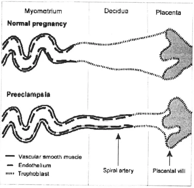
The placenta is considered the pathogenic focus for ail manifestations of PE, because delivery is the only definitive cure for this disease. Defective placentation and placental vascular insufficiency are the starting point of PE. Early in normal gestation, trophoblast celis invade placentai bed, leading to remodeiing of the spiral arteries into maximaily dilated low resistance vascular channels, unable to constrict to vasoactive stimuli [93, 94], thereby guaranteeing a higli flow volume to the uteroplacental bed (see Fig.?) [73]. In women who eventualiy develop PE, the invasion of the uterine spiral arteries is incomplete, with the vessels remaining thick-walled and muscular [95], and with failure
of endothelium-dependent relaxation [96]. The cytotrophoblasts in preeclamptic women
seem to fail to express vascular-type adhesion molecules, which impair their ability to form sufficient connections with the uterine vessels [94. 97, 9$].
Norrna prwnancy
Figure 7. Trophobalst invasion into the spiral arteries in the placental bed in normal pregnancy and in preeclarnpsia. (from [73] VanWijk MJ et ai: Cardiovascular Research 47: 39, 2000.)
The endothelium, a monolayer of epithelial ceils, is in direct contact with the biood and constitutes a physical and metabolic barrier. According to the wide range of functions displayed by endothelial celis, it is an attractive hypothesis that endothelial celi dysfunction plays an important role in the pathophysioiogy of PE. To link the placentai
Prrpê
“I
— Va.wut.r
mXtrL
(flUCkEn&h&tiim
abnormalities to the generalized endothelial dysfunction seen in PE, the existence of a factor X, reieased from the placenta into the maternai circulation, was proposed [73]. The disturbed placentation supposedly leads to hypoperfusion of the placenta and ischemia, resulting in the release of one or more unidentified factors from the placenta. factor X then causes the late vascular dysfunction of PE, consisting mainly of generalized endothelial dysfunction, resulting in vasoconstriction, activation of the coagulation system and redistribution of fluids, the symptoms of PE, and often in fetal growth restriction [73].
Another hypothesis about the etiology of PE postulates that abnormal placentation, with failure of the trophoblast to induce physiological dilation and remodeling of spiral arteries, is caused by a maternai immunological response to foreign paternal antigens of the fetoplacental unit [100]. This theory is supported by the greater incidence of PE among multiparous wornen becoming pregnant with a new partner. Lymphocytes of preeclamptic subjects do not show the cellular hyporesponsiveness to fetal celis that is typical of normal pregnancy [101], the activity of circulating natural killer ceils, neutrophils and cytokines, such as TNF-ct, IL-6, IL-2 and IL-12 is increased [100, 102]. Besides, in PE HLA-G, a surrogate auto-antigen known to prevent recognition by natural killer celis is not expressed as general in the placenta as in normal pregnancy [103]. The resulting activation of leukocytes in the deciduas, can cause release of cytokines, elastase and oxygen free radicals, ail ofwhich cal interact with endothelial function.
A familial factor in the pathogenesis of PE bas been recognized for many years, but the exact mode of inheritance and the interaction between maternai and fetai genotypes are stili under discussion. Several studies have shown an increased frequency of PE in the sisters, daughters and granddaughters, but not the daughters-in-low, of women who have had the disease [104-106]. These data have been interpreted to mean that the putative susceptibility genes act through the mother. More recently it was suggested that PE is a polygenic trait [107]. Implicated in this process are the angiotensin gene, the endotheliai nitric oxide synthase gene, and genes involved in TNF-u-production, thrombophilic disorder, hypertension and obesity [10$-112]. Thus far most studies investigating the role
of genetics in PE have been small scale and a large database of genotypes, present n
women with PE and their chiidren, is needed to elucidate to which extent genetics are involved in PE.
1.4
Experimental Model ofPreeclampsia
PE bas been a recurrent research area for many years. The multitude of systems invoived in the physiopathology of PE and their complex interactions provide an intriguing challenge. A major probiem in PE research is the lack of a valid animal model that can enable a more thorough investigation of physiopathology this disease. Over the past decade, various mammalian moUds have been proposed to study the pathogenesis implicated in PE, including:
1. Inhibition ofnitric oxide (NO) synthesis [40]
2. Reduced ofthe uterine perfusion [41]
3. Transfecting animais with genes ofhuman renin-angiotensin system [46]
4. Stimulating the sympathetic nerve [47]
5. Administration of endotheÏin-l during late pregnancy [4$]
6. Administration of low doses ofendotoxin [49]
Although providing valuable information on relevant mechanisms, these models are not specific to the pregnant condition and occasionally are associated with fetal mortality [42]. Thus, since PE is a pregnancy-specific disease, none of these approaches would represent an adequate animal model.
An ideal animai modei ofPE should fulfiui some criteria:
• It shouÏd exhibit increase in arteriai pressure similar to that occurring in PE. • It should occur oniy in pregnant animal.
• It should manifest many ofthe physiologicai characters ofPE. • The PE-condition induced should be relieved by delivery ofpups. • It should be feasibie in smaii animals, ideally.
1.4.1 High Sodium Intake Model
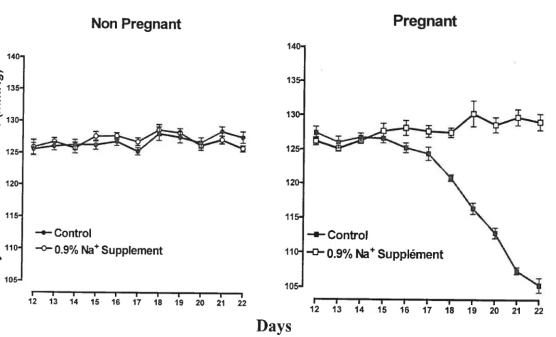
Sodium supplementation during gestation can provide an excellent animal model to study the mechanisrns impiicated in PE. It’s been demonstrated that high sodium intake
(0.9% NaCI) to the rats at the end of gestation, the last 7 days ofthe 22-days pregnancy
in this species, prevented the decrease of blood pressure and reversed the diminish of vascular reactivity normally observed in this period [42,74] (fig.8). In women, blood pressure already decreased by the end of the first trimester and retumed to pre gestational values approaching term [43]. In pregnant rats, blood pressure does flot change until the seventeenth and eighteenth days and then gradualiy decreases until term (23rd days) [44]. Thus, the cardiovascular changes observed in the last week of pregnancy in rats correspond to the ones occurring during the second trimester in pregnant women [42, 74]. Women who subsequently develop PE at the end of gestation commonly do flot show the characteristic decrease in blood pressure at the second trimester.
As shown in Figure 8, before sodium supplementation (day 12 to dayl4), systolic blood pressure was similar in both groups of pregnant rats. Sodium supplement 0.9% did flot affect systolic blood pressure in non-pregnant rats. However, pregnant rats receiving 0.9% NaCI supplement did not show the expected decrease of blood pressure observed in control pregnant rats [42]. It was aiso observed that sodium suppiement of 0.9% NaCI decreased the activity of renin-angiotensin-aldosterone system (RAAS) in non pregnant and pregnant rats. However, the decreased RAAS induced by high sait intake was flot associated with any change in systolic blood pressure in non-pregnant rats [45], but with suppression of the decrease in blood pressure in pregnancy. This resuit suggests that the mechanisms controliing blood pressure are easily perturbed by high sodium intake during pregnancy [42].
Non Pregnant
12 13 14 15 16 17 18 19 20 21 22
Figure 8. Arterial pressure of the rats in normai diet or in diet with sodium
supplement (Adapted from
1421
Beausejour et ai: A.J.P. 285: 11375, 2003)
In a parallel study, contractile response to vasoconstrictors (phenylephrine, KC1, arginine vasopressin) in the aortic rings of normal pregnant and pregnant rats on high sodium intake have also been measured, and increased responses to these vasoconstrictors in aortic rings of pregnant rats on 0.9% sodium supplement compared with pregnant rats on normal water was observed [74]. This demonstrated that augmented sodium intake during gestation in the rat is linked with the reversal of gestational-associated resistance to vasopressors and indicates that this is an experimental model showing some features ofgestational hypertension [74].
Pregnant
140 Q) X 135 E 120 oo
115 o —e—Control ‘ 110 —°—O.9% NaSuppIement >1 cl, 105 140- 135-130— 125- 120- 115- 110-105-Days
-‘-Control ° 0.9% Na Supplément I I I I I I I I I I I 12 13 14 15 16 17 18 19 20 21 221.4.2 Stress in Pregnancy Model
Stress in pregnancy rats was here proposed as animal model for human PE [51]. This model is based on epiderniological data on human populations, showing that stress has unfavorable effects on pregnancy and resuits in a greater incidence of PE and intrauterine growth retardation [52. 53]. In establishing this experimental model, the rats were exposed to some sound stimulus associated with overpopulation in cages between days 7 and 14 over 22 ofpregnancy. Pregnant female Wister rats experienced more manifestations from intense stress than did the non-pregnant animais, such as high arterial blood pressure, increased proteinuria, higher adrenal weight, iower endothelium-derived relaxing factor activity, decreased weight gain, lower fetal weight and greater number of fetuses. The alterations found in the rats were similar to those occurring in human PE [51].
As we know, PE occurs spontaneously only in the human species. Humans are the most intellectually endowed living beings and lead a complicated life in society in which behaviorai rules control most primitive emotional reactions. Thus, in some individuais, many aspects of daily routine may lead to intense chronic stress. Given these considerations, Nitol et ai believe that intense stress during pregnancy may provoke or favor PE [51].
1.4.3 Other Model: Adaptive Transfer ofActivated Thi Celis
Experimentally induced PE on the basis of immunological imbalances associated with endothelial ceIl dysfunction has also been reported. This immunologicai approach proposed that the immune system is important in the course of pregnancy and that it possibly participates in the etiology of PE [50]. This animal model was developed by transferring activated BALB/c Thi-like splenocytes into allogeneically pregnant BALB/c female mice during late gestation (on days 10 and 12 of pregnancy). The authors hypothesized that activated Thi ceils will negatively affect late allogeneic and induce PE like symptoms [50]. The model mimicked the symptoms of PE, i.e. incrcased blood
pressure and glomerulonephritis accompanied by proteinuria, which were flot detectable in non-pregnant recipients of activated Thi -like celis. Adoptive celi transfer adversely affected the outcome of pregnancy by increasing fetal rejection, with uterine immune ceils showing an inflammatory profile. This excessive maternai inflammatory response leads to generalized endothelial ceil dysfunction [50].
The study resuits showed that adoptive transfer of Th 1 -like celis induces an increased cytokine production (TNF- Πand IL-12) and Thi marker (CCR5) in uterine lymphocytes.
Thi cytokines can direcfly damage organs and destroy vessels [57], which is believed to
be induced by direct interaction of immune ceils (secreting Thi cytokines) with the vessels [50]. Before this, it’s also been reported that Thi celis play a detrimental role during mammalian pregnancy [54], [55]. And there is a close relationship between activated Thi immune ceils and PE in hurnans [56].
How the transfer of activated Thi-like ceiis provokes these physiological abnormalities exciusively in pregnant animais? It is believed that since the transferred celis produced predominantly Thi-type cytokines, they expand and stimulate host celis primed to paternal antigens toward the secretion of inflammatory cytokines [58]. Increased secretion of Thi cytokines by activated host celis or, in other words, an inflammatory host response, was in fact proposed to be the main cause leading to PE in humans [59], [60].
In summary, both of the latter two animai models, to some degree, may present some features and physiologicai changes that resemble those obseiwed in PE. But since PE is mainiy characterized by maternai hypertension, which is resulted from enhanced vesse! constriction, and is associated with enhanced vascular reactivity [38] and reduced activity
of RAAS [36], only the first mode! — 0.9% NaC1 supplementation, can represent these
aitered hernodynamic parameters sirnilar to those occurring in PE, and thus make a valuable tooi to study the mechanism implicated in PE [74].
2.
Research Objectives
The evidence that endogenous sodium pump ligands (SPL)/endogenous digitalis-like factors are increased during pregnancy and even more in PE [69,133] suggests that alteration in activity and expression of vascular sodium pump may contribute to these special physiological or pathological states. The purpose of this work is to investigate the inhibitory effects of ligands of the sodium pump and protein expression ofΠsubunit of
the NaIK-ATPase in isolated aorta of rats. The objectives are approached by the following ways:
1. Evaluate the contractile effect of the inhibition on the NaJK-ATPase with
ouabain in thoracic aorta of normal pregnant and experimental preeclamptic rats in normal physiological solution or in reduced potassium medium. We investigated the inhibitory effect of ouabain as the ftrst research object because it is a well-known compound and the first discovered cardiac steroid that selectively and potently blocks the plasmalemmal sodium pump in a variety of ceil types. Ouabain like compound was the first mammalian endogenous digitals-like factor to be purified [31]. The rat vascular sodium pump is very resistant to inhibition by cardiac glycosides ouabain, therefore high concentrations of this compound (0.1-f mM) were needed to observe a clear response [124-126].
2. Measure the inhibitory activity on KC1-induced relaxation by three sodium
pump ligands (ouabain, MBG and digoxin) in a K-free solution. The concentrations used of MBG (0.01-0.1tM) were based on the physiological concentration. Digoxin worked as a reference and similar concentrations (0.1
-1.0 iM) were applied.
3. Measure the protein expression of a-subunit of NaK-ATPase in the aorta of
3.
Materials and Methods
3.1
Animal
Female Sprague-Dawley rats (Charles River Canada. St-Constant, PQ, Canada) aged 10-11 weeks were mated with age-rnatched males. The morning on which vaginal smears were found to contain spermatozoa was labeled Dayl of pregnancy. The pregnant females were then placed in individuai caged until used on the 22d
day of gestation. Virgin rats of the saine age served as controis. Ail the animais were fed a normai diet containing 0.23% NaC1. Control animais (pregnant and non-pregnant) had tap water during ail the treatment period. The experimental group received 0.9% NaC1 solution as a beverage for 7 days, starting on day 15 of experimentation, corresponding to the last week of gestation, these are called experirnental preeclamptic rats. At the end of treatment (day 22 of gestation), the animais were decapitated, and thoracic aorta were coliected rapidly and placed in coid krebs bicarbonate solution (KBS) or stored at -80°C for western blot analysis.
3.2 Materials
Chemicals were purchased from Fisher Scientific (Montreal, PQ, Canada). Ail chemicals were of analyticai grade. Ouabain, Digoxin, Dimethisuifoxide (DMSO), Cocktail Protease Inhibitors and Hepes were purchased from Sigma Chemical Co. (St. Louis, MO), NP-40 Alternative was from Calbiochem. Marinobufagenin (MBG) was kindly provided by Dr. Bagrov (NIH/NIA, Baltimore. MD). Phenylephrine hydrochloride, Carbacol were obtained from Research Biochemical International. Anti-NaIK-ATPase ai, a3 subunit antibodies were purchased from Upstage Biotechnology. Mouse monoclonal [AC-15] to f3-actin-ioading control was from Novus Biologicals. Anti-mouse HRP-linked second antibody, moiecular weight markers and ECL Western blot detection solution were purchased from Amersham Biosciences. The BioRad protein assay kit was from BioRad (Hercuies,CA).
3.3 Organ Bath Assay
Isolated thoracic aorta was cleaned of fat and connective tissues and cut into consecutive
rings of 3-4 mm. The rings were mounted on stainless steel hooks and placed in individualjacketed tissue baths (10 ml; Radnotti Glass, Monrovia, CA) maintained at 37 (+ 0.5) °C and oxygenated. In order to exclusively study the regulation of Na pump in smooth muscle of the vessels, the vascular endothelium was removed by gently rubbing with injection needle.
The aortic rings were equilibrated for 60 min under 2.0 g passive tension with frequent washing and tension adjustment. The tissues were bathed in KBS (PH 7.4) of the following composition: 118 mM NaCY, 4.65 mM KC1, 25 mM NaHCO3, 2.5 mM CaC12, 1.1$ mM MgSO4, 1.18 mM KH7PO4, 5.5 mM dextrose. Normal KBS contained 5.83 mM of K, with reduced K-KBS had 2.0 mM K’. Tension was measured by isometric force displacement transducers (FI-03; Grass Instruments, Quincy, MA) and recorded on a computerized data acquisition system using Work Bench software (Kent Scientific, Litchfield, CT). Figure 9 illustrates the experiment of organ bath assays.
To confirm the removal of endothelium, after equilibration, the tissues were challenged with 1.0 1iM phenylephrine (PhE). At plateau responses, Carbacol (10 1iM) was added. There should be no relaxation present. If any relaxation was observed, the endothelium was further rubbed. The experiments with ouabain were performed under a sodium lamp to prevent photo-degradation of this substance.
3.4 Concentration
—Response Curves to KCI
b evaluate the contractile effect of ouabain on sodium pump, concentration — response
curves to KC1 were measured in the presence and absence of ouabain in normal physiological solution ([Kf] 5.8mM) or in reduced potassium medium ([Kj 2.OmM). Eight aortic rings from each of non-pregnant, pregnant or experimental preeclamptic rats were used and divided into two groups; four rings were incubated in normal KBS and the
1
n
-
C
z z (f2 (f2 r,)other four were bathed in reduced potassium KBS. Afier initial response to PhE and Carbacol, the solution of halfthe rats were changed to Iow potassium KBS. Afler 60 min stabilization period, one ring of each group served as control while the other three were challenged with increasing concentrations of ouabain (0.1, 0.3. 1.0 mM). Fifleen min later, KC1 (2.0 mM to 100 mM) was cumulatively added to the baths (Complete protocol’ see Fig.10). figure 11 shows a typical experimental record ofKCl-induced concentration.
Isolated thoracic aorta was cut into consecutive rings of 3-4 mm,
which were endothelium denuded and installed in individual tissue bath fihled with oxygenated Krebs solution (KBS).
8
The rings were equilibrated for 60 minutes under 2.0 g passive tension. Tension was measured by force-dispiacement transducers and was recorded on a computerized data acquisition system.
8
Afier equilibration, tissues were challenged with phenylephrine 1.0 1iM and carbacol 0. lmM to verify removal of endothelium.
8
After 60 minutes’ relaxation, the aortic rings were incubated with ouabain (0.1, 0.3, 1.0 mM). fifteen minutes later, KCI (2.0 to 100 mM) was cumulatively added to the bath.
3.5 Relaxation Curves to KCI
To further measure the inhibitory activity of Na pump ligands, another indicator of the functional activity of the NaJK-ATPase, K- induced relaxation in a K- free environment
was conducted in aortic rings of pregnant and non—pregnant rats. Afier equilibration, ail
eight rings were exposed to K- free solution. The K- free buffer solution (PH 7.4) was
prepared by substituting KH2PO4 and KC1 with NaH2PO4 and NaCI, respectively, on an equimolar basis. Then, one ring used as control, three were treated with different concentrations ofouabain: 0.01, 0.03, 0.1 mM, two were treated with digoxin I j.tM and 0.1 j.iM and 2 tissues with marinobufagenin (MBG) 0.1 jiM and 0.01 11M. Afier 5 mm, they were made to contract with PhE 1.0 11M. At plateau response, KC1 was gradually re— added into the tissue bath in a cumulated fashion (0.1-6.0 mM) to induce a relaxation curve. The relaxation response afier each addition of KC1 reached a steady state within
3-5 min. In the present experiment, Phenylephrine (lliM) was used for two times. The first
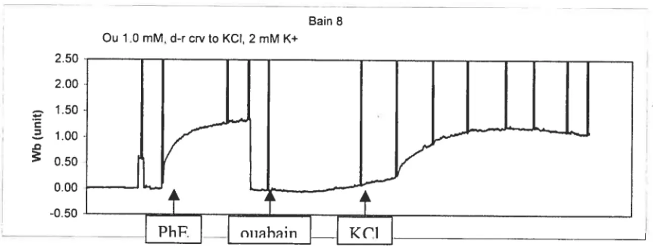
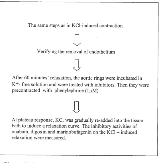
application was to conform the removal of endothelium, and the second one was to produce a contraction before the KC1-induced relaxation. (Complete experimental protocol see fig.12). f igure 13 shows a typical record ofKCl-induced relaxation.
250 Ou 1.0 mM, d-rcrvto KCI, 2 mM K+ Bain 8 C 2.00 1.50 1.00 0.50 0.00 -0.50
The same steps as in KC1-induced contraction
1
Verifying the rernoval of endothelium
8
Afier 60 minutes’ relaxation, the aortic rings were incubated in
K+ free solution and were treated with inhibitors. Then they were
precontracted with phenylephrine (1 tM).
I
At plateau response, KC1 xvas gradually re-added into the tissue bath to induce a relaxation curve. The inhibitory activities of ouabain, digoxin and marinobufagenin on the KCI — induced
relaxation were measured.
Figure 12. Experiment protocol ofKCI-induced relaxation
3.6
Tissue Protein Preparation
Five aortas of non-pregnant, pregnant or experimental PE rats were collected, frozen and powdered in liquid nitrogen. Tissue were homogenized in ice-cold buffer
t
Hepes 20 mM, NP-40 Alternative 1%, PH 7.4) containing 10% cocktail protease inhibitors (Table 2). Nonidet P-40 is a nonionic mild detergent which can solubilize the target proteins in an immunoreactive and undegrated form. The homogenates were centrifuged at 3000 rpm tHermie Z360K) for 10 min at 4°C to get rid of the nuclei and debris. The supematant fraction was recovered and the protein concentration was rneasured by the Bradfrod method (BioRad). Then the quantified protein sampies were mixed with electrophoresisindicator Laernrnli to make a final protein concentration of 1 .5 mM. The extracts were kept ice cold during this experiment and were stored in -20°C for further use.
Bain 3
Oua 003 mM, d-rcrvto KCI, K+ free
%[/_L
•
t
rK7
0.00 -0.50t t
-inhihitnrh
PhF.Figure 13. Experiment record of KC1-induced relaxation
Table 2. Cocktai] Protease Inhibitors
Inhibitors Concentration (mM) Proteases
AE 3Sf 2 Saine
ED+A 1 Metallo
Bestatin 0.13 Amino peptodases
E-64 0.0 14 Cysteines
Leupetine 1 Serin-cysteine
A protein 0.0003 Saine
3.7
Western Blot
Rat kidney microsorne (Upstate Biotechnology) was used as positive control for the al isofonn ofNaJK-ATPase and brain microsome for the a3. The sample was denatured by heating to 100°C for 5 min in boiling water. Then 30 — 45 ig protein of thoracic aorta
(10ig. BioRad) were loaded into wells in a BioRad mini-protean II electrophoresis celi and separated on poÏyacrylarnide gel with Tris-Glycine-SD running buffer (in M: Tris base-0.25, glycine-1.92 and SDS-l%). Electrophoresis lasted for about 30 min at 150 mV until the front of samples ran to the edge of the gels. The gels were removed from the glass plates and transferred to Hybond-ECL nitrocellulose membranes (Amersham Biosciences) for 2 hours at 75mV at 4°C, using a mini Trans-Blot Transfer Celi System (BioRad). The transfer buffer contains (in mM): Tris-base-50, glycine-380 and methano 20%. Then the membrane was blocked overnight at 4°C in TBS-Tween solution (in mM: Tris-base-50, NaCY-150, Tween 20-0.1%, PH 7.5) with 5% powdered non-fat milk. Next, the membrane was incubated and shacked for 60 min at room temperature with primary antibody anti-al (Upstate Bioeclmology) mouse monoclonal IgG (1:4000 dilution), anti a3 (Upstate Bioteclmology) rabbit polyclonal antibody (1:1000 dilution) and J3-actin-loading control (1:15000) (Table 3). The antibody dilutions were optimized to maximize signal and minimize background. Afier washing with TBS-Tween buffer for three times (3x10 min washes), the membranes were incubated for 30 min with second antibody —
anti-mouse (1:2000 dilution) or anti-rabbit (1:4000 dilution) IgG antibody conjugated to horseradish peroxidase (Amersham International). The membranes were thorough washed for two tirnes (each 10 mm) and were visualized with an enhanced chemiluminescence detection kit (Amersham Biosciences) and exposed to ECL Hyperfilm (Amersham Bioscience) for 30s to 60s (f3-actin for 6s). The films were then developed.
Quantification of the relative protein content was determined by scanning the blots and measuring the spot density using Aiphalmager program. Because of differences in background intensity, the abundance of the detected protein was nonnalized to that of
f3-actin.
3.8 Data Ana]ysis
Vasoconstrictor responses induced by KC1 were expressed as a ratio of the contraction to KCI related to that previously obtained with phenylephrine (PhE) 1 iM (data not shown).
Table 3. Western Blot Antibodies
Exposure Time Antibody Dilution (s) al anti-mouse monoclonal 1 :4000 50 Primar’ a3 anti-rabbit polyclonal I : 1000 120 Antibody anti-mouse f3-actin-loading control 1:15000 6 anti-mouse 1:2000 Second Antibody anti-rabbit 1:4000Each concentration-response curve to KC1 was analyzed by computer fitting to a 4-pararneter logistic equation with the program prism 4.0 version (Graphpad software, San Diego, CA) using non-liner regression analysis, to evaluate the concentration producing 50% of the maximal response (EC5O) and the maximum response (Emax). Data are expressed as mean experimental points with their standard error (SEM), together with the best-fitted curve to these points. Statistical significance was determined with a 2-tailed test comparing the means of independent sample groups. In K-induced relaxation curves, 2-way ANOVA was used for comparison of remaining contractions among groups.
b determine the protein expression of a isoforms of the NaIK-ATPase, resuits are
expressed as relative density of protein bands in each isoform to those of Ç3-actin on the same gel. Preliminary experiments showed that increasing the loaded protein concentrations gave proportional f3-actin and al signals. Data were analyzed with Student’s t-test for unpaired experiments. A P value of less than 5% was considered significant.
4.
Results
4.1
Contractile Effect of Ouabain
figure 14 shows contractile responses induced by ouabain in isoÏated aortic rings of non pregnant and pregnant rats with or without sait supplement and obtained in normai ([Kj 5.$3mM) or Ktreduced ([Kj 2.0 mM) physiological solution. In normal physiological medium (Fig.14A), aortic ring contracted to ouabain (0.3 mM) similariy in the different groups except in non-pregnant rats with NaC1 suppiement. Conversely, with 1.0 mM ouabain the response was similar in ail the groups, except pregnant rat on sodium supplement. This suggests that the inhibitory effect of ouabain on the sodium pump is reduced in the experimental preeclarnptic model.
In the presence of K-reduced solution (Fig.14B), the contractile effect of ouabain on aortic ring was increased in ail conditions, except in normai pregnant rats at 1.0 mM, compared to normal physiologicai solution (Fig.14A), but there was no statistical difference between the 2 concentrations of the inhibitor of the sodium pump. In addition, ouabain 0.1 mM induced contraction in each group (except in normal pregnant rats), which was absent in normal physiological medium. These results suggest that the pump
+
activity is increased upon bathing aortic rings in low K solution, and that pregnant animal is resistant to this increase in activity, not the preeclarnptic ones.
4.2
Vasoconstrictory Response to KCI
Tables 4 and 5 report the effects of 0.9% NaCl supplement on maximal responses and sensitivity to KCI without and with ouabain 0.3 or 1.0 mM pre-contraction in aortic rings ofnon-pregnant and pregnant rats in normal and low K physiologicai solution. figurel5 and 16 show the concentration-response curves to KC1. In normal physiological solution, the maximal response to KC1 in the absence of ouabain is reduced in aortic rings of pregnant compared with non-pregnant rats on normal sait intake (ftom 1.14± 0.06 to
A
[K+] 5.83 mM
Ouabain 0.3 mM Ouabain 1.0 mMj
•t’: ç’0 ç.0 C> C> 1.0-0.8 o 4-Q) > 4-0.4 oJ:
1.1-0.9 0.7 0.5 Q) 0.3 o 4-Q 0.1 o -0.1Figure 14.
Contraction induced by ouabain on the aortas of non
pregnant and pregnant rats in normal and increased sodium intake.
Rings were incubated in a solution containing 5.83 mM (A) or 2.0 mM
(B) of [K+].
B
[K+]2.OmM
Ouabain 0.1 mM Ouabain 0.3 mM Ouabain 1.0 mM & C> C> ç.0 ç’O qO) ç.0.98 ± 0.03, P < 0.01, table 4) but no change during 0.9% Naci supplementation.
Sensitivity to KCI is also decreased (from 1.85 ± 0.06 to 1.58 + 0.03, P< 0.01), again with
no change in 0.9% NaCl intake. High salt intake in pregnant rats (experimental preeclamptic rats) did flot affect maximal response or sensitivity to KCÏ.
Concentration-response curves to KC1 that were obtained afier stimulation by ouabain in non-pregnant 0.9% sait intake and normal pregnant rats shifted to the lefi, showing that ouabain potentiated the response to KC1 in these two groups. For example, in the former group, maximal responses to KC1 in the presence of both o.3 and 1.0 mM ouabain significantly increased compared with control (from 1.16 ± 0.04 to 1.40 ± 0.04, P <0.05
and to 1.46 ± 0.05, P < 0.05, respectively, table 4), and the sensitivity to KC1 is also
higher in the pre-incubation of 1.0 mM ouabain (from 1.86 ± 0.04 to 2.04 ± 0.06, P <
0.05). In normal pregnant group, maximal response to KC1 is statisticaily enhanced (from 0.98 + 0.03 to 1.10 ± 0.03, P < 0.05), and sensitivity also increased in the presence of
both 0.3 and 1.0 mM oubain (from 1.58+ 0.03 to 1.70+ 0.02, P <0.05 and to 1.91 + 0.04,
P <0.01, respectively). However, ouabain did flot produce potential effect on reactivity to KC1 in experimental preeclamptic rats.
These data reveai that the aortic reactivity to KC1 was diminished during normal pregnancy but not in PE. Ouabain increased the vascular reactivity to KC1 in non pregnant high sodium intake rats and pregnant rats in regular diet. Experimental PE obliterated this enhanced response to KC1.
In low K solution. similar to normal physiological medium, the maximal response and sensitivity to KC1 are reduced in pregnant rats compared to non-pregnant rats in normal diet (from 1.24 ± 0.04 to 1.03 ± 0.02, P< 0.05, and from 1.76 + 0.03 to 1.54 ± 0.03, P<
0.05, respectively, table 5). But in experimental preeclamptic group, there were no significant changes in maximal response or sensitivity to KC1 compared to normal pregnant rats. In addition, low potassium medium also enhanced the maximal response to KC1 in each group.
Table 4. Parameters of concentration-response curve to KCI in aortic
rings, bathed in normal physiological solution, from non-pregnant and
pregnant rats on normal and increase sodium intake.
Basal responses Emax EC5O
Np Ctl -0.004 + 0.10 (10) 1.14 ± 0.06 0.014 (1.85 ± 0.06) oua0.3 0.04±0.08 (9) 1.11±0.04 0.014(1.85±0.05) oua 1.0 0.34 ± 0.09* (9) 1.24 ± 0.05 0.011 (1.94 ± 0.07) NpO.9% -0.01 ±0.07 (8) 1.16±0.04 0.014(1.86±0.04) oua0.3 0.14±0.07 (8) 1.40±0.04* 0.011(1.96±0.03) oua 1.0 0.36 ± 0.09* (8) 1.46 ± 0.05* 0.009 (2.04 ± 0.060* Pg Ctl -0.02 ± 0.03 (11) 0.98 ± 0.03 0.026 (1.52± 0.03) otia 0.3 0.02 ± 0.04 (11) 0.99 ± 0.03 0.020 (1.70 ± 0.02)* oua 1.0 0.22 ± 0.04* (11) 1.10 ± 0.03* 0.013 (1.91 ± 0.04)* Pg 0.9% 0.00± 0.08 (6) 1.08 ± 0.07 0.024 (1.63 ± 0.09) oua 0.3 0.02 ± 0.10 (6) 1.20± 0.07 0.019(1.72± 0.05) oua 1.0 0.16 ± 0.10 (6) 1.16± 0.08 0.020 (1.69 ± 0.07)
Values are means ± SE (n=$). Basal response is the tone left afier 15 min preincubation
with ouabain; EC5O, in molar concentration with logEC5O in parenthesis; Emax in relative response to PhE (luM). * vs respective control and . vs non pregnant control.
Figure 15. Concentration-response curves to KC1 in aortic rings from
non-pregnant and pregnant rats in normal diet and on 0.9% NaCI
supplement. Rings were incubated in normal physiotogical solution.
Np • ctI D 0.3 mM Ouabain É 1.0 mM Quabain Pg • ctI D 0.3 mM Ouabain 1.0 mM Ouabain 1 .5-w 1.0
-o
o tu 0.5-C) 0.0 -I .5-w 1.0-o o C) > tu 0.5-C) 0.0-3 -2Log [KCI] (moIIL) Np 0.9% NaCI 1 .5-W 1 .0--t o C) > tu 0.5-C) 0.0-1.5 W 1.0 -t
o
o C) > tu 0.5 C) 0.0 • ctI D 0.3mM 1.OmM -3 -2 -1Log [KCI] (mol/L)
Pg 0.9% NaCI
• cu
D 0.3 mM Ouabain 1.0 mM Ouabain
-3 -2 -1
Log
[KCI] (mol/L)-3 -2
Log [KCI] (molIL) —1
![figure 2. Blood pressure profiles (sitting and lying) during pregnancy. (From [38]](https://thumb-eu.123doks.com/thumbv2/123doknet/2055541.5700/21.918.133.814.182.797/figure-blood-pressure-profiles-sitting-lying-pregnancy.webp)
![Figure 4. Putative three-dimensionaÏ model of the topological structure of the NaIK ATPase (From [120] Mobasheri A et ai: Bioscience Reports](https://thumb-eu.123doks.com/thumbv2/123doknet/2055541.5700/25.918.130.654.536.974/figure-putative-dimensionai-topological-structure-mobasheri-bioscience-reports.webp)
![Figure 6. Structure ofthe ouabain analogs (from [99] Manunta P, Hamilton BP, Hamlyn JM: Hypertension 37 (part 2): 472, 2001.)](https://thumb-eu.123doks.com/thumbv2/123doknet/2055541.5700/30.918.254.680.143.792/figure-structure-ouabain-analogs-manunta-hamilton-hamlyn-hypertension.webp)