access to biological analysis for urinary albumin. However, other screening methods such as the Micral-Test II®, based on semiquantitative strip assays, have been developed and proven useful in screening for microalbuminuria. Unfortunate-ly, Clinitek®Microalbumin has not been evalu-ated against the earlier Micral-Test II®strips.
Furthermore, the decision to use a strip for screening for microalbuminuria is dependent on the cost of the strip and the cost of analysing any positive samples using a biological assay. Although the Clinitek®Microalbumin strip appears to be a useful tool for screening for microalbuminuria, further information is needed before it can be utilized in local diabetes clinics.
References
1. Parving H-H, Lehnert H, Brochner-Mortensen J et al. The effect of irbesartan on the development of diabetic nephropathy in patients with type 2 diabetes. N Engl J Med 2001; 345: 870–8.
2. Mogensen CE, Viberti GC, Peheim E et al. Multicenter evaluation of the Micral-Test II test strip, an immuno-logic rapid test for the detection of microalbuminuria. Diabetes Care 1997; 20: 1642–6.
Summary and Comment: Elisabeth Mathiesen, Copenhagen, Denmark
Thymus, central T cell
self-tolerance and type 1
diabetes
Original articles:
Self-antigen-presenting cells expressing diabetes-asso-ciated autoantigens exist in both thymus and periph-eral lymphoid organs. Pugliese A, Brown D, Garza D, Murchison D, Zeller M, Redondo M, Diez J, Eisenbarth GS, Patel DD, Ricordi C. J Clin Invest 2001; 107: 555–64. Differential splicing of the IA-2 mRNA in pancreas and lymphoid organs as a permissive genetic mechanism for autoimmunity against the IA-2 type 1 diabetes autoantigen. Diez J, Park Y, Zeller M, Brown D, Garza D, Ricordi C, Hutton J, Eisenbarth GS, Pugliese A. Dia-betes 2001; 50: 895–900.
Summary
In the first paper, the authors characterized individual cells synthesizing major type 1 dia-betes-associated autoantigens such as
(pro)insulin, glutamic acid decarboxylase and the tyrosine phosphatase-like protein IA-2 in the human thymus and other lymphoid organs
(spleen, lymph nodes). Since these cells also express CD83, CD11c, CD40, CD14, CD80, CD86 and HLA class II, they may be qualified as antigen-presenting cells, mainly dendritic cells (DC) and macrophages. In addition, using the TUNEL (terminal deoxynucleotidyl trans-ferase-mediated dUTP nick-end labelling) method, apoptotic lymphocytes were identified in close vicinity to these cells.
In the second paper, the same group describes a differential splicing of the IA-2 mRNA
between the pancreas and lymphoid organs (thy-mus and spleen). Pancreatic islets express full-length mRNA and two alternatively spliced transcripts. Thymus and spleen exclusively express an alternatively spliced transcript lacking exon 13. This exon of IA-2 encodes for trans-membrane and juxtatrans-membrane domains that contain several target autoantigenic determi-nants of type 1 diabetes. This finding identifies a mechanism of gene expression that could play a permissive role in the development of autoim-munity through the absence of tolerance to IA-2 epitopes not expressed in lymphoid organs.
Comment
There is more and more evidence that the intrathymic expression of tissue-specific self-anti-gens plays a prominent role in the establishment of immunological self-tolerance. In this regard, these two papers lend further support to the con-cept that the central immunological self-tolerance of β-cell identity may be induced during thymic T cell differentiation [1]. The thymus is a very unique organ and constitutes the only site of per-manent confrontation between the repertoire of self-antigens presented by major histocompatibili-ty complex (MHC) proteins and the diversihistocompatibili-ty of T cell receptors for antigen (TCRs) generated by random recombination of gene segments encod-ing a TCR variable part. The thymus is also the major lymphoid organ located at the crossroads between the immune and neuroendocrine sys-tems. In this organ responsible for thymopoiesis (T lymphocyte generation) [2], the neuro-endocrine system regulates the process of T cell differentiation from the very early stages. In addition, inside this organ, which is physically separated from non-self-infectious antigens, T lymphocytes undergo a complex educative process that establishes the central T cell self-tolerance of neuroendocrine principles [3, 4]. Contrary to common assumption, the thymus functions throughout life and plays a fundamen-tal role in the recovery of a repertoire of
15
International Diabetes Monitor
Volume 14, Number 2, 2002
competent T cells after intensive chemotherapy or during highly active antiretroviral therapy [5–7].
From the first paper and a number of other observations [1, 8–11], we may draw some definitive conclusions on the intrathymic expres-sion of insulin-related genes (INS, IGF1 and IGF2). The members of this family are all expressed in the thymus environment according to a precise hierarchy and topography: IGF2 (thymic epithelial/nurse cells [TEC/TNC]) > IGF1 (thymic macrophages) >> INS (thymic DC). At the protein level, this hierarchical pat-tern is of great importance since the tolerogenic response primarily concerns the dominant epi-topes of a protein family [12]. Contrary to (pro)insulin, the blockade of thymic insulin-like growth factor (IGF)-mediated signalling, at the level of the IGF ligands (in particular IGF-II) or IGF receptors, severely interferes with the early stages of T cell differentiation [13].
In the second paper, a regulation in the estab-lishment of immunological self-tolerance is sug-gested by the very interesting observation of a dif-ferential splicing of IA-2 mRNA between thymus
and pancreas. Such a difference explains why some important antigenic determinants derived from IA-2 proteins are not tolerated and become strongly autoantigenic. Such shaping of the self-reactive T cell repertoire has already been shown for a splice variant of a proteolipid self-protein expressed in the murine thymic epithelium [14, 15]. The preprotachykinin A gene (PPT-A) is also transcribed by rat TEC, but thymus-specific mRNA processing gives rise to only one of the PPT-A-encoded peptides (neurokinin A) and not the other (substance P) [16]. With regard to the neurohypophysial genes, oxytocin (OT) and vaso-pressin (VP), both OT and VP transcripts are detected in TEC/TNC from different species. At the peptide level, however, thymic OT concentra-tions are much higher than VP concentraconcentra-tions. Consequently, more and more observations sug-gest the existence of thymus-specific post-tran-scriptional mechanisms [17].
Clinical and pharmacological implications
As already hypothesized by Burnet in 1973 [18], the pathogenesis of autoimmune diseases could form the appearance of ‘forbidden’ self-reactive clones in the peripheral lymphocyte repertoire.
16 International Diabetes Monitor Volume 14, Number 2, 2002 Clinical reviews
Thymus physiology
❖ Negative selection of self-reactive T cells (> 95% of precursor T cells) ❖ Development of T cells self-tolerant and competent against non-self
IGF-II = thymic dominant self-antigen precursor of insulin family
Thymus pathophysiology
❖ Abnormal cytoarchitecture (NOD mice, BB rat, man) ❖ Absence of intrathymic transcription of IGF2
Selection and enrichment of T repertoire with ‘forbidden’ self-reactive T cells to the insulin family
Lymphopenia and regulatory T cell deficiency
❖ Role of environment (thymic innervation, steroids, virus?)
Molecular ‘bridge’ between self-reactive T cells and ß-cells ❖ Genetic susceptibility (role of MHC +++)
❖ Environmental factors: molecular mimicry, nutrition, stress T
➔➔
Langerhans islet β-cells
Fig. 1: Role of the thymus in the development of the autoimmune response against islet insulin-secreting β-cells.Together with abnormalities of thymic cytoarchitecture, the thymus-specific defect of IGF2 expression is responsible both for lymphopenia (with decreased regulatory T cells) and for the absence of central T cell self-tolerance of the insulin family.The influence of environmental factors (such as enterovirus infection) could be exerted at the level of thymic T cell differentiation, as well as in the periphery through the establishment of a bridge between the β-cell autoantigen and ‘forbidden’ self-reactive T cells. NOD, non-obese diabetic; BB, bio-breeding. (Reprinted from [23] with permission from Bentham Science Publishers.)
Since the thymus is the primary site for induc-tion of self-tolerance, thorough investigainduc-tion of a defective thymic censorship should provide the scientific community with important keys to understand the mechanisms underlying the development of autoimmune responses. A num-ber of abnormalities in thymic morphology and cytoarchitecture have been described for several autoimmune disorders. In accordance with this hypothesis, INS transcripts were measured at lower levels in the thymus of human fetuses with short class-I VNTR (variable number of tandem repeats) alleles, a genetic trait of type 1 diabetes susceptibility [19, 20]. The expression of
insulin-related genes was also analysed in the thymus, liver and brain of a common animal model of type 1 diabetes, the biobreeding (BB) rat. A thymus-specific defect of Igf2 expression was evidenced in more than 80% of diabetes-prone BB rats (BBDP) [21]. This defect
explains both the lymphopenia (including a lack of antigen-specific regulatory/suppressive T cells generated in the thymus [22]) and the absence of central self-tolerance of insulin family in BBDP rats. As a consequence of the defective thymic censorship, self-reactive T cells bear-ing TCRs oriented against dominant epitopes of insulin-related peptides continuously migrate from the thymus to the periphery with a
poten-tial cytotoxic power against the islet β-cells. Under certain environmental influences, a mole-cular ‘bridge’ could be installed between the tar-get autoantigenic epitopes leading to activation of such a self-reactive T cell pool and subse-quent β-cell destruction (Fig. 1).
The investigation of neuroendocrine genes expressed in the thymus has led to the identifi-cation of neuroendocrine self-peptides. Through the study of insulin-related gene expression in the thymus, IGF-II — a prominent fetal growth fac-tor — was identified as the dominant self-pep-tide precursor of the insulin family expressed in the thymus. This observation is in close accor-dance with the theory of self-recognition which, according to F.M. Burnet, is not an inherited property but is gradually acquired in the course of fetal life. Although the tolerogenic properties of neuroendocrine self-peptides remain to be further documented, they are strongly suspected from what is known about the tolerance of clas-sic hormones. The development of specific anti-bodies by active immunization (or experimental-ly induced breakdown of tolerance) revealed that tolerance of IGF-II is higher than of IGF-I, and much greater than tolerance of insulin. Insulin is a primary autoantigen tackled by the autoimmune response observed in type 1 dia-betes and this could result from its very low
17
International Diabetes Monitor
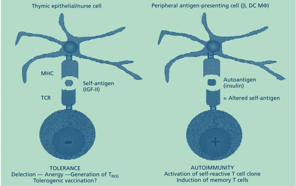
Volume 14, Number 2, 2002 Clinical reviews MHC TCR Self-antigen (IGF-II) Autoantigen (insulin) = Altered self-antigen AUTOIMMUNITY
Activation of self-reactive T cell clone Induction of memory T cells TOLERANCE
Delection — Anergy —Generation of TREG
Tolerogenic vaccination?
Peripheral antigen-presenting cell (β, DC MΦ) Thymic epithelial/nurse cell
expression in the thymus. Moreover, autoimmu-nity has never been observed against IGF-II, and the strong tolerance of this protein, linked to its high expression in the thymus, can be con-sidered as a consequence of the evolutive pres-sure to protect a fundamental process such as fetal development.
Thus, while insulin behaves as an immuno-genic autoantigen of the insulin gene family, IGF-II is the tolerogenic self-peptide precursor of this family. It is now probably appropriate to establish a distinction between an autoanti-gen and a self-antiautoanti-gen. According to this distinc-tion, an autoantigen can be considered as an ‘altered’ self-antigen. Though they are highly homologous, they are not absolutely identical and this biochemical difference theoretically drives opposing immune responses (i.e.
immunogenic vs. tolerogenic responses) (Fig. 2). The powerful tolerogenic properties of the thy-mus should soon be exploited to cure and/or prevent severe autoimmune diseases, such as type 1 diabetes, which constitute the heavy toll paid for the diversity and efficiency of the immune system.
Acknowledgments
Our studies have been supported by the Liège University Special Research Fund, the Fonda-tion Léon Fredericq (Liège University Hospi-tal), the National Fund for Scientific Research (Belgium), the Belgian Federation against Can-cer, the Belgian Diabetes Association, the Euro-pean Association for the Study of Diabetes (Düsseldorf), the Fondation Vaugrenier (Gene-va), and the Juvenile Diabetes Research Federa-tion (New York).
References
1. Geenen V, Achour I, Robert F et al. Evidence that insulin-like growth factor 2 (IGF-2) is the dominant thymic peptide of the insulin superfamily. Thymus 1993; 21: 115–27.
2. Kong FK, Chen CH, Cooper MD. Thymic function can be accurately monitored by the level of recent T cell emigrants in the circulation. Immunity 1998; 18: 514–8.
3. Geenen V, Robert F, Martens H et al. The thymic edu-cation of developing T cells in self neuroendocrine principles. J Endocrinol Invest 1992; 15(8): 621–9. 4. Martens H, Goxe B, Geenen V. The thymic repertoire
of neuroendocrine self-peptides: physiological implica-tions in T-cell life and death. Immunol Today 1996; 17: 312–7.
5. Mackall CL, Fleisher TA, Brown MR et al. Age, thymo-poiesis, and CD4+T-lymphocyte regeneration after
intensive chemotherapy. N Engl J Med 1995; 32: 143–9.
6. Douek DC, Macfarland RD, Keiser PH et al. Changes in thymic function with age and during the treatment of HIV infection. Nature 1998; 396: 690–5.
7. Poulin JF, Viswanathan MN, Harris JM et al. Direct evidence for thymic function in adult humans. J Exp Med 1999; 190: 479–86.
8. Smith KM, Olson DC, Hirose R, Hanahan D. Pancre-atic gene expression in rare cells of thymic medulla: evi-dence for functional contribution to T cell tolerance. Int Immunol 1997; 4: 1355–65.
9. Throsby M, Homo-Delarche F, Chevenne D et al. Pan-creatic hormone expression in the murine thymus: localization in dendritic cells and macrophages. J Clin Endocrinol Metab 1998; 139: 2399–406.
10. Heath VL, Moore NC, Parnell SM, Mason DW. Intrathymic expression of genes involved in organ spe-cific autoimmune disease. J Autoimmunity 1998; 11: 309–18.
11. Kecha O, Martens H, Franchimont N et al. Characteri-zation of the insulin-like growth factor axis in the human thymus. J Neuroendocrinol 1999; 11: 435–40. 12. Sercarz EE, Lehmann PV, Ametani A et al. Dominance
and crypticity of T cell antigenic determinants. Annu Rev Immunol 1993; 11: 729–66.
13. Kecha O, Brilot F, Martens H et al. Involvement of insulin-like growth factors in early T cell development: a study using fetal thymic organ cultures. Endocrinology 2000; 141: 1209–17.
14. Klein L, Klugmann M, Nave KA et al. Shaping of the autoreactive T cell repertoire by a splice variant of self protein expressed in thymic epithelial cells. Nat Med 2000; 6: 56–61.
15. Klein L, Kyewski B. ‘Promiscuous’ expression of tissue antigens in the thymus: a key to T-cell tolerance and autoimmunity? J Mol Med 2000; 78: 483–94. 16. Ericsson A, Geenen V, Frobert F et al. Expression of
preprotachykinin-A and neuropeptide-Y messenger RNA in the thymus. Mol Endocrinol 1990; 4: 1211–8. 17. Geenen V, Kecha O, Martens H. Thymic expression of
neuroendocrine self-peptide precursors: role in T cell survival and self-tolerance. J Neuroendocrinol 1998; 11: 811–22.
18. Burnet FM. A reassessment of the forbidden clone hypothesis of autoimmune diseases. Aust J Exp Biol Med Sci 1973; 50: 1–9.
19. Vafiadis P, Bennett ST, Todd JA et al. Insulin expres-sion in human thymus is modulated by INS VNTR alleles at the IDDM2 locus. Nat Genet 1997; 15: 289–92.
20. Pugliese A, Zeller M, Fernandez A Jr et al. The insulin gene is transcribed in the human thymus and transcrip-tion levels correlate with allelic variatranscrip-tion at the INS VNTR-IDDM2 susceptibility locus for type 1 diabetes. Nat Genet 1997; 15: 293–7.
21. Kecha-Kamoun O, Achour I, Martens H et al. Thymic expression of insulin-related genes in an animal model of autoimmune type 1 diabetes. Diabetes Metab Res Rev 2001; 17: 146–52.
22. Shevach EM. Regulatory T cells in autoimmunity. Annu Rev Immunol 2000; 18: 423–9.
23. Geenen V, Martens H, Brilot F et al. Central self-toler-ance by thymic presentation of self-antigens and autoimmunity. Curr Med Chem Immunol Endocr Metab Agents 2001; 1(1): 47–60.
Summary and Comment: Vincent Geenen, Liège, Belgium
18 International Diabetes Monitor Volume 14, Number 2, 2002 Clinical reviews