J. Clin. Invest.
© The American Society for Clinical Investigation, Inc. 0021-9738/96/04/1924/07 $2.00
Volume 97, Number 8, April 1996, 1924–1930
Stromelysin-3 Expression Promotes Tumor Take in Nude Mice
Agnès C. Noël,* Olivier Lefebvre,* Erik Maquoi,‡ Leen VanHoorde,§ Marie-Pierre Chenard,¶ Marc Mareel,§ Jean-Michel Foidart,‡ Paul Basset,* and Marie-Christine Rio*
*Institut de Génétique et de Biologie Moléculaire et Cellulaire (IGBMC), Centre National de la Recherche Scientifique/Institut National de la Santé et de la Recherche Médicale U184/Université Louis Pasteur, BP 163, 67404 Illkirch Cedex, C.U. de Strasbourg, France;
‡Laboratory of General Biology, University of Liège, 4000 Sart-Tilman, Liège, Belgium; §Laboratory of Experimental Cancerology,
University Hospital, Ghent, B-9000, Belgium; and ¶Service d’Anatomie Pathologique Générale, Centre Hospitalier Universitaire de
Hautepierre, 67098 Strasbourg Cedex, France
Abstract
Stromelysin-3 (ST3) is a matrix metalloproteinase ex-pressed in human carcinomas in ways suggesting that it may play a role in tumor progression. To test this possibil-ity, we have performed gene transfer experiments using both anti-sense and sense ST3 expression vectors, and ma-lignant cells either expressing (NIH 3T3 fibroblasts) or not (MCF7 epithelial cells) endogenous ST3. We have com-pared the ability of parental and transfected cells to cause subcutaneous tumor development in nude mice. 3T3 cells expressing anti-sense ST3 RNA showed reduced tumorige-nicity, and MCF7 cells expressing mouse or human ST3 were associated with reduced tumor-free period leading to a significant increased tumor incidence (P, 1024). However,
once established, the ST3-expressing tumors did not grow faster than those obtained with the parental MCF7 cell line. In addition, tumors obtained after sub-cutaneous injection of ST3-expressing or nonexpressing cells did not exhibit ob-vious histological differences, and careful examination did not reveal any local invasive tissue areas nor systemic me-tastases. These in vivo observations were in agreement with those obtained in vitro showing that ST3 expression did not modify proliferative nor invasive properties of transfected cells. Altogether, these results indicate that ST3 expression promotes tumor take in nude mice, presumably by favoring cancer cell survival in a tissue environment initially not per-missive for tumor growth. These findings represent the first experimental evidence showing that ST3 can modulate can-cer progression. (J. Clin. Invest. 1996. 97:1924–1930.) Key words: cancer cell invasion/survival • metalloproteinases • tumorigenicity
Introduction
Matrix metalloproteinases (MMPs)1 are members of a family
of zinc-dependent endopeptidases with a broad spectrum of proteolytic activities toward extracellular matrix (ECM) com-ponents (1–3). These enzymes include collagenases,
gelati-nases, stromelysins, and membrane-type MMPs (1–7). They are expressed as proenzymes which are processed to active forms through proteolytic cleavage, and their activities are controlled by physiological tissue inhibitors of metalloprotein-ases (TIMPs). It has been proposed that imbalance between MMPs and TIMPs is implicated in various physiological and pathological tissue remodeling processes, notably those enabling tumor progression and leading to metastasis (8–9). Experi-mental evidences supporting this possibility were presented by Khokha et al. (10) who demonstrated that the down-regulation of TIMP-1 levels by anti-sense (AS) RNA transfection in 3T3 cells increased their tumorigenic and metastatic potential in nude mice. Inversely, the secretion of functional TIMP-2 in stably transfected cells was found to result in a marked de-crease in tumor growth rate and local invasiveness (11). More recently, matrilysin over-expression in human prostate cancer cells was reported to increase their invasive potential in SCID mice (12). Gene transfer experiments using both sense (S) and AS expression vectors have also shown that the level of matri-lysin expression was a critical factor in the aptitude of colon cancer cells to migrate through basement membrane-like ma-trices in vitro (13) and to generate tumors in vivo, in nude mice (14). Similarly, the expression of gelatinase A or B was found to confer a metastatic phenotype to transfected transformed cells (15, 16).
Stromelysin-3 (ST3) was first described in fibroblastic cells of invasive breast carcinoma and classified as a member of the MMP family on the basis of sequence homologies (17). ST3 ex-pression was thereafter observed in most other types of human invasive carcinomas and in some precursor lesions (18, 19, and references therein). Strong ST3 gene expression has been cor-related with increased local aggressiveness of tumors (20, 21), and high levels of ST3 RNA were found to be predictive of re-currence in breast carcinomas (22). ST3 over-expression was also shown to be associated with tissue remodeling occurring in physiological conditions such as embryonic development (23), amphibian metamorphosis (24), mammary gland involu-tion (25), wound healing (20), and cycling endometrium (26). Although these observations suggest that ST3 may play a role in ECM remodeling, putative mature forms of human ST3 (hST3) appear unable to degrade any major ECM component (27, 28). Furthermore, it has been recently demonstrated that ST3 was predominantly secreted as a potentially active form, pro-ST3 being intracellularly processed by a furin-dependent proteolytic cleavage (29). ST3 thus differs from other MMPs which must be activated extracellularly, indicating that ST3 A. Noël and O. Lefebvre should be considered as equal first authors.
Address correspondence to Marie-Christine Rio, IGBMC, BP 163, Illkirch Cedex, 67404, France. Phone: 34-24; FAX: 88-65-32-01.
Received for publication 27 November 1995 and accepted in re-vised form 31 January 1996.
1. Abbreviations used in this paper: AS, anti-sense; ECM, extracellular matrix; h, m ST3: human, mouse stromelysin-3; MMP, matrix metal-loproteinase; S, sense; S.C., subcutaneous; TIMP, tissue inhibitor of metalloproteinase.
may have a unique role in tissue remodeling processes, includ-ing those associated with tumor progression.
In the present study, we have subcutaneously (S.C.) in-jected nude mice with malignant cells stably transfected with either AS or S ST3 cDNA constructs, in order to evaluate the effect of ST3 expression on tumorigenicity in vivo. Using the same cells, we have also tested whether ST3 could modulate cell proliferation and invasion in vitro.
Methods
Construction of anti-sense and sense ST3 expression vectors. The pCMVAS-mST3 plasmid was obtained by inserting in the AS orientation, a 360-bp BamHI fragment (nucleotides 1 to 360) of mouse ST3 (mST3) cDNA (24) into the pCMV vector in which expression is under the control of the cytomegalovirus (CMV) promoter (provided by B. Vo-gelstein, Baltimore, MD) (30). To generate the pCMVmST3 plasmid, a 1504-bp NsiI fragment, containing the entire coding sequence of mST3, was excised and ligated to a 8-mer BamHI–NsiI adaptor and subcloned in the S orientation into the BamHI site of the pCMV vec-tor. To generate the pCMVhST3 plasmid, a 1680-bp SpeI–XhoI frag-ment of hST3 cDNA (17), containing the entire coding sequence, was inserted in S orientation into the compatible NheI/XhoI sites of the pCMV vector modified by insertion of a polylinker containing BamHI, NheI, and XhoI restriction sites. To generate the pSG5hST3 plasmid, a 1681 bp EcoRI fragment of hST3 cDNA was inserted in S orientation into the EcoRI site of the pSG5 vector in which expres-sion is under the control of the SV40 promoter (31).
Obtention of stably transfected cell lines. Malignant NIH 3T3 (ob-tained from B. Pettmann, Strasbourg, France) (32) and MCF7 (ATCC HTB 22) cells were cultured in Dulbecco’s modified Eagle’s medium (DME) containing 10% fetal calf serum. 3T3 cells were electropo-rated with linearized pCMVASmST3 construct (10 mg) or linearized pCMV vector alone (10 mg), using a BioRad apparatus at 400 volts and 125 mF. Similar conditions were used for MCF7 cell transfection with linearized pCMVmST3 or pCMVhST3 S constructs, or linear-ized pCMV vector alone. Linearized pSG5hST3 S construct (8 mg) was co-transfected into MCF7 cells with linearized pSV2neo vector (ATCC 37149) (2 mg). Transfectants were then selected with the neo-mycin analogous G418 (400 µg/ml; GIBCO BRL, Gaithersburg, MD) through the neomycin-resistance gene of the pCMV vector, or through the cotransfected pSV2neo vector.
RNA isolation and analysis. Total RNA was prepared from cul-tured cells using acid guanidinium thiocyanate-phenol-chloroform extraction (33). RNAs were fractionated by electrophoresis through 1% agarose gel in the presence of formaldehyde, and transferred to nylon membranes (Hybond N; Amersham). Filters were acidified for 10 min in 5% CH3COOH and stained for 10 min (0.004% methylene blue, 0.5 M CH3COONa, pH 5.0) before hybridization. Northern blots were hybridized under stringent conditions (50% formamide, 428C) with mST3 (24), hST3 (17) and 36B4 (34) cDNA probes 32P-labeled
by random priming. Washings were performed in 2 3 SSC, 0.1% SDS at 228C, followed by 0.1 3 SSC, 0.1% SDS at 558C.
Protein analysis. Conditioned media were obtained by incubating subconfluent cells in serum-free DME. After 48 h incubation, media were collected, cleared by centrifugation at 5000 g for 30 min and pre-cipitated at 48C with 80% ammonium sulfate. After centrifugation at 10000 g for 2 h, protein pellets were resuspended in 50 mM Tris-HCl (pH 7.4), 100 mM NaCl, 5 mM CaCl2, 1 µM ZnCl2 and dialyzed
against this buffer at 48C, for 3 h. Protein concentrations were deter-mined using the BioRad kit (BioRad Laboratories). Protein samples were analyzed by SDS-PAGE (12%) under reducing conditions. For immunoblot analysis, proteins were transferred after electrophoresis to nitrocellulose filters which were incubated with monoclonal anti-body 5ST-4C10 which recognizes the ST3 catalytic domain (35). Bound antibodies were visualized using a peroxidase-labeled goat
an-tibody raised against mouse IgG, followed by Enhanced Chemilumi-nescence detection (ECL kit; Dupont NEN, Boston, MA).
Tumorigenicity assay. Subconfluent NIH 3T3 cells were trypsinized, centrifugated at 1000 g for 5 min, and resuspended in serum-free DME. A total volume of 0.2 ml containing 5 3 105 cells was S.C.
in-jected into 6–8-week-old female nude MF1 nu/nu mice (Harlan, France). Subconfluent MCF7 cells were trypsinized, washed twice, and harvested by centrifugation at 1000 g for 5 min. Cells resus-pended in cold serum-free DME were mixed with an equal volume of cold matrigel (10 mg/ml) prepared from the Engelbreth-Holm-Swarm tumor, as previously described (36). A total volume of 0.5 ml containing 5 3 104 cells was S.C. injected into 6–8-week-old female
nude mice, previously implanted with Silastic capsules (Dow Corn-ing) containing estradiol (37).
Injected mice were examined every 2 d for tumor apparition and tumor volume was calculated as previously described (37). Tumor in-cidence was defined as the percentage of mice presenting a tumor, in considering only tumors having a volume of at least 100 mm3. The
data were statistically analyzed with the log Rank test (38). P values lower than 0.05 were considered as significant. All mice were autop-sied. One half of each tumor was immediately frozen in liquid nitro-gen for RNA analysis. The second tumor half, the lungs and the liver were fixed in phosphate buffered formalin (4%) and embedded in paraffin. Histological examination was performed on hematoxilin-eosin stained sections. Immunohistochemical localization of mST3 was performed using polyclonal antibody 612, obtained by rabbit im-munization with recombinant mST3 extracted from bacterial inclu-sion bodies (28), whereas the immunohistochemical localization of hST3 was performed using rabbit polyclonal antibody 349 as previ-ously described (21). Tissue sections were also stained with a rabbit polyclonal antibody raised against human pS2 (39), in order to check for the presence of metastatic MCF7 cells in mouse lungs and liver.
Cell proliferation and in vitro invasion assays.Cell proliferation was evaluated by counting trypsinized cells every 2 d using a Coulter counter (Coultronics France).
Chemoinvasion assays (40) were performed using Transwell chambers (Costar Corp., Cambridge, MA) with polycarbonate mem-brane filters (6.5 mm diameter, 8 mm pore size) coated with matrigel (50 mg/filter) (35). Cells (3 3 104) were harvested by trypsinization,
resuspended in serum-free DME containing 0.2% bovine serum albu-min (ICN, Flow) and added to the upper compartment of Transwell chambers. The lower compartment was filled with 600 ml DME con-taining 20% fetal calf serum and 2% bovine serum albumin as chemoattractants. After 48 or 72 h of incubation at 378C, filters were rinsed in phosphate buffered saline, fixed in paraformaldehyde 4% and permeabilized in ice-cold methanol before staining with hema-toxylin-eosin. Cells on the upper face of the filters were wiped away with a cotton swab, and those on the lower face were counted on 24 fields/filter at a magnification of 3400. Three independent experi-ments were conducted, each using three Transwell chambers.
For collagen invasion assays, cells were seeded onto a polymer-ized type I collagen gel. After 24 h incubation at 378C, the number of cells that had invaded into the collagen gel was counted as previously described (41).
Invasion assays into chick heart were based on the in vitro con-frontation between cell aggregates and precultured chick heart frag-ments in organ culture (42). The interaction between tested cells and chick heart fragments was evaluated by microscopic examination of serial sections fixed in Bouin Holland’s solution, embedded in paraf-fin, and stained with hematoxylin and eosin.
Results
Decreased tumor incidence using cells down-regulated for mST3 expression. A malignant mouse NIH 3T3 fibroblastic cell line, which constitutively expresses mST3 RNA (Fig. 1, lane 1), was stably transfected with the pCMVASmST3 vector (see
Meth-ods) in order to downregulate the expression of the mST3 en-dogenous gene. Different neomycin resistant clones were es-tablished. Three of them were found to express AS transcripts with the expected size, either at high (3T3/7.5 and 3T3/4.6) or low (3T3/3.6) levels (Fig. 1, lanes 4–6). The other clones, in-cluding the 3T3/2.1 and 3T3/1.1 clones, did not express AS transcripts (Fig. 1, lanes 7 and 8). In addition, the NIH 3T3 cell line was transfected with the pCMV vector alone and gave rise to the 3T3/16.1 and 3T3/14.3 control clones (Fig. 1, lanes 2 and 3).
Nude mice were S.C. inoculated with an identical number of cells of each clone. After 3 wk, tumors were observed in 50 and 20% of injection sites for the parental NIH 3T3 cell line, and the 3T3/16.1 control cells, respectively (Table I). At 6 wk, the tumor incidence for these 2 cell lines reached 80% and
100%, respectively, demonstrating their high propensity to im-plant and proliferate into nude mice. Surprisingly, the second control clone, 3T3/14.3, did not give rise to any tumor until 6 wk after cell injection, and led to a tumor incidence of only 14% at 8 wk (Table I). However, as shown in Fig. 1 (lane 3), the endogenous mST3 RNA level of this clone was much lower than those of the other 3T3 clones, suggesting clonal variation in the parental 3T3 cell population, or that a re-combination event leading to decreased ST3 gene expression, has occurred during transfection of these cells. Among the
pCMVASmST3 transfected cells, the clones 3T3/3.6, 3T3/2.1 and 3T3/1.1, which expressed very low levels or no AS mST3 RNA (Fig. 1, lanes 6–8), behaved similarly to the parental cell line, although tumor appearance was slightly delayed with the 3T3/1.1 clone, which exhibited a tumor incidence of 71% after 8 wk of observation (Table I). The two other clones (3T3/7.5 and 3T3/4.6), however, exhibited a different pattern. First, no tumor could be detected 3 wk after cell injection. Second, tu-mor numbers were still low at 6 (14–20%) and at 8 (30–57%) weeks (Table I). Since these clones expressed the highest lev-els of AS mST3 RNA (Fig. 1, lanes 4 and 5), these findings sug-gested that decreased mST3 expression could explain their reduced tumorigenicity. Low levels of mST3 secretion by pa-rental 3T3 cells together with a poor stability of mST3 (28, 43) did not permit to precisely quantify the decrease in mST3 se-cretion in 3T3 cells transfected with AS mST3 RNA. However, using Western blot analysis, we could detect a low molecular weight form of mST3 at 28 kD in 4 out of 10 samples of culture media conditioned by parental 3T3 cells, but in none (0 out of 10) of those obtained from 3T3/7.5 transfected cells (data not shown).
To further investigate the contribution of ST3 to tumorige-nicity, we performed reverse experiments in which MCF7 cells, which do not express the endogenous ST3 gene (Fig. 2 A, lane 1), were stably transfected with a S mST3 cDNA in order to produce recombinant mST3.
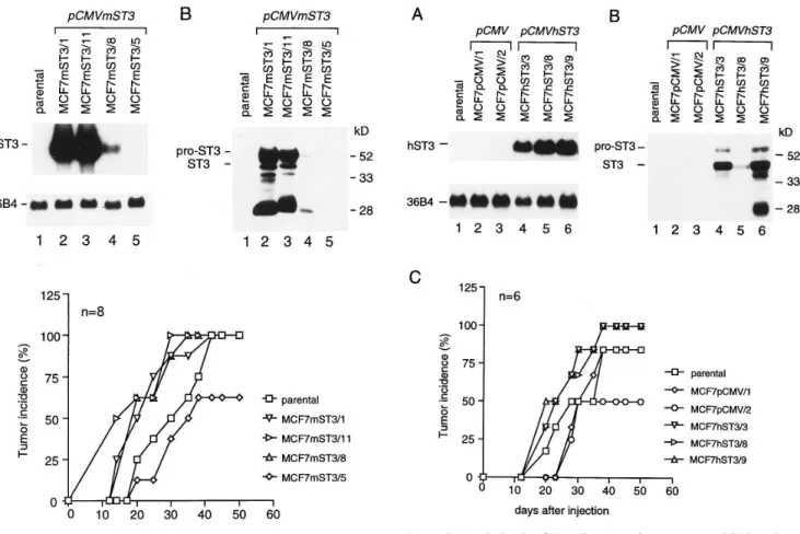
Increased tumor incidence using cells over-expressing mST3. MCF7 breast cancer cells were stably transfected with the pCMVmST3 vector (Methods), and neomycin resistant clones were screened for expression of mST3 using Northern blot analysis. Three clones were found to express high (MCF7mST3/1 and MCF7mST3/11; Fig. 2 A, lanes 2 and 3) or Figure 1. Northern blot analysis of NIH 3T3 cells transfected with an
AS mST3 cDNA construct. (Lane 1) parental cells; (lanes 2 and 3) pCMV transfected cells; (lanes 4–8) pCMVASmST3 transfected cells. The position of the endogenous sense (S) and exogenous anti-sense (AS) mST3 transcripts are indicated. The 36B4 probe was used as a control for loading and transfer; autoradiography was for 2 d (mST3) and 18 h (36B4).
Table I. Tumorigenicity of Parental, pCMV and pCMVASmST3-transfected NIH 3T3 Cells, after S.C. Injection into Nude Mice
Clones Transfected vector S RNA* AS RNA*
Tumor incidence after injection‡
3 wk 6 wk 8 wk 3T3 2 1 2 5/10 (50%) 8/10 (80%) 9/10 (90%) 3T3/16.1 pCMV 1 2 1/5 (20%) 5/5 (100%) 5/5 (100%) 3T3/14.3 pCMV 1/2 2 0/7 (0%) 0/7 (0%) 1/7 (14%) 3T3/7.5 pCMVASmST3 1 1 0/10 (0%) 2/10 (20%) 3/10 (30%) 3T3/4.6 pCMVASmST3 1 1 0/7 (0%) 1/7 (14%) 4/7 (57%) 3T3/3.6 pCMVASmST3 1 1/2 3/7 (43%) 7/7 (100%) 7/7 (100%) 3T3/2.1 pCMVASmST3 1 2 6/12 (50%) 9/12 (80%) 11/12 (92%) 3T3/1.1 pCMVASmST3 1 2 1/7 (14%) 3/7 (43%) 5/7 (71%)
*S and AS mST3 RNA expression was evaluated by Northern blot analysis as illustrated in Fig. 1; (1), high, (1/2), low and (2), undetectable levels. ‡Each ratio in the table represents the number of tumors larger than 100 mm3 vs. the total number of injected mice, at the indicated time after cell in-jection; corresponding percentages are indicated in parentheses.
moderate (MCF7mST3/8; Fig. 2 A, lane 4) levels of mST3 RNA, whereas we could not detect any mST3 RNA in the MCF7mST3/5 clone (Fig 2. A, lane 5), even after a longer ex-posure time (data not shown). By Western blot analysis, we could detect mST3 protein in culture media conditioned by MCF7mST3/1, MCF7mST3/11 and MCF7mST3/8 cells (Fig. 2
B, lanes 2–4), but not by MCF7mST3/5 cells (Fig. 2 B, lane 5). Both pro- and mature mST3 forms were detected together with some degradation products, including the 28-kD form.
In vivo tumorigenicity of these cells was tested by S.C. in-jection in nude mice. As shown in Fig. 2 C, the tumor incidence in mice injected with MCF7 cells expressing mST3 was signifi-cantly increased (log Rank test, P, 2 3 1025). Thus, 20 d after cell injection, MCF7mST3/1, MCF7mST3/11 and MCF7mST3/ 8 cells gave rise to a tumor incidence of 50–62%, whereas pa-rental MCF7 cells and MCF7mST3/5 cells led to a tumor inci-dence of 25 and 12%, respectively. This difference in tumor in-cidence, which was still observed 35 d after cell injection, was no longer apparent after 40 d when all cells, excepting MCF7mST3/5, led to 100% tumors. Thus, the variation in tu-mor incidence which was observed in these experiments in fact
corresponds to a reduction of the tumor-free period when mST3-expressing cells were S.C. injected in mice.
These findings were in good agreement with our hypothesis that mST3 could promote tumorigenicity in nude mice. How-ever, since differences have been observed between the func-tional properties of mST3 and hST3 (28, 43), we tested whether similar results could be obtained using MCF7 cells expressing hST3.
Increased tumor incidence using cells over-expressing hST3.
Using the same protocol as described above, we obtained 3 MCF7 clones stably transfected with the pCMVhST3 vector and expressing hST3 RNA (MCF7hST3/3, MCF7hST3/8 and MCF7hST3/9; Fig. 3 A, lanes 4–6). These cells secreted both pro- and mature hST3 in their culture media (Fig. 3 B, lanes 4–6). In addition, we obtained two control clones (MCF7pCMV/1 and MCF7pCMV/2) transfected with the pCMV vector alone, which did not express hST3 (Fig. 3, A and B, lanes 2 and 3). As observed for mST3, hST3 expression was found to promote tu-mor incidence in nude mice (log Rank test, P , 4 3 1024) (Fig. 3 C). Similar results were also obtained using a clone (MCF7hST3/13) stably transfected with a construct in which hST3 was expressed under the control of the less potent SV40 Figure 2. Analysis of MCF7 cells expressing exogenous mST3 under
the control of the CMV promoter. (A) Northern blot analysis: autora-diography was for 1 day (mST3) and 18 h (36B4). (B) Western blot analysis of conditioned media: the positions of molecular weight stan-dards, and of pro- and mature ST3 are indicated on the right and left, respectively. (A and B) lane 1, parental cells; lanes 2–5, pCMVmST3 transfected cells. (C) tumor incidence after S.C. injection of parental and pCMVmST3 transfected MCF7 cells; n indicates the number of injected mice for each cell clone.
Figure 3. Analysis of MCF7 cells expressing exogenous hST3 under the control of the CMV promoter. (A) Northern blot analysis: autora-diography was for 1 d (hST3) and 18 h (36B4). (B) Western blot anal-ysis of conditioned media: the positions of molecular weight stan-dards, and of pro- and mature ST3 are indicated on the right and left, respectively. (A and B) lane 1, parental cells; lanes 2–3, pCMV trans-fected cells; lanes 4–6, pCMVhST3 transtrans-fected cells. (C) tumor inci-dence after S.C. injection of parental, pCMV and pCMVhST3 trans-fected MCF7 cells; n indicates the number of injected mice for each cell clone.
promoter of the pSG5 vector, instead of the CMV promoter. Low levels of hST3 RNA (Fig. 4 A, lane 2) and of the hST3 28kD form (Fig. 4 B, lane 2) could be detected in MCF7hST3/13 cells and culture media, respectively. Despite this low level of hST3 expression, MCF7hST3/13 cells led to an increased tumor inci-dence (log Rank test, P , 4 3 1025). Thus, 20 d after cell
injec-tion, 75% of mice injected with MCF7hST3/13 cells exhibited tumors whereas parental MCF7 cells gave rise to only 25% (Fig. 4 C).
ST3 over-expression did not modify cell growth and inva-siveness. To define what could be the process leading to a
re-duction of the tumor-free period and thus to an increased tumor incidence after S.C. injection of MCF7 cells expressing either mST3 or hST3, we first evaluated tumor growth and cell prolif-eration rates. We observed that once the tumors were estab-lished, the evolution of tumor volumes was identical from one
cell clone to another, independently of the levels of ST3 ex-pression. Consistently, no major differences were observed for the in vitro cell proliferation rates whether or not the trans-fected cells expressed mST3 or hST3 (data not shown).
We next tested the invasive properties of transfected MCF7 cells, both in vivo and in vitro. All clones grew as well delineated tumor masses, and no evidence of local invasion could be detected regardless of the tumor size. Moreover, careful anatomopathological examination of mice did not re-veal any lung or liver metastases, even after pS2 immunostain-ing allowimmunostain-ing the detection of isolated MCF7 cells (39). No his-tological differences, notably concerning their vascular status, were observed between tumors obtained in nude mice after in-jections of either parental MCF7 cells or cells transfected with
pCMV, pCMVmST3 or pCMVhST3. Although the tumor
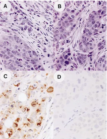
stroma was not as well developed as that of human breast car-cinomas, we could observe areas where tumoral epithelial cells were surrounded by fibroblastic cells after injection of ST3-expressing (Fig. 5 A) or control MCF7 cells (Fig. 5 B). Endoge-nous mST3 could not be detected in these fibroblasts, using an-tibody 612 directed against mST3 (data not shown). MCF7 cells expressing recombinant ST3 protein were found to be ho-mogeneously distributed throughout tumors, and staining was restricted to the cytoplasm of transfected MCF7 cells (Fig. 5 C). Northern blot (data not shown) and immunohistochemical Figure 4. Analysis of MCF7 cells expressing exogenous hST3 under
the control of the SV40 promoter. (A) Northern blot analysis: autora-diography was for 4 d (hST3) and 18 h (36B4). (B) Western blot anal-ysis of conditioned media: the positions of molecular weight stan-dards are indicated on the left. In panels A and B: lane 1: parental cells; lane 2, pSG5hST3 transfected cells. (C) tumor incidence after S.C. injection of parental and pSG5hST3 transfected cells; n indicates the number of injected mice for each cell clone.
Figure 5. Histological and immunohistological analyses of tumors de-rived from MCF7hST3/9 and control MCF7pCMV/1 cell clones. Tu-mor sections were stained with hematoxylin and eosin (A and B) (X 60), or immunostained with polyclonal antibody 349 raised against hST3 (C and D) (360). (A and C) hST3-expressing MCF7hST3/9 cells; (B and D) control MCF7pCMV/1 cells.
(Fig. 5, C and D, and data not shown) analyses showed that the levels of tumoral exogenous mST3 and hST3 RNAs and pro-teins were in good agreement with those observed for the cor-responding MCF7 clones, in vitro. Finally, the invasive proper-ties of control and ST3-expressing cells were compared in the type I collagen (41) and chick heart (42) invasion assays. The expression of exogenous ST3 did not modify cell invasiveness. Thus, neither ST3-expressing nor control cells invaded type I collagen gels, and they resulted in the development of an orga-nized non-invasive epithelioid layer at the periphery of the heart tissue. We also tested the capacity of parental and trans-fected cells to pass through matrigel-coated filters by using Transwell chamber (40). Once again, no significant differences were observed between cells expressing or not mST3 or hST3, suggesting that ST3 expression did not confer cell invasive properties, in these in vitro assays (data not shown).
Discussion
In the present study, we investigated the role of ST3 in the de-velopment of tumors after S.C. injection of malignant cells in nude mice. We demonstrated that the incidence of tumors ob-tained using malignant mouse fibroblastic NIH 3T3 cells, which endogenously express ST3, was decreased when high levels of AS mST3 RNA were expressed in these cells. In-versely, we observed that human epithelial MCF7 breast can-cer cells, which do not express endogenous ST3, showed signif-icant increased tumorigenicity in nude mice when stably transfected in order to produce mST3 (P , 2 3 1025). Similar observation was made using MCF7 cells stably transfected in order to express hST3 at either high levels under the control of the CMV promoter (P , 4 3 1024) or at lower levels under the control of the SV40 promoter (P , 4 3 1025). These studies showed that the increased tumor incidences induced by either mST3 or hST3 resulted from a shortening of tumor-free peri-ods indicating that, in nude mice, ST3 is involved in tumor for-mation rather than in tumor growth.
How could ST3 modulate tumor development in nude mice? In vitro analyses of cell proliferation did not show any obvious differences between ST3-expressing and non-express-ing cells, suggestnon-express-ing that the observed increase in tumor inci-dence could not be ascribed to an increased growth rate of ST3-expressing cells. Consistently, once the tumors were es-tablished in nude mice, their proliferation rates appeared to be independent of their levels of ST3 expression. In addition, ST3 does not seem to be implicated in cell invasion, since MCF7 cells expressing ST3 did not exhibit any modifications of their capacity to invade interstitial-like or basement membrane ma-trices. Accordingly, no local invasion or metastatic spreading was detected by histological analysis of tumors, lungs and liv-ers of nude mice injected with ST3-expressing MCF7 cells. Taken together, these findings indicate that ST3 expression did not favor tumor incidence in nude mice by increasing cell proliferation or invasion, but rather by increasing tumor take. This is consistent with our finding that the increase in tumor incidence observed for ST3-expressing MCF7 cells was more pronounced in the first weeks after cell injection. Thus, we propose that ST3 contributes to cell survival and implantation in host tissues. This hypothesis may appear paradoxical with regards to previous observations showing that ST3 was ex-pressed during processes involving epithelial cell apoptosis such as mouse mammary gland involution (25), mouse limb,
tail and snout morphogenesis (23) and frog metamorphosis (24). However, while unwanted cells are eliminated during ap-optotic events, some neighboring cells are selected for survival (44 and references therein). In this respect, we note that ST3 gene expression is also observed at the time of epithelial cell proliferation occurring in the neoformation of mammary ducts following mammary gland involution (25), in developing limb, tail and snout until birth (23), and in proliferative endometrium (26). Interestingly, comparable observations have been re-ported for matrilysin, another MMP implicated in cancer pro-gression (1, 3). Thus, while SW480 colon cancer cells stably transfected to express matrilysin did not show in vitro any modification of their proliferative or invasive properties, these cells exhibited increased tumorigenicity in vivo, suggesting that the most significant role of matrilysin was at early stage of tumor progression (14).
Tumors that are obtained by S.C. injection of malignant cells into nude mice should be regarded as a model for meta-static implantation rather than for primary tumor development (45). Indeed, S.C. injected cells mimic metastatic cells after ex-travasation, when these cells have to successfully implant at the metastatic site before being able to proliferate and invade (45, 46). This process is dependent upon the cells’ capability to survive in host tissues, either in an independent manner or by the recruitment of required factors from their near vicinity. In this context, ST3 may represent a local factor contributing to the survival and implantation of ST3-expressing MCF7 cells S.C. injected in nude mice. It appears reasonable to believe that this might be also the case in human tumors, where ST3 is produced by tumor stromal cells and not by the cancer cells themselves (17–22). ST3 is a secreted protein which is specifi-cally expressed by fibroblastic cells located in the vicinity of cancer cells, suggesting that the same effects should be ob-served irrespective of the source of the protein. Thus, in hu-man carcinomas ST3 could act as a paracrine host factor con-tributing to cancer cell survival outside of their compartment of origin. Consistently, in normal embryonic and adult tissues ST3 expression has been reported to occur during tissue re-modeling processes, notably when, as in malignant processes, the integrity of the basement membrane which separates epi-thelial cell compartments from mesenchymal cells showed fail-ure, leading to epithelial/stromal cell contacts (20, 23, 25, 26). Although further studies are required to evaluate this possibil-ity, a role for ST3 in favoring cell survival is consistent with ob-servations demonstrating that high ST3 expression levels were associated with metastatic propensity in human carcinomas (22, 47).
Acknowledgments
We thank P. Chambon for his support and interest in this study. We acknowledge I. Stoll, C. Wendling and S. Vicaire for excellent techni-cal assistance, J. Byrne for crititechni-cal reading of the manuscript and B. Vrijens for statistical analysis.
This work was supported by funds from the Institut National de la Santé et de la Recherche Médicale, the Centre National de la Recherche Scientifique, the Centre Hospitalier Universitaire Régional, the Mu-tuelle Générale de l’Education Nationale, the Groupement de Re-cherches et d’Etudes sur les Génomes (Grant 94/50), the Association pour la Recherche sur le Cancer, the Ligue Nationale Française con-tre le Cancer and the Comité du Haut-Rhin, the Fondation pour la Recherche Médicale Française, the Fondation de France, and a grant to P. Chambon from the Fondation Jeantet. O. Lefebvre was a
recipi-ent of a Ph.D. studrecipi-entship from the Association pour la Recherche sur le Cancer. A. Noël was a recipient of post-doctoral fellowships from the European Community, the Fondation Léon Frédericq, the Fondation Rose et Jean Hoguet, the Fondation cancérologique St. Michel and the Fondation Braconier-Lamarche, all from Belgium.
References
1. Matrisian, L.M. 1992. The matrix-degrading metalloproteinases.
BioEs-says. 14:455–463.
2. Docherty, A.J.P., J. O’Connell, T. Crabbe, S. Angal, and G. Murphy. 1992. The matrix metalloproteinases and their natural inhibitors: prospects for treating degenerative tissue diseases. Trends Biotechnol. 10:200–207.
3. Birkedal-Hansen, H. 1995. Proteolytic remodeling of extracellular ma-trix. Curr. Opin. Cell Biol. 7:728–735.
4. Freije, J.M.P., I. Diez-Itza, M. Balbin, L.M. Sanchez, R. Blasco, J. To-livia, and C. Lopez-Otin. 1994. Molecular cloning and expression of collage-nase-3, a novel human matrix metalloproteinase produced by breast carcino-mas. J. Biol. Chem. 269:16766–16773.
5. Sato, H., T. Takino, Y. Okada, J. Cao, A. Shinagawa, E. Yamamoto, and M. Seiki. 1994. A matrix metalloproteinase expressed on the surface of invasive tumour cells. Nature (Lond.). 230:61–65.
6. Will, H., and B. Hinzmann. 1995. cDNA sequence and mRNA tissue dis-tribution of a novel human matrix metalloproteinase with a potential trans-membrane segment. Eur. J. Biochem. 231:602–608.
7. Takino, T., H. Sato, A. Shinagawa, and M. Seiki. 1995. Identification of the second membrane-type matrix metalloproteinase (MT-MMP-2) gene from a human placenta cDNA library. J. Biol. Chem. 270:23013–23020.
8. Alexander, C.M., and Z. Werb. 1991. Extracellular matrix degradation.
In Cell Biology of Extracellular Matrix. E.D. Hay, editor. Plenum Press, New
York. 8:255–302.
9. Stetler-Stevenson, W.G., S. Aznavoorian, and L.A. Liotta. 1993. Tumor cell interactions with the extracellular matrix during invasion and metastasis.
Annu. Rev. Cell. Biol. 9:541–573.
10. Khokha, R., P. Waterhouse, S. Yagel, P.K. Lala, C.M. Overall, G. Norton, and D. Denhardt. 1989. Antisense RNA induced reduction in murine TIMP levels confers oncogenicity on Swiss 3T3 cells. Science (Wash. DC). 243: 947–950.
11. DeClerck, Y.A., N. Perez, H. Shimada, T.C. Boone, K.E. Langley, and S.M. Taylor. 1992. Inhibition of invasion and metastasis in cells transfected with an inhibitor of metalloproteinases. Cancer Res. 52:701–708.
12. Powell, W.C., J.D. Knox, M. Navre, T.M. Grogan, J. Kittelson, R.B. Na-gle, and G.T. Bowden. 1993. Expression of the metalloproteinase matrilysin in DU-145 cells increases their invasive potential in severe combined immunodefi-cient mice. Cancer Res. 53:417–422.
13. Yamamoto, H., F. Itoh, Y. Hinoda, and K. Imai. 1995. Suppression of matrilysin inhibits colon cancer cell invasion in vitro. Int. J. Cancer 61:218–222.
14. Witty, J.P., S. McDonnell, K.J. Newell, P. Cannon, M. Navre, R.J. Tressler, and L.M. Matrisian. 1994. Modulation of matrilysin levels in colon car-cinoma cell lines affects tumorigenicity in vivo. Cancer Res. 54:4805–4812.
15. Docherty, A.J.P., M.I. Cockett, M.L. Birch, S. Chander, N. Willmott, J.P. O’Connell, T. Crabbe, A. Mountain, J.R. Morphy, T.A. Millican et al. 1994. Gelatinase inhibitors for treatment of cancer. Clin. Exp. Metast. 12:12.
16. Bernhard, E.J., S. Gruber, and R.J. Muschel. 1994. Direct evidence link-ing expression of matrix metalloproteinase 9 (92 kDa gelatinase/collagenase) to the metastatic phenotype in transformed rat embryo cells. Proc. Natl. Acad. Sci.
USA. 91:4293–4297.
17. Basset, P., J.P. Bellocq, C. Wolf, I. Stoll, P. Hutin, J.M. Limacher, O.L. Podhajcer, M.P. Chenard, M.C. Rio, and P. Chambon. 1990. A novel metallo-proteinase gene specifically expressed in stromal cells of breast carcinomas.
Na-ture (Lond.). 348:699–704.
18. Rouyer, N., C. Wolf, M.P. Chenard, M.C. Rio, P. Chambon, J.P. Bel-locq, and P. Basset. 1994. Stromelysin-3 gene expression in human cancer: An overview. Invasion Metastasis. 14:269–275.
19. Basset, P., J.P. Bellocq, P. Anglard, M.P. Chenard, O. Lefebvre, A. Noël, A. Okada, N. Rouyer, M. Santavicca, I. Stoll, C. Wolf, and M.C. Rio. 1995. Stromelysin-3 in breast cancer: the importance of epithelial-stromal inter-actions during tumor progression. In Breast Cancer: Cellular and Molecular Bi-ology. Vol. V. R.B. Dickson and M.E. Lippman, editors. Boston: Kluwer Aca-demic Publishers. In press.
20. Wolf, C., M.P. Chenard, P. Durand de Grossouvre, J.P. Bellocq, P. Chambon, and P. Basset. 1992. Breast-cancer-associated Stromelysin-3 gene is expressed in basal cell carcinoma and during cutaneous wound healing. J.
In-vest. Dermatol. 99:870–872.
21. Muller, D., C. Wolf, J. Abecassis, R. Millon, A. Engelmann, G. Bronner, N. Rouyer, M.C. Rio, M. Eber, G. Methlin, P. Chambon, and P. Basset. 1993. Increased stromelysin-3 gene expression is associated with increased local inva-siveness in head and neck squamous carcinomas. Cancer Res. 53:165–169.
22. Engel, G., K. Heselmeyer, G. Auer, M. Backdahl, E. Eriksson, and S. Linder. 1994. Correlation between stromelysin-3 mRNA level and outcome of
human breast cancer. Int. J. Cancer. 58:830–835.
23. Lefebvre, O., C. Régnier, M.-C. Chenard, C. Wendling, P. Chambon, P. Basset, and M.C. Rio. 1995. Developmental expression of mouse stromelysin-3 mRNA. Development. 121:947–955.
24. Patterton, D., W. Pär Hayes, and Y.B. Shi. 1995. Transcriptional activa-tion of the matrix metalloproteinase gene stromelysin-3 coincides with thyroid hormone-induced cell death during frog metamorphosis. Dev. Biol. 167:252–262. 25. Lefebvre, O., C. Wolf, J.M. Limacher, P. Hutin, C. Wendling, M. LeMeur, P. Basset, and M.C. Rio. 1992. The breast cancer-associated strome-lysin-3 gene is expressed during mouse mammary gland apoptosis. J. Cell Biol. 119:997–1002.
26. Rodgers, W.H., L.M. Matrisian, L.C. Giudice, B. Dsupin, P. Cannon, C. Sviteck, F. Gorstein, and K.G. Osteen. 1994. Patterns of matrix metalloprotein-ase expression in cycling endometrium imply differential functions and regula-tion by steroid hormones. J. Clin. Invest. 94:946–953.
27. Pei, D., G. Majmudar, and S.J. Weiss. 1994. Hydrolytic inactivation of a breast carcinoma cell-derived serpin by human stromelysin-3. J. Biol. Chem. 269:25849–25855.
28. Noël, A., M. Santavicca, I. Stoll, C. L’Hoir, A. Staub, G. Murphy, M.C. Rio, and P. Basset. 1995. Identification of structural determinants controlling human and mouse stromelysin-3 proteolytic activities. J. Biol. Chem. 270: 22866–22872.
29. Pei, D., and S. Weiss. 1995. Furin-dependent intracellular activation of the human stromelysin-3 zymogen. Nature (Lond.). 375:244–247.
30. Baker, S., S. Markowitz, E.R. Fearon, J.K.V. Willson, and B. Vogel-stein. 1990. Suppression of human colorectal carcinoma cell growth by wild-type p53. Sciences (Wash. DC). 249:912–915.
31. Green, S., I. Issemann, and E. Sheer. 1988. Versatile in vivo and in vitro eukaryotic expression vector for protein engineering. Nucleic Acids Res. 16:369. 32. Janet, T., G. Labourdette, and B. Pettmann. 1992. Mitogenic growth factors regulate differentially early gene mRNA expression: a study on two clones of 3T3 fibroblasts. Exp. Cell Res. 198:305–314.
33. Chomczynski, P., and N. Sacchi. 1987. Single-step method of RNA isola-tion by acid guanidinium thiocyanate-phenol-chloroform extracisola-tion. Anal.
Bio-chem. 162:156–159.
34. Masiakowski, P., R. Breathnach, J. Block, F. Gannon, A. Krust, and P. Chambon. 1982. Cloning of cDNA sequences of hormone-regulated genes from MCF7 human breast cancer cell line. Nucleic Acids Res. 10:7895–7903.
35. Santavicca, M., A. Noël, M.P. Chenard, Y. Lutz, I. Stoll, J. P. Segain, N. Rouyer, C. Wolf, J.P. Bellocq, and P. Basset. 1995. Characterization of mono-clonal antibodies against stromelysin-3 and their use to evaluate stromelysin-3 levels in breast carcinoma by semi-quantitative immunohistochemistry. Int. J.
Cancer. 64:336–341.
36. Noël, A., A. Callé, H. Emonard, B. Nusgens, J.M. Foidart, and C.M. Lapière. 1991. Lack of invasion of reconstituted basement membrane matrix by tumor cells. Cancer Res. 51:405–414.
37. Noël, A., N. Simon, J. Raus, and J.M. Foidart. 1992. Basement mem-brane components (matrigel) promote the tumorigenicity of human breast ade-nocarcinoma MCF7 cells and provide an in vivo model to assess the responsive-ness of cells to estrogen. Biochem. Pharmacol. 43:1263–1267.
38. Cox, D.R. 1972. Regression models and life tables. J. R. Statist. Soc. 187: 187–220.
39. Rio, M.C., J.P. Bellocq, B. Gairard, U.B. Rasmussen, A. Krust, C. Koehl, H. Calderoli, V. Schiff, R. Renaud, and P. Chambon. 1987. Specific pression of the pS2 gene in subclasses of breast cancers in comparison with ex-pression of the estrogen and progesterone receptors and the oncogene ERBB2.
Proc. Natl. Acad. Sci. USA. 84:9243–9247.
40. Hendrix, M.J.C., E.A. Seftor, R.E.B. Seftor, and I.J. Fidler. 1987. A sim-ple quantitative assay for studying the invasive potential of high and low meta-static variants. Cancer Lett. 38:137–147.
41. Vakaet, L., K. Vleminckx, F. Van Roy, and M. Mareel. 1991. Numerical evaluation of the invasion of closely related cell lines into collagen type I gels.
Invasion Metastasis. 11:249–260.
42. Mareel, M., J. Kint, and C. Meyvisch. 1979. Methods of study of the in-vasion of malignant C3H mouse fibroblasts into embryonic chick heart in vitro.
Virshows Arch. B: Cell Pathol. 30:95.
43. Murphy, G., J.P. Segain, M. O’Shea, M. Cockett, C. Ionnou, O. Lefeb-vre, P. Chambon, and P. Basset. 1993. The 28-kDa N-terminal domain of mouse stromelysin-3 has the general properties of a weak metalloproteinase. J. Biol.
Chem. 268:15435–15441.
44. Raff, M.C., B.A. Barres, J.F. Burne, H.S. Coles, Y. Ishizaki, and M.D. Jacobson. 1993. Programmed cell death and the control of cell survival: lessons from the nervous system. Science (Wash. DC). 262:695–700.
45. Mareel, M., P. DeBaetselier, and F.M. Van Roy. 1990. Bioassays for in-vasion and metastasis. In Mechanisms of Inin-vasion and Metastases. CRC Press, Boca Raton, FL. 41–72.
46. Rusciano, D., and M.M. Burger. 1992. Why do cancer cells metastasize into particular organs? BioEssays. 14:185–194.
47. Porte, H., E. Chastre, S. Prevot, B. Nordlinger, S. Empereur, P. Basset, P. Chambon, and C. Gespach. 1995. Neoplastic progression of human colorec-tal cancer is associated with overexpression of the stromelysin-3 and BM-40/ SPARC genes. Int. J. Cancer. 64:70–75.