THÈSE
En vue de l’obtention du
DOCTORAT DE L’UNIVERSITÉ DE TOULOUSE
Délivré par :Université Toulouse 3 Paul Sabatier (UT3 Paul Sabatier)
Présenté et soutenu par :
Jin-Hui WANG
Le 29 septembre 2017
Titre
:Rhenium complexes based on triazolyl derivatives:
from synthesis, structural and theoretical characterization to application as
radiopharmaceuticals or fluorophores
Ecole doctorale et discipline ou spécialité :
ED SDM : Chimie Organométallique et de coordination – CO043
Unité de recherche :
LSPCMIB - UMR CNRS 5068
Directeur(s) de Thèse :
M. Eric BENOIST
Rapporteurs :
M. Ali OUADI, IR-HDR CNRS, Institut Pluridisciplinaire Hubert CURIEN, Strasbourg M. Federico CISNETTI, MCF-HDR, Université Clermont Auvergne
Autre(s) membre(s) du jury :
Acknowledgement
First and foremost I want to thank my advisor Prof. Eric Benoist for his support of my PhD study and related research. I appreciate his contribution of time, idea and supervision to make my PhD experience interesting and full of challenges. He is patient, knowledgeable and gives me lot of suggestions for writing the thesis. He also helps me a lot in daily life, otherwise I could not adapt to the country so quickly.
I would like to express my sincere gratitude to my thesis collaborator Dr. Suzanne Fery-Forgues. She has taught me how to manage multi-subjects and contributed so much as an advisor. She is humorous, active, productive and well planned. Her joy and enthusiasm for research is contagious and inspires me to move forward even in tough times.
The members of SOMAB team have left me a deep impression both in personally and professionally. I got not only advice and collaboration of experiments but also friendship. I am especially grateful to Nadine Leygue who treats me as a friend and family member. I would like to acknowledge these honorary team members, Atriz, Romain and Gaetan. They are smart, creative and full of passion. You will never feel boring or dull when with them. They also help me settle many daily issues in the lab and in life.
I am beholden to many other members of the lab: Jean-Marc and Isabella give many suggestions in the synthesis and purification methods. Nathalie and Tessa keep the regular supply of the lab. Muriel did many administrative things for me. Great thanks should be also given to Morgane, Beartrice, Corinne, Chantal and Claude Picard. They all accompanied me in the three years.
In regards to the crystals, I would like to thank Sonia Mallet-Ladeira andNathalie Saffon in X-ray diffraction Service where the X-ray structures were measured. The computational studies discussed in this thesis would not be possible without the calculation of Dr. Mariusz Wolff at University of Silesia (Poland). I also appreciate Nicolas Lepareur from the Nuclear Medicine Department of Centre Eugène Marquis (Rennes, France). He and Romain carried out the radiolabeling experiments.
My sincere gratitude goes to two reporters of my committee members: Federico Cisnetti and Ali Ouadi, for their time, interest and insightful comments. I am also greatly indebted to the two examiners of my jury: Christine Goze and Fabienne Alary, for their time and questions which widen my view of research.
I gratefully acknowledge the funding of China Scholarship Council that made my PhD possible. Thanks Service de l'Education de l'Ambassade de Chine who affords much convenience and offers tremendous help to me.
My time in Toulouse was of great joy due to the many friends. I appreciate the time spent with them: the memory of travel with Yayu, the experience of exercise with Lin and the regular activities with Longqi, Zhihua, Chongwei and Qinghuage. My time in France was also enriched by Mengchen, Lin Liu, Xu and Juan. Thanks for their great help and friendship, which is an invaluable treasure in my life.
At the end, I would like to express my greatest thanks to my parents who raised me up and taught me with wisdom and full of love. They always lighten the way when I am lost.
Chapter I. Literature Review and Project Objectives
... 51. Radioactive rhenium complexes as radiopharmaceuticals ... 6
1.1. Rhenium (and technetium) radiolabeling ... 6
1.2. Design of Re-based radiopharmaceuticals ... 8
1.2.1. Re(V) complexes as potential radiopharmaceuticals ... 9
1.2.2. Re(I) complexes as potential radiopharmaceuticals ... 12
2. Design of photoactive tricarbonyl rhenium complexes for applications in imaging and therapy ... 14
2.1. Rhenium(I) complexes ... 14
2.2. As imaging agents ... 14
2.2.1. Photophysical properties of Re(I) complexes ... 14
2.2.2. Unconjugated complexes for imaging applications ... 17
2.2.3. Bioconjugated complexes ... 18
2.2.4. Sensing ... 19
2.2.5. Correlations with radioimaging studies ... 19
2.2.6. Bimodal infrared and luminescence imaging ... 20
2.3. From photoactivity to the use as phototherapeutic agents ... 21
2.3.1. Photocytotoxicity and generation of active oxygen species ... 21
2.3.2. Photogeneration of carbon monoxide ... 21
3. Click chemistry, “Click-to-Chelate” concept, Click, then Chelate approach... 28
3.1. Copper(I)-catalyzed azide-alkyne cycloaddition (CuAAC reaction) ... 28
3.1.1. Copper catalysts ... 29
3.1.2. The CuAAC mechanism ... 32
3.1.3. Experimental conditions vs. azodiphobia ... 33
3.2. Click-to-Chelate ... 34
3.2.2. Inverse/regular click ligands ... 36
3.2.3. Extension of the “Click-to-Chelate” approach ... 37
3.3. Examples of technetium- and rhenium-specific click ligands ... 40
3.3.1. Tricarbonyltechnetium(I) complexes via a “Click-to-Chelate” approach ... 40
3.3.2. Tricarbonylrhenium(I) complexes via a Chelate, then Click approach ... 43
4. Project of the thesis... 45
4.1. Based on a N2O tripodal system ... 45
4.2. Based on a pyta scaffold ... 46
5. References ... 49
Chapter II. Development of a New Family of Semi-rigid Multidentate
Ligands for
99mTc/Re Complexation
... 571. Design, synthesis and reactivity of multidentate ligands with rhenium(I) and rhenium(V) cores ... 60
1.1. Introduction ... 60
1.2. Syntheses of the ligands and their corresponding rhenium complexes ... 61
1.2.1. Syntheses of the ligands ... 61
1.2.2. Syntheses of rhenium complexes ... 67
1.3. Tricarbonylrhenium(I) complexes: structural and computational studies ... 68
1.3.1. X-ray diffraction study ... 71
1.3.2. Optimized structural geometries ... 75
1.4. Oxorhenium(V) complex: structural characterization and first 99mTc-radiolabeling 77 1.4.1. Structural characterization ... 77
1.4.2. Preliminary 99mTc-radiolabeling ... 78
1.5. Conclusion ... 79
2. Dirhenium(I) hexacarbonyl complexes bridged by a 1,2,3-triazole ligand: Synthesis, structural and spectroscopic characterization ... 81
2.1. Introduction ... 81
2.3. Structural characterization and computational studies ... 84
2.3.1. Spectroscopic analysis ... 84
2.3.2. X-ray diffraction ... 87
2.3.3. Computational studies ... 90
2.4. Conclusion ... 966
3. Tricarbonylrhenium(I) complexes functionalized by a nitro or metronidazole group as potential imaging agents for hypoxia ... 97
3.1. Introduction ... 97
3.2. Syntheses and characterization ... 102
3.2.1. Syntheses of the ligands ... 102
3.2.2. Syntheses and characterization of corresponding rhenium complexes ... 104
3.3. Electrochemical studies ... 107
3.4. Conclusion ... 109
4. Experimental Section ... 111
4.1. Materials and equipment ... 111
4.2. Syntheses ... 111
4.2.1. Syntheses for Part 1 ... 111
4.2.2. Syntheses for Part 2: Dirhenium(I) complexation ... 122
4.2.3. Syntheses for Part 3 ... 123
4.3. X-ray crystallography and data analysis ... 126
4.4. Computational details ... 126
4.5. Radiochemistry ... 127
4.6. Electrochemical studies ... 128
5. References ... 129
Chapter III. Aggregation-Induced Emission in Tricarbonyl Rhenium
Complexes Based on a Benzoxazole-Substituted Pyridyl-Triazole Ligand
. 134 1. Introduction... 1352. Studies of complexes ReL8 and ReL9: Influence of the pyta ligand’s structural
isomerism ... 139
2.1. Syntheses of the ligands and corresponding rhenium complexes ... 139
2.2. Structural characterization ... 140
2.2.1. Spectroscopic and spectrometric analysis ... 140
2.2.2. X-ray diffraction ... 142
2.3. Computational studies ... 146
2.4. Electrochemical studies ... 155
2.5. Spectroscopic properties ... 159
2.5.1. UV-vis absorption spectroscopy of the four compounds in solution ... 160
2.5.2. Emission spectroscopy of ligands L8 and L9 in solution ... 160
2.5.3. Fluorescence emission of complexes ReL8 and ReL9 in dilute solution with excitation around 300 nm ... 162
2.5.4. Fluorescence emission of ReL8 in methanol and acetonitrile with excitation at 370 nm ... 163
2.5.5. Aggregation-induced phosphorescence emission in concentrated solution .. 163
2.5.6. Aggregation-induced phosphorescence emission in the solid-state ... 165
2.6. Energy diagrams and photophysical processes ... 166
2.7. Evaluation of cytotoxicity and cell uptake ... 168
2.8. Summary ... 171
3. Synthesis and spectroscopic studies of PBO-based 1,2,4-triazole derivatives and corresponding Re(I) complexes ReL10 and ReL11: toward improved photoluminescent properties ... 172
3.1. Structural description ... 172
3.2. Synthesis of the ligands and their corresponding rhenium complexes ... 172
3.3. Structural characterization ... 174
3.3.1. Spectroscopic and spectrometric analysis ... 174
3.3.2. X-ray diffraction ... 175
3.4.1. UV-visible absorption spectra in solution ... 178
3.4.2. Emission spectra in dilute solutions ... 179
3.4.3. Emission spectra in concentrated solution ... 180
3.4.4. Emission spectra in solid state ... 181
4. Conclusion ... 183
5. Experimental Section ... 185
5.1. Materials and Equipment ... 185
5.2. Experimental Procedure ... 185
5.2. X-ray crystallography ... 191
5.3. Computational details ... 192
5.4. Electrochemistry ... 192
5.5. Spectroscopy and photophysics ... 193
5.6. Cytotoxicity and dye uptake evaluation ... 194
6. References ... 196
Introduction
Despite the progress achieved in modern medicine and treatment methods, human malignant tumor diseases still threaten human life expectancy. According to the International Agency for Research on Cancer,i about 14.1 million new cancer cases and 8.2 million cancer-related deaths occurred in 2012. Additionally, the number of new cancer cases per year is predicted to increase to 19.3 million by 2025. So, in the last decades, massive research efforts have been focused on early diagnosis and efficient treatment.
To diagnose accurately the disease stage, numerous imaging techniques have been applied, including X-ray radiography, Magnetic Resonance Imaging (MRI), ultrasound and nuclear medicine imaging that uses specific radiotracers so-called radiopharmaceuticals.ii A huge advantage of the latter imaging modality is that it can easily be adapted to therapeutic purposes. Whereas the radionuclides used for nuclear medicine imaging emit gamma rays, which can penetrate deeply into the body and consequently give a whole-body image, the radionuclides used for targeted therapy must emit radiation with a relatively short path length ( - or emitter) in order to only irradiate the cancer cells. Among the radioisotopes that can be used, technetium-99m ( -emitter) and rhenium-188 (β--emitter) represent an interesting pair of radionuclides for diagnosis and therapy, the technetium being the most widely used for medical applications. Both compounds belong to the group 7 of the Periodic Table and have very similar coordination behaviors. Moreover, non-radioactive rhenium complexes have drawn a lot of attention due to their rich photophysical and photochemical properties that make them attractive luminescent imaging probes and photoactive agents for the controlled generation of carbon monoxide (PhotoCORMs). Therefore, a number of research groups, including ours, have been committed to the development of new rhenium complexes as a new class of imaging/therapeutic agents.
Among the many oxidation states of rhenium (ranging from –I to VII), Re(I) and Re(V) have been the most extensively investigated, in particular the [Re(CO)3]+ and [ReO]3+ cores, respectively. Until now, a large number of Tc/Re-specific chelators for the stabilization of both
i GLOBOCAN 2012 project: Estimated Cancer Incidence, Mortality and Prevalence worldwide in 2012 (see http://globocan.iarc.fr/Default.aspx).
ii Radiopharmaceuticals have been defined as radioactive drugs that, when used for the purpose of diagnosis or therapy, typically elicit no physiological response from the patient (definition from the Nuclear Medicine
cores have been described. Their structure, their denticity and the nature of the donor atoms depend on the considered rhenium cores. Among the plethora of synthetic strategies reported for the preparation of such chelators, the “Click-to-Chelate” concept, developed in 2006 by Schibli’s groupiii has especially attracted our attention. This group has showed that the 1,4-disubstitued 1,2,3-triazole obtained by click chemistry was an efficient chelator for the [Re(CO)3]+ core. Following this result, we developed several ligands including a triazole ring in the chelating cavity for the development of imaging and therapeutic agents based on tricarbonyltechnetium-99m and tricarbonylrhenium cores, respectively.iv
The present work is a continuation of our group’s investigations about Tc/Re-complexes. Based on the “Click-to-Chelate” or the analogous “Chelate, then Click” approach, our objective is to design a new series of click ligands for [Re(CO)3]+ and [ReO]3+ cores, and to assess their potential as imaging/therapeutic probes.
Before detailing our results, the first chapter presents a bibliographic state of the art related to our research project. In a first part, we outline the interest of rhenium complexes and provide a quick overview of their use as radiopharmaceuticals and a detailed review on their photophysical behavior. Focusing on the [ReO]3+ and [Re(CO)
3]+ cores, we give many examples of applications as imaging and therapeutic agents in the biomedical field. The main emphasis is placed on identifying the characteristic features that make the most efficient compounds. In a second part, particular attention is paid to synthesis and to the advantages of the copper(I)-catalyzed azide-alkyne cycloaddition so-called CuAAC reaction, including the elegant “Click-to-Chelate” or Chelate, then Click approaches for the conception of Re/Tc-specific click ligands.
The second chapter is focused on the design of new radiopharmaceuticals and it is an extension of previous works based on a semi-rigid N2O scaffold that have been carried out in the group.v Two different routes are investigated (i) the preparation of potentially tetradentate click ligands in order to develop original click chelators to stabilize the [188ReO]3+ core; (ii) the development of targeted 99mTc-imaging probes by combining the semi-rigid N
2O framework
iii See Ref 96, Chapter I.
iv See Ref 124 and 126, Chapter I. v See Ref 126c, Chapter I.
with metronidazole or nitrophenyl groups, 99mTc-complexes provided with an appended nitro moiety being potential hypoxia imaging agents. Interestingly, an original dinuclear rhenium complex including for the first time one 1,2,3-triazole group as a bridging ligand is also reported, the starting click chelator resulting from an unexpected reaction which occurred during the preparation of our tetradentate chelating species.
The third chapter deals with the solid-state emission properties of new tricarbonyl rhenium(I) complexes, based on the pyridine-triazole frameworkvi (so called pyta), an analog to the bipyridine bidentate ligand. The photophysical properties of such complexes in solution have been extensively studied in the literature, but curiously, the study of their solid-state emission properties is still in its early stages. Thus, the main objective of this chapter was to design highly emissive rhenium(I) luminescent probes. To do so, an organic fluorophore which displays excellent optical properties has been combined with the pyta unit. Different structural combinations were achieved and the photophysical properties of these hybrid systems (containing an organic scaffold plus a coordination complex) have been thoroughly studied in solutions and in the solid state. Results were supported by electrochemical data, and preliminary imaging test have been achieved. We try to explain how the emission properties are linked to the geometry of the complexes and we show the appearance of a phenomenon called aggregation-induced emission (or AIE), which has rarely been reported for Re complexes.
In both chapters II and III, a combined experimental and theoretical study was performed in most of the investigations. Finally, we conclude by briefly foregrounding the most pertinent results of this work, and giving the directions for future research that stem from the project.
Chapter I
The Group 7 element rhenium covers various oxidation states from –I to VII. This chemical diversity contributes to a number of corresponding complexes whose applications have been widely investigated, particularly as catalysts, imaging agents and radiopharmaceuticals. From a chemical point of view, the rhenium cores which lead to stable complexes involve either the higher oxidation states, such as [ReO]3+, [ReO
2]+, [ReN]2+ and [ReNR]3+ cores, or the lower oxidation states, especially the [Re(CO)
3]+ core. The former cores are usually stabilized by strong π-donors like oxide, nitride or halide, whereas the latter requires π-acceptors such as carbonyls, phosphines and cyanide.[1] Among all the oxidation states, rhenium(V) and rhenium(I) complexes have been extensively developed and studied in bioinorganic chemistry due to their great stability underphysiological conditions. For instance, radioactive 186/188Re-complexes have been emerged as promising therapeutic radiopharmaceuticals. In addition, the attractivephotophysical and photochemical properties of non-radioactive rhenium(I) complexes make them ideal candidates as imaging/therapeutic agents.
In this chapter, only Re(V) and Re(I) complexes will be considered. In the first two sections of this chapter, a brief overview on rhenium radiopharmaceuticals and a more detailed presentation of photoactive tricarbonyl rhenium complexes for applications in imaging and therapy will be given. In these parts, some important criteria for the design of efficient rhenium complexes for biomedical applications will be illustrated. Then, an exhaustive focus on two elegant chemical strategies, “Click-to-Chelate” and Click, then Chelate approaches, that allow the rapid development of rhenium complexes will be presented before listing the objectives of this work.
1. Radioactive rhenium complexes as radiopharmaceuticals
1.1. Rhenium (and technetium) radiolabeling
In nuclear medicine, radioisotopes of technetium and to a lesser extent rhenium have attracted increasing interests as diagnostic or therapeutic radiopharmaceuticals.[2] Despite the fact that technetium has no stable isotopes, metastable isotope 99mTc has been widely explored. This radionuclide is the “workhorse” of nuclear medicine, and some of 99mTc-complexes were currently used as diagnostic imaging agents in the clinic. However, 186Re (t
188Re (t
1/2 = 17.0 h) radionuclides have not acquire equal success, no matter in diagnosis and therapy.[3] This is probably due to their less stability under biological relevant conditions and low specific activity compared to the common therapeutic isotopes of 90Y and 177Lu.[ 4 ] Furthermore, the radioactive rhenium and technetium are usually available as perrhenate or pertechnetate ions, they must be reduced before coordinated to the ligand systems. But unfortunately 186/188ReO
4- is usually more difficult to reduce and easier to oxidize under chemical or biological relevant conditions than 99mTcO
4-, which also makes rhenium not as popular as technetium. In spite of these drawbacks, radioactive isotopes of rhenium still remain as a promising radionuclide for nuclear medicine with two principal reasons: (a) excellent penetration ability into solid tumors by high emission energy of β--emitter (1.069 MeV for 186Re and 2.12 MeV for 188Re); (b) recognizable -emitter energy (137 eV for 186Re and 155 eV for 188Re) during radiotherapy in most hospitals. These make 186/188Re potential usage as radiopharmaceuticals for radiotherapeutics and single photon emission computed tomography (SPECT) imaging agents (theranostic probes).[5]
Additionally, as 99mTc, 188Re are readily available and inexpensive.[6]The preparation of the short half-life radionuclide 99mTc (t
1/2 = 6 h, -emission of 140 keV) is generated by a β- -emitter 99Mo (t
1/2 = 66 h). This 99Mo/99mTc-generator can almost continuously provide eluted 99mTcO
4- solutions from an alumina chromatography column, which contains MoO42– ions. Similarly, 188Re is produced from the 188W/188Re-generator. The half-life of 188W is 69.4 days, making it possible to generate 188Re during a period of 6-12 months (depending on rated activity of generator).[7]
As mentioned before, to prepare the final 99mTc or 186/188Re radiopharmaceuticals, the negative permetallate ions have to be reduced and coordinated with ligand systems. The ligands should coordinate and stabilize the reduced metal cores, such as M(I) or M(V) cores (M = 99mTc or 186/188Re). Furthermore, the ligands must be designed elaborately in order to have the specific targeting properties or biological distribution patterns. For the reducing agents, one common strategy is by using so-called “instant kits”. This procedure consists to use a commercial set containing the prefabricated ligands, reductants (e.g. Sn2+ ion or boranocarbonate) and some other related reagents, in which an appropriate amount of the radioactive 99mTcO
4– or 186/188ReO
4– solutions is added. As prerequisites for pharmaceuticals, the resulting radiolabeled complexes should be in high yields and purities and do not need further purification steps. So the reaction conditions must be optimized so as to obtain the readily injected pharmaceuticals.
Based on these, a great progress has been achieved in the 99mTc and 186/188Re containing diagnostic and therapeutic radiopharmaceuticals nowadays. The best example is certainly the development of the Isolink KitTM by the Paul Scherrer Institute.[8] This fully aqueous-based kit allowing the preparation of the organometallic technetium precursor [99mTc(H
2O)3(CO)3]+ under mild reaction conditions. Similarly, The optimized preparation of the corresponding rhenium precursor [188Re(H
2O)3(CO)3]+, was also developed by the same authors.[8b]
1.2. Design of Re-based radiopharmaceuticals
The formation of a radiopharmaceutical showing high chemical stability and inertness under physiological conditions is the sine qua none condition for its industrial development. For example, a large variety of 99mTc-based radiopharmaceuticals have been developed for assessing disease stages and determining organ functions, and some of them have been approved by Food and Drug Administration (FDA), including 99mTc bicisate (Neurolite), 99mTc disofenin (Hepatolite), 99mTc exametazine (Ceretec), 99mTc mebrofenin (Choletec), 99mTc medronate (MDP-25, MDP Multidose), 99mTc mertiatide (Technescan MAG3™), 99mTc oxidronate (Technescan™ HDP), 99mTc red blood cells (UltraTag™), 99mTc tetrofosmin (Myoview™), 99mTc sestamibi (Cardiolite)[9] and so on.
With the fact tha rhenium and technetium elements are located in Group 7 of the Periodic Table, they exhibits very similar coordination behaviors. So it is expected to see parallel coordination chemistry and labelling results between technetium and rhenium. Besides, non-radioactive rhenium complexes were often used as surrogates for technetium-99m in order to indirectly confirm the structures of analogous 99mTc-compounds (by HPLC comparison with the corresponding non-radioactive rhenium complex). So the methods originally developed for 99mTc-radiochemistry should be proper for 188Re-radiochemistry.
Based on this strategy, some 186/188Re radiopharmaceuticals started to be tested in preclinical investigations, with some moved to clinic.[10] Specifically,for malignant gliomas, the 188Re-labelled humanized monoclonal antibody Nimotuzumab has been evaluated in the locoregional treatment.[10b] 188Re–tin colloid has been used to improve magnetic resonance imaging (MRI) in refractory rheumatoid arthritis patients.[10c] 188Re-Lipiodol was investigated in the treatment of inoperable hepatocellular carcinoma (HCC).[10d] 188Re-HEDP complexes have been used in palliative therapeutics of bone metastases.[11]
As mentioned in the beginning of this part, a high in vivo stability is required for such compounds. So it is necessary to design suitable coordinated ligands for the stabilization of Re cores. Depending on the oxidation states of rhenium, while different approaches and chelating systems have been developed, here we only focus on the two most stable oxidation states, Re(I) and Re(V) complexes.
1.2.1. Re(V) complexes as potential radiopharmaceuticals
The coordination chemistry of rhenium(V) complexes often presents a five- or six-coordination arrangement, the molecular geometry ranging from tetrahedral to distorted octahedral. As illustrated above, a large number of π-donor systems for Re(V) complexes have been chosen to develop new chelating agents, especially for [ReN]2+ and [ReO]3+ cores.
Based on these two prerequisites, many interesting compounds have been reported by changing the functional groups that coordinated the rhenium center.
- [ReO]3+ ([ReO
2]+) core
According to Pearson theory,[12] a plethoria of systems including “soft” and “borderline” ligand classes was developed. Thus, ligands with N, O and S chelating atoms are well reported due to their high donating properties. For example, several 188Re complexes with S and N donors have been used as potential drugs to treat hepatocellular carcinoma (Figure 1, compounds a, b, c).[13]
In these multidentate ligand systems, tetradentate ligands (Figure 1,compounds c, d, e) represent the most developed and appropriate chelators for Re(V) cores. Even though the design of such ligand requires multi-step synthesis, the corresponding rhenium complexes exhibit generally a better in vivo stability than those based on tridentate or bidentate ligands. As examples, the tetradentate N3S ligands for stable oxorhenium complexes have been used as interesting scaffolds for radiolabeled antibodies (Figure 1, compound e).[14] Another interesting alternative, so-called “3+1” mixed-ligand system, which combines different functionalized groups was also reported (Figure 1, compound b).[13b] In this case, the tripodal SNS-type ligand is generally commercially available or readily synthetized and the fourth coordination site is occupied by a monodentate ligand. The latter monodentate ligand usually bears either a functional group or a biomolecule directed toward a biological target (target-specific
radiopharmaceuticals). Finally, some pentadentate Re(V) complexes were applied in the labeling of proteins with both 186/188Re radioisotopes (Figure 1, compound e).[15]
Figure 1. Representative [186/188ReO] tracers based on different chelating systems that have been used as potential radiopharmaceuticals: (a) S4 donors with DMSA or DMSA derivatives;[13a] (b) “3+1” mixed-ligand system with NS2/S donors;[13b] (c) N2S2 ligand with amine and amidodithiolato donors;[13c] (d) S4 ligands from meso-dimercaptosuccinic acid; [13d] (e) N3S donors; [14] (f) pentadentate N2O3 ligands.[15]
More recently, the framework of N-heterocyclic carbene (NHC) has been applied to the coordination of 186/188Re. Wagner et al. synthesized with a high radiochemical purity (>98%), a radioactive dioxocomplex in which four NHC units chelate the metal (Scheme 1).[16] Although the final dioxorhenium(V) complex was not stable under physiological conditions, it paved a new way to design this kind of 188Re complexes.
Scheme 1. Synthetic pathway of NHC-based 188Re complexes (Scheme extracted from reference [16]).
The rhenium(V) nitride, [Re≡N]2+constitutes a characteristic functional moiety in which the Re5+ ion is multiply bonded to a nitride nitrogen atom (N3-).[17] It represents one of the most stable chemical scaffolds in all the rhenium(technetium) complexes and exhibits inertness toward oxidation and reduction reactions. With the [Re≡N]2+ core, numerous 186/188Re complexes were designed by the fine tuning of chelating ligands to various biomolecules. Using the “3+2” mixed-ligand system, the groups of Tisato then Refosco developed a series of phosphinoamine ligands (PNP) which were used for the development of [188ReN]2+-based target-specific radiopharmaceuticals. Different [Re(N)(PNP)]2+ mixed-ligand systems were described in Figure 2.[18]
Figure 2. Several examples of [188ReN]2+ complexes by the “3+2” strategy (Figure extracted from reference
[112]).
In the same manner, by using the “3+1” approach, a new class of 188Re-diagnostic and therapeutic agents was described recently.[ 19 ] This includes the mixed-ligand [M(N)(SNS)(PPh3)] complexes (M = Tc, Re) with a tridentate π-donor and one monodentate π-acceptor (PPh3) (Figure 3, compound a).[20] A similar structure with triphenylphosphine as monodentate chelating site and a tridentate NOS ligand which acted as the π-donor system, was synthesized by the same group (Figure 3, compound b).[21]
Figure 3. Two “3+1” mixed-ligand systems for [Re≡N]2+ core
1.2.2. Re(I) complexes as potential radiopharmaceuticals
Over the past two decades, the coordination chemistry of tricarbonyl technetium(I) and tricarbonyl rhenium(I) cores has been intensively studied, mainly due to the easy production of the hydrophilic air-stable fac-[M(CO)3(H2O)3]+ precursor from the corresponding permetallates MO4- (M = 99mTc or 186/188Re). From a chemical point of view, as reviewed by R. Schibli and P. A. Schubiger,[8a] the fac-[M(CO)
3]+core (M = Tc, Re) is very compact and the metal center is hidden by the chelating system plus the three carbonyl groups which form a shield around the metal, protecting it against further ligand attack or re-oxidation (fac-octahedral geometry). In contrast, in an oxorhenium (or oxotechnetium) complex, the [MO]3+ (M = Tc, Re) core is not completely protected by the chelating system and thence to decompose. Consequently, the chelating systems required for the stabilization of the Re(I) core are easier to develop than those for Re(V) cores. With all these reasons, one of the major challenges facing coordination chemists today is the synthesis of low-oxidation-state rhenium (or technetium) complexes that exhibit high in vivo inertness.
Various chelating systems for the [M(CO)3]+ core (M = 99mTc and 188Re) were developed as described above (Figure 4). Some of the rhenium-188 compounds gave promising results as targeted radiopharmaceuticals.[ 22 ] The vast majority of these ligands concern bidentate or tridentate systems which contain nitrogen and oxygen donor atoms. For nitrogen atoms, N-heterocycles are generally favoured. Moreover, as shown by Schibli, 99mTc- and 188 Re-complexes based on tridentate ligands were more in vitro and in vivo stable than those based on bidentate ones.[8a]
Figure 4. Representative examples of bidentate and tridentate chelators for technetium- and rhenium-tricarbonyl cores (Figure extracted from reference [8a]).
The possibility of obtaining high specific activity for 188Re, as well as the fact that this radionuclide is readily available (generator) and possesses both and - emissions, make it ideal for applications in nuclear medicine. While the preparation of 188Re-radiopharmaceuticals requires improvement (optimize the reduction step of the [ReO4]-), the simple and versatile synthetic strategies for the design of new chelating systems, in particular for the tricarbonyl rhenium core, are still in progress.
2. Design of photoactive tricarbonyl rhenium complexes for applications in
imaging and therapy
2.1. Rhenium(I) complexes
Non-radioactive rhenium(I) complexes have very attractive features that make them ideal candidates for applications in the biological and biomedical fields. Most of them are air and water stable so that they can be easily handled. The cytotoxicity of the Re(I) core is generally very low, although toxicity may be associated with certain ligands.[ 23 ] The coordination sphere lends itself to multiple modifications that impact the physical and biological properties of the complexes. Functionalization may be carried out in a stepwise manner, allowing a number of physical features to be tuned independently, and then combined with one another, until the desired properties are reached. Since the pioneering work of Wrighton and Morse in the 1970s,[24] these complexes have been known as light emitters. Thereafter, they have been widely studied as luminophores that allow detection in the visible range, in particular for application in live cell imaging through fluorescence microscopy which is a very powerful tool in modern biology and medicinal science. The chemical analogy of Re with 99mTc allows obtaining non-radioactive probes that mimic the behavior of “hot” probes within the cells and enable their localization by luminescence imaging. The Re(I) complexes also absorb strongly in the middle infrared where light penetration in tissues is optimal, hence their potential use for multimodal bioimaging. Finally, these compounds are also photoactive and can be used in the frame of phototherapy. A quick overview of these multiple applications will be given in the following parts.
2.2. As imaging agents
2.2.1. Photophysical properties of Re(I) complexes
Before we go any further, it is important to briefly describe the photophysical behavior of Re(I) complexes, which is well documented.[ 25 ] From a general point of view, the photophysics of transition metal complexes is dominated by a strong metal-induced spin-orbit coupling, which results in fast intersystem crossing processes. Consequently, the singlet state S1 that is initially formed after absorption of a photon passes non-radiatively to the lowest triplet state T1 after a very short period of time. Transition from T1 to the singlet ground state S0 is also
partially allowed. These processes lead to long-lived phosphorescence (i.e. light emission from the triplet state) that is often observable at room temperature. This explains why transition metal complexes present distinct advantages for imaging with respect to conventional organic dyes. Phosphorescence spectra being strongly red-shifted with respect to the excitation wavelength, there is no re-absorption effect. Quite high concentrations of complexes can be used without self-quenching and detection is facilitated by wavelength filtering.
Many luminescent transition metal complexes show good photostability and allow prolonged or repeated illumination by the microscope beam. Since phosphorescence is characterized by lifetimes up to the microsecond scale, interferences with cell autofluorescence (in the nanosecond scale) can be avoided using time-gated detection. Although time resolved microscopy is not as widely available as standard confocal fluorescence microscopy, the sensitivity of phosphorescence lifetime imaging microscopy (PLIM) is very high.[ 26 ] One drawback of these complexes is their sensitivity to the presence of oxygen that quenches the emission of phosphorescence.
Like other physical properties, the spectroscopic and photophysical properties of Re(I) complexes closely depend on the coordination sphere. As an example, we may consider tricarbonyl Re(I) complexes, whose photophysics is particularly rich and has been well studied for a long time.[25] The [Re(CO)
3]+ core possesses three facial positions available for substitution by various organic ligands. According to the nature of these ligands, a clear progression in the photophysical properties of the complexes can be distinguished.
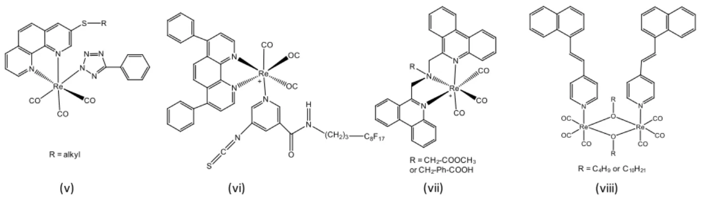
The simplest complexes are based on 2-2’-bipyridine, triazole and pyridyl-imidazole ligands (Figure 5).[27] Phosphorescence generally appears around 550 nm, and it is characterized by its very low emission efficiency in solutions (10-4-10-3), which does not hinder applications as bioimaging agents. It is noteworthy that in the structures presented in Figure 5, the R group on the monodentate ligand is frequently used to attach a recognition moiety directed toward a biological target.
Figure 5. Examples of Re(I) complexes based on simple ligands derived from bipyridine or analog.
Extending the π-conjugated system of the chelating groups is a very popular strategy for enhancing the photophysical properties of the complexes.[ 28 ] They generally contain polypyridyl ligands (e.g. phenanthroline and dipyridophenazine derivatives) and conjugated pyridyl groups (Figure 6). Very variable emission efficiencies are reported in solutions, the photoluminescence quantum yield often being in the 10-2 range. The quantum yield may also be drastically reduced due to quenching by energy transfer from 3MLCT to 3IL, as occurs in compound (viii).
Figure 6. Examples Re(I) complexes based on ligands with extended electron systems.
Complexes that incorporate an organic fluorophore non-conjugated to the chelating ligands are still quite rare (Figure 7). In this case, chelation with ReI has no direct impact on the electron system of the fluorophores. The excitation energy can be shifted to long wavelengths. Emission wavelength, Stokes shift and lifetime depend on the relative energy levels. With fluorophores that absorb at short wavelengths and emit poorly,[29] the optical properties are close to those of the compounds above. In contrast, with properly-chosen fluorophores, quenching by the 3MLCT state is prevented. Emission is therefore fluorescence, arising from the organic fluorophore, i.e. naphthalimide in the case of compound (xi). The position of the emission spectrum depends totally on the nature of the fluorophore, the quantum yield may be
(i) (ii) (iii) (iv)
(v) (vi) (vii) (viii)
R = CH2-COOCH3 or CH2-Ph-COOH R = alkyl
very high, and there is no quenching by oxygen.[30] In this case, advantage is not taken from the intrinsic emission properties of the rhenium center. These compounds have been originally designed to mimic the behavior of 99mTc radioactive probes and study their behavior and localization in biological medium.
Figure 7. Examples of Re(I) complexes incorporating an organic fluorophore moiety.
2.2.2. Unconjugated complexes for imaging applications
Whatever their emission properties, Re(I) complexes are often biocompatible. They have been tested on various types of cells, including yeasts[31] and parasitic flagellates.[23,30b] It appeared that they are particularly well uptaken by mammalian cells and, most interestingly, by cancer cells.[28,29] However, non-specific staining of the cytoplasm is not ideal for imaging applications. In a review article, Balasingham et al. have tried to understand the factors governing the cell uptake and localization of various metal complexes.[32] Regarding Re(I), a systematic study from Fernández-Moreira et al. allows some basic principles to be identified.[31] This study compares various complexes of type (iii) in which the bipyridyl group is responsible for the spectroscopic properties, while the ancillary pyridyl ligand is used for varying charges and lipophilicities. This allows the behavior of the complexes in living medium to be controlled, in particular for membrane permeability, cell uptake and localization in cell components, with minor interferences with the photophysical properties. It appears, with a few exceptions, that cationic species are taken up well by passive diffusion. Highly lipophilic complexes are easily incorporated into dead cells, while the other species require a healthy membrane potential to facilitate uptake. Simple cationic lipophilic complexes are highly membrane permeable, they localize in the cytoplasmic membrane and in the lipophilic sites of organelles.[31,33] However, when lipophilicity becomes very high, for example after incorporation of a fluorous chain in
(vi), the compound tends to self-aggregate in aqueous solutions and shows low cellular uptake
efficiency.[28b] Electrophilic complexes localize in the mitochondria. Anionic polar species associate with the outer face of the plasma membrane, while anionic lipophilic species show no or little uptake.[28,33] Neutral complexes such as (v) also exhibit good cellular uptake by live cancer cells and non-specific perinuclear localization.[28a] Finally, when the ligand incorporates a large planar aromatic system like dipyridophenazine, the resulting complex easily intercalates between two DNA bases.[28f,34]
2.2.3. Bioconjugated complexes
Cell localization may be markedly improved by biological targeting. As recently reviewed by Lo whose research group has done a lot of work in the field, bioconjugated complexes become efficient biological sensors and precise imaging reagents.[35] Most often, conjugation with a biomolecule is achieved on the pyridinyl group, so that the photophysical properties are affected as little as possible. It is classically made via a functional group, i.e. isothiocyanate and aldehyde that can react on primary amines, or maleimide and iodoacetamide that react with the sulfhydryl groups of biomolecules. For instance, Re(I) complexes have been directly used in solid-phase peptide synthesis to afford labeled neurotensin.[28d] They have been used to tag gluthathione and proteins like bovine serum albumin (BSA), with successful results in imaging (Figure 8).[28b] Luminescent biotin complexes [Re(N N)(CO)
3(py-biotin)]+ have been developed.[35c] Contrary to classical organic biotin-fluorophores that suffer from self-quenching, they show an increase of the emission intensity and elongation of the lifetime upon binding to avidin, the natural receptor of biotin. They have therefore been very useful to study the recognition processes between these two biologically important molecules. The luminescence properties of Re(I) complexes conjugated with estradiol have also been exploited for imaging of hormone-dependent breast cancer cells.[35d] Similarly, Re(I) complexes conjugated to glucose have been imagined to study the uptake and transportation of glucose in cancer cells, that generally overexpress the sugar receptors.[36] Actually, the fructose conjugates have shown enhanced uptake by breast cancer cells.[ 37 ] Complexes of the type [Re(N N)(CO)3(py-indole)]+ have been developed to study the interactions with indole-binding proteins such as BSA and lysozyme.[38] Chromone derivatives such as complexes (ix) have been designed to recognize regulatory enzymes engaged in the biochemical signal transduction pathways.[29]
Figure 8. False-color fluorescence (left), differential interference contrast (middle), and overlaid (right) laser-scanning confocal microscopy images of HeLa cells incubated with the BSA conjugate of complex (vi) (10 μM) at 37 °C for 2 h, λex = 405 nm.(Figures are extracted from reference [28b])
2.2.4. Sensing
Re(I) probes have also been used as biochemical sensors. For example, Re(I) complexes appended to a dipicolylamine unit displayed enhanced emission and elongation of the phosphorescence lifetime in the presence of zinc(II) and cadmium (II) ions. These compounds have been shown to be potentially useful for monitoring the intracellular concentration of these ions, although the ion-concentration range where they can be used in their present form is very narrow.[28c] It is also noteworthy that some probes have been successfully developed for the sensing of nitric oxide (NO), a small endogenously-produced molecule that has recently been recognized as a major signaling agent whose unregulated production leads to diseases of the immune, cardiovascular and nervous.[39]
2.2.5. Correlations with radioimaging studies
As already mentioned above, Re(I) probes are often used for correlations with radioimaging techniques, whose main modalities are single photon emission computed tomography (SPECT) and positron emission tomography (PET). As mentioned previously, since Re is the closest chemical analog of Tc, non-radioactive isotope of Re allow the development of complexes adapted to biological applications. Moreover, the luminescence properties allow the identification of cellular targets using confocal fluorescence microscopy, whereas the insufficient resolution of radioimaging techniques does not permit access to this crucial information.
In this field, we must underline the effort made by the group of Valliant and Zubieta to develop a library of metallopeptides that can be used in the preparation of a variety of
bioconjugates.[40] It is curious to note that very few probes developed in this aim exploit the intrinsic luminescence properties of Re(I).[27b,30a] They are often associated to organic fluorophores, i.e. naphthalimide in the case of compound (xi)[30b] and 2-phenylbenzothiazole in complexes designed as potential imaging probes for the β-amyloid plaques that are formed in the course of Alzheimer’s disease.[ 41 ] In the latter case, both techniques are well complementary, since confocal fluorescence imaging has been used to detect β-amyloid plaques at the cellular level, while in vitro autoradiography provided images of their distribution in large brain sections.
2.2.6. Bimodal infrared and luminescence imaging
When rhenium complexes incorporate carbonyl ligands, the unique vibrational signature of the CO group enables detection by infrared and Raman spectroscopies. These vibrational spectroscopies are of particular interest for bio-imaging because radiations in the infrared cause little damage to living tissues, contrary to excitation in the UV and visible range. The problem of fluorophore photobleaching does not arise. Resolution reached the subcellular level.[42] As recently reviewed by Clède and Policar,[43] Re-carbonyl probes are very attractive candidates for these spectroscopies, especially because they absorb in the middle-infrared, a wavelength range comprised between 2200 and 1800 cm-1 where the biological medium is almost transparent.
IR-imaging at the sub-cellular level has first been made using synchrotron radiation FTIR spectromicroscopy (SR-FTIR SM) and Re-carbonyl probes that contain a cyclopentadienyl group linked to a tamoxifen moiety.[ 44 ] Thereafter, vibrational and luminescence spectroscopies have been combined, taking advantage of the intrinsic luminescence properties of the Re(I) core. Using a pytavii-based Re(I) complexes, provided with various alkyl chains that enhance cell uptake by cancer cells, the authors have shown a good correlation between IR mappings and fluorescence imaging at the cell level.[27b, 45 ] Bioconjugation with an estrogen analog allowed visualization by both techniques of estrogen-dependent breast cancer cells.[45b]
vii Pyta means 2-pyridyl-1,2,3-triazole entity. This term was used first by Obata in 2008 (see M. Obata et al., Dalton
To our knowledge, no rhenium probes have been used for bimodal imaging using Raman spectroscopy, contrary to Mn probes,[46] probably because this powerful non-invasive method is still in its early stages.
2.3. From photoactivity to the use as phototherapeutic agents
2.3.1. Photocytotoxicity and generation of active oxygen species
During imaging studies using Re(I) complexes, two interesting features have emerged. The first one is the slow photobleaching of these complexes with respect to common organic dyes, although remarkable exceptions have been reported. The second one is their photocytotoxic action.[35,37] It results in membrane blebbing that appears after multiple image collection under the confocal fluorescence microscope, and leads to cell death. The origin of photocytoxicity has rarely been investigated, although the photochemistry of Re(I) complexes has been thoroughly investigated.[25a,25c] It is well known that these complexes are quenched by molecular oxygen and may generate highly reactive oxygen species such as singlet oxygen 1O
2.[ 47 ] In the case of dipyridophenazine Re(I) complexes intercalated in DNA, the photocleavage of DNA would be rather due to direct oxidation by the photoexcited complex, or to the photoproduction of superoxide (O2-) and hydroxyl (OH.) radicals, depending on the ligand structure.[34] The easy uptake of Re(I) complexes by cancer cells combined with their phototoxic activity suggests that they could be used for theranostics that is the combination of diagnosis and therapeutics.
2.3.2. Photogeneration of carbon monoxide
Besides, a characteristic feature of Re(I) complexes is their ability to release carbon monoxide (CO) photochemically. They could therefore serve as new agents for the delivery of CO to biological targets or in the frame of a therapy based on the use of this gas, namely CO-therapy.
2.3.2.1. From CORMS to photoCORMs in CO-therapy
The toxic nature of carbon monoxide has been well known for a long time. This small molecule, often referred to as the “silent killer”, strongly binds to hemoglobin, thus reducing the oxygen-carrying capacity of the blood and impairing respiration. The salutary effects of CO have only recently been recognized.[48] Much like its toxic twin nitric oxide, CO has been
identified as an important endogenously-generated signaling molecule that participates in a variety of physiological processes. It is now well established that CO plays a crucial role in immune and anti-inflammatory responses, as well as in vaso-relaxation. Most spectacular of the CO-mediated effects is its capacity to promote graft survival during organ transplantation. CO also helps treating cardiovascular disease, promotes wound-healing and apoptosis of cancer tissues, and has antibacterial activity. Exogenously applied CO gas is thus increasingly considered as a therapeutic agent. According to the desired therapeutic effect, low doses (100-250 ppm) or moderate doses (> (100-250 ppm) may be used. Unfortunately, the implementation of CO gas in hospital setting raises technical and safety-related issues. These issues have prompted the quest for exogenous CO-releasing molecules (CORMs) to deliver controlled amounts of CO to biological targets. But, controlling when and where the CO is released is also of major importance. In these regards, triggering by light has rapidly appeared as an excellent strategy, because it allows fine temporal and spatial control of CO release. This led to the concept of light-triggered CORMs (PhotoCORMs), the development of which has expanded significantly in the last half-decade, as recently reviewed.[49]
Like CORMs, the majority of photoCORMs are based around metal carbonyl complexes, which offer a direct route to the release of CO. The photochemical reaction induces the release of at least one CO molecule that is replaced by a solvent molecule, e.g. water, in the coordination sphere. The first photoCORMs have been reported by Motterlini et al. in 2002.[50] They were simple metal carbonyls, i.e. Fe(CO)5 and Mn2(CO)10, and were not truly suitable for biological use because their required excitation in the ultraviolet (UV) that damages living cells. Since then, a lot of systems have been explored and the design of photoCORMs has been markedly improved. The ideal photoCORM is stable in the dark under ambient conditions, soluble in water and non-toxic before and after CO-release. It releases CO efficiently under illumination by visible light, preferentially red light that has the best penetration of the living tissues. Organometallic complexes have therefore been developed to meet these specifications. Various metal d6 centers such as Mn(I), Re(I), Fe(II) and Ru(II) that ensure overall stability of the carbonyl complexes have been investigated, with particular attention to Mn(I). Collectively, experimental and theoretical works have allowed several principles to emerge regarding the choice of ligands and their disposition in the coordination sphere.[51] It has been shown that CO release is favored in complexes in which electron transfer from electron-rich metals to π* orbitals of the ligand (metal-to-ligand charge transfer, MLCT) is easy. In other words, low-lying orbitals on the ligand system, hence strong MLCT absorption in the visible or near
infrared, should be associated to good CO dissociation. For example, when the 2-2’-bipyridine ligand of a Mn complex is replaced by a ligand with increased π-electron system, the excitation wavelength passes from 350-450 nm to 520 nm, with increased photolability of CO.[ 52 ] Auxillary ligands also modulate the energy of the occupied orbitals depending on their electron-donating abilities. A small number of CO ligands in the complexes also favors good photochemical activity.
2.3.2.2. Design of Re(I)-based photoCORMs
As specifically concerns rhenium complexes, interest in these compounds has grown considerably in recent years. Their stability and biocompatibility are attractive. Since these complexes were already known for their photoluminescence properties, the idea was also to take advantage of both their imaging and therapeutic abilities. However, the use of Re salts as photoCORMs raises three important issues concerning photoreactivity, as well as the optimization of photoluminescence properties and excitation wavelength.
First of all, many Re complexes are not very photoreactive. Complexes of the type fac-[ReCO3(X)(bpy)] (bpy = 2,2’-bipyridine; X= Cl or Br) and fac-[ReCO2(X2bpy)(py)]+ (py = pyridine, X = H, CF3), based on either -donating or moderately π-accepting ancillary ligands, are relatively photoinert, unless highly energetic irradiation by UV-B light is used.[53] Koike et al. have thoroughly studied the mechanisms of photochemical ligand substitution in this series of complexes and they have explained the reason for this photostability. Most interestingly, they have also shown that the introduction of a strongly π-acidic ancillary ligand, such as a phosphine, makes the axial CO photolabile with low-energy UV-A light (Figure 9).[53] For instance, complexes of type (xii) undergo photorelease of one CO molecule upon irradiation in the near-UV, and substitution by a molecule of organic solvent. Complexes of type (xiii), derived from 1,10-phenanthroline, readily delivered CO under illumination by light. Four of them could even be excited by low-power UV light and liberate none, one or all three molecules of CO depending on the nature of an ancillary ligand.[54] Complexes (xiv) and (xv) derived from 2-(2-pyridyl)-benzothiazole also undergo rapid release of CO under low-power UV illumination (Figure 9).[55]
It is interesting to notice that in these complexes built on a large -conjugated ligand, a phosphine ligand is no longer compulsory to observe CO-photorelease. Photoreactivity
therefore depends totally on the proper choice of the ligands. Compared to Mn complexes, Re complexes such as (xvi), (xvii), (xviii) and (xix) (Figure 9) are moderately photoreactive.[51,56, 57] The presence of the bromide ligand in (xix) leads to facile intersystem crossing to 3MLCT excited state due to spin-orbit coupling, i.e. heavy atom effect, and this prevents the dissociation of the metal-CO bond.
Figure 9. Chemical structure of some photoCORMs.
Rhenium photoCORMs may indeed be used as photoluminescent trackers and generally show good internalization by cells. For example, several type (xiii) complexes, as well as complex (xvi) have been used to show the uptake of this type of compounds by human breast cancer cells.[54,56] Ideally, it should be possible to take advantage of the photoluminescence properties to monitor the release of CO. The pioneering work of Ford and co-workers has paved the way for this.[58] As seen above, phosphine complexes are well photoreactive. But, they are often poorly soluble in water. Owing to the P(CH2OH)3 ligand, complex (xx) is water-soluble and releases CO efficiently. This complex was readily internalized by human prostatic cancer cells with no apparent cytotoxicity and could be used to visualize cell uptake using a
(xii) (xiii) (xiv) (xv)
(xvi)
(xvii)
(xviii)
(xix)
fluorescence microscope. Interestingly, the starting compound emitted in the green while the photoproduct emitted in the blue inside the cells (Figure 10). This color change suggested the possibility to track both the localization of the compounds and the release of CO. However, the spectral overlap between the two emission bands hampers this simultaneous detection. Thereafter, significant contributions have been made by the Mascharak group, with the aim to make the luminescence signals as distinct as possible. Complexes (xiv) and (xv) have been shown to release CO under low-power UV illumination. Both complexes are photoluminescent in the orange. In (xv), the loss of one CO molecule results in complete extinction of the luminescence signal. The delivery process was then monitored through the decrease of photoluminescence.[55b] In contrast, in (xiv), the CO release is accompanied by a change in photoluminescence from orange to deep blue. This compound was used successfully to track in vitro the uptake into cancer cells and the end of CO delivery within the target.[55a]
Figure 10. Top: Chemical structure and photoreactivity of a photoCORM. Bottom: Confocal fluorescence microscopy images of PPC-1 cells that were incubated for 60 min with 50 μM of (xx). The top image (in blue, λem = 465−495 nm) was collected with minimal photolysis from the 405 nm excitation source and indicates the incorporation of (xx) into the cellular cytosol. The bottom image (in green, λem > 660 nm) was collected after 405 nm photolysis for 15 min and indicates the transformation of (xx) to its photoproduct. (Figures extracted from
reference [58]).
(xx)
Finally, the biggest challenge in the field of Re-photoCORMs remains to shift the excitation wavelength to the biocompatible range (500 nm – 900 nm).[49] Complexes of type
(xii), as well as (xvi) and (xvii) were efficiently excited around 360 nm.[56,58] Complex (xx) was excited around 405 nm.[58] Curiously, contrary to their Mn analogs, complexes of type (xiii) and complexes (xiv), (xv), (xviii) and (xix) released CO only with illumination by low-power UV light, and were insensitive to visible light despite the presence of a MLCT band in the visible.[54,55a,57] This shows that the presence of a strong MLCT band in the visible region is not sufficient to insure good photolability of the CO ligands.[51] At the moment, the reported complexes must be excited in the near-UV or, at the best, at the edge of the visible range. Excitation at these wavelengths allows in vitro cell experiments, but it would be highly desirable to move the excitation wavelength to the red for in vivo applications. Only a small number of complexes have been explored to date and a lot of developments can still be made in this direction. In any event, these systems can be very finely tuned and obviously constitute very attractive candidates for theranostics.
2.3.2.3. Vectorization
Very few examples of incorporation of photoCORMs in nanoparticles can be found in the literature. To our knowledge, Rhenium photoCORMs have only been used once in nanoparticles. Complex (xv) incorporated in the mesoporous silica nanoparticles has been shown to be well endocytosed in vitro by human breast cancer cells, leading to their rapid eradication.[55b] According to the exciting results obtained with Mn-based photoCORMs incorporated in mesoporous silica nanoparticles and phospholipid-functionalized poly(ethyleneglycol) (PEG) layers,[59] many developments with Re(I) probes can be expected in this field in a near future.
The most important thing to remember is that Re(I) complexes allow to play on many fields at a time. Not only the versatility of the ligand structure allows precise targeting to be achieved, but multimodal imaging and theranostic can also be envisaged. Consequently, a rational design of the complexes is absolutely necessary so that the various functionalities can be harmoniously combined. Additionally, the simplicity and modularity of using different bifunctional chelators and radiometals facilitate the creation of a wide variety of imaging agents. To do so, it is obvious that the use of general synthetic approaches allowing the readily preparation of a large family of rhenium-specific chelates is also crucial. Among modular and
versatile chemistry reactions, in our opinion, the Copper(I)-catalyzed azide-alkyne cycloaddition seems the more suitable approach.
3. Click chemistry, “Click-to-Chelate” concept, Click, then Chelate approach
3.1. Copper(I)-catalyzed azide-alkyne cycloaddition (CuAAC reaction)
Click chemistry, a term coined in 1998 and reported in 2001 by Sharpless et al., is a particular useful synthetic method for the formation of carbon-heteroatom bonds by joining small molecular building blocks in a rapid, facile, and selective reaction.[60] According to Sharpless’ criteria, click transformations are usually performed under mild aqueous conditions and with readily available starting materials and reagents, in high yields and exhibited high
atom economy and generated little or no byproducts. Among these click reactions,viii the
copper(I)-catalyzed azide-alkyne cycloaddition so-called CuAAC reaction, reported independently in 2002 by the groups of Sharpless and Fokin[61] and Meldal,[62] has emerged as the perfect example of click chemistry. Compared to non-catalyzed azide/alkyne reaction first described by Michael[63] and later investigated by Huisgen[64] which lead to a mixture of two isomers (1,5-disubstituted and 1,4-disubstituted 1,2,3-triazoles), the CuAAC reaction of terminal alkynes proceeds selectively in the formation of the 1,4-disubstituted 1,2,3-triazole and does not require high temperature and/or harsh conditions (Scheme 2).
Scheme 2. Synthesis of 1,2,3-triazole via Huisgen 1,3-dipolar cycloaddition and CuAAC.
viii Non exhaustive list of click chemistry reactions: nucleophilic ring opening reactions of epoxides and aziridines, non-aldol type carbonyl reactions such as formation of hydrazones, Diels-Alder and inverse electron demand Diels-Alder reactions, Michael addition…[61]
Due to the intense research interests focused on CuAAC, numerous papers involving some reviews have been reported.[65] The CuAAC reaction has become ubiquitous since Fokin and Sharpless publication in 2002. The reasons for this success are mainly due to the versatility of this “click” reaction which is easy to perform and appropriate in a wide range of conditions. It may be conducted in aqueous solution at room temperature, using a large variety of catalytically Cu(I) species and exhibited a high functional group tolerance. In addition, numerous applications of the CuAAC reaction have been reported. This modular synthetic approach was used in distinct scientific disciplines such as drug discovery,[66] biochemistry,[67] dendrimer,[68] polymer materials[69] and so on. The CuAAC reaction was currently considered as the “cream of the crop” of click reactions and more than 3000 publications with “click chemistry” in the title have come out in a decade and a half.[70]
As mentioned above, the CuAAC reaction is an extraordinarily robust reaction, which could be performed under a wide variety of conditions and almost any copper source can be used as a pre-catalyst.[ 71 ] Before detailing the mechanism of this reaction, different used catalytic copper systems will be briefly discussed.
3.1.1. Copper catalystsix - Cu(II) salts
If Cu(II) ion is the most thermodynamically stable among the three most common states of copper (0, +1, and +2), its reduction is required to be used as copper catalyst in the CuAAC reaction. In 2002, Fokin and co-workers introduced sodium ascorbate as a convenient reductant for Cu(II) ion.[61] Its combination with a copper(II) salt, such as the stable and commercial available CuSO4.5H2O or Cu(OAc)2.H2O, constituted the method of choice for the preparation of 1,2,3-triazoles, the main advantage of this protocol being its compatibility with oxygen and water, allowing the application of CuAAC in biological media. Classically, this reaction is carried out with two to ten equivalents of the Cu(II) species in a mixture of water and organic solvent, the most commonly used being aqueous alcohol, THF or DMSO to ensure the solubility of hydrophobic reactants.
ix Only examples of “molecular” catalytic systems will be presented in this section. For catalytic nanoreactors (like dendrimers or zeolite) or copper-based nanoparticules, see C. Deraedt et al., J. Am. Chem. Soc. 136 (2014) 12092-12098 or S. Chassaing et al., Org. Lett. 9 (2007) 883-886 or F. Alonso et al., Acc. Chem. Res. 48 (2015) 2516-2528, respectively.
![Figure 2. Several examples of [ 188 ReN] 2+ complexes by the “3+2” strategy (Figure extracted from reference](https://thumb-eu.123doks.com/thumbv2/123doknet/2085993.7246/21.892.127.758.398.865/figure-examples-ren-complexes-strategy-figure-extracted-reference.webp)

![Figure 15. Examples of “Click-to-Chelate” chelating systems and their corresponding rhenium complexes, developed by Schibli and Mindt (figure extracted from reference [112])](https://thumb-eu.123doks.com/thumbv2/123doknet/2085993.7246/50.892.115.777.475.989/examples-chelate-chelating-corresponding-complexes-developed-extracted-reference.webp)

![Figure 3. The molecular structure of complex [Re(CO)3Cl(L2)]. Displacement ellipsoids are drawn at 50%](https://thumb-eu.123doks.com/thumbv2/123doknet/2085993.7246/82.892.147.752.102.440/figure-molecular-structure-complex-cl-displacement-ellipsoids-drawn.webp)

