HAL Id: hal-02403722
https://hal.archives-ouvertes.fr/hal-02403722
Submitted on 11 Dec 2019HAL is a multi-disciplinary open access archive for the deposit and dissemination of sci-entific research documents, whether they are pub-lished or not. The documents may come from teaching and research institutions in France or abroad, or from public or private research centers.
L’archive ouverte pluridisciplinaire HAL, est destinée au dépôt et à la diffusion de documents scientifiques de niveau recherche, publiés ou non, émanant des établissements d’enseignement et de recherche français ou étrangers, des laboratoires publics ou privés.
Noninvasive vascular occlusion with HIFU for venous
insufficiency treatment: preclinical feasibility experience
in rabbits
N Barnat, A Grisey, B Lecuelle, J. Anquez, B Gerold, S. Yon, J.-F Aubry
To cite this version:
N Barnat, A Grisey, B Lecuelle, J. Anquez, B Gerold, et al.. Noninvasive vascular occlusion with HIFU for venous insufficiency treatment: preclinical feasibility experience in rabbits. Physics in Medicine and Biology, IOP Publishing, 2019, 64 (2), pp.025003. �10.1088/1361-6560/aaf58d�. �hal-02403722�
1
Noninvasive vascular occlusion with HIFU for venous insufficiency treatment:
1preclinical feasibility experience in rabbits
2AUTHORS/INSTITUTIONS:
3
N. Barnat1, 2, 3, 4, A. Grisey4, B. Lecuelle5, J. Anquez4, B. Gerold4, S. Yon4, J.-F. Aubry1, 2, 3
4
1- INSERM U979, Institut Langevin, Paris, France;
5
2- ESPCI Paris, PSL Research University, Institut Langevin, Paris, France;
6
3- CNRS UMR 7587, Institut Langevin, Paris, France;
7
4- Theraclion, Malakoff, France;
8
5- Ecole nationale vétérinaire d'Alfort, CRBM, Maisons-Alfort, France
9 10
Abstract
11
Venous insufficiency is a common disease arising when veins of the lower limb become
12
incompetent. A conventional surgical strategy consists in stripping the incompetent veins.
13
However, this treatment option is invasive and carries complication risks. In the present
14
study, we propose noninvasive high-intensity focused ultrasound (HIFU) to treat lower limbs
15
venous insufficiency, in particular incompetent perforating veins (mean diameter between 2-6
16
mm).
17
Sonication parameters were designed by numerical simulations using the k-Wave toolbox to
18
ensure continuous coagulation of a vein with a diameter superior or equal to 2 mm. The
19
selected ultrasound exposures were 4 seconds pulses in continuous wave mode. Two types
20
of sonication were studied: (1) fixed pulses and (2) moving pulses at constant speed (0.75
21
mm.s-1) across the vein.
22
The potential of these exposures to thermally occlude veins were investigated in vivo on
23
rabbit saphenous veins. The impact of vein compression during ultrasonic exposure was also
24
investigated.
25
Fifteen rabbits were used in these trials. A total of 27 saphenous veins (mean diameter 2.0 ±
26
0.6 mm) were sonicated with a transducer operated at 3 MHz. After a mean 15 days
2
up, rabbits were euthanized and venous samples were extracted and sent for histologic
28
assessment. Only samples with the vein within the HIFU lesion were considered for analysis.
29
Simulated thermal damage distribution demonstrated that fixed pulses and moving pulses
30
respectively placed every 1.5 and 0.5 mm along the vein and delivered at an acoustic power
31
of 85 W and for 4 seconds were able to induce continuous thermal damages along the vein
32
segments.
33
Experimentally, both treatment parameters (1) and (2) have proven effective to occlude veins
34
with a success rate of 82%. Occlusion was always observed when compression was applied.
35
Our results demonstrate that HIFU can durably and non-invasively occlude veins of
36
diameters comparable to human veins.
37
Keywords: HIFU; ultrasound; thermal occlusion; vascular occlusion; vein
38
Introduction
39
Venous insufficiency is a common medical condition arising when a high pressure is
40
transmitted from deep to superficial venous system of the lower limbs. High-pressure
41
transmission frequently occurs through retrograde flow. This elevated venous pressure
42
causes the vein to become tortuous and dilated over time. Besides the unsightly appearance
43
of varicosities, venous disease is associated with pain and clinical signs may vary from
44
asymptomatic telangiectasia to achy ulcer [1]. The strategy to treat venous disease consists
45
in ceasing reflux by eliminating the involved incompetent vein segment.
46
Conventional surgical techniques consist in removing the vein. The invasive nature of
47
surgery leads to the development of minimally invasive procedures such as radiofrequency
48
ablation [2] or endovenous laser ablation [3]. In both cases, after introducing a catheter into
49
the vein, thermal energy is delivered, in order to denature collagen and induce a shrinkage of
50
the vein [4,5].
51
Although these techniques proved their efficacy, they are not appropriate to treat all leg
52
veins. In case of incompetent perforating veins, thermal ablation is appropriate only for large
53
(>3.5 mm) perforators and may treat perforators only superficially [6]. Furthermore, treating
3
pathologic veins can be technically challenging and requires experienced physicians
55
especially for the insertion of the catheter in a deep or tortuous vein.
56
In this context, high-intensity focused ultrasound treatments (HIFU) could provide a
non-57
invasive alternative to occlude incompetent veins.
58
Previous studies have demonstrated HIFU occlusion in blood vessels of small diameters only
59
(range 0.5-1.5 mm) [7,8,17–21,9–16]. The occlusion of large vessels (≥2 mm) has not been
60
reported to the best of our knowledge. We demonstrate here on a rabbit model that HIFU can
61
occlude large veins (≥2mm), paving the way for a clinical treatment of venous insufficiency
62
with focused ultrasound.
63
For this purpose, sonication strategies including placement and trajectory of pulses were first
64
identified by numerical simulations and then evaluated on an in vivo model of rabbit veins
65 (range: 1.1-3.7 mm). 66
Methods
67Ultrasound equipment
68Experiments were performed with the Echopulse® HIFU device (Theraclion, Malakoff,
69
France) used clinically for human breast fibroadenomas [22] and benign thyroid nodules [23]
70
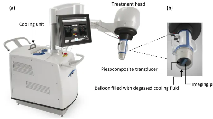
treatments (Figure 1(a)). The system encompasses a treatment head mounted on an
71
articulated arm which includes a single element piezocomposite therapy transducer and a
72
linear confocal imaging probe for real-time monitoring of the treatment (Figure 1(b)). The
73
therapy transducer, operated at 3 MHz, has a spherical cap shape with a curvature radius of
74
38 mm and an outer diameter of 56 mm. The RF signal applied to the transducer comes from
75
a frequency generator (JJ&A Instruments, Model RFG-200T1) with an electric power ranging
76
from 10 to 215 W. The ultrasound imaging probe working at a frequency of 6.2 MHz
77
(Vermon, Tours, France) was connected to an Aixplorer® ultrasound system (SuperSonic
78
Imagine, Aix-en-Provence, France) to guide treatments with B-mode images. By
79
construction, the therapeutic focus is located within the imaging plane. A fluid-filled balloon
4
(EPack, Theraclion®, Malakoff, France) was fixed to the transducer to ensure acoustic
81
coupling between the transducer and the target. The coupling liquid was circulated and
82
cooled during all the procedure to avoid transducer overheating and skin burns. Cooling is
83
ensured by built-in Peltier modules (Figure 1 (a)). A dedicated software implemented in
84
MATLAB (MathWorks, Inc., Natick, Massachusetts, United States) was used to drive
85
sonications.
86
87
Figure 1: Ultrasound equipment: (a) Echopulse, (b) HIFU transducer.
88
Treatments simulation
89
Simulations were performed to adjust ultrasound exposures layout to adequately sonicate a
90
vein of 2 cm in length and 2 mm in diameter. Two types of pulses were studied: (1) fixed
91
pulses and (2) moving pulses at constant speed (0.75 mm.s-1) along a line orthogonal to the
92
longitudinal axis of the vein. In both cases, we used 4-second sonications delivered in
93
continuous wave mode at an acoustic power of 85 W (typical power used in clinics and in our
94
experiments) and with the focus located at 15 mm under the skin. Acoustic pressure field,
95
temperature rise and, ultimately, thermal damages extent were simulated.
96
Balloon filled with degassed cooling fluid Piezocomposite transducer
Imaging probe
(a) (b)
Cooling unit
5
Geometrical model
97
The modelled HIFU transducer has the same geometry than the therapy transducer
98
described in the Ultrasound equipment section. The frame of reference of the transducer was
99
defined as follows: the origin corresponds to the HIFU transducer focus. X is the axis of the
100
imaging transducer and oriented along the left-to-right direction of the generated images. Z
101
denotes the main ultrasound propagation axis, pointing towards the subject anatomy. Y
102
results from the cross product of Z and X, for the frame (X, Y, Z) to be orthonormal and
right-103
handed.
104
In the simulations, the vein was considered as collapsed and the blood flow abolished. Thus,
105
the vein itself was not modelled. We assumed that the ultrasound beam propagates through
106
3 layers, from top to bottom: (1) the cooling liquid contained in the balloon fixed to the
107
transducer; (2) the skin and (3) the muscle. Skin was modelled as a 2-mm thick layer. For
108
illustration purposes, a 2D view of the geometrical model is shown in Figure 2.
109 110 111
Cooling liquid
Skin
Muscle
Z
X
Y
HIFU transducer
Imaging probe
6
Figure 2: Model used in the simulations
112
Acoustic simulation
113Acoustic simulations were performed using the k-Wave toolbox 1.2 [24]. The toolbox solves
114
the system of coupled first-order partial differential equations comprising momentum
115
conservation, mass conservation and pressure-density relation.
116
To solve the system of coupled acoustic equations, k-Wave uses a k-space pseudospectral
117
method where spatial gradients are computed using the Fourier collocation spectral method
118
[24]. Although the mathematical approach used in k-Wave is efficient, the 3D simulation of
119
the HIFU beam is costly in terms of memory. In our case, at 3 MHz and considering a
120
domain of 606046mm, the simulation of the nonlinear propagation up to the fifth
121
harmonic with the minimum of two points per wavelength requires a grid of points.
122
Hence, to reduce memory requirements, we used a “layer by layer” approach [25]. The
123
computational grid is divided into finer and finer layers along the main propagation axis and
124
we simulated ultrasound propagation from the transducer to 3 mm beyond the focus. We
125
respectively used one layer for the cooling liquid, one layer for the skin and five layers for
126
muscle tissue. Nonlinearities were modeled up to the fifth harmonic in the widest layer.
127
The size of the smallest layer was 4 x 4 x 4 mm (corresponding to 8 x 8 x 8 wavelengths),
128
with a spatial grid step of 49.8 m. A perfectly matched layer was used to avoid reflections at 129
the boundaries of the domain.
130
The Courant-Friedrichs-Lewy (CFL) number was set to 0.5 resulting in a smallest time step
131
of 14.8 ns. The pressure acoustic source term was computed as:
132
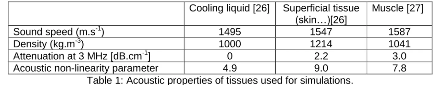
The cooling liquid, skin, fat and muscle tissue were modeled as homogenous media with the
133
acoustic properties listed in
134 135
7
Tissue properties were assumed to remain constant for the full procedure period. Variability
136
of parameters with temperature was neglected for all simulations.
137 138
Cooling liquid [26] Superficial tissue (skin…)[26]
Muscle [27]
Sound speed (m.s-1) 1495 1547 1587
Density (kg.m-3) 1000 1214 1041
Attenuation at 3 MHz [dB.cm-1] 0 2.2 3.0 Acoustic non-linearity parameter 4.9 9.0 7.8
Table 1: Acoustic properties of tissues used for simulations.
139
Thermal simulation
140Tissue heating was modeled using the Pennes’s bioheat equation [28]. The equation was
141
solved in 3D using a first-order explicit Euler scheme in time and a centered scheme in
142
space.
143
The time step was 50 ms and the spatial step was 200 m. The grid size was 38 ×38×29
144
mm.
145
The heat source term was computed as stated in [25]. The classic attenuation model
146
(absorption + scattering) was considered, and thermal rise was generated by the absorbing
147
component. 148
The tissue initial temperature was set to
T
0
39
.
5
°C which is the mean body temperature for149
a rabbit [29] and Dirichlet boundary conditions were used with Tboundaries = T0. We modelled
150
skin cooling as a heat flux at skin surface considering that the temperature of the cooling fluid
151
was 10°C (value achieved during experiments) and the heat transfer coefficient between skin
152
and liquid was set to 320 W.m2.K-1 based on previous experiments. To determine this value,
153
temperature of skin was first measured several times with a thermocouple slipped between
154
skin and the balloon in which circulated cooled liquid. Thereafter by assuming that heat flow
155
at skin is proportional to the difference of temperatures between skin and a characteristic
156
temperature of the cooling liquid, the value of 320 W.m².K-1 has been assigned. As
157
experimentally, the cooling time between subsequent sonications was set to 15 seconds.
8
The tissue properties used in thermal simulations are listed in Table 2 and were assumed
159
invariant with temperature in simulations.
160
Muscle[30] Specific heat capacity (J.kg-1.K
-1)
3600
Thermal conductivity (W.m-1.K-1) 0.47 Perfusion (s-1) 0.018
Table 2: thermal properties of media used in thermal simulation.
161
Thermal damage
162To achieve vessel sealing, sufficient thermal damage shall be induced to the vessel wall.
163
Modelling of vascular thermal damage [31] suggests that vessel thermal damage correlates
164
with the denaturation of collagen fibrils, which begins at 62°C [32].
165
In this study, extent of thermal damage to vessels was evaluated based on a dedicated
166
thermal damage model.
167
To assess the level of damage induced to vessels, we implemented the model described by
168
Agah [31]. By considering denaturation of collagen as a first-order reaction, damage was
169
modelled using a general Arrhenius equation:
170
Where:
171
(t) is the damage parameter. , are the concentrations of the undamaged
172
molecules at the beginning and at time respectively. is a coefficient of rate process
173
(frequency factor), the activation energy, the universal gas constant, and the
174
temperature. and were set to s-1 and kJ.mol-1 according to
175
[31].
176
Degree of vessel damage is appraised by (t), where defines the first noticeable
177
irreversible damage and formally corresponds to the denaturation of 63% of the native
178
proteins [31]. As we are looking for an extensive damage to the vein, we set the threshold
179
for thermal damage to , which formally corresponds to the denaturation of 99% of
180
the collagen.
9
Animals
182
All animal work was approved by the institutional ethics committee for animal experiments
183
(agreement number CE16/2016082512126208). Experiments were carried out according to
184
the guidelines of the French National Comity for animal trials C.N.R.E.E.A (Comité national
185
de réflexion éthique sur l'expérimentation animale). New-Zealand white rabbits (n=15), 5
186
males and 10 females, 2 to 4 months (3 ± 1) old and weighing 2.7 to 4.5 kg (3.5 ± 0.5), were
187
used for the experiments. HIFU exposures were applied on medial saphenous veins. Prior to
188
sonications, the animals were anesthetized intramuscularly with a mixture of Ketamine (0.33
189
mg/kg), Domitor (0.5 mg/kg) and Valium (0.17 mg/kg). They were then intubated with an
190
endotracheal tube and ventilated with isoflurane to be kept under anesthesia throughout the
191
procedure. The fur of their hind limbs was shaved with a hair trimmer and then carefully
192
depilated with a depilatory cream to ensure good acoustic coupling and prevent skin burns.
193
HIFU procedure
194
Before the procedure, the rabbit was placed on the table in supine position to access the
195
targeted vein. Ultrasonic gel (Polysonic Parker Laboratories, Inc., Fairfield, NJ) was applied
196
to the area to be treated to ensure appropriate coupling. B-mode ultrasound and color
197
Doppler imaging were used to position the focus on the vein.
198
27 medial saphenous veins (average diameter of 2 mm) were exposed to HIFU.
199
Blood flow is known to hamper thermal therapies by cooling down the vein wall [33–37].
200
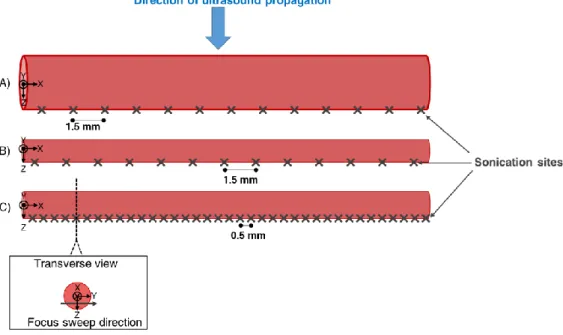
Therefore, several configurations based on previous simulations were tested during the HIFU
201
treatments: (1) 5 veins were treated with multiple pulses (median 1.5 mm spacing) distributed
202
along the target vein (Figure 3A)); (2) 14 veins underwent the same treatment protocol than
203
the one used in configuration(1) but the hind limb was elevated to about 25 degrees from the
204
horizontal to diminish blood flow (Figure 3 B)); and (3) 8 veins were sonicated with moving
205
pulses (0.75 mm.s-1 along a 3-mm line transecting the vein) (Erreur ! Source du renvoi 206
introuvable.) and were mechanically compressed by pressing a finger on the groin during
207
exposures to stop blood flow and minimize its heat sink effect. Rear limbs elevation empty
10
veins of their intraluminal blood and induce venous spasm (illustrated in Erreur ! Source du 209
renvoi introuvable. for configurations (2) and (3)). The average diameter of the vein after
210
proximal compression was of 1 mm. According to simulations, for configuration 3, the
211
spacing between moving pulses was reduced to 0.5 mm to account for the smaller lesion
212
size in the longitudinal direction (Figure 3C)
213
Thus, the number of sonications was increased to sonicate the same length of 2 cm. 214
215
Figure 3: Schema illustrating dispositions of HIFU exposures. A), B) and C) stand for
216
configurations (1), (2) and (3) respectively.
217
For all cases, HIFU sonications were applied for 4 seconds and at an acoustic power of 83 ±
218
10 W (obtained by multiplying electric power by the 70% efficiency of the transducer). The
219
corresponding spatial-peak intensity (linear extrapolation from low power hydrophone
220
measurement in water is 28 kW.cm-2. A minimum time period of 15 s was set between two
221
sonications to allow the skin to cool down. Experimental protocols are summarized in Table
222
3.
223
Experimental protocol ID 1 2 3 Number of HIFU exposed veins 5 14 8
Sonication types
Fixed pulses 1.5 mm apart
Fixed pulses 1.5 mm apart
Linear track with sites 0.5 mm apart Hind limb elevated
11
Table 3: Experimental conditions
224
Histological protocol
225
After the experiments, the rabbits were recovered from anesthesia and were monitored for at
226
least 15 days after treatment, to assess the sustainability of the effects of HIFU exposures on
227
veins. Animals were then euthanized by intravenous injection of Pentobarbital (1 mL/kg).
228
The day of euthanasia, the animals underwent a post mortem dissection and the targeted
229
vessel was harvested. In some cases, the perivenous conjunctive tissue was adherent and
230
the vein could not be separated. In such cases, the vein was sent for analysis with the
231
attached perivenous tissue.
232
The proximal tip of each venous section was sutured, preserved in identified cassettes, fixed
233
in 4% formalin and sent for histologic examination. Samples were then cross-sectioned into 3
234
subsections and included in paraffin. 4-µm slices were cut and stained with
haematoxylin-235
eosin (HE) for evaluation. Each section was evaluated without knowledge of the detailed
236
treatment status.
237
Excision of the proper vein segment is difficult since other veins branch in the vicinity of the
238
target. Therefore, samples were taken into account only when a vein was present on the
239
slide and was located within the HIFU lesion.
240
A venous sample from untreated region was also extracted and served as a control.
241
Changes were graded as: normal vein (0), reactive endothelium (1), myoendothelial
242
proliferation (2), acute fibrinous thrombi (3) and thrombus organization (4).
243
12
Results
244
Numerical model
245
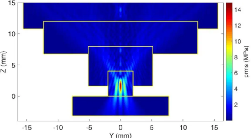
Ultrasound pressure field
246
Figure 4 shows the rms acoustic pressure field profile in the YZ plane simulated with the
247
“layer by layer” approach. Simulations were ran at 3 MHz and at an acoustic power of 85 W.
248
249
Figure 4: Simulated rms pressure field of the geometrical model in the YZ plane simulated.
250
Pressure is expressed in MPa. Yellow rectangles represent the limits of the subsequent
251
layers.
252
Thermal damages
253
Temperature elevation at the expected location of the upper vein wall (2 mm above the focal
254
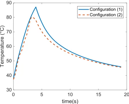
point centered on the lower vein wall) is plotted on Figure 5 for configurations (2) and (3).
13 256
Figure 5: Simulated temperature elevation at the location of the vein wall during 4 s
257
sonification followed by 15 s cooling for unitary fixed pulse (solid line) and moving pulse
258
(dash line).
259
For unitary fixed and moving pulse, the extent of the coagulated zone as defined using the
260
thermal damage model was 1.2 mm and 0.6 mm along the vein respectively and 1.8 mm and
261
2.7 mm in the orthogonal direction to the vein respectively.
262
To induce homogeneous and extensive heating of a vein 2 cm in length and 2 mm in
263
diameter, we considered: (1) for fixed pulses, 13 sonications with a spacing of 1.5 mm and
264
(2) for moving pulses, 37 sonications moving along the cross sectional axis with a spacing of
265
0.5 mm along the longitudinal axis of the vein.
266
Figure 6 and Figure 7 show the results of thermal damage distribution for such treatments:
267
(1) 13 fixed pulses and (2) 37 moving pulses respectively. An iso-contour representing the
268
area corresponding to denaturation of 99% of native proteins is displayed.
269
Distribution of thermal damage simulations are displayed using maximum intensity
270
projections.
14 272
Figure 6: Maximum intensity projected damage map at the end of a treatment comprising
273
thirteen 4-s fixed pulses delivered 1.5 mm apart at an acoustic power of 85 W and with the
274
focus located at 15 mm under skin. Iso-contour corresponding to: the thermal damage where
275
99% of native proteins denature is displayed in black.
276
277
Figure 7: Projected damage map at the end of a treatment comprising 37 4-s pulses during
278
which the focus was moved at 0.75 mm.s-1. Sonications were delivered at an acoustic power
279
of 85 W, with a spacing of 0.5 mm and with the focus located at 15 mm under skin.
Iso-280
contour corresponding thermal damage where 99% of native proteins denature is displayed
281
in black.
282
15
To quantify the impact of the trajectory, attention has been given to the extent of the region
283
corresponding to protein denaturation.
284
Damaged zones in the XY plane of the vein for treatment layouts (1) and (2) are reported in
285
Table 4.
286
Treatments ID Damaged zone Lesion area
(1) 19.2 × 2.4 mm² 42.8 mm²
(2) 19.2 × 4.2 mm² 72.0 mm²
Table 4: Simulated results in terms of damaged areas for treatment plannings (1) and (2)
287
After HIFU exposures, vessel damage can be observed through vessel shrinkage as
288
illustrated in the following ultrasound image (Figure 8).
289
290
Figure 8: Acute vascular shrinkage following HIFU pulses
291
Histological findings
292
Clinical follow-up of the animals showed that recovery after HIFU treatments were well
293
tolerated. All animals survived until the day of euthanasia and no abnormal behavior was
294
noted postoperatively. All animals continued their normal pattern of feeding.
295
As mentioned in Histological protocol, only samples satisfying acceptance criterion were
296
considered in the study.
297
16
In total and excluding the control sample, 17 samples (63%) were taken into account in this
298
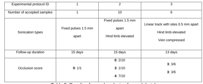
study. Histological results are reported in Table 5.
299
Experimental protocol ID 1 2 3 Number of accepted samples 1 10 6
Sonication types
Fixed pulses 1.5 mm apart
Fixed pulses 1.5 mm apart Hind limb elevated
Linear track with sites 0.5 mm apart Hind limb elevated
Vein compressed
Follow-up duration 15 days 15 days 13 days
Occlusion score 0: 1/1 0: 2/10 3: 1/10 4: 7/10 3: 3/6 3: 3/6
Table 5: Results of vessel occlusion for each batch
300
The follow-up duration indicated in Table 5 corresponds to the median value. It is however
301
necessary to emphasize that follow-up duration extended from 7 days for the shortest one to
302
19 days for the longest one due to the availability of the animal facility.
303
For all samples treated with fixed pulses and with a score superior or equal to 3, histological
304
findings revealed a thrombo-vasculitis of the saphenous vein characterized by a transmural
305
thickening and collapse of the lumen. Fibrinous thrombi were also present. These lesions are
306
irreversible in nature or expected to be irreversible (as assessed by the histopathologist) for
307
the samples with a follow-up duration inferior to 13 days. Lesions were expected to evolve
308
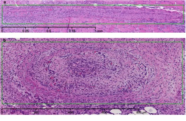
towards the formation of large fibrotic scars incorporating saphenous veins. Figure 9 shows
309
illustrative HE stained histologic cross sections.
17
311
Figure 9: HE sections (magnification 5×) of representative saphenous veins (green
312
rectangles) showing a transmural thickening and collapse of their lumens. The sections (a)
313
and (b) respectively correspond to veins treated according to protocols (2) and (3).
314
The lesions observed on samples sonicated with moving pulses were histologically identical
315
to those treated with fixed pulses. However, variability in the severity of the lesions was
316
observed between samples. The majority (73%) of samples treated with fixed pulses showed
317
a severe thrombo-vasculitis while lesions were evenly distributed between severe and
318
moderate for samples sonicated with moving pulses.
319
Discussion
320
The feasibility of inducing sustainable vascular occlusion with HIFU thermal effects was
321
demonstrated since histology reported 82% of vascular occlusion. Occlusion was successful
322
in all cases where compression was applied.
323
Histological analysis systematically showed severe lesions for samples which underwent
324
fixed point-by-point sonications and moderate lesions for half of the ones that were treated
325 a
18
by moving pulses. With moving pulses, the energy delivered is more spread and results in
326
less severe lesions.
327
However, vein samples that showed no occlusion were treated with fixed pulses. According
328
to simulations in the orthogonal direction to the vein diameter, coagulated zone after fixed
329
pulses is barely enough to cover the vein. Fixed pulses are effective when the vein wall is
330
completely encompassed in the lesion.
331
After fixed pulses, the coagulated zone is 2.4 mm in diameter and does not cover the whole
332
circumference of all targeted veins (diameter range between [1.1 – 3.7 mm].
333
Conversely, moving pulses cover an area 1.8 times wider which reliably damage the whole
334
circumference of all the veins. However, treatment with moving pulses required nearly three
335
times more sonications than treatment with fixed pulses to treat a same vein segment.
336
The lack of symmetry observed in the figure 5 resulted from heat accumulation effect. As
337
sonications are delivered, thermal energy deposited by previous pulse is added to energy
338
deposition of current pulse. There is therefore an overlap in heat accumulation between
339
successive pulses.
340
Although temperature dependence of ultrasound parameters has been demonstrated
341
experimentally [38,39,48,49,40–47], constant acoustic parameters were used in the
342
simulation as done by other teams[50–53]
343
In this study, we did not investigate focus movement speeds higher than 0.75 mm.s-1 but
344
further work may be performed to assess the highest focus speed that can lead to both a fast
345
and an effective occlusion.
346
Considering the good correlation between simulations and histologic findings, the model
347
used to assess thermal damage in the vein, based on the denaturation of collagen, appears
348
to be consistent. This is supported by the study conducted by Fujiwara et al [54] where
349
vessel occlusion was observed when peak temperature of the insonated sample hit 98°C but
350
not at 47°C or 54°C.
19
Area corresponding to denaturation of 99% of native proteins correlates with the region
352
where temperature reached 85°C. This temperature is also the level recommended by
353
radiofrequency closure ablation to seal veins.
354
By heating homogeneously the intima to 85°C, we would mostly likely achieve total
355
coagulation of the vein wall.
356
In our study, the three occlusion-free samples correspond to treatments without
357
compression. We hypothesize that blood flow could have acted as a heat sink which
358
prevented the temperature elevation of the vein wall from reaching sufficient levels.
359
Compression should be considered for future clinical treatments as it helps to abolish blood
360
flow and to decrease lumen area.
361
The high temperature elevations obtained in the simulations indicate that in our study,
362
thermal effects of HIFU are expected to play a major role in our venous occlusions. Thermal
363
mechanism compares favorably to the use of purely mechanical occlusion effects as
364
reported by Hwang et al [55], which lead to a recanalization of the veins after the treatment.
365
Hwang et al [55] used pulsed HIFU exposures with a low duty cycle in synergy with
366
ultrasound contrast agent. The treatment was effective but could not maintain rabbit auricular
367
vein occlusion over 14 days. With similar pulsing regime (9 MPa peak rarefaction pressure,
368
1-Hz pulse repetition frequency), Zhou et al [15] demonstrated a sustainable occlusion with
369
mechanical effects of HIFU but only by injecting pro-inflammatory agents corresponding to
370
the detergents found in sclerosing agents [56].
371
Groups who used ultrasound alone reported a temporary occlusion that did not exceed one
372
week after treatments [8,54,57]. Notwithstanding that they have demonstrated occlusion in
373
small veins (<1.5 mm), no long-term occlusion was proved.
374
Our exposure ultrasound parameters without additives were sufficient to induce venous
375
closure. To date and to the best of our knowledge, no other study reported noninvasive
376
permanent occlusion on veins of similar size, with the use of thermal effects of HIFU.
20
Conclusion
378
In this study, two sonication strategies have been identified and evaluated in a rabbit model.
379
In vivo experiments have demonstrated that thermal ablation with HIFU can induce
380
permanent sealing of veins of up to 3.7 mm [1.1-3.7 mm] in diameter.
381
HIFU is thereby a promising alternative to treat incompetent perforating veins. The treatment
382
parameters used in this study provide insights for subsequent clinical trials.
383
Vein compression was identified as a key factor contributing to successful vascular occlusion
384
since it avoided the cooling effect of the blood flow.
385
Future work should demonstrate the ability to coagulate larger veins (in a larger animal
386
model) similar to the great saphenous vein and its tributaries.
387
References
388
[1] Alam M, Nguyen T H and Dover J S 2006 Treatment of Leg Veins (Elsevier)
389
[2] Goldman MP 2000 Closure of the great saphenous vein with endoluminal
390
radiofrequency thermal heating in combination with ambulatory phlebectomy:
391
preliminary 6-month follow-up Dermatol Surg26 452–6 392
[3] Navarro L, Min R J and Bone C 2001 Endovenous laser: a new minimally invasive
393
method of treatment for varicose veins-preliminary observations using an 810 nm
394
diode laser Dermatol Surg27 117–22 395
[4] Manfrini S, Gasbarro V, Danielsson G, Norgren L, Chandler J G, Lennox A F, Zarka Z
396
a. and Nicolaides A N 2000 Endovenous management of saphenous vein reflux J.
397
Vasc. Surg.32 330–42
398
[5] Bergan J, Kumins N, Owens E and Sparks S 2002 Surgical end endovascular
399
treatment of lower extremity venous insufficieency J. Vasc. Interv. Radiol.13 563–8 400
[6] Dillavou E D, Harlander-Locke M, Labropoulos N, Elias S and Ozsvath K J 2016
401
Current state of the treatment of perforating veins J. Vasc. Surg.4 131–5 402
[7] Delon-Martin C, Vogt C, Chignier E, Guers C, Chapelon J Y and Cathignol D 1995
21
Venous thrombosis generation by means of high-intensity focused ultrasound
404
Ultrasound Med. Biol21 113–9
405
[8] Hynynen K, Chung A H, Colucci V and Jolesz F A 1996 Potential adverse effects of
406
high-intensity focused ultrasound exposure on blood vessels in vivo Ultrasound Med.
407
Biol 22 193–201 408
[9] Hynynen K ;, V C, Chung A and Jolesz F 1996 Noninvasive arterial occlusion using
409
MRI-guided focused ultrasound Ultrasound Med. Biol22 1071–7 410
[10] Rivens I H, Rowland I J, Denbow M, Fisk N M, Ter Haar G R and Leach M O 1999
411
Vascular occlusion using focused ultrasound surgery for use in fetal medicine Eur. J.
412
Ultrasound9 89–97
413
[11] Denbow M L, Rivens I H, Rowland I J, Leach M O, Fisk N M and ter Haar G R 2000
414
Preclinical development of noninvasive vascular occlusion with focused ultrasonic
415
surgery for fetal therapy J Obs. Gyneco 387–92
416
[12] Ishikawa T, Okai T, Sasaki K, Umemura S ichiro, Fujiwara R, Kushima M, Ichihara M
417
and Ichizuka K 2003 Functional and histological changes in rat femoral arteries by
418
HIFU exposure Ultrasound Med. Biol.29 1471–7 419
[13] Fujiwara K, Takeuchi H, Itani K, Yoshinaka K, Sasaki A, Azuma T, Sakuma I,
420
Matsumoto Y, Medical H A, High T, Focused I and Aloka H 2011 Real time HIFU
421
beam imaging Proc. 2001 Symp. Ultrason. Electron.32 583–4 422
[14] Hwang J H, Tu J, Brayman A A, Matula T J and Crum L A 2006 Correlation between
423
inertial cavitation dose and endothelial cell damage in vivo Ultrasound Med. Biol 32 424
1611–9
425
[15] Zhou Y, Zia J, Warren C, Starr F L, Brayman A A, Crum L A and Hwang J H 2011
426
Targeted long-term venous occlusion using pulsed high-intensity focused ultrasound
427
combined with a pro-inflammatory agent Ultrasound Med Biol37 1653–8 428
[16] Hwang J H, Brayman A A, Reidy M A, Matula T J, Kimmey M B and Crum L A 2005
429
Vascular effects induced by combined 1-MHz ultrasound and microbubble contrast
430
agent treatments in vivo Ultrasound Med. Biol31 553–64 431
22
[17] Hwang J H, Zhou Y, Warren C, Brayman A A and Crum L A 2010 Targeted venous
432
occlusion using pulsed high-intensity focused ultrasound. IEEE Trans. Biomed. Eng.
433
57 37–40 434
[18] Ichizuka K, Ando S, Ichihara M, Ishikawa T, Uchiyama N, Sasaki K, Umemura S,
435
Matsuoka R, Sekizawa a, Okai T, Akabane T and Kushima M 2007 Application of
436
high-intensity focused ultrasound for umbilical artery occlusion in a rabbit model.
437
Ultrasound Obstet. Gynecol. 30 47–51
438
[19] Ichiharan Mitsuyoshi, Sasaki K, Umemura S-I, Kushima M and Okai T 2007 Blood
439
flow occlusion via ultrasound image-guided high-intensity focused ultrasound and its
440
effect on tissue perfusion Ultrasound Med. Biol33 452–9 441
[20] Shaw C J, ter Haar G R, Rivens I H, Giussani D A and Lees C C 2014
442
Pathophysiological mechanisms of high-intensity focused ultrasound-mediated
443
vascular occlusion and relevance to non-invasive fetal surgery J. R. Soc. Interface11 444
[21] Zhang M, Fabiilli M L, Haworth K J, Fowlkes J B, Kripfgans O D, Roberts W W, Ives K
445
A and Carson P . 2010 In vivo investigation of acoustic droplet vaporization for
446
occlusion in canine kidney Ultrasound36 1691–703 447
[22] Kovatcheva R, Guglielmina J-N, Abehsera M, Boulanger L, Laurent N and Poncelet E
448
2015 Ultrasound-guided high-intensity focused ultrasound treatment of breast
449
fibroadenoma-a multicenter experience J. Ther. Ultrasound 3 1
450
[23] Esnault O, Franc B, Leenhardt L, Rouxel A, Ménégaux F and Lacoste F 2006
High-451
Intensity Focused Ultrasound (Hifu) Treatment For Thyroid Nodules: Experimental and
452
First Clinical Studies 6th International Symposium on Therapeutic Ultrasound vol 911
453
(AIP) pp 98–103
454
[24] Treeby B E and Cox B T 2010 k-Wave: MATLAB toolbox for the simulation and
455
reconstruction of photoacoustic wave fields J. Biomed. Opt.15 021314-1: 021314-12
456
[25] Grisey A, Yon S, Letort V and Lafitte P 2016 Simulation of high-intensity focused
457
ultrasound lesions in presence of boiling J. Ther. Ultrasound4 11
458
[26] Grisey A, Heidmann M, Letort V, Lafitte P and Yon S 2016 Influence of Skin and
23
Subcutaneous Tissue on High-Intensity Focused Ultrasound Beam: Experimental
460
Quantification and Numerical Modeling Ultrasound Med. Biol42 2457–65 461
[27] Duck F A 1990 Acoustic Properties of Tissue at Ultrasonic Frequencies Phys. Prop.
462
Tissues 73–135
463
[28] Pennes H H 1948 Analysis of tissue and arterial blood temperatures in the resting
464
human forearm. J. Appl. Physiol.1 93–122 465
[29] Kozma C, Macklin W, Cummins L M and Mauer R 1974 The Biology of the Laboratory
466
Rabbit ed S H Weisbroth, R E Flatt and A L Kraus (United States of America:
467
Academic Press Inc. (London) Ltd.)
468
[30] Duck F A 1990 Thermal Properties of Tissue Phys. Prop. Tissues 9–42
469
[31] Agah R, Pearce J A, Welch A J and Motamedi M 1994 Rate process model for arterial
470
tissue thermal damage: Implications on vessel photocoagulation Lasers Surg. Med. 15 471
176–84
472
[32] Pankhurst K 1947 Incipient Shrinkage of Collagen and Gelatin Nature159 538
473
[33] Chent L, Ter Haart G, Hill C R, Dworkint M, Carnochant P, Young H and Benstedt J P
474
M 1993 Physics in Medicine & Biology Effect of blood perfusion on the ablation of
475
liver parenchyma with high-intensity focused ultrasound Phys. Med. Biol38 1661–73 476
[34] Goldberg S N, Stein M C, Gazelle G S, Sheiman R G, Kruskal J B and Clouse M E
477
1999 Percutaneous Radiofrequency Tissue Ablation: Optimization of Pulsed-
478
Radiofrequency Technique to Increase Coagulation Necrosis J. Vasc. Interv. Radiol.
479
907–16
480
[35] Heisterkamp J, Hillegersberg R van, Mulder P G ., Sinofsky E L and Ijzermans J N .
481
1997 Importance of eliminating portal flow to produce large intrahepatic lesions with
482
interstitial laser coagulation Br. J. Surg. 1245–8
483
[36] Patterson E J, Scudamore C H, Eng E, Owen D A, Nagy A G and Buczkowski A K
484
1998 Radiofrequency Ablation of Porcine Liver In Vivo Effects of Blood Flow and
485
Treatment Time on Lesion Size Ann. Surg.227 559–65 486
[37] Dorr L N and Hynynen K 1992 The effects of tissue heterogeneities and large blood
24
vessels on the thermal exposure induced by short high-power ultrasound pulses Int. J.
488
Hyperth. 8 45–59
489
[38] Chato J . 1985 Measurement of thermal properties of biological materials Heat Transf.
490
Med. Biol. 1 167–92 491
[39] Valvano J W, Cochran J R and Diller K R 1985 Thermal Conductivity and Diffusivity of
492
Biomaterials Measured with Self-Heated Thermistors Int. J. Thermophys.6 301–11 493
[40] Valvano J . and Chitsabesan B 1987 Thermal conductivity and diffusivity of arterial
494
wall and atherosclerotic plaque Lasers Life Sci.1 219–29 495
[41] Gammell P M, Le Croissette D H and Heyser R C 1979 Temperature and frequency
496
dependence of ultrasonic attenuation in selected tissues Ultrasound Med. Biol5 269– 497
77
498
[42] Damianou C A, Sanghvi N T, Fry F J and Maass-Moreno R 1997 Dependence of
499
ultrasonic attenuation and absorption in dog soft tissues on temperature and thermal
500
dose J. Acoust. Soc. Am.102 628–34 501
[43] Gertner M R, Wilson B C and Sherar M D 1997 Ultrasound properties of liver tissue
502
during heating Ultrasound Med. Biol.23 1395–403 503
[44] Techavipoo U, Varghese T, Zagzebski J A, Stiles T and Frank G 2002 Temperature
504
Dependence of Ultrasonic Propagation Speed and Attenuation in Canine Tissue
505
Ultrason. Imaging 24 246–60
506
[45] Zderic V, Keshavarzi A, Andrew M A, Vaezy S and Martin R W 2004 Attenuation of
507
porcine tissues in vivo after high-intensity ultrasound treatment Ultrasound Med. Biol.
508
30 61–6 509
[46] Choi M J, Guntur S R, Lee J M, Paeng D G, Lee K I L and Coleman A 2011 Changes
510
in ultrasonic properties of liver tissue in vitro during heating-cooling cycle concomitant
511
with thermal coagulation Ultrasound Med. Biol.37 2000–12 512
[47] Ghoshal G, Luchies A C, Blue J P and Oelze M L 2011 Temperature dependent
513
ultrasonic characterization of biological media J. Acoust. Soc. Am.130 2203–11 514
[48] Jackson E, Coussios C and Cleveland R 2014 Nonlinear acoustic properties of ex
25
vivo bovine liver and the effects of temperature and denaturation Phys. Med. Biol 59 516
3223
517
[49] Bamber, J.C Hill C . 1979 Ultrasonic attenuation and propagation sped in mammalian
518
tissues as a function of temperature Ultrasound Med. Biol.5 149–57 519
[50] Constans C, Mateo P ., Tanter M and Aubry J . 2018 Potential impact of thermal
520
effects during ultrasonic neurostimulation : retrospective numerical estimation of
521
temperature elevation in seven rodent setups Phys. Med. Biol.63 522
[51] Eikelder H H ten;, Bosnacki D D, Elevelt A, Donato K, Tullio A D, Breuer B B, Wijk J
523
van;, Dijk E E van;, Modena D, Yeo S S Y and Grüll H H 2016 Modelling the
524
temperature evolution of bone under high intensity focused ultrasound Phys. Med.
525
Biol.61 526
[52] Pinton G, Aubry J-F, Fink M and Tanter M 2011 Effects of nonlinear ultrasound
527
propagation on high intensity brain therapy Med. Phys.38 1207–16 528
[53] Meaney P M, Clarke R L, Ter Haar G R and Rivens I H 1998 A 3-D finite-element
529
model for computation of temperature profiles and regions of thermal damage during
530
focused ultrasound surgery exposures Ultrasound Med. Biol.24 1489–99 531
[54] Fujiwara R, Sasaki K, Ishikawa T, Suzuki M, Umemura S-I, Kushima M and Okai T
532
2002 Arterial Blood Flow Occlusion by High Intensity Focused Ultrasound and
533
Histologic Evaluation of Its Effect on Arteries and Surrounding Tissues J Med
534
Ultrason.29 85–90
535
[55] Hwang J H, Zhou Y, Warren C, Brayman A A and Crum L A 2010 Targeted venous
536
occlusion using pulsed high-intensity focused ultrasound IEEE Trans Biomed Eng 57 537
37–40
538
[56] Loffroy R, Guiu B, Cercueil J-P and Krause D 2009 Endovascular Therapeutic
539
Embolisation: An Overview of Occluding Agents and their Effects on Embolised
540
Tissues Curr. Vasc. Pharmacol.7 250–63 541
[57] Hynynen K, Colucci V, Chung A and Jolesz F 1996 Noninvasive arterial occlusion
542
using MRI-guided focused ultrasound Ultrasound Med. Biol22 1071–7 543
26 544