CD40-stimulated B Lymphocytes Pulsed with Tumor Antigens Are Effective
Antigen-presenting Cells That Can Generate Specific T Cells
1Re´jean Lapointe,
2Ange´lique Bellemare-Pelletier, Franck Housseau, Jacques Thibodeau, and Patrick Hwu
3National Cancer Institute, NIH, Bethesda, Maryland 20892 [R. L., F. H., P. H.], and Laboratoire d’immunologie mole´culaire, De´partement de Microbiologie et Immunologie, Universite´ de Montre´al, Montre´al, Que´bec, Canada [A. B-P., J. T.]
ABSTRACT
Although they are considered as antigen-presenting cells, the role of antigen-unspecific B lymphocytes in antigen presentation and T-lympho-cyte stimulation remains controversial. In this paper, we tested the capac-ity of normal human peripheral activated B cells to stimulate T cells using melanoma antigens or melanoma cell lysates. B lymphocytes activated through CD40 ligation and then pulsed with tumor antigens efficiently processed and presented MHC class II-restricted peptides to specific CD4ⴙT-cell clones. This suggests that CD40-activated B cells have the functional and molecular competence to present MHC class II epitopes when pulsed with exogenous antigens, thereby making them a relevant source of antigen-presenting cells to generate T cells. To test this hypoth-esis, CD40-activated B cells were pulsed with a lysate prepared from melanoma cells and used to stimulate peripheral autologous T cells. Interestingly, T cells specific to melanoma antigens were generated. Ad-ditional analysis of these T-cell clones revealed that they recognized MHC class II-restricted epitopes from tyrosinase, a known melanoma tumor antigen. The efficient antigen presentation by antigen-unspecific activated B cells was correlated with a down-regulation in the expression of HLA-DO, a B cell-specific protein known to interfere with HLA-DM function. Because HLA-DM is important in MHC class II peptide loading, the observed decrease in HLA-DO may partially explain the enhanced antigen presentation after B-cell activation. Results globally suggest that when they are properly activated, antigen-unspecific B-lymphocytes can present exogenous antigens by MHC class II molecules and stimulate peripheral antigen-specific T cells. Antigen presentation by activated B cells could be exploited for immunotherapy by allowing the in vitro generation of T cells specific against antigens expressed by tumors or viruses.
INTRODUCTION
B lymphocytes have the capacity to present exogenous proteins to T cells after antigen-specific internalization by their surface immuno-globulins. This MHC class II-restricted presentation is an important step in the establishment of the humoral response (1, 2). In addition, B cells can endocytose and present antigens in an immunoglobulin-independent fashion. For example, EBV-transformed B cells have the capacity to present tumor antigens by MHC class II (3, 4). In addition, both DCs4
and B lymphocytes from spleen can present a peptide derived from hen egg lysosyme by MHC class II after the injection of the protein (5). However, the in vivo consequence of antigen process-ing by unspecific B cells remains elusive, and there is conflictprocess-ing evidence whether these B cells interact with T cells in a tolerogenic or immunogenic fashion (6 –10).
Previous studies have shown that in some conditions, resting B cells can be tolerogenic (7, 8, 10, 11). Activation signals may be required to condition unspecific B lymphocytes to become competent APCs for T-cell activation and antigen presentation. Stimulatory signals can be provided to B cells from different sources, such as from activated T cells expressing CD40L and secreting cytokines such as IL-4. CD40-activation of B cells increases different aspects of APC function (7, 10, 11). Specifically, activation of a B-cell line by the CD40 receptor increased MHC class II-restricted antigen presentation (12). The rea-son for enhanced T-cell recognition of activated B cells is not clear, but may be a function of altered expression of proteins involved in MHC class II antigen processing and presentation.
Antigen presentation by the MHC class II pathway is dependent on the involvement of several key proteins implicated in MHC class II maturation and sorting, as well as proteins favoring the loading of stable peptides. Among them, the invariant chain is known to asso-ciate with the newly synthesized MHC class II molecules in the endoplasmic reticulum to chaperone transport to the MIIC (13). In MIICs, the invariant chain is degraded leaving a small fragment called CLIP in the peptide binding site of the class II molecules (14). An accessory molecule, HLA-DM, catalyzes the removal of the CLIP fragment and peptides having low affinity to MHC molecules, which promotes the loading of peptides with higher stability (15–17). Al-though HLA-DM is expressed in all of the APCs, B lymphocytes also express HLA-DO, another nonclassical MHC accessory molecule (18). By interacting with HLA-DM (19) and possibly impairing its function, HLA-DO modifies the peptide repertoire associated with surface MHC class II molecules in favor of low affinity peptides (20, 21). Indeed, HLA-DO was postulated to indirectly favor presentation of antigens taken up by the surface immunoglobulins by inhibiting class II loading of nonspecific proteins in early compartments of the endocytic pathway. Because HLA-DO changes the peptide/MHC class II repertoire, its presence or absence could impact the B- and T-lymphocyte interaction.
In this study, antigen-unspecific B lymphocytes from peripheral blood lymphocytes of human donors were stimulated with CD40L and IL-4 in a primary cell culture system, and analyzed for their ability to present exogenous antigens by MHC class II. Data presented suggest that such stimulation of B cells increased the presentation of exoge-nous antigens by MHC class II molecules. Furthermore, B-cell acti-vation provoked a consistent decrease in the expression of HLA-DO. As a source of antigen, we used lysates from tumor cells, which are known to contain a number of weakly immunogenic antigens, to stringently test the immunogenicity of these B cells. Importantly, CD40-activated B cells pulsed with melanoma lysates had the capac-ity to generate antigen-specific CD4⫹T cells from resting PBMCs. This data suggests that when properly activated, antigen-unspecific B cells can be efficient APCs.
MATERIALS AND METHODS
Cell Culture. Immortalized EBV-B cells from patient 1088, and melanoma cell lines 1088mel, 888mel, and 553mel were prepared as described previously (4). The melanoma line SK23mel and the breast cancer line MDA231 were obtained from the American Type Culture Collection (Manassas, VA). EBV-B Received 10/17/02; accepted 3/25/03.
The costs of publication of this article were defrayed in part by the payment of page charges. This article must therefore be hereby marked advertisement in accordance with 18 U.S.C. Section 1734 solely to indicate this fact.
1J. T. was supported by a fellowship from the Canadian Institutes for Health Research. This work was funded in part by grants from the Canadian Institutes for Health Research and the Cancer Research Society Inc. (to J. T.).
2Present address: Centre de recherche du Centre Hospitalier de l’Universite´ de Montre´al, Montre´al, Que´bec H2L 4M1, Canada.
3To whom requests for reprints should be addressed, at National Cancer Institute, NIH, Building 10, Room 2B42, 10 Center Drive, Bethesda, MD 20892. Phone: (301) 402-1156; Fax: (301) 435-5167; E-mail: Patrick_Hwu@nih.gov.
4The abbreviations used are: DC, dendritic cell; APC, antigen-presenting cell; PBMC, peripheral blood mononuclear cell; IL, interleukin; MIIC, MHC class II compartment; MLR, mixed leukocyte reaction; GM-CSF, granulocyte macrophage colony-stimulating factor.
cells and tumor lines were cultured in RPMI 1640 (Invitrogen, Gaithersburg, MD) supplemented with 10% heat-inactivated fetal bovine serum (Invitrogen), 1 mM glutamine, 50 units/ml penicillin, 50g/ml streptomycin (all from Biofluids Inc., Rockville, MD), and 50g/ml gentamicin (Invitrogen).
B lymphocytes and some of the T cells were cultured in complete medium consisting of Iscove’s Modified Dulbecco’s medium (Invitrogen) supple-mented with 10% human AB serum (male, heat inactivated; Gemini Bio-Products, Calabasas, CA), 1 mMglutamine, 50 units/ml penicillin, 50g/ml streptomycin, and 50g/ml gentamicin (all from Invitrogen).
To generate CD40-activated B-cell cultures, purified B cells (using a negative selection system; StemCell Technologies Inc., Vancouver, British Columbia, Canada) or bulk PBMCs were cultured with either 500 ng/ml of a soluble trimeric CD40L (soluble trimeric CD40L; Immunex Corporation, Seattle, WA). Cells were cultured in complete medium complemented with 200 units/ml of recombinant human IL-4 (Peprotech, Rocky Hill, NJ). When using the StemCell enrichment system, we routinely obtained⬎95% B lym-phocytes with no detectable T cells or monocytes. Fresh complete medium was added again on day 3 with IL-4 (and 500 ng/ml soluble trimeric CD40L if used). After the first round of proliferation (days 5– 8), cells were either frozen for future use or restimulated every 2–3 days when the culture reached a density of 1.5–2 ⫻ 106cells/ml. They were replated at about 3–5 ⫻ 105 cells/ml of medium (containing 500 ng/ml of CD40L) and 200 units/ml IL-4. The anti-gp100 clone was prepared by limiting dilution of a bulk T-cell culture after 2 stimulations with gp100-transduced CD34-derived DCs, and were cultured and characterized as described previously (22).
Phenotypic Analysis and MLR. Fluorescence-activated cell sorter analy-ses were performed using phycoerythrin- or FITC-labeled antibodies specific to human CD3, CD14, CD19, CD20, CD21, CD40, CD80, CD86, and HLA-DR (all from Becton-Dickinson, San Jose, CA), CD83 (Immunotech, Marseille, France), or isotype-matched controls (Becton Dickinson). Labeled cells were collected and analyzed using a FACScan cytofluorometer (Becton Dickinson), and analysis was done with CellQuest software (Becton Dickin-son). Propidium Iodine was used to exclude dead cells from analysis.
MLR was performed as described previously (22). Briefly, 1⫻ 105 allo-geneic-enriched T cells (human T-cell immunoaffinity columns; R&D Inc., Minneapolis, MN) were cocultured with increasing numbers of irradiated (1500 rads) PBMCs or CD40 and IL-4-activated B cells prepared from the same donor in 96-well flat-bottomed tissue culture plates for 6 days. T-cell proliferation was monitored by [3H]thymidine (DuPont New England Nuclear, Boston, MA) incorporation for the last 16 h. Results were corrected for [3H]thymidine incorporation by irradiated CD40-B cells and T cells alone.
Pulsing of CD40-activated B Cells and Recognition by CD4ⴙ T-Cell Clones. Cell lysates were prepared by five successive freeze/thaw cycles of 1⫻ 107cells/ml in Iscove’s based complete medium containing 10% human AB serum. CD40-activated B cells were plated at 1⫻ 105cells/well in 96-well plates in 100l or 1–2 ⫻ 106cells/well in 24-well plates in 500l in B-cell medium without IL-4. Lysates were added for 16 –20 h to reach a ratio of B cell:lysed cell of 1:1 to 1:0.5 depending on the toxicity of the lysate on B-cell viability as determined by trypan blue exclusion. For a recognition assay in 96-well plates, T cells were added for an additional 24 h. For a T-cell stimulation assay, pulsed B cells from 24-well plates were harvested and irradiated at 15 Gy.
In some experiments, chloroquine (Sigma, St. Louis, MO) was used at 100 Mfor 4 h before antigen pulsing on B cells. Cells were then washed and
cultured for 20 h in the presence of 5Mof chloroquine with or without gp100. Cells were washed once and fixed with 0.5% of formaldehyde for 5 min. Cells were then washed extensively three times and cocultured with T cells in a 96-well plate assay.
Western Blots. B lymphocytes were purified from PBMCs using a nega-tive selection system (StemCell Technologies Inc.). B cells were⬎95% pure based on flow cytometry analysis. Cells were left untreated or stimulated with 500 ng/ml of CD40L and 200 units/ml of IL-4 in B-cell culture medium for a different period of time as indicated in “Results.” Protein extracts were prepared as described previously (23). The amount of protein in each sample was determined by the Bradford method to ensure even loading on the gels. Samples were boiled for 5 min under nonreducing conditions in SDS loading buffer and loaded on 10% SDS-PAGE. Proteins were transferred on nitrocel-lulose membranes (Amersham Pharmacia Biotech, Buckinghamshire, Eng-land) and saturated in tris buffer saline-tween 0.190 (TT135)-milk for 1 h as
recommended by the manufacturer. Antibodies were incubated overnight at 4°C. Polyclonal antibodies were first incubated for 1 h on a blank membrane in TTBS-milk to remove background. After three washes in TTBS, a peroxi-dase-coupled antibody was added for 1 h. After three more washes, the peroxidase substrate [BM Chemiluminescence Blotting Substrate (POD); Roche Diagnostics Corporation, Indianapolis, IN] was added, and the mem-branes were exposed on Fuji Medical X-ray films (Fuji Photo Film CO., LDT, Tokyo, Japan).
Membranes were washed using the ReBlot-Western blot recycling kit (Chemicon International Inc., Temecula, CA) and reprobed using an actin-specific antibody to ascertain equal loading of proteins. The films were analyzed with Gel Pro Analyzer (1993–97; Media Cybernetics, Silver Spring, MD). This software measures gray densities of protein spots found on a scanned film. Values of stimulated cells were normalized compared with those of actin. These normalized values were compared with gross values of non-stimulated cells [Normalized value ⫽ (actin value of nontreated cells/actin values of treated cells)⫻ antigen presentation molecule values]. Ratios were then calculated with relevant combinations of molecules to observe expression differences after cell activation. Nonstimulated cell ratios were adjusted to 1, and stimulated cell ratios were calculated accordingly so they could be com-pared with the nonstimulated ratios.
Antibodies. DA6.147 is a monoclonal antibody (IgG1) directed against the cytoplasmic tail of the HLA-DR␣ chain (24). The rabbit antisera against the cytoplasmic tails of HLA-DO␣ or HLA-DM have been described previously (25). Mouse antiactin is a monoclonal antibody that recognizes the NH2 -terminal two-thirds of the cellular actin molecules (Chemicon International, Inc.). Secondary peroxidase-coupled antibodies are directed against the Fc part of mouse immunoglobulins (used with primary monoclonal antibodies) or heavy and light chain of rabbit immunoglobulins (used with primary poly-clonal antibodies; Jackson ImmunoResearch Laboratories, Inc., Mississauga, Ontario, Canada).
Stimulation of Autologous T Lymphocytes with Pulsed CD40-activated B Cells. In the first T-cell generation experiment, the T-cell donor was a melanoma patient from the Surgery Branch (National Cancer Institute) who previously received two i.m. injections of Vaccinia-gp100 and one injection of FowlPox-gp100 on Institutional Review Board-approved protocols. Irradiated CD40-activated B cells (1 ⫻ 105) pulsed with a lysate prepared from the melanoma line 1088mel were cocultured with 1⫻ 105autologous PBMCs in a 96-well plate in complete Iscove’s based medium containing 10% human AB serum. Cell cultures were restimulated with 1⫻ 1051088mel lysate-pulsed CD40-activated B cells per well on day 11. IL-2 (Chiron, Emeryville, CA) was added on day 13 and again every 3– 4 days at 600 IU/ml. Individual wells were assayed by coculture with 1⫻ 105CD40-activated B cells pulsed with lysates prepared from 1088mel or a control cell line MDA231. After 16 –24 h, GM-CSF secretion was evaluated by ELISA from culture supernatants using coupled antibody pairs from (Endogen, Woburn, MA) using the protocol provided by the manufacturer. Streptavidin-peroxidase (Research Diagnostic Inc., Flanders, NJ) was added at a dilution of 1:4000 followed by peroxidase substrate (Dako, Carpinteria, CA).
In the second T-cell generation study, the T-cell donor was a melanoma patient from the Surgery Branch (National Cancer Institute), and cells were obtained before any immunizations. Irradiated washed CD40-activated B cells (2⫻ 105) pulsed with a lysate prepared from 1088mel were cocultured with purified CD4⫹T cells (5⫻ 105; human T-cell immunoaffinity columns; R&D Inc.) in complete Iscove’s base medium containing 10% human AB serum in 1 well of a 24-well plate. Cultures were restimulated with 1088mel lysate CD40-activated B cells on day 11, and IL-2 was added on day 13 and again every 3– 4 days at 150 IU/ml. Cultures were restimulated again on day 17 using 1088mel lysate-pulsed B cells. Culture cells were cloned by limiting dilution on day 28 as described previously (22, 26). Clones were cultured in complete medium using AIM-V (Invitrogen) supplemented with 5% human AB serum, 1 mMglutamine, 50 units/ml penicillin, 50 g/ml streptomycin, 50 g/ml gentamicin, and 300 IU/ml of IL-2.
All of the recognition assays were carried out in 96-well plates (flat-bottomed) by coculture of 1⫻ 105target cells and 1⫻ 105responder T cells for 20 –24 h in 200 –250 l. Supernatants were harvested, and IFN-␥ or GM-CSF were assayed by ELISA using coupled antibody pairs from Endogen. 2837
RESULTS
B-Cell Presentation of an Exogenous Antigen by MHC Class II. B lymphocytes were first enriched from PBMCs prepared from nor-mal donors. B-lymphocyte purity and phenotype were additionally confirmed by flow cytometry analyses. After the negative selection enrichment procedure, ⬎95% of the cells expressed B-cell markers such as CD19, CD20, and CD21 (Fig. 1A), which were additionally confirmed by the absence of markers for T cells or monocytes (Fig. 1B). Before stimulation, purified B cells showed the following phe-notype: CD19⫹/CD20⫹/CD21⫹ (B-cell markers), CD3⫺ (T-cell markers), CD14⫺ (monocyte/macrophage markers), HLA-DR⫹, CD40⫹, CD80low
, CD86low
, and CD83⫺ (APC markers). After
CD40L and IL-4 stimulation, the phenotype was CD19⫹/CD20low
/ CD21low
(B cell markers), CD3⫺(T cell markers), CD14⫺ (mono-cyte/macrophage markers), HLA-DR⫹, CD40low
, CD80low
, CD86⫹, and CD83⫹ (APC markers). Importantly, CD40L/IL-4 stimulation resulted in the generation of a highly homogeneous population of CD19⫹/CD20⫹B cells.
CD40L and IL-4-stimulated B lymphocytes were efficient in allo-geneic T-cell stimulation in MLR as presented in Fig. 1D, consistent with a previous report (27).
The capacity of activated human primary B cells to present an exogenous tumor antigen by MHC class II molecules was then eval-uated. B cells enriched from PBMCs were stimulated with CD40L and IL-4 individually or in combination. At a variety of time points, activated B cells were pulsed for 18 –24 h with recombinant gp100, a melanoma antigen. Class II-restricted presentation of gp100 was as-sessed using a T-cell clone specific for an HLA-DR1ⴱ0701 gp100-epitope (22). Purified B cells pulsed with antigen on day 0 that were either nonstimulated (NS d0) or stimulated with IL-4 alone (IL-4 d0) were weakly recognized by the gp100-specific CD4⫹ T-cell clone (Fig. 2A). However, the recognition was enhanced when gp100-pulsed B cells were stimulated with CD40L alone (CD40L d0) or a combi-nation of CD40L and IL-4 (CD40L/IL-4 d0). The recognition of B cells cultured with CD40L and IL-4 for 5 days before gp100 antigen exposure was better compared with any other condition. These obser-vations suggest that short-term stimulation with CD40L is sufficient to increase the capacity of B cells to present exogenous antigens by MHC class II.
In a second experiment, purified B cells cultured with CD40L or a combination of CD40L and IL-4, pulsed with gp100 antigen 2 days later were efficiently recognized by gp100-specific CD4⫹ T cells (Fig. 2B; CD40L d2 and CD40L/IL-4 d2). The recognition was specific to gp100 since CD40L/IL-4-stimulated B cells were not recognized when pulsed with the control protein NY-ESO-1 (Fig. 2, A and B), which is a recombinant protein prepared similarly to gp100. Again, gp100-pulsed B cells that were unstimulated or cultured in IL-4 alone were not efficiently recognized. Also, no recognition was observed when HLA-DR1ⴱ0701⫺ CD40-activated B cells were pulsed with gp100 (Fig. 2B), confirming the specificity of the MHC restriction element. B lymphocytes stimulated with either CD40L or IL-4 alone were not cultured for⬎3 days because of lack of prolif-eration.
Finally, to determine whether intracellular antigen processing was required for presentation by MHC class II, CD40/IL-4-activated B cells were first treated with chloroquine, which inhibits the processing of exogenous antigen and presentation by MHC class II by neutral-izing the pH of endosomes. As presented in Fig. 2C (left section), chloroquine treatment resulted in inhibition of MHC class II presen-tation of exogenous gp100, suggesting that intracellular processing was required for MHC class II presentation. Importantly, this inhibi-tion was not because of chloroquine toxicity because similar treat-ments of tumor cell lines did not inhibit MHC class I-mediated presentation of endogenous gp100 when using a CD8⫹T-cell clone specific for an HLA-Aⴱ0201 epitope.
These observations suggest that activation of human B cells with CD40L enhances the capacity to present an exogenous tumor antigen by MHC class II, and that intracellular antigen processing is required. MHC class II-restricted presentation of exogenous antigens was ef-fective in CD40-activated B cells cultured for ⬎16 days (data not shown). The reason for enhanced antigen presentation after B-cell activation is not clear, but may involve a differential expression of molecules involved in MHC class II antigen processing.
Fig. 1. Phenotypic analysis of B lymphocytes enriched from PBMCs. Expression of B lymphocyte markers (A), T lymphocyte or monocyte/macrophage markers (B), and APC markers (C) was assessed on PBMCs and purified B lymphocytes, before and after culture with CD40L and IL-4, as described in “Materials and Methods.” Results are representative of more than two independent experiments. B-cell purity after the enrichment procedure was routinely evaluated. D, CD40/IL-4-activated B lymphocytes or PBMCs from the same donor were irradiated and incubated with 1⫻ 105allogeneic T cells at various cell ratios as indicated. [3H]thymidine was added for the last 16 h of a 6-day proliferation assay, and incorporation was measured as described in “Materials and Methods” (repre-sentative of⬎15 similar experiments). White area represents fluorescence with isotype matched control conjugated-antibodies; N/A, not available.
Differential Expression of HLA-DR, -DM, and -DO Correlates with Increased Antigen Presentation Activity. The above results show that on activation, antigen-unspecific B cells can present soluble antigens to T cells. This could be caused by multifactorial events such as increased expression of costimulatory molecules, secretion of cy-tokines, increase in endocytosis, or enhancement in antigen process-ing and presentation. Interestprocess-ingly, the group of Liljedahl (28) dem-onstrated recently that HLA-DO expression in B cells inhibits the presentation of such soluble fluid-phase endocytosed antigens. HLA-DO is almost exclusively expressed in B lymphocytes and interacts with HLA-DM to form a heterotetrameric structure (19). As a consequence of its association with HLA-DM and the colocalization in the MHC class II peptide loading compartment (MIIC), HLA-DO modulates the MHC class II/peptide repertoire (20, 21, 29). We reasoned that CD40 activation of B cells would probably lead to a decrease in the expression of this nonclassical class II molecule in our system. To investigate this potential mechanism in the observed enhancement in antigen presentation after B-cell activation, we ana-lyzed the expression of molecules known to be critical for MHC class II antigen processing (15–17, 30). Analysis of HLA-DR, -DM, and -DO expression was performed by Western blotting on total cell extracts from control cells and B cells activated for various period of time. Importantly, resting B cells highly expressed HLA-DO, which probably contributes to their inability to efficiently present soluble antigens to T cells. After activation with CD40L and IL-4, a contin-uous decrease in the expression of HLA-DO was observed as early as
2 days after activation, with only a faint band visible 6 days after activation (Fig. 3A). Additional analysis of the autoradiograms re-vealed that the band intensity ratios for DR:DO and HLA-DM:DO gradually increased after B-cell activation (Fig. 3B). The ratios for nonstimulated cells were adjusted to 1 to facilitate the analysis of the results. The HLA-DR/DO band intensity ratios in-creased to 6 (donor A) and 4 (donor B) after 2 days of activation, and additionally increased to 50 (day 13 for donor A) and 31 (day 16 for donor B) at later time points. The HLA-DM:DO band intensity ratios also increased to 9 (day 9 for donor A) and 10 (day 6 for donor B) but at later time points compared with the HLA-DR:DO band intensity ratios.
These data suggest that the enhanced recognition of pulsed-acti-vated B cells by the specific CD4⫹T cells could be a consequence of a change in the expression of molecules involved in antigenic pres-entation by MHC class II, such as a decrease in HLA-DO expression. Generation of Melanoma Antigen-specific T Cells Using CD40-activated B Cells Pulsed with a Melanoma Lysate. Having dem-onstrated that activated B cells can present exogenous antigens by MHC class II and considering that they were efficient T-cell stimu-lators (Fig. 1D), B cells were next evaluated for their capacity to promote the generation of tumor-specific T cells from resting periph-eral lymphocytes. As a source of antigen, we used lysates from tumor cells, which are known to express a number of weakly immunogenic self-antigens (31), thereby rigorously testing the stimulatory capacity of the activated B cells.
Fig. 2. Presentation of gp100 by MHC class II by activated B cell. Purified B cells were concomitantly pulsed with recombinant gp100 protein and activated (d0) for 18 –24 h (A) or were preactivated for 2 days (d2) and pulsed with protein for an additional 18 –24 h (B) as described in “Materials and Methods.” B cells preactivated with CD40L and IL-4 for 5, 7, or 9 days were subsequently pulsed with protein for 18 –24 h as indicated (d5, d7, and d9, respectively). C, HLA-DR7⫹or -DR7⫺CD40L and IL-4-activated B cells (left section) or tumor cells (right section) were treated with 100Mchloroquine (CHL) or were left untreated (NT). Cells were then washed, and B cells were pulsed with gp100 as indicated for 20 h in the presence of 5Mof chloroquine. Cells were washed once and fixed with 0.5% formaldehyde for 5 min. Cells were then washed extensively three times. For A–C, antigen processing was assessed by coculturing the pulsed B cells with a CD4⫹T-cell clone specific to an MHC-DR1ⴱ0701-restricted epitope of gp100 (A–C, left section) or a CD8⫹T-cell clone specific for an HLA-Aⴱ0201 epitope of gp100 (C, right section) for an additional 18–24 h. Culture supernatants were collected and IFN-␥ was measured using an ELISA assay (pg/ml). ESO-1, NY-ESO-1 recombinant protein; gp100, gp100 recombinant protein; DR7, HLA-DR1ⴱ0701.
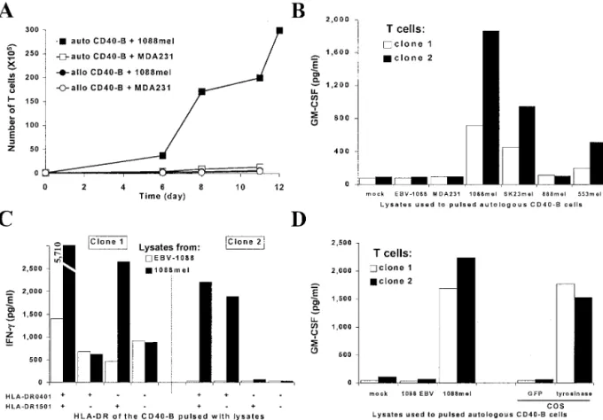
CD40-activated B cells were pulsed with a lysate prepared from a melanoma cell line and were used to stimulate autologous T cells from blood. In experiment 1, PBMCs were stimulated in a 96-well plate with autologous CD40-activated B cells pulsed with a lysate prepared from the melanoma cell line 1088mel. After three stimulations with 1088mel lysate-pulsed B cells, all of the 96-wells were individually tested against CD40-activated B cells pulsed with lysates prepared from 1088mel or from the irrelevant breast cancer line MDA231. Cells from 5 of the 96 wells were more reactive against the 1088mel lysate-pulsed B cells (Fig. 4A) compared with those pulsed with the MDA231 lysate. These results suggest that T cells reactive against antigens expressed in 1088mel were generated.
In a second experiment, CD4⫹T cells purified from a different donor were stimulated three times with 1088mel lysate-pulsed autol-ogous CD40-activated B cells 10 days apart in 1 well of a 48-well plate. The T-cell culture was cloned by limiting dilution and from 80 clones screened, 2 were consistently reactive against CD40-stimulated B cells pulsed with 1088mel lysate but not against those pulsed with a lysate prepared from MDA231 (Fig. 4B). The recognition was specific for melanoma because both clones failed to recognize a lysate prepared from EBV-immortalized B cells generated from patient 1088, excluding the possibility of recognition of allogeneic antigens. Both T-cell clones were expanded using B cells pulsed with 1088mel lysate as stimulators. Clone 2 specifically expanded when stimulated with autologous B cells pulsed with 1088mel lysate (Fig. 5A), but failed to proliferate when stimulated with autologous B cells pulsed with a lysate from MDA231 or allogeneic B cells pulsed with either lysates from 1088mel or MDA231. Additional characterization of these two T-cell clones revealed that the antigen recognized was shared by at least two other melanoma cell lines (SK23mel and 553mel) when lysates were pulsed on B cells (Fig. 5B). Also, the recognition of CD40-activated B cells pulsed with 1088mel lysate was inhibited using a blocking pan-MHC class II antibody or an anti-HLA-DR antibody (data not shown), suggesting that the recognition was HLA-DR restricted. HLA-restriction was additionally character-ized using CD40-activated B cells from HLA-DR-matched donors pulsed with 1088mel lysates. Clone 1 recognized only pulsed CD40-activated B cells prepared from HLA-DR1ⴱ1501 donors (Fig. 5C).
In contrast to clone 1, clone 2 recognized only pulsed CD40-activated B cells prepared from HLA-DR1ⴱ0401 donors, demonstrating that recognition by clones 1 and 2 was, respectively, HLA-DR1ⴱ1501 and HLA-DR1ⴱ0401 restricted.
To determine the antigen recognized, lysates were prepared from COS cells transfected with a panel of known melanoma tumor anti-gens (4, 22, 32). By using these pulsed B cells as targets for clones 1 and 2, tyrosinase was identified to be the antigen recognized by both clones (Fig. 5D). This observation was confirmed using B cells pulsed with lysates prepared from COS cells infected with a fowlpox virus vector expressing tyrosinase (data not shown).
In summary, T cells reactive against melanoma were generated using CD40-activated B-lymphocytes pulsed with lysates prepared from melanoma cells. These data suggest that CD40-activated B cells are efficient APCs in the presentation of exogenous antigens by MHC class II molecules and have the capacity to generate antigen-specific T cells.
Finally, clone 2 was used to examine whether B-cell stimulation
Fig. 3. Differential expression of HLA-DR, -DM, and -DO in CD40-activated B lymphocytes. Purified B cells were cultured in vitro in the absence (nonstimulated for 2 days; NS) or presence of 500 ng/ml of soluble trimeric CD40L and 200 units/ml of IL-4 for the indicated periods of time. A, extracts were prepared and analyzed by Western blots using antibodies specific to the indicated molecules. Representative blots done on 2 donors are presented. B, the band intensities from the autoradiograms were evaluated and the relative ratios for HLA-DR compared with -DO (DR/DO) and HLA-DM compared with -DO (DM/DO) were calculated.
Fig. 4. Generation of 1088mel-specific T lymphocytes using CD40-activated B cells pulsed with a tumor lysate. CD40-activated B cells were pulsed for 18 –24 h with a lysate prepared from the melanoma line 1088mel. A, autologous PBMCs were stimulated three times with irradiated pulsed CD40-activated B cells as described in the “Materials and Methods.” Single wells were assayed using B cells pulsed with lysates prepared from 1088mel or MDA231. Two representative negative cultures are shown on the left side. B, purified CD4⫹T cells were stimulated three times with irradiated pulsed autologous CD40-activated B cells, and cloning was done by limiting dilution as described in “Materials and Methods.” Two clones were consistently reactive against CD40-stimulated B cells pulsed with a lysate prepared from 1088mel and failed to secrete GM-CSF when incubated with B cells pulsed with lysates prepared from MDA231 or EBV-B cells from donor 1088. For A and B, T cells and targets were cocultured for 20 –24 h, supernatants were collected and GM-CSF secretion was evaluated by ELISA (pg/ml).
was required for the presentation of the HLA-DR1ⴱ0401-restricted tyrosinase epitope. Purified B cells were left unstimulated or were activated by CD40L, IL-4, or a combination of these. B cells were then pulsed with lysates from 1088mel and used to stimulate clone 2. Clone 2 was reactive against B cells pulsed with antigen on day 0 and stimulated with CD40L alone (Fig. 6A; CD40L d0) or a combination of CD40L and IL-4 (CD40L/IL-4 d0), but failed to recognize B cells that were either nonstimulated (NS d0) or stimulated with IL-4 alone (IL-4 d0). The recognition of B cells cultured with CD40L and IL-4 for 5 days before antigen exposure was better compared with any other condition. These observations confirmed that short-term stimu-lation with CD40L is sufficient to increase the capacity of B cells to present exogenous antigens by MHC class II, as presented previously in Fig. 2A. Similarly, purified B cells cultured with CD40L or a combination of CD40L and IL-4, pulsed with 1088mel lysate 2 days later were efficiently recognized by clone 2 (Fig. 6B; CD40L d2 and CD40L/IL-4 d2). The recognition was specific to tyrosinase, because CD40L/IL-4-stimulated B cells were not recognized when pulsed with control lysate prepared from 1088-EBV-B cells (Fig. 6, A and B), which do not express tyrosinase.
These data additionally validate the original observation suggesting that activation of human B cells with CD40L enhances the capacity to present an exogenous tumor antigen by MHC class II.
DISCUSSION
In the present work, we studied the consequences of CD40-activa-tion on antigen presentaCD40-activa-tion and T-cell stimulatory capacity of normal
human antigen-unspecific B lymphocytes. Results presented show that CD40-activation of B cells increased their ability to present exogenous antigens by MHC class II. Indeed, B lymphocytes have critical functions of antigen uptake, processing, and presentation in the context of the humoral response, where antigens are taken up by the rare B cells expressing antigen-specific surface immunoglobulins, a situation that has been extensively studied. In the present paper, we show that after CD40-activation, nontransformed normal human B lymphocytes can uptake, process, and present exogenous antigens independent of the surface immunoglobulins. Interestingly, this ex-perimental system allows the culture of primary B cells for up to 3 weeks, and a direct correlation was found between the length of activation and their ability to stimulate T cells. These CD40-activated B cells pulsed with a lysate prepared from melanoma cells had the capacity to generate T cells specific to melanoma. Our data suggest that indeed, when properly activated, B cells can present exogenous antigens by MHC class II molecules and stimulate antigen-specific T cells.
B-cell activation modulates the activity of other processes involved in antigen presentation such as increase in fluid-phase endocytosis, increase in the expression of costimulatory molecules (as shown in Fig. 1C demonstrating increased CD86 expression; Refs. 9, 33), and secretion of immunomodulatory cytokines (34). It is possible that activation of B cells by CD40L results in a number of changes leading to enhanced antigen presentation and T-cell stimulatory capacity. In this study, MHC class II antigen presentation by B cells correlated with the differential expression of classical and nonclassical MHC
Fig. 5. T-cell clones generated by stimulation with melanoma lysate-pulsed activated B cells recognized tyrosinase epitopes presented by HLA-DR1ⴱ0401 and HLA-DR1ⴱ1501.
A, proliferation of clone 2 (1⫻ 105on day 0) after stimulation with autologous or HLA-DR-unmatched (allogeneic) CD40-activated B cells pulsed with 1088mel or MDA231 lysates. Viable cells were counted on the basis of trypan blue exclusion using a hemacytometer. B–D, clones 1 and 2 were cocultured for 18 –24 h with (B) autologous CD40-activated B cells pulsed with lysates prepared from 1088-EBV-B cells or from different tumor lines as indicated (MDA 231 is a breast cancer line, all others are melanoma lines), (C) CD40-stimulated B cells prepared from HLA-DR matched donors pulsed with lysates prepared from 1088-EBV-B cells or 1088mel, and (D) autologous CD40-activated B cells pulsed with lysates prepared from 1088-EBV-B cells, 1088mel, or COS cells expressing green fluorescent protein or tyrosinase. Supernatants were collected and GM-CSF (B and D) or IFN-␥ (C) were assayed by ELISA (pg/ml). auto CD40-B, autologous CD40-activated B cells; allo CD40-B, allogeneic CD40-activated B cells.
class II molecules after activation by CD40L. The expression of HLA-DO, a molecule described previously to alter the MHC class II/peptide repertoire and to inhibit the presentation of nonspecific exogenous soluble antigens, was decreased after B-cell activation with CD40L. By using B cells stimulated with an antibody specific to IgM compared with unstimulated cells, Roucard et al. (35) have reported a 50% decrease in the expression of HLA-DO, but no functional anal-ysis of antigen processing was performed. In contrast to our work, no decrease in HLA-DO expression was observed when stimulating B cells with IL-4 and an antibody against CD40. Perhaps the decrease in HLA-DO that we observed after activation was because of B-cell stimulation with the more physiological soluble trimeric form of CD40L compared with the use of an antibody against the receptor. An increase in the HLA-DR:DO and HLA-DM:DO ratios should allow the loading of a broader repertoire of peptides, which bind with more stability to surface HLA molecules. Additionally, antigen-unspecific B cells that become activated by T cells may also increase their capacity to load antigens in less acidic compartments because of the diminished expression of HLA-DO. The down-regulation of HLA-DO might be one of several events leading to improved APC functions in addition to other mechanisms enumerated earlier. The exact mecha-nisms by which DO is down-regulated and potentially influences antigen presentation are currently under investigation.
The results presented suggest that, after CD40-activation, antigen-unspecific normal human B cells can efficiently present exogenous antigens through MHC class II. CD40L is mainly expressed on activated CD4⫹T cells (36). In vivo, unspecific B lymphocytes could
receive CD40 stimulation from activated CD4⫹ T cells during an immune response. We hypothesize that antigen-unspecific B cells receive stimulatory signals from activated CD4⫹T cells, and as a consequence, B cells present exogenous antigens to T cells. The involvement of antigen-unspecific B cells in T-cell activation could enhance the magnitude of the T-cell response in an immune reaction. Although we used weakly immunogenic tumor cells as a source of antigen, B cells may also play a role in presenting more immunogenic foreign antigens after viral or bacterial infections. Additional work is needed to define the relevance of T-cell activation by antigen-unspe-cific B cells in vivo.
CD40-activated B cells can present exogenous antigens by MHC class II, and they are effective in the generation of antigen-specific T cells. For in vitro use, B cells have a number of advantages compared with other APCs, such as DCs, including a high proliferation rate, which allows the generation of a many activated B cells, even from cryopreserved PBMCs. This would allow the generation of an effec-tive source of APCs when a small amount of blood is available (27). Antigen-specific T cells generated from lysate-pulsed activated B cells could be used to identify new tumor antigens from common cancers for vaccine development. In addition, tumor antigen-pulsed CD40-activated B cells could be used to generate specific T cells for adoptive immunotherapy, which has been demonstrated recently to result in high response rates in melanoma patients (37).
ACKNOWLEDGMENTS
We thank Dr. Angela Samaan for providing antibodies and Dr. Suzanne L. Topalian for help in the characterization of the antityrosinase T-cell clones and critical review of the manuscript. We also thank Diane Beauseigle and Ste´phanie Lepage for technical assistance.
REFERENCES
1. Rock, K. L., Benacerraf, B., and Abbas, A. K. Antigen presentation by hapten-specific B lymphocytes. I. role of surface immunoglobulins. J. Exp. Med., 160: 1102–1113, 1984.
2. Lanzavecchia, A. Antigen-specific interaction between T and B cells. Nature (Lond.),
314: 537–539, 1985.
3. Topalian, S. L., Rivoltini, L., Mancini, M., Ng, J., Hartzman, R. J., and Rosenberg, S. A. Melanoma-specific CD4⫹ T lymphocytes recognize human melanoma antigens processed and presented by Epstein-Barr virus-transformed B cells. Int. J. Cancer, 58: 69 –79, 1994.
4. Topalian, S. L., Rivoltini, L., Mancini, M., Markus, N. R., Robbins, P. F., Kawakami, Y., and Rosenberg, S. A. Human CD4⫹ T cells specifically recognize a shared melanoma-associated antigen encoded by the tyrosinase gene. Proc. Natl. Acad. Sci. USA, 91: 9461–9465, 1994.
5. Zhong, G., Sousa, C. R., and Germain, R. N. Antigen-unspecific B cells and lymphoid dendritic cells both show extensive surface expression of processed antigen-major histocompatibility complex class II complexes after soluble protein exposure in vivo or in vitro. J. Exp. Med., 186: 673– 682, 1997.
6. Ozaki, M. E., Coren, B. A., Huynh, T. N., Redondo, D. J., Kikutani, H., and Webb, S. R. CD4⫹ T cell responses to CD40-deficient APCs: defects in proliferation and negative selection apply only with B cells as APCs. J. Immunol., 163: 5250 –5256, 1999.
7. Evans, D. E., Munks, M. W., Purkerson, J. M., and Parker, D. C. Resting B lymphocytes as APC for naive T lymphocytes: dependence on CD40 ligand/CD40. J. Immunol., 164: 688 – 697, 2000.
8. Bennett, S. R., Carbone, F. R., Toy, T., Miller, J. F., and Heath, W. R. B cells directly tolerize CD8(⫹) T cells. J. Exp. Med., 188: 1977–1983, 1998.
9. Akiba, H., Oshima, H., Takeda, K., Atsuta, M., Nakano, H., Nakajima, A., Nohara, C., Yagita, H., and Okumura, K. CD28-independent costimulation of T cells by OX40 ligand and CD70 on activated B cells. J. Immunol., 162: 7058 –7066, 1999. 10. Buhlmann, J. E., Foy, T. M., Aruffo, A., Crassi, K. M., Ledbetter, J. A., Green, W. R.,
Xu, J. C., Shultz, L. D., Roopesian, D., Flavell, R. A., Fast, L., Noelle, R. J., and Durie, F. H. In the absence of a CD40 signal. B cells are tolerogenic. Immunity, 2: 645– 653, 1995.
11. Croft, M., Joseph, S. B., and Miner, K. T. Partial activation of naive CD4 T cells and tolerance induction in response to peptide presented by resting B cells. J. Immunol.,
159: 3257–3265, 1997.
12. Faassen, A. E., Dalke, D. P., Berton, M. T., Warren, W. D., and Pierce, S. K. CD40-CD40 ligand interactions stimulate B cell antigen processing. Eur. J. Immunol.,
25: 3249 –3255, 1995.
Fig. 6. Presentation of tyrosinase by MHC class II by activated B cell. Purified B cells were concomitantly pulsed with a lysate prepared from 1088mel and activated (d0) for 18 –24 h (A) or were preactivated for 2 days (d2) and pulsed with lysate for an additional 18 –24 h (B) as described in “Materials and Methods.” B cells preactivated with CD40L and IL-4 for 5 or 12 days were subsequently pulsed with lysate for 18 –24 h as indicated (d5 and d12, respectively). Antigen processing was then assessed by coculturing pulsed B cells with the tyrosinase-reactive T-cell clone 2 for an additional 18 –24 h. Culture supernatants were collected, and IFN-␥ was measured using an ELISA assay (pg/ml).
13. Bakke, O., and Dobberstein, B. MHC class II-associated invariant chain contains a sorting signal for endosomal compartments. Cell, 63: 707–716, 1990.
14. Ghosh, P., Amaya, M., Mellins, E., and Wiley, D. C. The structure of an intermediate in class II MHC maturation: CLIP bound to HLA-DR3. Nature (Lond.), 378: 457– 462, 1995.
15. Denzin, L. K., and Cresswell, P. HLA-DM induces CLIP dissociation from MHC class II␣  dimers and facilitates peptide loading. Cell, 82: 155–165, 1995. 16. van Ham, S. M., Gruneberg, U., Malcherek, G., Broker, I., Melms, A., and Trowsdale,
J. Human histocompatibility leukocyte antigen (HLA)-DM edits peptides presented by HLA-DR according to their ligand binding motifs. J. Exp. Med., 184: 2019 –2024, 1996.
17. Sherman, M. A., Weber, D. A., and Jensen, P. E. DM enhances peptide binding to class II MHC by release of invariant chain-derived peptide. Immunity, 3: 197–205, 1995.
18. van Ham, M., van Lith, M., Griekspoor, A., and Neefjes, J. What to do with HLA-DO? Immunogenetics, 51: 765–770, 2000.
19. Liljedahl, M., Kuwana, T., Fung-Leung, W. P., Jackson, M. R., Peterson, P. A., and Karlsson, L. HLA-DO is a lysosomal resident which requires association with HLA-DM for efficient intracellular transport. EMBO J., 15: 4817– 4824, 1996. 20. van Ham, M., van Lith, M., Lillemeier, B., Tjin, E., Gruneberg, U., Rahman, D.,
Pastoors, L., van Meijgaarden, K., Roucard, C., Trowsdale, J., Ottenhoff, T., Pappin, D., and Neefjes, J. Modulation of the major histocompatibility complex class II-associated peptide repertoire by human histocompatibility leukocyte antigen (HLA)-DO. J. Exp. Med., 1127–1136, 2000.
21. van Ham, S. M., Tjin, E. P., Lillemeier, B. F., Gruneberg, U., van Meijgaarden, K. E., Pastoors, L., Verwoerd, D., Tulp, A., Canas, B., Rahman, D., Ottenhoff, T. H., Pappin, D. J., Trowsdale, J., and Neefjes, J. HLA-DO is a negative modulator of HLA-DM-mediated MHC class II peptide loading. Curr. Biol., 7: 950 –957, 1997. 22. Lapointe, R., Royal, R. E., Reeves, M. E., Altomare, I., Robbins, P. F., and Hwu, P.
Retrovirally-transduced human dendritic cells can generate T cells recognizing mul-tiple MHC class I and class II epitopes from the melanoma antigen gp100. J. Immu-nol., 167: 4758 – 4764, 2001.
23. Lapointe, R., Lemieux, R., Olivier, M., and Darveau, A. Tyrosine kinase and cAMP-dependent protein kinase activities in CD40-activated human B lymphocytes. Eur. J. Immunol., 26: 2376 –2382, 1996.
24. Guy, K., Van Heyningen, V., Cohen, B. B., Deane, D. L., and Steel, C. M. Differ-ential expression and serologically distinct subpopulations of human Ia antigens detected with monoclonal antibodies to Ia␣ and  chains. Eur. J. Immunol., 12: 942–948, 1982.
25. Brunet, A., Samaan, A., Deshaies, F., Kindt, T. J., and Thibodeau, J. Functional characterization of a lysosomal sorting motif in the cytoplasmic tail of HLA-DO. J. Biol. Chem., 275: 37062–37071, 2000.
26. Dudley, M. E., Nishimura, M. I., Holt, A. K., and Rosenberg, S. A. Antitumor immunization with a minimal peptide epitope (G9 –209-2M) leads to a functionally heterogeneous CTL response. J. Immunother., 22: 288 –298, 1999.
27. Schultze, J. L., Michalak, S., Seamon, M. J., Dranoff, G., Jung, K., Daley, J., Delgado, J. C., Gribben, J. G., and Nadler, L. M. CD40-activated human B cells: an alternative source of highly efficient antigen presenting cells to generate autologous antigen-specific T cells for adoptive immunotherapy. J. Clin. Investig., 100: 2757– 2765, 1997.
28. Liljedahl, M., Winqvist, O., Surh, C. D., Wong, P., Ngo, K., Teyton, L., Peterson, P. A., Brunmark, A., Rudensky, A. Y., Fung-Leung, W. P., and Karlsson, L. Altered antigen presentation in mice lacking H2-O. Immunity, 8: 233–243, 1998. 29. Kropshofer, H., Vogt, A. B., Thery, C., Armandola, E. A., Li, B. C., Moldenhauer, G.,
Amigorena, S., and Hammerling, G. J. A role for HLA-DO as a co-chaperone of HLA-DM in peptide loading of MHC class II molecules. EMBO J., 17: 2971–2981, 1998.
30. Alfonso, C., and Karlsson, L. Nonclassical MHC class II molecules. Annu. Rev. Immunol., 18: 113–142, 2000.
31. Renkvist, N., Castelli, C., Robbins, P. F., and Parmiani, G. A listing of human tumor antigens recognized by T cells. Cancer Immunol. Immunother., 50: 3–15, 2001. 32. Touloukian, C. E., Leitner, W. W., Topalian, S. L., Li, Y. F., Robbins, P. F.,
Rosenberg, S. A., and Restifo, N. P. Identification of a MHC class II-restricted human gp100 epitope using DR4-IE transgenic mice. J. Immunol., 164: 3535–3542, 2000. 33. Cassell, D. J., and Schwartz, R. H. A quantitative analysis of antigen-presenting cell function: activated B cells stimulate naive CD4 T cells but are inferior to dendritic cells in providing costimulation. J. Exp. Med., 180: 1829 –1840, 1994.
34. Schultze, J. L., Michalak, S., Lowne, J., Wong, A., Gilleece, M. H., Gribben, J. G., and Nadler, L. M. Human non-germinal center B cell interleukin (IL)-12 production is primarily regulated by T cell signals CD40 ligand, interferon␥, and IL-10: role of B cells in the maintenance of T cell responses. J. Exp. Med., 189: 1–12, 1999. 35. Roucard, C., Thomas, C., Pasquier, M., Trowsdale, J., Sotto, J., Neefjes, J., and van
Ham, M. In vivo and in vitro modulation of HLA-DM and HLA-DO is induced by B lymphocyte activation. J. Immunol., 167: 6849 – 6858, 2001.
36. Armitage, R. J., Fanslow, W. C., Strockbine, L., Sato, T. A., Clifford, K. N., Macduff, B. M., Anderson, D. M., Gimpel, S. D., Davis-Smith, T., and Maliszewski, C. R. Molecular and biological characterization of a murine ligand for CD40. Nature (Lond.), 357: 80 – 82, 1992.
37. Dudley, M. E., Wunderlich, J. R., Robbins, P. F., Yang, J. C., Hwu, P., Schwartzentruber, D. J., Topalian, S. L., Sherry, R., Restifo, N. P., Hubicki, A. M., Robinson, M. R., Raffeld, M., Duray, P., Seipp, C. A., Rogers-Freezer, L., Morton, K. E., Mavroukakis, S. A., White, D. E., and Rosenberg, S. A. Cancer regression and autoimmunity in patients after clonal repopulation with antitumor lymphocytes. Science (Wash. DC), 298: 850 – 854, 2002.