Université de Montréal
Devetoprnent ofa nove! approach for brain de!ivery:
Dendritic nanocarriers for tue enhanced de!iveiy of
metitotrexate to the brain turnors
par Renu Singh
Faculté de Pharmacie
Thèse présentée à la Faculté des études supérieures en vue de l’obtention du grade de Philosophiae Doctor (Ph. D.)
en Sciences Pharmaceutique option Technologie Pharmaceutique
]uly. 2007
ZOOB © Renu Singh. 2007
Q
Université
de Montréal
Direction des bib1othèques
AVIS
L’auteur a autorisé l’Université de Montréal à reproduite et diffuser, en totalité ou en partie, par quelque moyen que ce soit et sur quelque support que ce soit, et exclusivement à des fins non lucratives d’enseignement et de
recherche, des copies de ce mémoire ou de cette thèse.
L’auteur et les coauteurs le cas échéant conservent la propriété du droit
d’auteur et des droits moraux qui protègent ce document. Ni la thèse ou le mémoire, ni des extraits substantiels de ce document, ne doivent être imprimés ou autrement reproduits sans l’autorisation de l’auteur.
Afin de se conformer à la Loi canadienne sur la protection des
renseignements personnels, quelques formulaires secondaires, coordonnées
ou signatures intégrées au texte ont pu être enlevés de ce document. Bien
que cela ait pu affecter la pagination, il n’y a aucun contenu manquant. NOTICE
The author of this thesis or dissertation has granted a nonexciusive license allowing Université de Montréal to reproduce and publish the document, in part or in whole, and in any format, solely for noncommercial educational and
research purposes.
The author and co-authors if applicable retain copyright ownership and moral
rights in this document. Neither the whole thesis or dissertation, flot
substantial extracts from it, may be printed or otherwise reproduced without the author’s permission.
In compliance with the Canadian Privacy Act some supporting forms, contact
information or signatures may have been removed from the document. While this may affect the document page count, it does not represent any loss of
Université de Montréal Faculté des études supérieures
Cette thèse intitulée
Development ofa nove! approach for brain delivery: Dendritic nanocarriers for the enhanced de!ivery ofmethotrexate to the brain tumors
présentée par: Renu Singh
a été évaluée parunjury composé des personnes suivantes:
Dr. Françoise Winnik, président-rapporteur Dr. Patrice Hi!dgen, directeur de recherche Dr. Jean-françois Bouchard. membre du jury
Dr. Grégoire Leclair, examinateur externe représentant du doyen de la FIS
111
Résumé
Dans ce projet, un nouveau dendrirnère polyester-co-polyéther (PEPE) a été conçu et évalué comme système de livraison de médicament ayant la capacité de traverser la barrière hémato-encéphalique (BHE). Une série de dendrimères PEPE dont le noyau est l’acide butane tétracarboxylique a été synthétisée. Des variations de l’architecture ont été faites en changeant le nombre de branches, d’unités de branchement, de générations, les groupes terminaux, et la longueur des chaînes de polyoxyéthylène (PEO). La biocompatibilité du dendrimère a été vérifiée in vitro en mesurant la cytotoxicité sur des cellules endothéliales (bEnd.3) et leur potentiel hémolytique sur des hématies de rat (RBCs). L’adsorption des proteines plasmatiques à la surface des dendrimères a aussi été évaluée. Le mécanisme d’intemalisation a été élucidé en étudiant le devenir dans des cellules bEnd.3, de dendrimères marqués à la rhodamine. Le méthotrexate (MTX) a été choisi comme modèle thérapeutique en raison de sa faible perméabilité à travers la BHE mais aussi pour sa bonne activité contre les tumeurs cérébrales. La capacité de ces dendrimères de traverser la BHE a été vérifiée en utilisant un modèle in vitro de culture conjointe de bEnd.3 et de cellules U373 MG. Finalement, l’efficacité des dendrimères chargés avec du MIX a été établie sur une couche monocellulaire de gliome humain (U$7 MG et U343 MG-A) et des sphéroïdes avasculaires de gliome humain. Les dendrimères n’ont montré aucune toxicité sur bEnd.3 à des concentrations aussi hautes que 10 mg/ml. Ils ont provoqué moins de 10% d’hémolyse à 5 mg/ml. Les protéines d’opsonisation tels que les immunoglobulines et les facteurs du complément ne s’adsorbent pas à la surface des dendrimères et le taux de liaisons aux protéines plasmatiques est très bas. Ces dendrimères sont capables de traverser la BHE en grande quantité et d’augmenter significativement la quantité de MTX disponible comparée à du MTX libre. La glycosylation des dendrimères PEPE augmente également le passage trans-BHE comparéà celui des dendrimères non glycosylés. L’1C50 du MIX après encapsulation dans les dendrimères est plus faible que celle du MTX libre, ce qui suggère un meilleur potentiel. De même, les dendrimères
iv
glycosylés ont montré une meilleure activité quand on mesure la réduction de volume des sphéroïdes tumoraux. Ces dendrimères sont même capables de tuer des cellules résistantes au MTX, mettant ainsi en lumière une possibilité de contrer cette résistance. Aussi intéressante est la possibilité d’atteindre même les régions centrales et non oxygénées des tumeurs non-vascularisées. Finalement les dendrimères PEPE glycosylés constituent un système de livraison de médicament très prometteur pour le traitement des gliomes comme pour d’autres agents thérapeutiques dans le cerveau.
V
Abstract
In the present project, the suitability of novel polyether-co-polyester (PEPE) dendrimers for drug delivery across the blood brain barrier (BBB) and in the treatment of gliomas was evaluated. A series of PEPE dendrimers consisting of butane tetracarboxylic acid as the core molecule were synthesized. Modifications in the architecture were accomplished by varying the number of branches, branching units, terminal functional groups, generation and the chain length of polyethylene oxide (PEO) linking the branches to the interior cavity. The biocompatibility of the resulting dendrirners was evaluated in vitro by assessing their cytotoxicity on brain endothelial celis (bEnd.3) and their hemolytic potential against rat red blood ceils (RBCs). Plasma protein adsorption on the surface of dendrimers was also evaluated. The intemalization of rhodamine Iabeled dendrimers into bEnd.3 celis was studied to comprehend the mechanism of uptake. Methotrexate (MTX) was selected as mode! chemotherapeutic agent because of its poor BBB permeability but at the same time good activity against brain tumors. The ability of the dendrimers to cross BBB was ascertained by using an in vitro model consisting of co-culture of bEnd.3 and U373 MG celis. Finally, the cfficacy of MTX Ïoaded dendrimers was established against monolayer human glioma ceil lines. narnely U$7 MG and U343 MG-A and their avascular human glioma tumor spheroids. PEPE dendrimers showed no toxicity towards the bEnd.3 celis at concentration as high as 5 mg/mL and also produced less than 10% lysis of rat RBC’s even at 5 mg/mL. Opsoninic proteins like immunoglobulin and complement factors were flot adsorbed on the surface of the dendrimers; additionally, the total amount of protein adsorbed was also low. The dendrimers were able to permeate BBB in large amount and thus. significantly increase the availability of MIX across BBB mode! as compared to free MIX. Glucosylation of PEPE dendrimers was found to further increase their permeation across BBB as compared to non-glucosylated dendrimers and hence. transport of MTX into the receiver compartment. Ihe 1C50 of MTX afier loading in dendrimers was lower than that of free MTX, suggesting that loading MIX in PEPE dendrimers increased
vi
its potency. Similar higher activity of MTX loaded glucosyÏated and non-glucosylated dendrimers was found in the reduction of volume of turnor spheroids. The MIX loaded dendrimers were able to kili even MTX resistant ceils, highlighting, there ability to overcome MIX resistance. Most interestingly, glucosylation augmented the rate and extent of delivery of dendrirners to the central and hypoxic regions of the avascular turnor spheroids. Thus. glucosylated PEPE dendrimers can serve as a prornising delivery system for the treatment of gliomas and also for delivery of other therapeutic agents into the brain.
vii
Table of Contents
List of figures xv
List of tables xxi
List of schemes xxii
List of abbreviations xxiii
List of equations xxvii
ACKNOWLEDGEMENTS xxix
INTRODUCTION 1
1.1.Cancer 2
1.2. Brain cancer 3
1.3. Physiological barriers in delivery oftherapeutics to the brain tumors 5
1.3.1. BÏood-brain barrier 5
1.3.2. Blood-cerebrospinaïfluid barrier (3CR) 5
1.3.3. BÏood-tumor barrier (BTB,) 6
1.3.4. Effiux transporters 6
1.4. Drug transport at the BBB 8
1.4.1. Diffusion $
1.4.2. Endocytosis $
1.5. Standard therapies for brain tumors 10
1.5.1. Surgery 10 1.5.2. Radiation therapy 11 1.5.3. Chernotherapy 12 1.5.4. Irnrnunotherapy 13 1.5.5. Gene therapy 15 1.5.6. Antiangiogenic therapy 16 1.5.7. Combination therapy 17
1.6. Challenges in delivery to brain tumors 1$
viii
1.6.1.]. Modification in the drug molecules . 20
1.6.1.2. Prodrug approaches 23
1.6.1.2.1. Lipophilic prodrugs 23
1.6.1.2.2. Antibody-directed enzymeprodrug therapy (ADEP7) and Gene directed enzyme
prodrug therapy (GDEFT) 24
1.6.1.3. Chemical deÏivery systems 25
1.6.1.4. Disruption ofBBB 25
1.6.1.5. Conjugating vectors ofcarrier-mediated endocytosis 26 1.6.1.6. Conjugating vectors ofreceptor-mediated endocytosis 27
1.6.2. Physical based stralegies 27
1.6.2.1. Catheter delivery systems 28
1.6.2.2. Implantable Pumps 28
1.6.2.3. Convection enhanced delivery (CED) 2$
1.6.2.4. Polymer based delivery systems /InterstitiaÏ chemotherapy 29
1.6.3. IntravascuÏar techniques 30
1.6.3.1. High-dose intravenous chemotherapy 30
1.6.3.2. Intra-arterial chemotherapy 30
1.6.3.3. CoÏloidal carriers 31
1.6.3.3.1. Polymeric micelles 31
1.6.3.3.2. Liposomes 32
A). FEGylated liosonies 32
B). Cationic liposonies 33
C). Targeted lzposonies 33
1.6.3.3.3. Nanoparticles 34
A). Coated nanoparticles 35
B). PEGyÏated nanoparticles 36
1.6.3.3.4. Nanogel 36
ix
A). Molecular structure of the dendrimers . 3$
B,). Synthesis ofdendrimers 39
a). Divergent Synthesis 40
b). Convergent Synthesis 41
C). Froperties ofdendrimers 42
D). Dendrimers in drug deliveiy 43
ci,). Dendrimer drug conjugates 43
b,). Complexation ofdrugs with polar groups on the dendrimer surface 44
c). Encapsulation ofdrugs withinci dendrimer 45
i). Unimolecular micelles 46
ii). Cored dendrimers 47
iii). Dendritic box 48
iv,). Dendrimer-based block copolymers 49
y,). Dendrimers with PEG grafts 51
E). Limitations ofdendrimers- toxicity and biodistribution 52
ci). Toxicity 53
b). Biodistribution 54
F). Designing dendrimers based on PEG 54
1.7. Application ofdendrimers in delivery to brain tumors 55
1.8. Methotrexate 56
HYPOTHESIS 5$
OBJECTIVE 61
CHAPTER ONE
Synthesis and evaluation of novel dendrimers with hydrophilic interior as nanocarriers for
drug delivery 80
2.1. Abstract $1
2.2. Introduction $2
X
2.4. Experimental procedures . $5
2.1.]. Characterization techniques $5
2.4.2. Synthesis ofthe core (3,): 85
2.4.3. Synthesis ofpoÏy’ôxyethylene) monomethacrylate carboxyÏic acid (4): $6
2.4.4. Synthesis ofPOE-DH3A (6): 87
2.4.5. Synthesis ofdendron], FOE-DHBA-PEG (7) 8$
2.4.6. Synthesis ofden-1-(G2) (8): $9
2.4.7. Synthesis ofden-1-(G2)-OH (9): $9
2.4.8. Synthesis ofden-1-(G3) (10): 90
2.4.9. Synthesis ofPOE-gaÏÏate (1]): 91
2.4.10. Synthesis of dendron 2, POE-gaÏÏate-PEG (12): 92
2.4.11. $ynthesis ofden-2-(’G2,) (13): 92
2.4.12. Determination ofacid value ofthe core 93
2.4.13. Determination ofhydroxyl value of den-1-(’G2,)-OH 97
2.4.14. Particle size measurements 97
2.4.15. Dfferential scanning caÏorimetiy (DSÇ.) 98
2.4.16. Atomic force microscopy (AFM) 9$
2.4.17. Encapsulation studies 9$
2.4.18. Release studies 99
2.5. Resuits and discussion 100
2.5.1. Synthesis and characterization ofdendrimers 100
2.5.2. Farticle size measurements 106
2.5.3. Thermal properties 109
2.5.4. Atomic force microscopy 110
2.5.5. Encapsulation and release studies 111
2.6. Conclusion 118
2.7. Acknowledgernents 119
xi
CHAPTER TWO
Influence of molecular architecture of polyether-co-polyester dendrirners on the
encapsulation and release of methotrexate 125
3.1. Abstract 126 3.2. Introduction 127 3.3. Experimental 129 3.3.1. Materials 129 3.3.2. Synlhesis ofdendrirners 129 3.3.3. Characterization ofdendrimers 130
3.3.4. Hydrodynarnic size measurements 131
3.3.5. Cytotoxicity $tudies 131
3.3.6. DSCstudies 131
3.3.7. Drugloading 132
3.3.8. Mechanisrn ofdrug encapsulation 132
3.3.9. Release studies 133
3.4. Results and Discussion 133
3.4.1. Synthesis and characterization ofdendrirners 136
3.1.2. Hydrodynarnic size 139 3.1.3. Cytotoxicity ofdendrirners 139 3.1.1. Encapsulation studies 141 3.1.5. DSC studies 142 3.1.6. Mechanisrn of encapsulation 144 3.4.7. MTXrelease 14$ 3.5. Conclusion 151 3.6. Acknowledgements 152 3.7. References 153 3.8. Supporting information 157
xii
CHAPTER THREE
Polyether-co-polyester dendrimers for delivery across the blood brain barrier 164
4.1. Abstract 165 4.2. Introduction 166 4.3. Experimental 16$ 1.3.1. Materials 16$ 1.3.2. Methods 169 4.3.2.1. Dendrirners evaluated 169
4.3.2.2. fluorescent labeling ofdendrimers 172
4.3.2.3. Cytotoxicity ofdendrimers 172
1.3.2.4. Hernolytic potential ofdendrimers 173
1.3.2.5. Adsorption ofplasrna prote ins on dendrimers 173
4.3.2.6. Endocytosis of dendrirners 174
4.3.2.7. Mechanism ofendocytosis 175
1.3.2.8. Microscopic exarnination ofendocytosis 176
1.3.2.9. 333 model 176
A). F-gp expression assay 177
B,). Bioelectric properties andperrneability assessment 177
4.3.2.10. Transport ofdendrirners across 333 17$
4.4. Results and Discussion 17$
4.1.1. Cytotoxicity ofdendrirners 17$
4.4.2. Hemolyticpotential ofdendrimers 180
4.4.3. Adsoiption ofplasrna proteins on dendrirners 180
4.4.4. Endocytosis ofdendrimers in brain endothelial celis 181
1.1.5. Characterization of333 model 186
4.1.6. Transport ofdendrirners across 333 189
4.5. Conclusion 191
xlii
4.7. References. 193
4.8. Supporting Information 199
CHAPTER FOUR
Methotrexate loaded polyether-co-polyester dendrimers for the treatment of gliomas: enhanced efficacy and intraturnoral transport capability 201
5.1. Abstract 202 5.2. Introduction 203 5.3. Experimental 205 5.3.1. Materials 205 5.3.2. Methods 206 5.3.2.]. Dendrimers evaluated 206
5.3.2.2. Conjugation ofglucosamine to dendrimers 206
5.3.2.3. fluorescent ÏabeÏing ofthe dendrimers 207
5.3.2.1. MTX encapsulation 207
5.3.2.5. Internai ization ofdendrimers by glioma cells 20$
5.3.2.6. Intraceilular localization ofdendrimers 20$
5.3.2.7. Development ofMTXresistant U87 MG ceils 209
A). Enzyme assay 209
B,). MTX-fITC accumulation 209
5.3.2.8. Antirohferative activity ofdendrimers against giioma cells 210
5.3.2.9. Transport ofdendrimers andMTXacross BBB 210
5.3.2.10. Avascular human giioma tumor spheroids 211
A). Growth inhibition oftumor spheroids 211
B). Determinationofccii viability in tumorspheroids 212
C). Diffusion ofdendrimers into tumor spheroids 212
5.4. Resuits 213
5.1.1. Conjugation ofglucosamine to the dendrimers 213
xiv
5.4.3. Internalization ofdendrimers by gÏioma celÏs .213
5.4.1. CharacterizationofMlXresistant U87MG ceils 215 5.4.5. Antiproliferative activity ofdendrimers against gÏioma ceÏÏs 21 $
5.4.6. Transport ofdendrimers and MTX across 333 218
5.4.7. Interaction with avascular huinan glioma tumor spheroids 220
5.5. Discussion 224
5.6. Acknowledgements 235
5.7. References 236
5.8. Supporting information 242
DISCUSSION 244
6.1. Synthesis and characterization of dendrirners 246
6.2. Biological characterization 250
6.3. Drug loading in PEPE dendrimers 252
6.4. Release of encapsu!ated drug 253
6.5. Understanding mechanism ofuptake by brain endothelial celis 254 6.6. Permeability of PEPE dendrimers across BBB mode! 256 6.7. Efficacy against glioma ceil unes and avascular tumor spheroids: Enhanced delivery by
D-g!ucosamine tigand 259
CONCLUSION 264
xv
List of figures
Figure 1.1. Distribution of ail primary brain and CN$ tumors by histology 4
Figure 1.2. BBB and its anatomical characteristics 7
Figure 1.3. The various transport processes that may occur at the BBB 10 Figure 1.4. Permeability of various chemotherapeutic agents across BBB in relation to
partition coefficient 13
Figure 1.5. Various strategies that have been used to improve delivery of therapeutic
molecules to brain 21
Figure 1.6. Pictorial representation ofthe molecular architecture ofdendrirner 38 Figure 1.7. ta) Layer-block and (b) Segment-biock dendrimers 39 Figure 1.8. Divergent and convergent synthetic methods for dendrimer synthesis 41 Figure 1.9. Unimoiecular micelle consisting of 4, 4-bis(4’-hydroxyphenyl) pentanol core
and PEG mesylate sheil 47
Figure 1.10. Synthesis of cored dendrimers 48
Figure 1.11. Dendritic box of poiy(propyleneimine) dendrimers 49 Figure 1.12. Poly(ethylene glycol)-bÏock-poly(L-lysine) dendrimer 51 Figure 1.13. PEGylated PAMAM dendrimer with PEG chains attached to the terminal
amino groups 52
Figure 2.1. Structure ofden-1-(G2) 102
Figure 2.2. FTIR spectra’s of PEG monomethacrylate, POE-DHBA, dendron 1 and
dendrimer (den-1-(G2)) 103
Figure 2.3. 1HNMR spectra of(a) PEG-methacryiate, (b) POE-DHBA and (c) POE-DHBA
PEG in DM50 at 400 MHz 103
Figure 2.4 1HNMR spectrum of dendrimer (Den-1-(G2)) in DMSO at 400 MHz 105 Figure 2.5. ‘H HNMRspectra showing disappearance ofallylic cis and trans protons during oxidation to hydroxyl group, (1) with out any oxidation, (2) afier approxirnately 50 ¾
xvi
Figure 2.6. Topographical tapping mode AFM images of (a) den-1-(G2). (b) den-2-(G2). (e) den-1-(G2)-OH, and (d) den-1-(G3), spin coated on freshly cleaved mica surface. 112 Figure 2.7. Absorbance spectra of (a) rhodarnine and, (b) j3-carotene before and afler
encapsulation in dendrimer, determined to study the effect of encapsulation on the
fluorophore 114
Figure 2.8. 1H NMR spectra of (a) rhodamine (10 iM) and (b) rhodamine encapsulated in
dendrimer (Den-1-(G2)) (4 11M) measured in DMSO at 400MHz 114 Figure 2.9. FuR spectra of (a) free rhodamine and (b) encapsulated rhodamine (obtained by subtracting spectrum of den- 1 -(G2) from spectrum of rhodamine encapsulated in
den-1-(G2)) 117
Figure 2.10. Cumulative release profile of(A) rhodamine encapsulated in den-1-(G2); (B) f3-carotene encapsulated in den- I -(G2) (diamonds). and J3-carotene suspension
(squares) 117
Figure 3.1. Structures ofvarious dendrimers showing their architecture (a) den-1-(G2)-200. (b) den-1-(G2)-300. (e) den-1-(G2)-400, (d) den-2-(G2)-200. (e) den-2-(G2)-300. (f) den-2-(G2)-400, (g) den-3-(G2)-200. (h) den-3-(G2)-300, (i) den-3-(G2)-400, (j) den
1-(G3)-400, and (k) den-1-(G2)-OH-400 135
Figure 3.2. ‘HNMR spectra of dendrimers recorded at 400 MHz in DM50 137 figure 3.3. Cytotoxicity of dendrimers on murine macrophage cells, RAW 264.7 140 Figure 3.4. DSC thermograms of MTX and dendrimers loaded with MIX. Samples were scanned in a standard aluminum pan in the temperature range of O to 250°C at a heating rate of 1 0°C/min under nitrogen atrnosphere 143 Figure 3.5. UV spectra of MTX and dendrirners loaded with MIX in water. Concentration of MTX in water was 0.026 mg/mL and that of dendrimers loaded with MTX was
approximately 0.25 mg/mL 146
Figure 3.6. 1H NMR spectra of (a) MIX (50 11M) and (b) MTX encapsulated in
xvii
Figure 3.7. FTIR spectra of blank dendrimers and dendrimers loaded with MTX showing the changes in the IR spectral features of dendrimers in the presence of MTX. Samples were scanned at room temperature after lyophilization 147 Figure 3.8. Comparitive release profiles of MTX from (a) den-1-(G2)-400, den-l-(G2)-300 and den-1-(G2)-200; (b) den-2-(G2)-400, den-2-(G2)-300 and den-2-(G2)-200; (c) den-3-(G2)-400, den-3-(G2)-300 and den-3-(G2)-200; (d) (G3)-400,
den-1-(G2)-OH and den-1-(G2)-400 150
Figure 3.ls. DSC thermograms of blank dendrimers. Samples were scanned in a standard aluminum pan, in the temperature range of O to 250°C at a heating rate of 1O°C/min
under nitrogen atrnosphere 163
Figure 3.2s. Release of MIX from dialysis membrane (MWCO 3500 Da) in phosphate
buffer (pH 7.4) at 37°C 163
Figure 4.1. Chemical structures ofvarious dendrimers showing their architecture (a) den-1-(G2)-200, (b) den-1-(G2)-300, (c) den-1-(G2)-400, (d) den-1-(G2)-200, (e) den-2-(G2)-300, (f) den-2-(G2)-400, (g) (G2)-200, (h) den-2-(G2)-300, (i)
den-3-(G2)-400 170
Figure 4.2. Viability of bEnd.3 cells assessed by MIT assay afier 72 h of incubation with
PEPE dendrimers 179
Figure 4.3. Extent of hemolysis of rat RBCs produced by PEPE dendrimers afier 2 h of
incubation at 37°C 179
Figure 4.4. SDS-PAGE images of plasma proteins adsorbed on various dendrimers afier 1 h of incubation with rat serum at 3 7°C. Images show pattern and extent of adsorption of
different plasma proteins on dendrimers 182
Figure 4.5. lime (a) and concentration (b) dependent uptake of rhodamine labeled dendrimers by bEnd.3 celis. Ceils were incubated with 500 jig/rnL of dendrimers for different time or with different concentration of dendrimers for 4 h 124
xviii
Figure 4.6. Influence of various endocytotic inhibitors on the uptake of rhodarnine labeled dendrimers by bEnd.3 celis. Celis were incubated with 500 tg/mL ofdendrimers for 4
hat37°C 184
Figure 4.7. Epi-fluorescence images of bEnd.3 ceils incubated with rhodamine !abeled den 1-(G2)-400 (200 tg/mL) for 4 h. ta) DIC image of the celis (b) Overlay of red and blue fluorescent images. Image shows localization of dendrirner in the celis. Red fluorescence corresponds to rhodamine !abeled dendrimers and blue to nuclear stain
HOE 3325$ 187
Figure 4.8. (a) Retention ofrhodamine-123 inside bEnd.3 ceils afler 1 and 2 h of incubation in the presence and absence of verapamil (P-gp efflux inhibitor). Presence of veraparnil resulted in increased retention of rhodamine- 123 inside celis afler 2 h, suggesting expression of P-gp in the ceils. (b) Permeation of rhodamine-123 across BBB mode! in the presence and absence ofveraparnil 18$ Figure 4.9. Permeation of den-1-(G2)-400 across BBB mode! (bEnd.3 and U373 MG ce!!s co-cu!ture). Celis wcre grown on polycarbonate Transwell® inserts for 9-10 days and incubatcd with 500 tg/rnL ofrhodaminc !abeled den-1-(G2)-400 190 Figure 4.10. Percentage decrease in the TEER of BBB model afler incubation with 500 jag/mL ofrhodamine !abe!ed den-1-(G2)-400 in the donor compartment at 37°C.... 190
Figure 4.1 s. 1I{JMR spectra of den- 1 -(G2)-400, rhodamine and rhodamine labeled den-
1-(G2)-400 in DMSO at 300 MHz 199
Figure 4.2s. ‘T-[NMR spectrum ofrhodamine !abeled den-1-(G2)-400 obtained at 24 h from permeation experirnents. Permeation samples co!lected at 24 h were !yophi!ized and reconstituted in DMSO prior to recording NMR spectrum at 300 MHz 200 Figure 5.1. Extent of intemalization of rhodamine !abeled (A) den- I -(G2)-400 (B) den-
1-(G2)-400-G!u in the human g!ioma ce!ls 216
Figure 5.2. Confoca! microscope images of U343 MG-A celis incubated with (A) rhodamine !abe!ed den-1-(G2)-400 (B) rhodamine labe!ed den-1-(G2)-400-Glu 217
xix
Figure 5.3. Transport of rhodamine labeled dendrirners across the BBB mode! (co-culture
ofbEnd.3 and U373 MG ce!!s) at 37°C 2211
Figure 5.4. Cumulative amount of MTX permeating across the BBB mode! at 3 7°C. MTX or MTX !oaded dendrimers were placed in the donor compartrnent of Transwell® inserts and the amount of MTX permeating into the receiver compartment was
ana!yzed by HPLC 222
Figure 5.5. Confoca! microscope images of U$7 MG tumor spheroids incubated with (A) rhodamine labeled den- 1 -(G2)-400 (B) rhodamine !abe!ed den- 1 -(G2)-400-G!u as a
function oftime 225
Figure 5.6. Representative micrographs ofU$7 MG tumor spheroids (a) before treatment, 4 days afler treatment with (b) PBS (e) MIX (0.4 mM) (d) den-2-(G2)-400 loaded with MTX (0.4 mM) (e) den-1-(G2)-400-G!u loaded with MTX (0.4 mM) 226 Figure 5.7. Inhibition in the growth of (A) U343 MG-A (B) U87 MG tumor spheroids on treatment with MIX or MIX !oaded dendrimers. The diameter of tumor spheroids were measured using a microscope fitted with an ocular micrometer and the volume of
the spheroids was calcu!ated 227
Figure 5.8. Accumulation of ethidium bromide in U87 MG tumor spheroids subjected to different treatments. Ethidium bromide stains the DNA of dead celis and thus its
concentration relates to dead celi population 228
Figure 5.ls. 1T-INMR spectra of (A) Den-1-(G2)-400-G!u (glucosamine conjugated den-1-(G2)-400), (B) glucosamine and (C) Den-1-(G2)-400 in DMSO at 400 MHz 242 Figure 5.2s. Micrographs ofU87 MG and MTX resistant U$7 MG ceils 243 Figure 5.3s. Accumulation ofMIX-FIIC in the U87 MG and MTX resistant U$7 MG ceils
xx
List of tables
Table 1.1. Various approaches utilized to enhance the delivery of drugs to the brain 22 Table 2.1. Acid value ofthe core determined by titration method 102
Table 2.2. Characteristics ofdendrimers 107
Table 2.3. Details of thermal events for various dendrons and dendrimers obtained by DSC 10$ Table 2.4. Encapsulation ofrhodamine and 13-Carotene in den-1-(G2) 112
Table 3.1. Characteristics ofdendrimers 13$
Table 3.2. Hydrodynarnic size and loading of MTX in various dendrimers 140
Table 4.1. Characteristics of PEPE dendrimers 171
Table 4.2. Total amount of plasma proteins adsorbed on the surface ofdendrimers I $2 Table 4.3. TEER and permeability coefficients of theophylline and atenolol across bEnd.3 celis alone and co-cultures ofbEnd.3 and U373 MG cells 1$7 Table 5.1. Hydrodynarnic size and MTX loading in dendrimers 214 Table 5.2. 1C50 values of MTX and MTX loaded dendrimers against different human
xxi
List of schemes
Scheme 2.1. Synthesis ofcore 94
Scheme 2.2. Synthesis of den-1-(G2), den-1-(G2)-OH and den-1-(G3) 95
Scheme 2.3. Synthesis of den-2-(G2) 96
Scherne 3.ls. Synthesis ofcore 159
xxii
List of abbreviations
ADEPT Antibody-directed enzyme prodrug therapy
AfM Atomic force microscopy
AME Absorptive-mediated endocytosis ANOVA Analysis of variance
AUC Area under curve
B.C. Before christ
BBB Blood brain barrier
BBBD Blood brain barrier disruption
BCA Bicinchonic acid
BCB Blood-cerebrospinal fluid barrier BCNU ,3 -bis(2-chloroethyl)- Ï -nitrosourea BH3-THF Borane-tetra hydrofuran complex BHBA Bis(hydroxyl methyl) butyric acid BMEC Brain microvessel endothelial celis
BTB Brain tumor barrier
CED Convection enhanced delivery CDC13 Deuteriated chlorofrom
CNS Central nervous system
C02 Carbon dioxide
Cr03 Chromium trioxide
COX-2 Cycloxygenase-2
CSF Cerebrospinal fluid
13CNMR Carbon nuclear magnetic resonance imaging
Da Dalton
xxiii
DAE Diaminoethane
DHBA Dihydroxy benzoic acid
DHFR Dihydrofolate reductase
DIC Differential interference contrast DLS Dynamic light scattering
DMAP Dimethyl amino pyridine
DMEM Dulbeccos modified eagl&s medium
DMF Dimethyl formamide
DMSO Dimethyl sulfoxide
DNA Deoxyribonucleic acid
DSC Differential scanriing calorimetery
EDC 1 -Ethyl-3 -[3 -dirnethylaminopropyl] carbodiimide EDCU 1 -Ethyl-3 -[3 -dimethylaminopropyll carbodiimide urea EGfR Endothelial growth factor receptor
FBS F etal bovine serum
FDA United states food and drug administration FTIR Fourier transforrn infrared spectroscopy GDEPT Gene directed enzyme prodrug therapy
GLUT Glucose transporter
GPC Gel Permeation chromatography
1fTNMR Proton nuclear magnetic resonance imaging
H20 Water
H202 Hydrogen peroxide
HB$$ Hank’s balanced salt solution
HC1 Hydrochloric acid
HOBI N-Hydroxy benzotriazole
HPLC High performance liquid chromatograpy
xxiv
1C50 Inhibitory concentration-50%
IFN Interferon
LDH Lactate dehydrogenase
L-DOPA L-dopamine
MAb Monoclonal antibody
MALDI-TOF Matrix assisted laser desorption/ionization tirne-of-flight
MPS Mononuclear phagocytic system
mRNA Messenger ribonucleic acid
MIT 3 -(4,5 -Dirnethylthiazol-2-yl)-2,5 -diphenyltetrazolium brornide
MIX Methotrexate
MW Molecular weight
MWCO Molecular weight cut-off
NaC1 Sodium chloride
NAD Nicotinamide adenine dinucleotide
NADH Reduced forrn of nicotinamide adenine dinucleotide
NaOH Sodium hydroxide
NO Nitric oxide
02 Oxygen
OAT Organic anion transporters
ODN Oligonuleotide
P85 Pluronic 85
PBCA Polybutylcyanoacrylate
PBS Phosphate buffered saline
PEG Polyethylene glycol
PEG-PHDCA PEGylated-poly(hexadecyl cyanoacrylate)
PEI Polyethylenimine
PEO Polyethylene oxide
xxv
P-gp P-glycoprotein
PI Polydispersity
PMAM Polyamido amine
P5 Polysorbate
RBC Red blood ceils
RH Relative humidity
RM ANOVA Repeated measure analysis of variance RME Receptor-mediated endocytosis
RNA Ribonucleic acid
S.D. Standard deviation
SDS Sodium dodecyl suiphate
Si RNA Small interfering ribonucleic acid TEER Transendothelial electrical resi stance
Tg Glass transition temperature
THF Tetra hydrofuran
TMS Tetramethyl silane
TsOH p-Toluene sulfonic acid monohydrate
UV Ultra violet
VEGF Vascular endothelial growth factor
w/w Weight by weight
ZO Zonula occludens protein
Wavelength
2em Emission wavelength
2ex Excitation wavelength
xxvi
List of equations
Acid value = (Weight ofacid}/(”NaOH normality)*(Volume ofNaOH) Equation 1 93
Hydroxylvalue = —‘bla,ik
)
XN x1.701)/WS(,,?IP/C Equation 2 97xxvii
xxviii
ACKNOWLEDGEMENTS
During the years of research at University of Montreal, I have worked with a great number of people whose contribution in assorted ways to the research and the making of the thesis deserved special mention. It is a pleasure to convey rny gratitude and appreciation to them ail, in my humble acknowledgement.
I am highly indebted to my advisor Professor Dr. Patrice Hiidgen who lias been very patient and supportive. The fteedom given by him in designing the project and conducting the research lias enriched my growth as a student and a researcher. Special thanks are due to Prof Jean-Christophe Leroux and bis group, who have aiways their instruments and expertise availabie to us. I aiso express gratitude to Profs. françoise Wiimik and Julian Zhu for providing me access to their instruments. I extend my sincere appreciation to Jolie Boivin at the Department of Chemistry, who lias seiflessiy extended her help and advice during MALDI-TOF and GPC experiments, which were very troublesome as well as crucial for this work. Thanks are also due to Samir Elouatik and Suzie Poulin at the Department of Chemistry and Ecole Polytechnique for their technical support in AFM and XPS studies. Many thanks go to Dr. Jean-Francois Bouchard and Anteneh Argaw at the School of Optometry, for their valuable time and suggestions in cpi-fluorescence and confocal laser scanning microscopy experiments.
I would also like to acknowledge ail the group members, Taha, Alexander, Jean Michel, Veronique, Nicolas, Salma, Hamza and Shilpa for providing a cordial atmosphere in the laboratory.
xxix
The financial support provided by Rx & D Health Research Foundation, Canadian Institutes of Health Research and Faculty of Pharmacy (University of Montreal) is also gratefully acknowledged.
Finally, to my dear farnilies, who always gave me the best support, love and persistent inspiration. I feel a deep sense of gratitude to my father and mother who formed part of rny vision and taught me the good things that are valuable in life and whose blessings and love have assisted me in every stride of life. It is their sacrifice and guidance that has aiways motivated me to work hard and competently. I extend my heartwarming thankftilness to my brother and sisters for endowing me love and the joys of sisterhood. I am grateful to my parent-in-laws who, in past few years have showered me with ail the possible love, care and support. Words are flot suffice to express my thanks rny husband, Anand for his immense love, exceptional constant support and encouragement at the times of failures and success alike; without him this Ph. D. would have not been possible.
2
1.1. Cancer
Cancer lias affticted humans throughout the recorded history (Gallucci, 1985; Diamandopoulus, 1996). Our oldest description of cancer dates back to approximately 1600 B.C. by Egyptian, Edwin Smith Papyrus, who described it as a disease with ‘no treatrnent.” Even physicians like Hippocrates (460-370 B.C.) in most part considered it an incurable disease. However, back then disease was viewed in terms of four bodily fluids—blood, phlegm, yellow bile, and black bile. Over the centuries nurnerous inventions and discoveries have set a new perspective for medicine and radical changes in the understanding of diseases have transpired. In the nineteenth century, rnany striking developments in science such as bacteriology, cell pathology, discovery of anesthesia, X rays and radioactivity immensely contributed to the growth of cancer research (Diamandopoulus, 1996). However, it was afier German zoologist, Theodor Boveri, related cancer to abnormal chromosomes that understanding of cancer and its treatments gained impetus (Balmain et al., 2003). It was later understood that cancer is a proliferative disease of cells, characterized by uncontrolied growth. Under normal physiological conditions, ail ceils divide and reproduce in an orderly and controlled maimer. In cancer, however, ceils muitipiy without proper control to form a lump (which is called a primary tumor). Sometimes cancer ceils detach from the tumor and travel to other parts of the body via
bloodstream or lymphatic system, called metastasis, where they may settie and start to develop into new tumors. These are known as secondary cancers or metastases. There are around 200 different types of cancer, some are very common while others are extremely rare.
In ternis of incidence, mortality, and prevalence in human population cancer is a dreadful disease. The World Health Organization Globocan database estimats that there were over 5$ million deaths worldwide in 2005, cancer accounted for 7.6 million (or 13%) of all deaths. Deaths from cancer in the world are projected to continue rising, with an
3
estimated 9 million people dying from it in 2015 and 11.4 million dying in 2030. (http://www.who.int/whosis). Advances in the cancer diagnosis and treatment together with the high mortality rate of cancer patients have resulted in vigorous research and significant progress have been made in the treatment of some neoplasms such as testicular germ celi tumors, choriocarcinoma, Burkitt’s lymphoma, Hodgkins disease and several childhood cancers. However, progress in treating solid malignancies of lung, colon, breast and brain cancer is less impressive (Giaccone, 2002). In fact, in no cancer has the inconsistency been more pronounced or more tragic than in the treatment of brain cancer. As of today, brain cancers stand as one ofthe most challenging cancer to treat.
1.2. Brain cancer
A brain tumor is a noncancerous (benign) or cancerous (malignant) growth in the brain; it can originate in the brain or spread (metastasized) to the brain from another part of the body. Although primary tumors of central nervous system (CNS) are uncommon, yet they are one of the most lethal forms of cancer. The patients with brain tumor, including those with certain “benign” brain tumors, have poorer survival rates than breast cancer patients. Each year approximately 190,000 people in the United States and 10,000 people in Canada are diagnosed with a primary or metastatic brain tumor (Parkin et al., 2005). A minority of patients (approximately 5%) are likely to survive after 5 years of diagnosis. Brain tumors are the leading cause of solid tumor death in children under age of 20 years and are the third leading cause of cancer death in young adults ages 20-3 9 years (Landis et al., 1999). Glial neoplasms represent one of the most common types of the brain tumors (Figure 1.1). They consist of astrocytomas, oligodendrogliomas, anaplastie oligoastrocytomas, ependymomas and glioblastoma multiforme and account for more than 60% ofthe primary brain tumors.
4
Ail other Glioblastomas
14% 21% Lymphomas 3% Nerve-seath turmrs 8% Carinopharygiomas Astrocytomas 1% 10%
Fltuitary tumors endyn7omas
6% 2% Oligodendrogliornas 4% Ernbryonal, including medulloblastomas 2%
f igure 1.1. Distribution of ail primary brain and CNS tumors by histology. Modified from (Landis et al., 1999) and (Parkin et al., 2005).
Due to the strategic location of these tumors they produce profound and progressive disability and lead to death in most cases (Morantz and Walsh, 1994). In addition, to the effect on performance and ability ofthe patient, brain tumors also exhibit unique challenges in treatment due to their location, aggressive biological behavior and diffuse infiltrative growth. So far, the partial control of tumor growth for a brief period of time has been possible. Most of the chemotherapeutic drugs reach these tumors in limited amounts. This restricted drug delivery is largely due to the biological characteristics of brain and its protective barriers namely, blood-brain barrier (BBB), blood-cerebrospinal-ftuid barrier (Scherrmann, 2002).
Meningiomas 29%
5
1.3. Physiological barriers in delivery of therapeutics to the brain tumors
1.3.1. Btood-brabz barrier
The blood-brain barrier (BBB) is a membranic structure that acts primarily to protect the brain from chemicals in the blood, while stili allowing essential metabolic functions. It is composed of endothelial celis which are packed very tightly in brain capillaries (figure 1.2). Outgrowths from astrocytes called astrocytic feet sunound the endothelial ceils, providing biochemical support to these celis. In addition to tight junctions which prevent transport in between epithelial celis, there are two barriers which prevent passive diffusion through the BBB (Wolburg and Lippoldt, 2002). The first barrier includes, guai ceils surrounding capillaries in the brain which pose a secondary hindrance to the hydrophilic molecules. The second barrier is the metabolism of certain molecules by endothelial ceils to prevent their entry into the central nervous system (Demeulea et al., 2002). For example, L-DOPA, the precursor to dopamine can cross the BBB, whereas dopamine itself caimot, as a resuit, L-DOPA is administered for dopamine deficiencies. The low concentration of interstitiai proteins in the brain is also reported to prevent access of hydrophilic molecules into the barin. BBB blocks ail molecules except those that cross celi membranes by means of iipid soiubility (such as ethanol and steroid hormones) and those that are allowed in by specific transport systems (such as sugars and some amino acids). Substances with a molecular weight higher than 500 Da generaiiy cannot cross the BBB.
1.3.2. Blood-cerebrospinalfluid barrier (BCB)
BCB is the additional obstacle that impedes the delivery to the CNS. It comprises mainly the choroidai and arachnoidal epithelium, giving access to the ventricular and subarachnoidai cerebrospinai fluid (CSf), respectiveiy (Masserman, 1935). The choriod
6
epithelial ceils, which une the ventricles and produce CSF form a tight boundary that regulates the transfer of molecules into the interstitial fluid that surrounds the brain parenchyma (Cserr, 1971). In addition to these tight junctions there are organic-acid transport receptors which efflux the chemotherapeutic molecules form the CSF. However, the (gap)-junctions ofthe choroid epithelium are more permeable than the tightjunctions of the BBB-endotheliurn (Felgenhauer et al., 1982). Moreover, blood flow in the choroid plexus blood capillaries seems to be 5 to 10 times higher than the mean cerebral blood flow. Ihus, it is more easily permeable barrier as compared to BBB.
1.3.3. Btood-turnor barrier (BTB)
It is a pathological barrier formed by the abnormally dilated and tortuous microvasculature of the brain tumors that limits the effectiveness of the brain tumor therapy (Groothuis, 2000). Though the blood capillaries in the tumors are leaky, the blood flow veÏocity is significantly low which results in increase in the intratumoral interstitial pressure; thus, compromising the drug delivery. Also the hypoxic environment caused by the erratic blood flow induces expression of many biologically reductive enzymes, allows tumors to become more resistant to chemo and radio-therapy.
1.3.4. Efflux transporters
Carrier mediated efflux is involved in extruding drugs from the brain and is a major obstacle for many pharmacological agents, with the ATP binding cassette transporter P glycoprotein (P-gp) being the principle efflux mechanism of these agents (Cordon-Cardo et al., 1989). There also exists efflux transporters for organic anions via multidrug resistance associated proteins (Kusuhara et al., 1998), and anionic and cationic cyclic peptide (lamai and Tsuji, 2000).
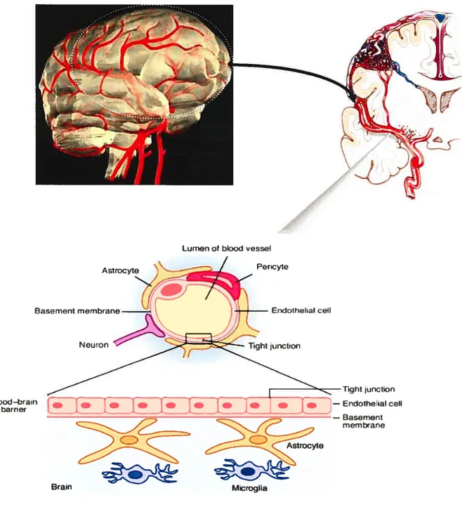
7 Tighl JJnction Endolheial ceil — Basement membane Astrocyte Basement membrane. N eu ton
Lumen ol blond vessel Pericyte Endothehal ceil Blood-brain barriet
ç
Microglia Brainfigure 1.2. BBB and its anatomical characteristics. Modified from http://www
8
1.4. Drug transport at the BBB
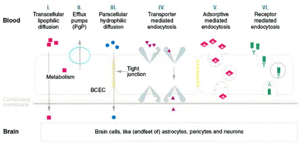
BBB has a number of highly selective mechanisms for transport of nutrients into the brain, as shown in figure 1.3.
1.4.1. Dffusioit
Diffusion of substances into the brain can occur by paracellular (i.e. between celis) and transcellular (i.e. across cells) routes, both of which are non-saturable and non competitive pathways. However, paracellular diffusion does flot occur to a great extent at the BBB due to the presence of tight junctions. On the other hand, transcellular diffusion is restricted to the substances with higher lipophilicity and small size (Pardridge, 199$). The smaller substances penetrate more rapidly and hence, small inorganic molecules (i.e. 02,
C02, NO, and H20) are highly permeable into the brain. For the transport of large molecules, the hydrogen bonding potential of a compound also plays a crucial role in the BBB permeability, substances with lower hydrogen bonding potential have greater membrane permeability (Chikhale et al., 1994).
1.4.2. Endocytosis
In addition to these diffusive mechanisms, entry of molecules into brain can occur by endocytosis. Bulk-phase endocytosis (pinocytosis), a non-saturable, nonspecific uptake of extracellular fluids (Simionescu et al., 1987) which occurs readily and to a large extent in other cells of the body. occurs to a very limited degree in the endothelial celis of brain microvasculature (Pardridge, 1995).
Absorptive-rnediated endocytosis (AME) is the mechanism by which positively charged substances are taken up by BBB. AME is reported to be triggered by the electrostatic interaction of positively charged moiety with the negatively charged plasma
9
membrane surface (i.e. glycocalyx) (Stieber et al., 1984). Due to its non-specific and non saturable nature, AME has been the focus for the development of many new drug delivery technologies for delivery across BBB (Pardridge, 1999).
Carrier-rnediaied endocytosis is one ofthe most prevailing modes ofuptake into the brain. It is used for the delivery of nutrients, such as monocarboxylates, hexoses (glucose), amines, amino acids, nucleoside, and purine bases to the brain (Pardridge, 1998). More than eight different nutrient transport systems have been identified at BBB, with each transporting a group of nutrients of the similar structure. Carrier-mediated transcytosis is substrate selective and the transport rate is dependent on the degree of occupation of the carrier.
Receptor-rnediated endocytosis (RME) provides a means for selective uptake of macromolecules into the brain. It occurs for substances, such as transferrin (f ishman et al., 1987), insulin (Duffy and Pardridge, 1987), leptin (Banks et al., 1996), and IGF-I & IGF-II (Duffy et al., 1988). RIVIE is a highly specific energy dependent transport mechanism. Substances that enter a cdl by means ofRME become bound to the receptors that collect in specialized areas ofthe plasma membrane known as coated pits (Moore et al., 1987). When bound to ligand these pits invaginate into the cytoplasm and then pinch free of the plasma membrane to form coated vesicles. These clathrin vesicle coats are rapidly removed to form smooth-coated endosomes, thereby allowing release of substances/ligand into the ccli (Stahi and Schwartz, 1986).
10
I. Il. III. IV. V. VI.
Transcellular Efflux Paracellular Transporter Adsorptive Receptor
Blaod lipophilic pumps hydrophilic mediated mediated mediated
diffusion (PgP) diffusion endocytosis endocytosis endocytosis ••. * Y • ]ight Metabolism Junction BCEC À À
Brain Brain ceils. like (endteet of) astrocytes, pericytes and neurons
Figure 1.3. The various transport processes that may occur at the BBB. Reproduced from (Pardridge, 1995).
1.5. Standard therapies for brain tumors
The treatment, a cancer patient receives is determined by many re!ated factors such as, the kind of ce!! from which cancer is derived, its size, the presence or absence of metastatic spread etc. Mu!tip!e treatment moda!ities have been deve!oped in an attempt to eradicate brain tumors. Some ofthe commonly used therapies areenlisted be!ow:
1.5.1. Surgery
Surgery is one of the first treatment modality used in clinics for the treatment of most of the cancers. It not on!y serves as a too! for patho!ogica! diagnosis, but also for immediate relief of the symptoms. Surgery reduces the number of cancer ce!ls requiring treatment and ofien serves to remove the hypoxic core of the tumors that are re!ative!y
11
resistant to radiation and inaccessible to chernotherapy (Siker and Mehta, 2007). However, damage to the neurological tissues during resection remains as a major challenge in the surgical procedures. A surgical resection with tumor free margins would increase the risk ofremoving normal brain tissue resulting in serious neurological damage (Nesbitt, 2007).
1.5.2. Radiation tlterapy
Few decades before, primarily in 1970 and 19$O’s postoperative irradiation of the organs was established as a standard protocol for patients with high-grade gliomas (Brada, 2006). It was believed that increasing the dose of radiation would improve the patient survival (Chang et al., 2007). But now, it is established that tumors reoccur in patients treated solely with radiation therapy. The major limitation to this preferred modality for the treatment of the tumors is the fact that the dose optimal for eradication of brain tumor might damage the surrounding brain tissue. Therefore, the adjuvant treatment with chemotherapeutic agents is ofien recornmended. Because of the limitations of whole brain radiotherapy, newer non-invasive methodologies called “stereotactic radiosurgery” have been developed for delivering radiation locally using techniques like Gliasite brachytherapy in which liquid radiation is placed into a balloon that is implanted in the surgical cavity of a resected tumor (Laperriere et al., 199$; Souhami et al., 2004). Alternative radiation delivery methods for stereotactic radiotherapy including, intensity modulated radiotherapy, implanted seeds and Novalis have been developed to improve the efficacy of the radiotherapy and reduce toxicity. Drugs called radiation sensitizers which sensitize the tumor tissue to radiations so that effect can be obtained at significantly lower dose of radiation have also been developed (Brada, 2006). They are given at the time of radiation and also are used in a clinical research setting as part of a clinical trial (Huncharek, 199$; Huang et al., 2007). These stereotatic methods of radiotherapy allow outpatient procedure, decrease recovery period from radiosurgery and improve patients’ quality of life due to reduced side-effects and thus, are been increasingly used in brain tumor treatments.
12
1.5.3. Chenwtherapy
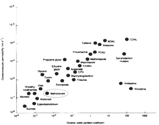
Chernotherapies have been used to control and ideally inadiate the infiltrative tumor ceils. However, the amount of drug reaching the CNS is difficuit and incomplete due to the BBB. There are oniy a few chemotherapeutic agents that readily cross 333 in therapeutic amounts (Figure 1.4). and these include nitrosoureas like 1.3-bis(2-chloroethyl)-1-nitrosourea (BCNU). procarbazine. topotecan and temozolomide (Muldoon et al., 2007). Nitrosoureas were the first single agent therapy used for the treatment of gliornas because. they readily cross the BBB due their small size, non-ionized state and high lipid solubility. BCNU becarne the most commonly used single-agent chernotherapeutic agent for recurrent gliomas (Komblith and Walker, 1982). Later, lipophilic derivatives of BCNU were produced in order to increase its efficacy by enhancing the BBB permeability; however, these agents showed similar resuits as 3CNU. It lias been reported that radiotherapy and BCNU produce superior resuits than radiotherapy alone (Walker et al.. 1980). A recent analysis suggests that nitrosourea based adjuvant therapy has modest benefit for patients with anaplastie gliomas and smaller for those with glioblastorna (Levin et al., 1990). Procarbazine is another cliemotherapeutic agent which crosses BBB readily. As a single agent, it lias been reported to increase the long-term survival in patients with malignant gliomas as cornpared to BCNU (Newton et al., 1993). Temozolomide, another agent that cross 333, lias shown significantly better survival in patients with gliobastoma multiforme as compared to procarbazine (Macdonald et al., 2005). It is also reported to be effective in patients with newly diagnosed malignant glioma. Unlike other cliemotherapeutic agents, temozolomide can be given orally due to its high oral bioavailability. The promising resuits with temozolomide have establislied it as new, modestly improved standard of care for patients with newly diagnosed glioblastoma multiforme (Nagasubramanian and Dolan. 2003). Methotrexate (MTX) is another cbemotlierapeutic agent, which at higli dose and in combination with Citrovorum Factor Rescue (Lleucovorin®) lias clinically treated patient witli various brain turnors including glioblastoma multifonTie (Abelson et al., 1981 a).
13
paclitaxel, vincristine etc. have flot been extensively evaluated for agents for brain tumor therapy due to their poor BBB permeability (Muldoon et al., 2007). Resistance to chemotherapy remains a central reason for the failure to cure patients with cancer. And in the case of brain tumors it is another challenge that needs to be overcome besides poor BBB permeability. Uro nhwni
. •
•
— flrIO1•
• •
Iii0 __________________ — 1G4 ,fl_ 1O Odo w11.ç plrI,œ cœttcrwlFigure 1.4. Permeability of various chemotherapeutic agents across BBB in relation to partition coefficient. Reproduced from (Muldoon et al., 2007).
1.5.4. Irnmunotlterapy
Immune system is uniquely qualified to be an instrument used in cancer therapy because all the cancer patients including those with brain tumors have some degree of immunosupression (Wu et al., 2006). In fact, it is speculated that tumors may be able to
î—
1’s•
CHefr,e•.
Âr,pnr Prrrab.•
rcu P-nçm1i1•
•
M.2r•
ETh4-. F-mrur j0•
A.Irr • 5-EU•
Cumr•
•
•
•
Fonramr$o•
•
•
I Iii 1lOC14
grow because they evade the body’s immune surveillance. An immune response directed against celis bearing tumor antigens couÏd provide a specific and effective mechanism for killing of residual tumor ceils (Gross et al., 2004). One of the strategies in immunotherapy involves delivering antibodies against tumor specific antigens to inactivate and clear highly expressed substances on tumors celis and allow restoration of a more normal immune state. They also help to reduce the tumor-induced immunosuppression that is mediated by soluble factors (Yang et al., 2006a). Monoclonal antibodies (MAb) are also an attractive tool for targeting radio-isotopes, chemotherapy agents, or even activated lymphocytes to the tumor cells (Wu et al., 2006; Sathomsumetee et al., 2007). An antibody specific for tumor antigen conjugated to radio-isotope could deliver high-dose radiation to malignant celis, reducing the morbidity caused by less closely targeted radiotherapy. Problems associated with MAb therapy relate to creating truly tumor-specific antibodies and delivery of sufficient doses of the antibodies to the tumor. Most of the antigens expressed by tumor cells are not tumor specific (Sathomsumetee and Rich, 2006; Wygoda et al., 2006). The epidermal growth factor receptor (EGFR) is one target reported to be specific to the tumor cells, it is found in 17% of glioblastomas. Miyamoto et al., (Miyamoto et al., 1996), conducted a trial using 1251-labeled EGFR-425, a MAb to the human EGfR, as an adjuvant to conventional therapy for patients with anaplastic astrocytorna and glioblastoma multiforme and found it to improve patient survival with minimal toxicity. Injection of antibodies likc antitenascin into the resection cavity has also shown improvement in patient survival with moderate side effects (Reardon et al., 2007).
Other strategies in immunotherapy involve activation of immune response to the cancer ceils by delivering interferons (IFN), cytokines, interleukins etc. (Edwards et al., 1985; Hill et al., 1992; Boiardi et al., 1994). These agents are given to patients to imitate or influence the natural immune response either by directly altering the cancer cell growth or acting indirectly to help healthy cells to control the cancer. Scientists are also studying vaccines that would boost the bodys immune response to cancer cells and thereby prevent cancer from developing. The challenge in immunotherapy has been to identify the putative
15
target antigens and then direct and stimulate the patients’ immune system against their own
tumors (Lutz, 1983
).
The additional immunological disadvantage in the brain is the limited availability of antigen presenting ceils and immune stimulatory signais. The success of immunotherapy depends on careful selection of the target antigen, corresponding specific MAb, size and affinity of the antibody and its proper compartmental administration (Ribas et al., 2003). The major limitation of this modality remains the poor permeability of antibodies across BBB after systemic administration which limits its potential for frequent administration.1.5.5. Gene therapy
Gene therapy is meant to deiiver genetic material with a therapeutic goal of encoding proteins (e.g., enzymes) or siRNA to the ceils (Kouraklis, 2000). In addition, gene therapy provides the potential for a long-term effect following one single administration. However, genes are hydrophilic, charged, and large molecules that caimot pass celi membranes and tight cellular layers like the BBB. Therefore, their delivery to the desired site of action in brain is chailenging. Viruses have evolved over millions of years to obtain optimal mechanisms for gene delivery to host cells, which makes them applicable as a biological vector system to deliver genetic material to brain ceils. There is a broad range of viral vectors available, but the most commonly used are adeno-associated viral vectors and lentiviral vectors (Cascallo and Alemany, 2004). Current gene therapy for brain tumor patients utilizes scientffically engineered viruses made from strains of the cold sore virus (herpes virus) or common cold virus (adenovirus). When introduced into the human body these engineered viruses are abie to recognize cancer ceils and kili them. Engineered viruses are also able to self-multiply and thus have a long “life” to kill and keep on killing cancer cells during the time-frame of the virus? life span. However, the immunogenic and pro-inflammatory nature of viral vectors iimits their applicability (Tangney et al., 2006). Important issues in viral gene delivery are stable transgene expression, limited immunogenicity, induction of an inflammatory response, the lack of cell specific targeting
16
efficiency, safety, and toxicity. Due to these limitations of viral gene therapy, non-viral alternatives like liposomes and nanoparticles, for their delivery into brain have been explored (Dachs et al., 1997).
1.5.6. Antiangiogenic therapy
Angiogenesis is a normal process, the growth of new blood vessels, necessary for growth and for wound healing. It has recently been deterrnined that in order for a tumor to grow, it must develop its own blood supply (Folkman, 2006). In contrast to the traditional cancer treatments that attack tumor celis directly, angiogenesis inhibitors target at the formation of turnor-feeding blood vessels that provide continuous supply of nutrients and oxygen. Additionally, antiangiogenic therapies target the endothelial celis thus, delivery challenges are considerably reduced (Miller et al., 2001; Sezer et al., 2001). The formation of new blood vessels is also a hallmark of malignant gliomas. Vascular endothelial growth factor (VEGF) is a major regulator of angiogenesis in most of the tumors (Cébe-Suarez et al., 2006). Thus, anti-VEGF therapies have been evaluated clinically and have shown to improve survival. It lias been reported that endothelial cells of various tumors also express specific receptors like integrins, selectins etc. which are involved in tumor ce!! migration and invasion. These receptors are unregulated during neovascularization and tumor neo angiogenesis and thus targeting them would reduce or inhibit vessel development and tumor spread. 0f ail the integrins, a,83 integrins are expressed in small blood vessels surrounding glioblastoma (Gladson, 1996). Antibodies developed to this integrin have been shown to inhibit matrigel invasion of glioma ce!! !ines and primary cultures (Paulus and Toim, 1994). Recently, a number of naturally occurring endogenous inhibitors of angiogenesis have been discovered and these include moiecules !ike platelet derived factor 4 (Maione et al., 1990; Tanaka et al., 199$), or tbrombospondin-1 (Streit et a!., 1999) and anti-angiogenic factors such as endostatin (O’Reilly et al., 1997; Sasaki et a!., 1998). These antiangiogenic factors can a!so serve as potential molecules that can be delivered to the tumors for reducing angiogenesis. Antiangiogenic therapies have shown to reduce the
17
the studies but the cure of cancer from this therapy has neyer been reported. Indeed, in the clinic, antiangiogenic drugs used as monotherapies have littie discernible therapeutic (survival) benefit in the treatment of advanced-stage cancers. Thus, it bas been recognized that antiangiogenic therapies might not be effective alone in the treatment of the cancer, the combination witb chemotherapy would be needed to enhance their potential.
1.5.7. Combination therapy
Active research bas failed to identify a single therapeutic approach whicb can cure malignant gliomas. Due to the failure of single-agent delivery or radiotherapy alone to improve the survival in patients with brain tumors, efforts have been directed to use more than one chemotherapeutic agents alone or in combination with immunotherapy or antiangiogenic therapy (Krauseneck and Mertens, 1987). It bas been reported that the delivery of chemotherapeutic agent along with radiotherapy or combination of radiotherapy and immunotherapy are promising approaches. Radiation therapy treats cancer that is confined locally, while chemotherapy also kills cancer cells that may have spread. Sometimes radiation or chemotherapy is given before surgery to shrink a tumor, thereby making the complete removal of the tumor using surgery, or afier surgery to destroy any remaining cancer ceils (Wheeler and Kaufman, 1981; Scheda et al., 2007
).
The combination of radiotherapy with temozolomide therapy bas shown a promise for patients with newly diagnosed glioblastoma (Addeo et al., 2007). This study has confirmed that combination therapy can improve the outcome for patients with these devastating tumors. The combination of cytotoxic and cytostatic chemotherapeutic agents bas also been attempted for improving the efficacy ofthe treatment. The rationale for combining different chemotherapeutic agents is to use drugs that work on different parts of the cancer ceils life cycle, thereby increasing the likelihood that more cancer cells will be killed (Scheda et al., 2007).
When drugs with different toxicities are combined, each drug can be used at its optimal dose, helping avoid intolerable side effects. For instance, combination of18
temozolomide, BCNU and thalidomide has shown to improve patient survival than single agents therapy in clinical settings (Pamey and Chang, 2003).
A number of studies have shown that targeted antiangiogenic agents and chemotherapy ofien have additive or synergistic effects when used in combination. Combination of chemotherapy agent with antiangiogenic therapies have been shown to be effective in primary glioblastoma multiforme tumors, producing a patient survival time of 16 months (Wygoda et al., 2006). It has been shown that PTK-787 (a VEGF receptor inhibitor) combined with temozolomide produced a median time to progression of 15.1 weeks. The combination of antiangiogenic therapy has also shown to potentiate the response to radiation therapy. Now it is increasingly realized that combination therapy is one of the most effective way for cure of malignant and aggressive tumors. However, due to complications in dosing schedule, frequency of administration etc., combination therapy is not widely used. Infact, Hildebrand, 1981 lias reviewed a number of randomized combination therapy protocols and has concluded that based on the stage and the type of cancer, a specific regimen has to be developed for the success of combination therapy.
1.6. Challenges in delivery to brain tumors
0f all the brain tumors, malignant gliomas are most difficult to treat, this is largely because like most solid tumors they are variably resistant to treatment because of inadequate drug delivery, systemic toxicity, development of resistance to drugs (Krauseneck and Mertens, 1987). The specific considerations for gliomas are;
• BBB restricts the delivery of chemotherapy to brain • High cellular heterogeneity in the tumor celis • Defects in immune surveillance ad response • Low toxicity to non-tumor celis
19
Clinical failure of rnany potentially effective therapeutics for the treatment of brain tumors is usually flot due to the lack of drug potency, but rather due to the inability of drugs to cross BBB, i.e., can be attributed to shortcomings in the methods by which a drug is delivered to the brain and into the brain tumors (Penas-Prado and Gilbert, 2007
).
Ihe unique physiological and pathological barriers found in the CNS impede the delivery for the treatment of brain tumors. Orally and intravenously (i.v.) administered chemotherapeutics have difficulty in reaching therapeutic concentrations at the tumors site because ofthe BBB, BCB, and the BIB. It is recognized that BTB is more permeable than normal brain tissue (Groothuis, 2000). However, the breakdown of the BBB in the area of the tumor is variable and large areas exist where BBB remains intact. In these areas the amount of drug reaching the tumor remains severely restricted. In addition, the brain adjacent to the tumor is the area around the tumor that contains infiltrating turnor cells. Ihese sites may be associated with an intact BBB and drug delivery to these areas is difficult. Finally, although most brain tumors are highly vascular the aberrant tumor induced angiogenic process tends to create abnormal vessels such as blind loops and arteriole-venuole shunts, making parenchymal drug delivery even more inefficient (Folkman, 2006).Though imrnunotherapy had shown promise in treating these tumors it bad failed to deliver the therapeutic benefit mainly due to the inability to produce significant immune response following the administration. Even genetherapy bas not shown significant promise in clinical trials; further the need to deliver it invasively has limited its applicability (Kouraklis, 2000). Antiangiogenic therapy on the other hand has also failed to improve survival of patients with gliomas when administered alone. Thus, so far chemotherapy remains as the mainstay of treatment for patients with gliomas because, it bas played important role in the treatment of primary brain tumors. However, enhancing permeability of chemotherapeutics across BBB is the major challenge which bas been addressed
20
1.6. Strategies to enhance drug delivery across BBB
Several approaches have been implemented to address the limitations posed by both natural and pathological barriers in the CNS (Table 1.1, Figure 1.5). The strategies used to bypass these boundaries can be divided into chemical and physical based techniques.
1.6.1. Chemical strategies
They focus on improving drug delivery by altering the structure of the drug or by disrupting the BBB.
1.6.1.1. Modjfication b, the drtig molectites
BBB is permeable to small, electrically neutral and lipid-soluble molecules. Thus, increasing the lipophilicity of the drug molecule to increase its permeability is one of the preferred enhancement strategies. Such approach was used to enhance the delivery of BCNU by developing its lipophilic analogs like lomustine (CCNU) and semustine (methyl-CCNU). However, clinical trial evaluating the systemic administration of these agents showed no statistical improvement in the efficacy over BCNU in treating guai tumors (Chin et al., 1981; Ushio et al., 1984). Another strategy is linking the drug to
21
[Strategies for drug delivery to brain tumors
[
Chemical-based sttategiesJ
[
Physical-basedEe
IIe ascuhrsfrategi drJry Catheterbased iochemic systemsI
BBBDf
Implantable daICarrier-mediated polvmeric devices
ers transport vectors Receptor-mediated transport vectors ADEPT and GDEPT
ADEPT- Antibody-directed enzyme prodrug therapy GDEPT- Gene directed enzyme prodrug therapy BBBD- BIood-bran barrier disruøtion
Figure 1.5. Various strategies that have been used to improve delivery of therapeutic molecules to brain.
Table 1.1. Various approaches utilized to enhance the delivery of drugs to the brain Intraventicular/ intrathecal route Injections, catheters and pumps Biodegradable polymer wafers, microspheres and nanoparticles Drug delivery from biological tissues
Receptor/vector
mediated drug delivery Pegylated liposomes
Readily penetrate CNS
High drug residence time Specific drug delivery e.g. neuropeptides
Alters barrier inducing factors
Selective opening of brain turnor capillaries
Direct nose to brain transport and access to CSF
Bypasses BBB and provides immediate high CSF concentrations, longer drug haif-life. minimized protein binding and decreased enzymatic activity
Continuous delivery drug distribution can be maintained
Circumvent BBB, controlled delivery, easily implantable
Therapeutic proteins can be released from co-grafted ceils Allows designing linkers for functional needs Capable of receptor rnediated transport to 333
Poor aqueous solubility. molecular weight limit (400-600 Da), enhanced peripheral distribution
Possibility of reactive metabolite formation, poor selectivity and poor retention
Oxidative liability and hydrolytic instability limit the sheif life of these systems
Breaks down the defense mechanism of the brain, ofien leads to unfavorable toxic effects
Breaks down the defense mechanisrn of the brain
Enzymatically active. low pi-l, mucosal irritation or variability caused by nasal pathology
Increased intrathecal pressure leads to clinical incidences of hemorrhage. CSF leaks. neuro-toxicitv and CNS infections
Due to diffusion problems therapeutic agent like to reach only near by sites
Useful in very limited number of patients, due to diffusion problems therapeutic agent like to reach only near by sites
Inefficient transfection of host cells, nonselective expression of transgene and deleterious regulation of transgene by the host
Saturable process, enzymatic dependent release, attachment to BBB transport drugs renders some drugs inactive
Do not undergo significant transport in absence of vector rnediated delivery
Approaches Advantages Limitations
Lipophilic analogs Pro drug Chernical drug delivery Osmotic BBBD Biochemical BBBD Olfactory pathway transport specific