HAL Id: tel-02409881
https://tel.archives-ouvertes.fr/tel-02409881
Submitted on 13 Dec 2019HAL is a multi-disciplinary open access archive for the deposit and dissemination of sci-entific research documents, whether they are pub-lished or not. The documents may come from teaching and research institutions in France or abroad, or from public or private research centers.
L’archive ouverte pluridisciplinaire HAL, est destinée au dépôt et à la diffusion de documents scientifiques de niveau recherche, publiés ou non, émanant des établissements d’enseignement et de recherche français ou étrangers, des laboratoires publics ou privés.
Self-organized wave-like beating of actin bundles in a
minimal actomyosin system
Marie Pochitaloff-Huvale
To cite this version:
Marie Pochitaloff-Huvale. Self-organized wave-like beating of actin bundles in a minimal acto-myosin system. Chemical Physics [physics.chem-ph]. Sorbonne Université, 2018. English. �NNT : 2018SORUS318�. �tel-02409881�
Sorbonne Université
Ecole doctorale Physique en Ile-de-France
Laboratoire Physico Chimie Curie — Institut Curie
Auto-organisation de faisceaux d’actine oscillants
dans un système minimal d'actomyosine
Par Marie Pochitaloff-Huvalé
Thèse de doctorat de Physique
Dirigée par Pascal Martin
Présentée et soutenue publiquement le 16 Octobre 2018 Devant un jury composé de :
Mathilde Badoual Professeur Examinatrice Laurent Blanchoin Directeur de recherche Examinateur Kinneret Keren Professeur Rapportrice Pascal Martin Directeur de recherche Directeur de thèse Guillaume Romet-Lemonne Directeur de recherche Rapporteur
Manuscripts do not burn The Master and Margarita — Mikhail Bulgakov
Acknowledgements
Doing a PhD is often described as a marathon ending by a final sprint, this image well illustrates the perseverance and the tenacity that we have to reinforce through these very intense years. During this marathon, we encountered many difficulties that we had to overcome patiently, in that sense the PhD was also an obstacle course, during which I found a precious help among many people that I would like to deeply thank for their moral and scientific support.
I want to thank Pascal, my supervisor, to let me work on that project I found exciting, I am convinced that his will of perfection had really forced me to make my minimal system robust and improved my skills for presentation and writing.
I would like to thank the members of the jury for reviewing my work, it was a pleasure to discuss with you and receive your feedback on my work, it encourages me to continue in research ! First my examiners Kinneret Keren and Guillaume Romet-Lemonne, I am very grateful for your positive reports and your enthusiasm, thank you to the president of the jury Raphaël Voituriez, sorry for not having more theory in my work. I would like to thank in particular Mathilde Badoual and Laurent Blanchoin, who were also members of my jury but also followed my advances in my thesis committee and supported me during all my thesis.
It is the Thesis Advisory Committee (Cécile Sykes, Julie Plastino, Mathilde Badoual, Laurent Blanchoin and Nicolas Minc) that pushed me to go to the project I was the most excited about among the two initial projects I had at the beginning of my PhD. I chose the most ambitious and the most risky but it was worth it ! Thank you !
I would like to thank Laurent Blanchoin and his team in Grenoble who welcomed me, trained me and make me aware of the difficulties of working with an in vitro system, thanks Laurent for your support ad your advices during all my PhD; Christophe for never making fun of me despite my dummy questions and the ugly pictures I sent you when my experiments did not work; Hajer for sharing with me your energy, your motivation,
Acknowledgements Acknowledgements
you were doing experiments with me two weeks before your defense, you provided me a very precious help.
This work was made possible through the support I found in the lab, in fact the Sykes / Plastino team almost adopted me : I participated at their journal club, where I learned a lot on actin biochemistry. I was running every Tuesday to arrive on time at 12 pm not to have to make cakes. Julie and Cecile were always helpful when my experiments did not work and encouraged me a lot for not giving up. Thanks a lot ! Thank you Bassam for our nice discussions and the single molecule experiments we have done together, it was great to work with you ! I would like to thank Axel or his precious help at the end of my thesis for his sharp advices to improve my manuscript and my thesis presentation, thanks a lot Axel to have taken the time despite your tight schedule. I would like to thank the members of the Martin team, who created a good atmosphere; Mathieu, my predecessor, who trained me as a Jedi Master in Matlab and experiments, thanks for your patience and the fun we had in Nikon Center ! Even though we worked on different projects, I’m thankful to Atitheb, who developed Matlab codes for some of my PhD projects, good luck for your PhD ! and Mélanie, for her positive attitude and the absurd jokes we made together and continuing to encourage me through hard times even after the end of your PhD, thanks Mélanie, I hope our ways will cross again in the future. Thank you Jonathan and Martial as alumni of the team for all your support. I would like to thank Laurence for guiding me through administrative procedures. Thank you Brigitte for your kindness and your warm welcome every morning, it was a good start for working days !
My time at Curie was made enjoyable in large part due to the many friends and col-leagues I made along the way, and to the stimulating discussions we had, on a professional as well as personal, I want to express my gratitude. I would thus like to thank all my fellow labmates from UMR 168, including but not limited to: Tatiana, Mohamed, Chiara, Quentin, Bastien, Patricia, Alicia, Laura, Valentina, Fanny, Aude, Fabrice Thibault, Car-les, Remy, Jean-Patrick, Louis, Nadir, Koceila, Kotryna, Amanda, Simon, Perrine and many more. Special mention to my office-mates Patricia and Isabelle, my ballet-mate Sarah, who gave me precious advices for my manuscript, not be desperate when my experiments were not working, wedding organization, LaTex ... Julien who was always there to explain me actin biochemistry, myosin, TIRF, that ?black actin does not exist?. I am very thankful to Majdouline Antoine and Camille, who helped and supported me so much during my PhD (actin, pyrene assay, TIRF, Matlab) and have so kindly prepared me surprise with Julien for my PhD defense ! I wish you all the best for the future !
Acknowledgements Acknowledgements
There are all the friends from ESPCI with whom life was happy and carefree : Dou-nia, Simon, Simon Aymeric, Baptiste, Sacha, Romain, Alice, Alice, Eva, Marine, Elodie, Corentin, Hubert, Emeric, Romain friends with whom I spent quality time.
I would like to thank my family for their encouragements and support ! Last but not least, I would like to thank my husband Antoine ♥. My words lack when I want to express it all.
The Curie Institute is a truly exceptional environment to do research in, not least because of the access we have to great equipment and expertise. I gratefully acknowledge John and Fahima for the biochemistry platform, Vincent, Francois Ludovic and Lucie from the Nikon Center, for their training and their invaluable help with the microscopes. I would like to thank all the people who somehow contributed to my ending up here, teachers and mentors, and I would like to mention specifically: Timo Betz, Jacques Prost, Rob Philips, Jane Kondev, Hernan Garcia.
My thoughts also go to Maxime Dahan and his family. I want to address my profound gratitude to Maxime for helping me in difficult time in my PhD, he was a truly great mentor.
Abbreviations
• Arp2/3 complex: Actin-related proteins 2 and 3 complex • ATP/ADP: Adenosine TriPhosphate / Adenosine DiPhosphate • BSA: Bovine Serum Albumin
• DIC: Differential Interference Contrast microscopy • DRC: Dynein Regulatory Complex
• DTT: Dithiothreitol
• EDTA: Ethylene Diamine Tetraacetic Acid (chelating agent which sequesters Ca2+)
• EGTA: Ethylene Glycol-bis(β-aminoethyl ether)-N,N,N’,N’-Tetraacetic Acid. • F-actin: Filamentous-actin
• G-actin: Globular actin
• GFP: Green Fluorescence Protein
• GTP/GDP: Guanosine TriPhosphate / Guanosine DiPhosphate • HMM: Heavy Mero Myosin
• LMM: Light Mero Myosin
• NPF: "Nucleation Promoting Factor" (promoting factor of actin nucleation) • WASP: "Wiskott-Aldrich Syndrome Protein", (protein containing VCA domain,
which can activate Arp 2/3 complex and initiate nucleation with branching, notably in lamellipodia of motile cells)
• Pi: Inorganic phosphate
Abbreviations Abbreviations
• pWA: VCA domain with a polyproline region, used in vitro to activate Arp 2/3 complex and to initiate branching nucleation in presence of actin
• TIRF microscopy: Total Internal Reflection Fluorescence microscopy • Tris: Tris(hydroxymethyl)aminomethane
• SEM: Scanning Electron Microscopy
• VCA, WA: C-terminal domain of proteins of WASP/scar family. The region "ver-prolin homology", named V or W, is recruiting two actin monomers; the region cofilin homology,named C, and the acid sequence, named A respectively, is recruit-ing and activatrecruit-ing Arp2/3 complex
Contents
Acknowledgements v
Abbreviations ix
General Introduction 3
1 Self-organization and Spontaneous oscillations 5
1.1 Self-Organization . . . 5
1.1.1 What is self-organization ? . . . 5
1.1.2 Self-organization in biological systems . . . 6
1.1.3 Spontaneous oscillations . . . 10
1.2 Spontaneous beating of eukaryotic flagella . . . 14
1.2.1 Cilia and flagella : motile organelles . . . 14
1.2.2 The axoneme : the core structure of cilia and flagella . . . 15
1.2.3 Beating properties . . . 16
1.2.4 Existing theoretical approaches of flagellar beating . . . 25
2 The actomyosin system 31 2.1 The actin cytoskeleton . . . 31
2.1.1 Actin : From monomers to filaments . . . 31
2.1.2 Actin binding proteins . . . 36
2.2 The myosins . . . 40
2.2.1 Myosin topology . . . 41
2.2.2 The chemomechanical cycle of myosin . . . 43
2.2.3 Duty ratio and processivity . . . 45
2.2.4 Experiments to characterize motor action . . . 46
2.2.5 Recap of myosin II and myosin V properties (Table 2.3) . . . 51
3 Materials and Methods 53 3.1 Micropatterning of an actin nucleation promoting factor . . . 55
CONTENTS CONTENTS
3.1.1 Surface passivation . . . 55
3.1.2 Patterning of a nucleation promoting factor . . . 56
3.1.3 Experimental chambers . . . 57
3.2 The actomyosin polymerization mix . . . 58
3.3 Image acquisition . . . 60
3.4 Image analysis . . . 62
3.4.1 Tracking of the actin bundles . . . 62
3.4.2 Beating pattern analysis . . . 64
4 Self-organized wave-like beating of actin bundles 67 4.1 Architecture of the actin network with or without myosins . . . 67
4.1.1 In the absence of myosins . . . 67
4.1.2 In the presence of active myosins : assembly of tight actin bundles . 71 4.2 Dynamic analysis: Spontaneous oscillations . . . 74
4.2.1 General description of beating properties in the case of myosin II . 74 4.2.2 Comparing beating properties with myosin II to those with myosin V 78 4.2.3 Effect of bundle length on beating properties . . . 81
4.2.4 Varying the concentration of the molecular motors . . . 89
4.3 Distribution of myosin motors along a beating actin bundle . . . 91
5 Conclusion and Discussion 99 5.1 Comparison to other oscillating systems . . . 100
5.2 3D beating of actin bundles . . . 103
5.3 How does myosin sense the shape of actin bundles? . . . 105
6 Perspectives 109 Appendices 115 A Experimental protocol 117 A.1 Proteins preparation . . . 117
A.1.1 G actin solution . . . 117
A.1.2 Minimal set of proteins which ensures actin polymerization . . . 117
B References of purified proteins 119 B.1 Actin and actin-related proteins . . . 119
B.1.1 Preparation of the proteins . . . 119
B.1.2 Characteristics and storage conditions of the proteins . . . 119
CONTENTS CONTENTS
B.2.1 Methods of purification of the studied HMM myosins . . . 120
B.2.2 Characteristics and storage of the studied HMM myosins . . . 120
C References of chemical products 121 C.1 References and storage . . . 121
C.2 Preparation of Methyl cellulose . . . 121
C.3 Preparation of PLL-g-PEG solution for coverslip passivation . . . 122
C.4 Preparation of aliquots from chemical products . . . 122
D Role and Composition of Buffers 123 D.1 Buffer for monomeric actin : G Buffer . . . 123
D.2 Buffer for actin polymerization : 1X KMEI . . . 123
D.3 Buffer for myosin activity : 10X KEEI . . . 124
E French abstract 125 E.1 Analyse dynamique des oscillations spontanées . . . 127
E.1.1 Description générale des propriétés du battement dans le cas de la myosine II . . . 127
E.1.2 Comparaison des propriétés de battements avec la myosine II par rapport à celles avec la myosine V . . . 130
E.1.3 Effet de la longueur du faisceau sur les propriétés du battement . . 132
E.2 Distribution des moteurs le long d'un faisceau d'actine oscillant . . . 134
List of figures 140
List of tables 144
General Introduction
Living systems consume energy to move, change shape, divide and control their own morphology. To fulfill all these key processes, living systems are highly dynamic and require active mechanics over a broad range of length scales: from transport within cells at the molecular scale, mitosis and motility at the cellular scale, to morphogenesis of organs and whole animals at the multicellular scale. The diversity of the dynamics is ensured by the cytoskeleton. The cytoskeleton is composed of different biopolymers that are able to assemble and disassemble, resulting in networks of different architectures. The microtubule cytoskeleton is made of pipe-like filaments that are very rigid, whereas the actin cytoskeleton consists of cable-like filaments that are semi-flexible. Within the actin and microtubule cytoskeletons, motor proteins are present to exert forces that can deform, contract the network, or mediate transport of cargoes along the filaments, which provide tracks for these directed movements.
Understanding the incredible complexity of cellular processes constitutes a significant challenge for both biologists and physicists. To tackle this problem, two mainstream approaches have been developed. The top-down approach considers the cell in all its complexity and consists in perturbing the system to study the role of its molecular com-ponents. Development of genetic tools and super resolution microscopy has allowed to increase our knowledge with high precision. The second approach starts in the opposite way, by reducing the complexity of the system to identify general biophysical principles underlying cellular processes. To reach this goal, many in vitro model systems of puri-fied motors and filaments that lack biochemical regulation have been recently developed. This bottom-up approach of reconstructing biological functionality with minimal compo-nents is useful because it allows to delineate between generic (i.e. that do not depend on molecular details) and specific properties in the more complicated biological systems.
Cytoskeletal filaments and molecular motors have been extensively studied at the single molecule level : we have a pretty good idea about how cytoskeletal filaments assemble/dis-assemble, their mechanical properties, their structure, as well as the mechanism of force
General Introduction
production and movement by single molecular motors (Howard, 2001). However their collective behavior remains poorly understood and cannot be explained only from their single molecule properties.
The main goal of my thesis was to study an in vitro minimal system comprising a net-work of actin filaments and myosin motors. Micropatterning of a nucleation promoting factor of actin polymerization allowed for the geometrical control of the network architec-ture by governing the spatial assembly of the actin filaments, which emerged out of the pattern parallel to each others and with the same polarity. We worked with two types of myosin motor, myosin II and myosin V, that we added in bulk to study how molecular motors reorganized such a network.
This experimental configuration led to the self-assembly of tight actin bundles that displayed periodic wave-like beating resembling that observed in eukaryotic flagella, e.g. in sperm cells. My work provides a detailed description of this dynamic phenomenon, aiming at clarifying the underlying biophysical mechanism and discuss its relevance for more complex biological systems.
Chapter 1
Self-organization and Spontaneous
oscillations
1.1 Self-Organization
1.1.1
What is self-organization ?
Self-organization refers to the emergence of an overall order in time and space of a given system from the collective interaction of its many individual components (Camazine et al., 2001; Karsenti, 2008; Wedlich-Söldner and Betz, 2018). A large variety of examples are found in physical, chemical and biological systems, such as sand grains assembling into rippled dunes, chemical reactants forming swirling spirals, cells making up highly struc-tured tissues, or birds joining together in a flocking formation (Fig. 1.1). When wind blows over a uniform field of sand, a pattern of regularly spaced ridges (Fig. 1.1.A) is formed through a set of forces attributable to gravity and wind acting on the sand par-ticles (Anderson, 1990; Forrest and Haff, 1992). In the Belousov-Zhabotinsky chemical reaction, the mixture alternates between red and blue (Fig. 1.1.B) owing to changes in the charge state of the iron ions that catalyze the reaction. The mechanism for this reaction is very complex and is thought to involve around 18 components (Field and Foersterling, 1986). A group of animals can also show global order by synchronizing to some extent their behavioral state (Fig. 1.1.D) : In the case of starlings, they align their individual direction of motion, which results in flocks forming a tight sphere-like formation, fre-quently expanding and contracting and changing shape, seemingly without any sort of leader (Okubo, 1986). The pattern is an emergent property of the system rather than a property imposed on the system by an external ordering influence. Emergence refers to a process by which a system of interacting subunits acquire collectively qualitative
Self-organization and Spontaneous oscillations Self-Organization
new properties that cannot be understood and predicted as the simple addition of their subunit individual contributions.
Figure 1.1: Examples of self-organization at the macroscopic scale. A: Rip-pled sand dunes, the wavelength is approximately 8 cm (Ball, 2012). B: Spiral waves produced by the Belousov-Zhabotinsky chemical reaction in a 14-cm-diameter Petri dish, in which the mixture alternates between red and blue owing to changes in the charge state of the iron ions that catalyze the reaction (Ball, 2012). C: Pattern of black and white stripes on zebra coat (complexitylabs.io/ecological-self-organization.) D: Flock of starling birds (www.telegraph.co.uk/news/earth/wildlife/8315106/ Starling-flocks-how-they-form-into-incredible-wildlife-spectacles.html)
1.1.2
Self-organization in biological systems
Cellular systems Using the hydrolysis of Adenosine TriPhosphate (ATP) or Guanosine TriPhosphate (GTP) as a chemical energy source, cellular materials are known to produce a broad range of self-organized phenomena, most of which have evolved to perform a variety of cellular functions, including cell division, motility, shape changes, contraction (Karsenti, 2008; Wedlich-Söldner and Betz, 2018). All of them are supported by networks of cytoskeletal filaments and molecular motors, which form structures fundamentally out of equilibrium. This self-organization spans over several orders of magnitude in space and
Self-organization and Spontaneous oscillations Self-Organization
time and involves a complex interplay between biochemical and biophysical processes. For example, molecular motors and cytoskeletal networks interact to drive the cell cycle (Fig. 1.2.A) : they are responsible for cell-shape changes, chromosome segregation, and cleavage to form two daughter cells from a mother cell. The movement of keratocytes is ensured by an interplay between the well-orchestrated dynamics of the actin cytoskeleton assembly at the leading edge of the cell and myosin motors contracting the cytoskeleton at the trailing edge of the cell (Allard and Mogilner, 2013) (Fig. 1.2.B). At a multicellular level, the compaction of a 8-cell-stage embryo, which is characterized by the transformation of the embryo from a loose cluster of spherical cells into a tightly packed spheroïd (Fig. 1.2.C), is an important step of embryogenenesis that will trigger the establishment of the first tissue-like structure of the embryo (White et al., 2016).
Figure 1.2: Examples of self-organization in cellular systems A: During the cell cy-cle, the cell undergoes a large panel of shape changes (https://bigpictureeducation.com/ cell-division-images). Chromatin is labeled in red, the microtubules of the cytoskeleton in green. B: A keratocyte is a crawling cell, which protrudes its leading edge and contracts its trailing edge to move (Allard and Mogilner, 2013). Scale bar : 10 µm. Time (in s) is indicated at the top of each picture. C: Embryo compaction at 8 cell state (White et al., 2016). The embryo is shown before and after compaction, using Differential Interferential Contrast microscopy (DIC, top) and Scanning Electron Microscopy (SEM, bottom). Scale bars : 10µm for DIC and 15 µm for SEM.
Self-organization and Spontaneous oscillations Self-Organization
In vitro systems Designing molecular or cellular systems that mimic cellular processes in vitro, detached from the complexity of the cell and/or of the in vivo environement of the cell, is a powerful approach for dissecting complex cellular phenomena. In particular, in vitro experiments have shed light on the key role of geometrical constrains for the selection of the emergent structures of the cytoskeleton.
Figure 1.3: Examples of in vitro self-organization of cytoskeletal filaments due to ge-ometrical constrains. A: RPE1 cell visualized in phase contrast (a) and plated on a fibronectin crossbow micropattern (b). Using a fluorescent label on actin, one sees that actin polymerizes in membrane ruffles on the curved adhesive edge, but assembles in contractile stress fibers an-chored to fibronectin on the cell lateral edges. (c) Microtubule plus-end trajectories. Scale bar: 10 µm. Figure adapted from (Thery et al., 2006). B: Self-organization in the constrained geometry of micro-fabricated chambers etched in glass. Formation of a vortex as observed by dark-field microscopy in a chamber of diameter 90 µm and depth 5 µm. Microtubule polymerization has been initiated by heating the sample to 37 °C. After 0.5 min, microtubules nucleate uniformly in the sample (a). After 1.5 min, an aster forms in the centre of the chamber (b). After 3 min, a steady-state vortex structure is observed (c). Figure from (Nédélec et al., 1997). C: Actin net-work formed from a star-branch array of a nucleation promoting factor of actin polymerization. Panel (a) shows a fluorescence image of the actin network in a micropatterned network. Panel (b) depicts the diversity of actin network structures formed: actin meshwork (green) on the patterned region, and parallel (blue) and antiparallel (red) filaments out of the pattern. Scale bar: 10 µm. Figure adapted from(Reymann et al., 2010)
Self-organization and Spontaneous oscillations Self-Organization
At the single cell level, imposing adhesive constraints sets specific polarities and or-ganization of the cytoskeleton (Thery et al., 2006) (Fig. 1.3.A). When microtubules with molecular motors are confined in vitro in a cylindrical geometry (Fig. 1.3.B.(a)), the sys-tem will first form an aster-like structure that is positioned almost at the center (Fig. 1.3.B.(b)), but when the microtubules continue to grow in this confined geometry, a vortex-like structure then emerges (Fig. 1.3.B.(c)) (Nédélec et al., 1997). In the case of the actin network, filaments can become aligned in parallel or antiparallel configurations by controlling the orientation of their growth thanks to surface micropatterning of a nucleation-promoting factor; the pattern can precisely set the geometrical boundary con-ditions of filament growth and orientation (Fig. 1.3.C.(a)) (Reymann et al., 2010). On micropatterned regions, a branched meshwork is formed. Only non-branched filaments grow out of the micropattern, with their barbed ends oriented outward. Filaments out of the patterns align parallel to each other, orthogonal to the nucleation region due to steric interactions. Away from the nucleation region, filaments can form either parallel or antiparallel network (Fig. 1.3.C.(b)), in a reproducible manner. Moreover, reconstituted systems with purified proteins allow to control their biochemical composition, and thus to modify this composition to shed light on the role of molecular components on self-organization. One can this way observe transitions between different "states" by varying components. Such approach naturally allows to develop a phase diagram such as in ther-modynamics. For instance, one can study the effect of connectivity within the network and of motor concentrations on contractility of actin gels, by measuring the size of acto-myosin condensates at steady state and the speed of their formation (Fig. 1.4), (Alvarado et al., 2013; Soares e Silva et al., 2011). These actomyosin systems raised the concept of active gels, which rely on contractility through active force generation and dissipation in a viscoelastic environment (Prost et al., 2015).
Self-organization and Spontaneous oscillations Self-Organization
Figure 1.4: Phase diagram in a reconstituted system. The effect of motor and cross-link density on active coarsening of an actin network is characterized. A: Steady-state actin patterns observed in active networks containing varying concentrations of myosin motors and cross-links. Scale bar, 10 µm. B: The average size of actomyosin condensates increases with increasing motor density at fixed ratio of crosslinkers. Confocal images show that this size increase results from an increased degree of coalescence. Scale bars 5 µm. C: (top) The average speed of foci movement depends non monotonically on cross-link density at a fixed ratio of actin:myosin. Open squares represent individual data points; open diamonds represent the average speed for each cross-linker density. (bottom) The cross-linker density-dependence on the size of actomyosin condensates mirrors that of the contraction speed. All error bars represent Standard Deviations. Figure from (Soares e Silva et al., 2011)
1.1.3
Spontaneous oscillations
An important aspect of self-organization in living systems deals with the emergence of spontaneous oscillations. In cells, many of these oscillations arise from interactions be-tween cytoskeletal filaments and assemblies of molecular motors that consume energy from ATP or GTP hydrolysis to power the oscillatory movements. Oscillations involving molecular motors can be found in vivo in many instances.
Self-organization and Spontaneous oscillations Self-Organization
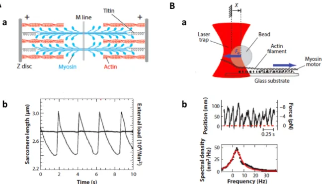
Figure 1.5: Oscillations with actin filaments and myosin motors. A: Sarcomere oscil-lations : (a) Schematic representation of the structure of a sarcomere. (b) Time course of a sarcomere length (gray line) in presence of an external load (black line) from (Beta and Kruse, 2017), adapted from (Guenther and Kruse, 2007). B: In vitro motor oscillations (a) Schematic of a gliding assay under elastic loading. Motors develop a force Fa on the actin filament held in an optical trap that exerts an elastic restoring force F . (b) Bead position as a function of time and corresponding spectral density from (Beta and Kruse, 2017), adapted from (Plaçais et al., 2009)
A classic example is given by sarcomeres (Okamura and Ishiwata, 1988), (Fig. 1.5.A), which are the basic functional units of skeletal muscles. Myosin ("thick") filaments inter-digitate with antiparallel ("thin") actin filaments such that the structure shortens when the motors are active. In presence of an external load on the sarcomere, this system can undergo periodic cycles of contractions and extensions can emerge. The asymmetric time course of the length of the oscillating sarcomere defines the system as a "relaxation os-cillator", with a faster extension phase than the contraction phase. It has been suggested that the oscillations come from a collective effect in elastically-coupled motors (Jülicher and Prost, 1997) and not from chemical (Ca2+) waves that drive the movement (Fabiato
and Fabiato, 1978). This hypothesis has been confirmed with an in vitro reconstituted system, where an actin filament held by optical tweezers was brought into contact with a substrate covered with Heavy-MeroMyosin myosin II motors and started to oscillate spontaneously (Plaçais et al., 2009) (Fig. 1.5.B).
Self-organization and Spontaneous oscillations Self-Organization
the cell membrane, in a region called the cell cortex (Bornens et al., 1989; Paluch et al., 2005; Salbreux et al., 2007). The cell cortex consists in a thin layer of actin beneath the cell membrane that myosin motors can contract by generating forces. The contractile stresses generated by myosin motors can destabilize the cortex and fracture it. For example, the cortex oscillates after depolymerization of all microtubules in suspended lymphocytes (Bornens et al., 1989), or in fibroblasts after loss of adhesion (Salbreux et al., 2007).
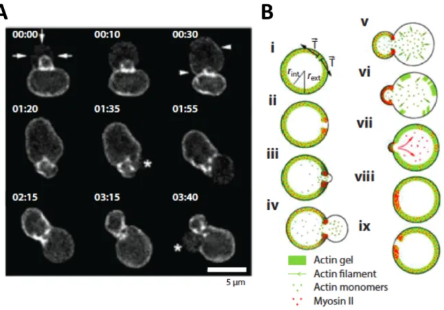
Figure 1.6: Cortex oscillations of lymphoblast fragments. A: Distribution of fluorescently labeled actin. B: Schematic description of the oscillation mechanism adapted from (Paluch et al., 2005)
In their studies on cell fragments, Paluch et al. showed that the oscillations in sus-pended cells resulted from localized breakage of the cortex (Paluch et al., 2005), which then retracted through the action of myosin, and subsequently reassembled (Fig. 1.6).
Other examples include oscillations of the mitotic spindle to determine the division plane in eukaryotic cells (Grill et al., 2005) and the regular beating of eukaryotic cilia and flagella (Howard, 2009; Satir and Christensen, 2007). Related to this last example, Sanchez et al.. developed a minimal model system composed of microtubules and molecular motors which self-assemble into active bundles exhibiting beating patterns resembling those found in eukaryotic cilia and flagella.
Self-organization and Spontaneous oscillations Self-Organization
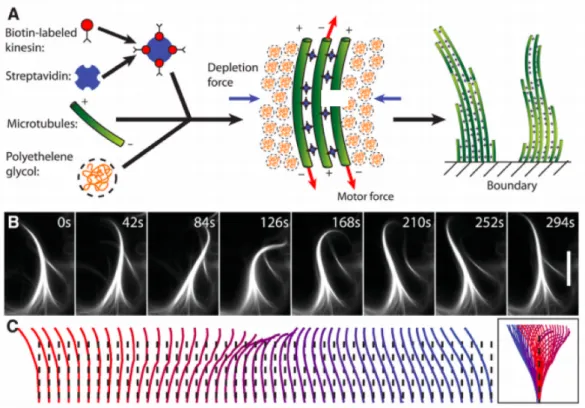
Figure 1.7: A minimal system of microtubules, molecular motors and depleting agent (polyethylene glycol) assembles into actively beating bundles. A: Schematic illustration of all components required for the assembly of active bundles. B: A sequence of images illustrating the beating pattern of an active bundle over one beat cycle. Scale bar is 30 µm. C: The conformations of the bending microtubule bundle indicates a fairly symmetric beating pattern that is reminiscent of those found in cilia and flagella. Figure from(Sanchez et al., 2011)
This observation allowed the authors to identify few essential components for the assembly of beating microtubule bundles: microtubules, kinesin assemblies and a depleting agent to promote filament bundling (Fig. 1.7). The examples shown in Figure 1.5B and in Figure 1.7 suggest that oscillations in in vitro systems may emerge as a generic property of the motor assemblies under elastic loading, as proposed on the basis of general theoretical models developed by Frank Jülicher and Jacques Prost. The oscillatory instability is associated to a region of negative slope in the force-velocity relationship of motor assembly (Jülicher and Prost, 1997). Another proposed mechanism for motor-induced oscillations relies on a force-dependence of the rate at which a motor detaches from a filament (Grill et al., 2005). If this rate increases with the applied force, the spontaneous detachment of a single motor leads to an increase of the load on all other motors, which in turn increase their detachment rates. This mechanism can result in the coordinated detachment of all the motors. This scenario is well-illustrated in everyday life by the tug of war, when one person in the team releases the rope, it becomes suddenly one step harder for the remaining people of the team to still hold the rope.
Self-organization and Spontaneous oscillations Spontaneous beating of eukaryotic flagella
1.2 Spontaneous beating of eukaryotic flagella
This section is a brief overview of biological flagellar beating in eukaryotes to later compare its phenomenology to that of beating bundles of actin filaments that I produced in vitro during my PhD using a minimal actomyosin system.
1.2.1
Cilia and flagella : motile organelles
Cilia and flagella are hair-like appendages that produce motility through repetitive episodes of bending-wave propagation. This rhythmic and undulating motion is called flagellar beating and can propel a cell through liquid media or displace a fluid over the surface of a group of cells in a tissue (Bruot and Cicuta, 2016; Ginger et al., 2008).
Figure 1.8: Diversity of cilia and flagella. A: Flagella of Chlamydomonas reinhardtii. Scale bar: 10 µm. B: Cilia of Paramecium tetraurelia. Scale bar: 10 µm. C: Flagellum of spermatozoan. Scale bar: 10 µm. Figure from (Rikmenspoel and Isles, 1985) D: Scanning electron micrographs of the epithelium lining the ventricles of a mouse brain (4 days old), showing many cilia. Scale bar: 1 µm. Figures (A,B,D) adapted from (Vincensini et al., 2011),
Although they are both called flagella, note that bacterial flagella and eukaryotic flag-ella differ in structure and mode of action: eukaryotic flagflag-ella contain linear motors that are uniformly distributed along the length of the flagellum whereas rotatory motors are located at the base of bacterial flagella. As a result, bacterial flagella are passive rigid helical structures that rotate whereas eukaryotic flagella actively bend to beat (Bray, 2001). Despite their different names, cilia and flagella are instead endowed with the same structure, which is called the axoneme. The typical length of eukaryotic cilia and flagella is 10 µm with a diameter of 0.2 µm diameter (Marshall, 2004; Neidhardt et al., 1996).
Self-organization and Spontaneous oscillations Spontaneous beating of eukaryotic flagella
Cilia tend to be shorter (around 7 µm) than flagella (from 12 µm (Chlamydomonas rein-hardtii) to 70 µm (sperm)) (Bruot and Cicuta, 2016). Typically, cells possess one or two long flagella at their extremity, whereas ciliated cells are covered with many short cilia, partially or all-over their surface (Alberts et al., 2008). Cilia and flagella cover a wide range of functions for animals and plants as diverse as clams and algae (Fig. 1.8) : They can for instance be used for feeding, reproduction or protection from infection in the bronchi of the lung.
1.2.2
The axoneme : the core structure of cilia and flagella
Cilia and flagella are highly ordered structures containing more than 650 different proteins (Pazour et al., 2005). The core of this structure is called the axoneme (Fig. 1.9). An axoneme is mostly composed of microtubules with a pipe-like structure, and dyneins. In an axoneme, dyneins are assembled uniformly onto a scaffold of nine cylindrically arranged doublets of microtubules. These nine microtubule doublets are surrounding a pair of single microtubules, called the central pair (Nicastro et al., 2006). Neighboring outer doublets are linked together through strands of proteins called nexin links. Those doublets are also connected to the central pair by radial spokes, acting as spacers to maintain the cylindrical geometry of the axoneme, and anchored to the basal body.
Figure 1.9: Cross section of the flagellar axoneme. Dyneins are assembled onto a scaffold of nine cylindrically arranged doublets of microtubules. These nine microtubules doublets are surrounding a pair of single microtubules, called the central pair. The neighboring outer doublets are linked together through strands of proteins called nexin links. Those doublets are connected to the central pair by radial spokes, and anchored to the basal body. Here, doublets 5 and 6 are connected by a rigid bridge that prevents their relative sliding; its position defines the beating plane (perpendicular to the bridge). Figure from (Lindemann and Lesich, 2010) DRC : dynein regulatory complex.
Self-organization and Spontaneous oscillations Spontaneous beating of eukaryotic flagella
In the axoneme, dyneins are bound to two outer neighboring doublets : the active longitudinal motion of the dyneins along the microtubule tracks causes the microtubule doublets to slide with respect to one another (Fig. 1.10.A) (Summers and Gibbons, 1971). This sliding movement is then converted into bending by the nexins linkers and the basal attachment of the microtubules, which both limit inter microtubule sliding and maintain the structural integrity of the axoneme (Fig. 1.10.B).
Figure 1.10: Bending of an axoneme. A: Isolated doublet can be obtained by exposing axonemes to trypsin, which breaks the linkages holding neighboring microtubules. The addition of ATP— the source of energy for the motors— allows dyneins to slide one pair of doublet microtubules with respect to the other pair. B: In intact axonemes (such as in sperm), the linking proteins (nexin) prevent the relative sliding of the doublets and motor action causes a bending motion. Figure adapted from (Alberts et al., 2008) and experiments from (Summers and Gibbons, 1971)
1.2.3
Beating properties
Taylor highlighted in 1951 that self-propelled cells work at very low Reynolds numbers (Re 1), so that viscous drag dominates and inertia can be neglected (Taylor, 1951). Cilia and flagella undergo periodic oscillatory motion, thanks to motors that coordinate an oscillatory bending motion of the axoneme. The beating pattern typically consists of rather symmetrical and propagating waves, often planar or helical. Their frequency typically varies from 5 to 100 Hz (Satir and Christensen, 2007). These waves normally travel from the base to the tip of the flagellum, although there are exceptions such as the flagella of the kinetoplastid Critidia oncopelti, which exhibit waves in the opposite direction (Douglas and Holwill, 1972). The simplest waveform seen in eukaryotic flagella is a planar sinusoidal wave that travels steadily from the anchored base to the free tip of the flagellum. The amplitude of the wave at any given point along the longitudinal axis of the flagellum is defined by the maximal tangent angle that the flagellum can reach (in
Self-organization and Spontaneous oscillations Spontaneous beating of eukaryotic flagella
deg) or by the maximal distance with respect to the reference, from which the flagellum is straight. This pattern of beating is seen in many animal sperm that are streamlined for efficient swimming. The most studied flagella of this type are from the sea urchin spermatozoan, the Chlamydomonas reinhardtii and the bull sperm (Fig. 1.11). I will briefly describe their properties and focus more into details on the bull sperm studies.
Figure 1.11: Beating pattern of three eukaryotic flagella. A: Sea urchin sperm. Scale bar: 10 µm. The snapshots are taken at 10-ms time intervals. Figure from (Rikmenspoel and Isles, 1985). B: Bull sperm. Scale bar 20 µm. The snapshots are taken at 10-ms time intervals. Figure from (Gray, 1957). C: Chlamydomonas reinhardtii. Scale bar: 5 µm. Figure from (Sanchez et al., 2011)
.
Table 1.1: Flagellar beating properties in different species
Species Length Frequency Speed Maximum Wavelength References of swimming amplitude
(µm) (Hz) (µm/s) (µm) (µm)
Chlamydomonas 14 70 60 5 14 (1-2) Sea urchin sperm 41 33 180 4 24 (3)
Bull sperm 55 6 100 5 54 (4-5)
Data from: (1) (Brokaw and Luck, 1983); (2)(Bayly et al., 2010); (3) (Gray, 1955); (4) (Rikmenspoel, 1984); (5) (Gray, 1957).
Self-organization and Spontaneous oscillations Spontaneous beating of eukaryotic flagella
Sea urchin sperm Historically, the beating waveforms of sea urchin sperm were the first studied (Gray, 1955) (Fig. 1.11.A). Sea urchin spermatozoa show planar beating when close to a surface, and the wave of the sperm tail travels with an almost constant amplitude along the tail. Bending waves are composed of nearly circular arcs separated by short straight regions. The wave speed along flagella is around 800 µm/s for a speed of propulsion around 200 µm/s. The amplitude of the wave is 4 ± 0.5 µm, the wavelength is 24 ± 2 µm for a flagellum length of 41 ± 2 µm.
Chlamydomonas reinhardtii For Chlamydomonas reinhardtii, waveforms exhibit an asymmetric pattern : the beat cycle starts with the "power stroke", where the flagellum straightens and changes its orientation, thus pushing the fluid. It returns to its initial position curled up, in the "recovery stroke", thus minimizing the interaction with the fluid (Bruot and Cicuta, 2016). The maximal amplitude of the wave is 5 µm, the wavelength is 14 µm for a flagellum length of 14 µm, suggesting that the length of the flagellum sets the wavelength. (Bayly et al., 2010; Brokaw and Luck, 1983). Sartori and collegues measured the flagellar beating waveforms in axonemes with high temporal and spatial precision (Sartori et al., 2016). To characterize the shape of the filament through the beating, they introduced the tangent angle ψ with respect to the horizontal axis of the laboratory frame (Fig. 1.12.A.(ii)). As shown in Figure 1.12.A.(iii), the beating of Chlamydomonas reinhardtii is periodic and Chlamydomonas axonemes swim counterclockwise in circles at a slow angular rotation speed (Fig. 1.12.A.(i-iii)). As the power spectrum of the tangent angle (averaged over the flagellar length) shows (Fig. 1.12.B.(i)), the beating can be decomposed in harmonics, but the peak at the fundamental frequency (n = 1) accounts for 90% of the total power, so the higher harmonics (n = 2; 3; 4; ...) can be neglected for reconstructing the flagellar shape. The static and fundamental modes provide a good description of the beats (Fig. 1.12.C). The amplitude ψ0 of the static mode (n = 0) is
presented in Figure 1.12.B.(ii), it decreases linearly over the length of the flagellum. This corresponds to an approximately constant static curvature and a time-averaged shape close to a semi-circular arc (radius ≈ 4 µm). This static curvature confers to the flagellum an asymmetric waveform. The amplitude and the phase of the fundamental mode (n =1) are shown in Figure 1.12.B.(iii-iv). The amplitude is almost uniform along the length of the flagellum, the argument, which determines the phase of the wave, decreases at a roughly constant rate, indicating that the beat is a traveling wave. Because the total phase shift is about 2π along the flagellum, the wavelength of the beat is approximately equal to the length of the axoneme.
Self-organization and Spontaneous oscillations Spontaneous beating of eukaryotic flagella
Figure 1.12: Beating properties of the Chlamydomonas reinhardtii. A: High-precision tracking of isolated, reactivated axonemes. (i) Inverted phase-contrast image of a wild type axoneme. The orange curve represents the tracked centerline. The points depict the basal end (red), the distal end (green) and the center position (black) of the axoneme. The green line depicts the trajectory of the basal end, which is the leading end during swimming. (ii) Same image as in A), magnified around the center region. The tangent angle ψ(s,t) is defined with respect to horizontal axis in the lab frame. (iii) Tangent angle at three different arc-length positions (depicted in A) as a function of time. The linearly increasing tangent angle corresponds to a counter-clockwise rotation of the axoneme during swimming. B: Fourier decomposition of the beat. (i) Power spectrum of the tangent angle averaged over the arc-length. The fundamental mode (n = 1) and three higher harmonics (n = 2; 3; 4) are labeled. (ii) Angular representation of the static (n = 0) mode as a function of arc-length. The approximately constant slope indicates that the static curvature is close to constant ˙ψ0 = C0. (iii) The amplitude and phase (argument) of the fundamental mode are shown in iii and iv, respectively. The approximately linear decrease in phase indicates that the magnitude of the wave vector does not vary during wave propagation. The data of a representative axoneme is highlighted in the panels ii-iv. C: Beat shapes of one representative beat cycle of the wild type axoneme highlighted in panel A (left panel, data) and shapes reconstructed from the superposition of the static and fundamental modes, neglecting all higher harmonics. The progression of shapes through the beat cycle is represented by the rainbow color code (see inset). Figure adapted from (Sartori et al., 2016)
Self-organization and Spontaneous oscillations Spontaneous beating of eukaryotic flagella
Bull sperm To study mammalian sperms, the bull spermatozoan served as a model system, first by analyzing pictures of bull sperm waveforms (Gray, 1958) (Fig. 1.11.B). In more recent studies using automated image analysis of the flagellum (Riedel-Kruse et al., 2007), the tangent angle along the flagellum ψ(s, t) was analyzed (Fig. 1.13.A). Tracking of the flagellum indicates a slight asymmetry in the beating pattern, resulting in curved trajectories of the swimming plane (Fig. 1.13.B). A space-time plot of ψ(s, t) reveals the periodicity of the flagellar beat (Fig. 1.13.C). In contrast to sea urchin spermatozoa, the wave amplitude for bull spermatozoa was found to increase as the wave propagated towards the end of the tail. The maximum amplitude of the wave is 5 µm, the wavelength is 54 µm for a flagellum length of 55 µm (Gray, 1957; Rikmenspoel, 1984).
Figure 1.13: Beating properties of the bull sperm. A: Snapshot of a beating bull sperm, superimposed on the image are red crosses tracing the contour of the flagellum as determined by the automated image analysis. The tangent angle ψ(s) is measured at each curvilinear abscissa salong the flagellum. The center of the red crosses are separated by 1.4 µm. Scale bar: 10 µm. Figure from (Riedel-Kruse et al., 2007). B: Beating patterns of a bull sperm, with clamped head. Figure from (Friedrich et al., 2010). C: Kymograph of the tangent angle ψ(s, t) reveals the periodicity of the flagellar beat, as well as the growth of the amplitude of angle oscillations as the wave propagates towards the tip of the flagellum. Figure from (Ma et al., 2014).
The time series of the tangent angle over time at a fixed curvilinear abscissa (s = 28 µm) exhibits stable oscillations over time (Fig. 1.14.A). By computing the power spectrum at each position, all time series were well-approximated by their zeroth and first Fourier temporal modes.
ψ(s, t) ≈ψ‹(0)(s) +ψ(s) e‹ iωt+ψ‹∗(s) e−iωt (1.1)
where the star indicates the complex conjugate. Thus, the motion of each point can be described by a sinusoidal oscillation of amplitude 2 | ψ | and angular frequency ω
Self-organization and Spontaneous oscillations Spontaneous beating of eukaryotic flagella
with an average angle ψ(0). The time-averaged tangent angle ψ(0)(s)as a function of the
curvilinear abscissa corresponds to the mean shape of the flagellum (Fig. 1.14.B). This relation characterizes the asymmetry of the mean shape of the flagellum (if the mean shape were symmetric the averaged angle would be 0). The amplitude | ψ |‹ increases
linearly along the flagellum below s ≈ 45 µm and then saturates (Fig. 1.14.C). The phase of the first mode decreases linearly along the curvilinear abscissa, the slope of the plot is inversely related to the wavelength of beating (the slope corresponds to −2π/λ), which is thus uniform over most of the length of the flagellum (Fig. 1.14.D).
Figure 1.14: Time series analysis and Fourier modes of beat patterns. A: Time series of the tangent angle ψ(s, t) at a given position (s = 28 µm) along the flagellum. The head of the flagellum was clamped. B: The time average of the tangent angle ψ(0) at each point along the flagellum. C, D: Amplitude and phase of the first mode ψe, respectively. Figures from
(Riedel-Kruse et al., 2007).
Effect of viscosity
It has long been demonstrated that the flagellar beat frequency declines when the viscos-ity of the surrounding medium is increased (Brokaw, 1966; Rikmenspoel, 1984; Woolley et al., 2009). The effect is associated with an increase in bending angle and a decrease in wavelength, in swimming velocity and bending-wave speed (Brokaw, 1966; Rikmenspoel, 1984; Woolley et al., 2009)
In sea urchin sperm When the sea urchin spermatozoan swims at unphysiologically high viscosities of the surrounding medium (1.5 to 4 Pa.s), it has been shown that the
Self-organization and Spontaneous oscillations Spontaneous beating of eukaryotic flagella
usual planar beat of sea urchin spermatozoa can change into a fully 3D helical beat pattern (Woolley and Vernon, 2001).
In bull sperm The frequency of the flagellar beating in bull sperm decreases almost exactly as the square root of viscosity (Rikmenspoel, 1984), as shown in Figure 1.15.
Figure 1.15: Beating frequency of bull sperm at 37 °C as a function of the viscosity of the medium. The black circles represent the measurements of (Rikmenspoel, 1984); The + symbol refers to data from (Rikmenspoel et al., 1973) reported previously. Each point represents the average over 8 to 12 sperm cells. The vertical bars are standard deviations. The line with a slope of - 0.48 was drawn by eye. (Rikmenspoel, 1984). These data suggest a power law relating flagellar frequency f to viscosity η : f ∝ 1/√η
Moreover, the flagellar waveform changes qualitatively at raised viscosity. The am-plitude near the base becomes progressively smaller, whereas the amam-plitude near the tip remains roughly constant over the entire viscosity range. This results in an increasingly trumpet-shaped appearance of the envelope of the flagellar wave (Fig. 1.16).
Self-organization and Spontaneous oscillations Spontaneous beating of eukaryotic flagella
Figure 1.16: Viscosity and beating pattern. Typical waveforms of bull sperm flagellum in media of raised viscosity from 1 to 3600 mPa.s. The forward progression of the sperm was removed. The flagellar waveform changes qualitatively at raised viscosity. The amplitude near the base becomes progressively smaller at higher viscosities, whereas the amplitude near the tip remains roughly constant over the entire range. This results in an increasingly trumpet-shaped appearance of the envelope of the flagellar wave. Figure adapted from (Rikmenspoel, 1984).
More recently, a Fourier analysis of the flagellar beat of a swimming bull sperm cell was computed for the case of normal viscosity η = 0.7 mPa.s and increased viscosity η = 10 mPa.s (Friedrich et al., 2010). It shows that the static shape of the flagellum, as determined from ψ0(s), is less curved at higher viscosities (Fig. 1.17.A). The linear fit of
the two curves gives the mean flagellar curvature K0, which drops from 19.1 rad.mm−1
to 6.5 rad.mm−1. The amplitude of the first mode decreases at higher viscosity and does
not exhibit saturation in contrast to what observed at normal viscosity.
Figure 1.17: Variation of beating properties at high viscosity. A: The time-averaged of tangent angle, ψ(0), at each point along the flagellum for the case of normal viscosity η = 0.7 mPa.s (black disks)) and increased viscosity η = 10 mPa.s (white disks). The linear fit gives the mean flagellar curvature K0. B , C: Amplitude and phase, respectively, of the first Fourier mode
e
ψ for the case of normal viscosity η = 0.7 mPa.s (black disks) and increased viscosity η = 10 mPa.s (white disks). The linear fits give respectively the amplitude rise A0 and the wavelength λof the wave. Figures from (Friedrich et al., 2010)
Self-organization and Spontaneous oscillations Spontaneous beating of eukaryotic flagella
A linear fit of the relation between the amplitude of the first mode and the curvilinear abscissa gives the amplitude rise of the flagellar bending waves A0 (Fig. 1.17.B). The
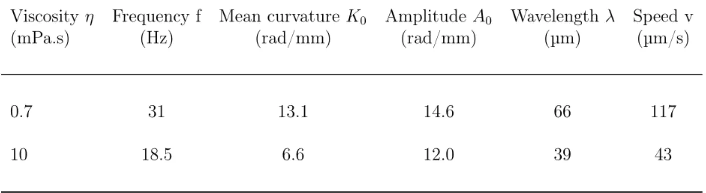
phase of the first mode, as a function of the curvilinear abscissa, indicates higher delays for wave propagation at higher viscosity (Fig. 1.17.C). The linear fit of the plots gives the wavelength λ of the principal flagellar bending wave, (the slope of the linear fit corresponds to 2π/λ). At higher viscosity the wavelength is almost divided by 2 compared to normal viscosity. The Table 1.2 summarizes the changes in the beating properties of the flagella.
Table 1.2: Changes of flagellar beat properties with viscosity
Viscosity η Frequency f Mean curvature K0 Amplitude A0 Wavelength λ Speed v
(mPa.s) (Hz) (rad/mm) (rad/mm) (µm) (µm/s)
0.7 31 13.1 14.6 66 117
10 18.5 6.6 12.0 39 43
Data from (Friedrich et al., 2010).
Effect of length on frequency
The effect of flagellar length on beating properties has been studied on demembraneted axonemes. When flagella are severed transversely, the proximal segments continue to beat but with an increased beat frequency, even if the flagellum length is reduced below the half of its original length (Gibbons, 1975; Woolley et al., 2009). The waveforms are generally similar, but the amplitude of the bends and the bending angle decrease with the length. Bending angle measurements indicate that for fragments shorter than 20 µm, the maximum angle varies linearly with the length of the fragment, as shown in Figure 1.18.
Self-organization and Spontaneous oscillations Spontaneous beating of eukaryotic flagella
Figure 1.18: Effect of flagellum length on beating properties. A: Movement of axonemal fragments of 28, 21, 16, 12, 8 µm. B: Relation between the maximal bend angles developed and the length of the fragments in (A). The upper (lower) curve shows angles of the principal (reverse) bends convex upwards (downwards) in the picture panels of (A). Figure adapted from (Gibbons, 1975)
1.2.4
Existing theoretical approaches of flagellar beating
The oscillatory pattern of the axoneme implies that the action of dyneins is coordinated. There must be two switching sets of dynein domains within the axoneme that are either active (mediating bending/sliding) or inactive (prevented to do the same by opposing shear forces) (Brokaw, 1975; Jülicher and Prost, 1997) to obtain bending alternatively in the two opposite directions (Fig. 1.19). How the motor activity is controlled to generate the observed wave-like beating of the axoneme remains unclear.
Figure 1.19: Asymmetric (a) and symmetric (b) motor filament pairs. The arrows indicate the polarity of the filaments. In case (a), spontaneous bending occurs in a steady state. In case (b) both filaments play identical roles, no spontaneous bending occurs. Figure from (Camalet and Jülicher, 2000)
Self-organization and Spontaneous oscillations Spontaneous beating of eukaryotic flagella
I briefly introduce the theory of Camalet and coworkers (Camalet and Jülicher, 2000; Camalet et al., 1999) to present the general concepts that are used in the field to describe the axonemal beat. The theory presented by Camalet describes the two-dimensional beat of an axoneme, where two filaments slide relative to each others. The centerline of the axoneme in space is given by r(s), where s is the arc length of the axoneme with s = 0 at the base, i.e. at the head of the cell, and s = L at the tip, the filament-separation is a (Fig. 1.20).
Figure 1.20: The complex structure of the axoneme is reduced to a pair of sliding filaments: Two filaments are spaced apart by the distance a. The filaments are connected at the base and free to slide at ∆. The arc-length of the filament pair is described by s. The motors (not shown) generate local forces f(s). Figure from (Camalet and Jülicher, 2000)
The relative sliding distance of the filaments ∆(s) at position s is determined by assuming that the filaments are incompressible (i.e. the filament-separation a remains constant) and by substracting the arc length of the filaments. Using the definition of curvature C(s) related to the tangent angle on the filament pair ψ(s) : ∂sψ(s) = C(s),
we get : ∆(s) = a Z s 0 C(s0)ds0 = a Z s 0 ∂ψ(s0) ∂s0 ds 0 = a(ψ(s) − ψ(0)) (1.2) Hence the tangent angle of the filament is linearly related to the internal sliding of the filament. The enthalpy functional G takes into account the bending of the filaments and the internal stresses due to active and passive elements inside the axoneme,
G =
Z L
Self-organization and Spontaneous oscillations Spontaneous beating of eukaryotic flagella
where L is the length of the flagellum, κ the bending elasticity of the flagellum (which is assumed to be the same at all positions), f is the effective active force per unit lengths exerted by the motors (drives bending) and Λ is the Lagrange multiplier ensuring that s is the arc length. After a partial integration and using equation (1.2), we find
G = Z L 0 [κ 2C 2− aF C + Λ 2∂sr 2 ]ds (1.4) where F (s) = − Z L 0 f (s0)ds0 (1.5) Determining the variation δG with respect to variation δr, which gives by definition the external forces applied to the axoneme:
δG
δr = ∂s[(κ∂sC − af )n − τ t] (1.6) where τ = Λ + κC2− aF C plays the role of a physical tension and n and t are the
normal and tangent on the outline r.
For simplicity, it is assumed that the hydrodynamics of the surrounding fluid can be described by two local friction coefficients ξk and ξ⊥ for tangential and normal motion
respectively. The condition of force balance reads : ∂tr = −( 1 ξ⊥ nn + 1 ξk tt).δG δr (1.7)
Here nn and tt are the normal and tangential projection operators.
Injecting the expression (1.6) of δG/δr into the dynamic equation (1.7), we can get a differential equation of the tangent angle ψ(s, t). To study the linear stability of the system, we work in the limit of small tangent angles. Moreover under the simplifying assumption of a symmetric beating. We have to exclude antisymmetric terms such as f ↔ −f and ψ ↔ −ψ. Then using equation (1.6) with ∂sF (s, t) = f (s, t)
ξ⊥∂tψ = −κ∂4sψ + a∂ 2
sf (1.8)
The second term on the right-hand side describes the active contribution of the motors to force balance. In a passive system, this term is zero and we are left with force balance between friction (on left-hand side of the equality) and elasticity (first term on the right-hand side); this equation, which is known as the elasto-hydrodynamic equation, describes the bending modes of slender rods (Howard, 2001).
Self-organization and Spontaneous oscillations Spontaneous beating of eukaryotic flagella
Four different boundary conditions have been discussed by Camalet : • clamped head, free tail
• fixed head, free tail
• swimming flagellum with viscous load ζ
• clamped head, external force applied to the tail.
To apply the boundary conditions to our system, it is more instinctive to work with transverse deformation h than with the tangent angle ψ, ∂sh = ψ, which can be injected
in equation 1.8 above:
ξ⊥∂th = −κ∂4sh + a∂ 2
sf (1.9)
I will focus on the first case where the head is clamped, which gives the following boundary conditions: h(0) = 0 ∂sh |s=0= 0 ∂2sh |s=L= 0 [κ∂3sh − af ] |s=L= 0 (1.10) In the case of oscillatory patterns, one can express h(s, t) = P+∞
−∞hn(s)einωt, and
f (s, t) =P+∞
−∞fn(s)einωt, as Fourier series in time (with h−n = h∗n, ensure that h is real)
where ω is the angular beating frequency which leads to:
κ∂4shn− a∂2sfn(s) = −iωξ⊥hn (1.11)
and the boundary conditions
hn(0) = 0
∂shn|s=0= 0
∂2shn |s=L= 0
[κ∂3shn− afn(s)] |s=L= 0
(1.12) The forcing term fn describes the activity of motors and the passive internal
visco-elastic elements. Three different mechanisms have been proposed to describe how the mechanical activity of the motors (through the term fn) is regulated by the shape of the
axoneme; the description assumes the existence of a linear response function between fn
Self-organization and Spontaneous oscillations Spontaneous beating of eukaryotic flagella
• Sliding control
fn(s) = χ(nω)∆n(s) (1.13)
where ∆n represents a Fourier mode of the sliding distance and χ(nω) is the
corre-sponding linear response.
In this model the activity of dyneins is regulated by the sliding between the two fil-aments within the axoneme. Assuming a load-dependent detachment rate of motors kof f = k0exp(fL/fc), where fLis the load and fC is a characteristic force giving the
scale of the load that can affect kof f, one can derive the following expression for χ
(Riedel-Kruse et al., 2007):
χ(nω) = k + inωλ − ρKCBΩ
iωα + (ω/α)2
1 + (ω/α)2 (1.14)
A similar expression was obtained from a generic two-state model of collective motor dynamics that does not explicitly assume load-dependent detachment of the motors (Camalet and Jülicher, 2000; Jülicher and Prost, 1997). Here k is the stiffness, λ is the friction per unit length of the passive internal elements, the last term is the linear response of the active motors themselves, with ρ the motor density, KCB is
the cross-bridge elasticity of the motors, α is the characteristic ATP-cycling rate and Ω plays the role of activity parameter.
As seen in Equation 1.14, the active term leads to both negative elasticity and fric-tion, which can precisely cancel the passive terms k and λ, respectively, resulting in an oscillatory instability called a Hopf bifurcation (where χ vanishes at a specific value of ω). This property is generic of large groups of molecular motors coupled elastically to the environment : such system can show a dynamic instability leading to oscillations (Brokaw, 1975; Jülicher and Prost, 1997). In vitro experiments have demonstrated that oscillatory instabilities can indeed emerge as an intrinsic prop-erty of motor groups under elastic loading (Okamura and Ishiwata, 1988; Plaçais et al., 2009)
• Curvature control
fn(s) = β(nω)∂sψn(s) (1.15)
where ∂sψnrepresents a Fourier mode of the curvature and β(nω) is the
correspond-ing linear response.
In this model of feedback, the activity of the dyneins is regulated by the local curvature of the axoneme i.e. its degree of bending. This was one of the earliest ideas (Brokaw, 1971, 1972a,b) after the proposal of the sliding bending mechanism.
Self-organization and Spontaneous oscillations Spontaneous beating of eukaryotic flagella
Many simulation studies were undertaken by Brokaw showing the feasibility of the approach.
• geometrical clutch
fn(s) = γ(nω)f⊥,n(s) (1.16)
where f⊥,nrepresents Fourier mode of the normal force felt by the motors and γ(nω)
is the corresponding linear response.
In this model of feedback, the dynein detachment is regulated by transverse forces separating curved adjacent doublets. This model proposed that in bending axoneme, transverse forces develop that pull neighboring microtubule doublets apart and that this increase in interdoublet spacing leads to a decrease in the probability of dynein engagement (Lindemann, 1994a,b). Such models are based on observations in elec-tron micrographs showing that the spacing between two neighboring microtubule doublets is larger when the dyneins are unbound than when they are bound (Gib-bons and Gib(Gib-bons, 1974). Although neither curvature nor geometrical clutch mech-anism is supported by a direct experimental evidence with microscopy, they have been shown to produce traveling waves in computer models (Lindemann, 2002). Dynamic curvature regulation has also recently been proposed to account for the symmetric and asymmetric beats of Chlamydomonas reinhardtii flagella (Sartori et al., 2016). Note that in the geometrical clutch model, the filament-separation a is not constant, unlike in the model used for the calculation, thus the general equation of the tangent angle and transverse deformation would be modified.
Note that χ, β and γ have real and imaginary parts that can become negative (Eq. 1.14), as the result of motor activity (Sartori et al., 2016).
Chapter 2
The actomyosin system
2.1 The actin cytoskeleton
The actin cytoskeleton is a network of semi-flexible filaments, which are active polymers that can elongate or shrink depending on the surrounding environment. As a result, the network can continuously reorganize in order to adapt to changing conditions. The actin cytoskeleton is involved in various key cellular processes such as motility, morphogenesis, polarity, transport and cell division.
2.1.1
Actin : From monomers to filaments
Actin structure
The actin protein exists in two forms : the monomeric Globular actin (G-actin) form and the polymerized Filamentous actin (F-actin) form. G-actin is a 43-kDa-protein composed of 4 subdomains (Kabsch et al., 1990), which binds to a divalent cation — Magnesium under physiological conditions (Blanchoin and Pollard, 2002) — and a nucleotide (ATP or ADP). G-actin is asymmetrical, hence the monomers are arranged head-to-tail in a filament. That confers polarity to the actin filament, i.e. both ends are structurally different: Subdomains 1 and 3 constitute the "barbed end" of the actin filament and subdomains 2 and 4 compose the "pointed end" (Fig. 2.1). The actin filament forms a double-stranded helix, with a twist repeating every 37 nm and diameter of a 5-9 nm (Howard, 2001).
The actomyosin system The actin cytoskeleton
Figure 2.1: Structure of G-actin. A: The asymmetric topology of G-actin, with its 4 subdo-mains represented in different colors, its binding site, its barbed end (formed by the subdosubdo-mains 1 and 3) and its pointed end (formed by the subdomains 2 and 4). Adapted from (Kim et al., 2006). B: The arrangement of monomers within the filament forms a double stranded helix, with a twist repeating every 37 nm and diameter of a 5-9 nm. C: Electron micrograph of negatively stained actin filament. (B,C) adapted from (Alberts et al., 2008).
The assembly of actin filament induces a conformational transition in the actin sub-unit: G-actin has a twisted conformation and F-actin has a flat conformation (Oda et al., 2009), obtained by a rotation of 20 degree of subdomains 1-2 with respect to subdomains 3-4 (Fig. 2.2).
Figure 2.2: Transition from the G-actin conformation to the flat conformation in F-actin. A: Front view. The structures of the subunits in the F-actin (cyan) and in the G-actin (yellow) are superimposed on subdomains 1 and 2. Subdomains 3 and 4 are rotated with respect to subdomains 1 and 2 about the rotational axis indicated by the red line in the direction indicated by the red arrow. B: Side view from the left-hand side of subdomains 3 and 4 in (A). Figure adapted from (Oda et al., 2009)
The actomyosin system The actin cytoskeleton
Actin mechanics
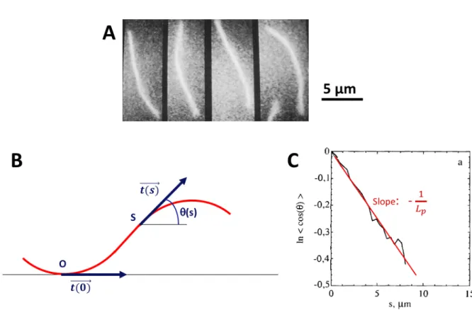
Just as thermal forces cause a spring to undergo fluctuations in length, thermal forces cause a flexible filament to undergo fluctuations in shape (Fig. 2.3.A). The persistence length Lp is a measure of the length scale below which a polymer keeps the memory
of the monomer orientation under thermal fluctuations. By comparing the value of the persistence length Lp to the filament length L, three classes of polymer can be defined :
• if Lp L: the filament is classified as a flexible polymer
• if Lp ∼ L : the filament is classified as semi-flexible
• if Lp L: the filament is classified as rigid
Figure 2.3: Persistence length of an actin filament. A: Recorded shape of an unstabilized F-ADP-actin filament undergoing thermal fluctuation at 6-s intervals. B: Schematic representa-tion of a filament. For each point of curvilinear abscissa s, the tangent angle θ(s) with respect to the tangent at the origin O is measured. As the distance between O and S increases, the tangent angle θ(s) and the tangent angle at the origin become uncorrelated, as the result of thermal fluctuations. C: The persistence length can be measured from the exponential decay of tangent-tangent correlation along a chain, i.e. the persistence length is the characteristic scale over which memory of the tangent angle is lost. The slope of the graph corresponds to − 1
Lp.
Figure adapted from (Isambert et al., 1995)
The persistence length can be measured from the decay of tangent-tangent correlation along a chain, i.e. the persistence length is the scale over which memory of the initial
The actomyosin system The actin cytoskeleton
tangent angle is lost (Howard, 2001), as seen in Figure 2.3.B and C. <−→t (s) . −→t (0) >=< cos(θ(s) − θ(0)) >= exp Ç − s Lp å (2.1) where θ and s represent the tangent angle and the curvilinear abscissa along the chain, respectively.
The persistence length is related to the flexural (or bending) rigidity K of the filament: Lp =
K kBT
(2.2) where kB is the Boltzmann constant and T is the temperature in Kelvin. The more flexible
the filament, the smaller the persistence length, the greater the curvature that the filament can achieve under thermal fluctuations. The persistence length of the actin filament, when it is not bound to other proteins, is 9 ± 0.5 µm (Isambert et al., 1995), which is in the range of the actin filament length in cells. Hence in a cell, actin is considered as a semi-flexible filament (Fig. 2.3). It has a flexural rigidity of K = 3.6 10−26 N.m2 (Howard,
2001; Isambert et al., 1995). Actin polymerization kinetics
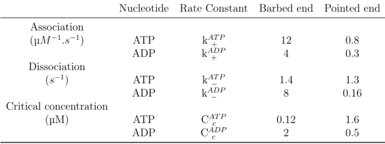
The actin filaments polarity is associated with the different dynamics of polymeriza-tion/depolymerization at the two filaments ends : at large concentrations of G-actin, actin filaments elongate and the barbed end is the fast growing end, whereas the pointed end is the slow growing end (Pollard, 1986). The barbed end of the actin filament has an association rate of G-actin (k+), ten times higher than that of the pointed end (Pollard,
1986). Nucleotides regulate the actin polymerization/ depolymerization process by tuning the values of association/dissociation rates at both ends of the filament (Fig. 2.4).
Figure 2.4: Association/dissociation rates of actin. G-actin is bound to ATP (respectively ADP) represented by the letter T (D). The association rates have units of µM−1.s−1. Dissociation rates have units of s−1. The ratio of the dissociation to the association rate gives Cc, the critical concentration. Figure from (Philips et al., 2009), adapted from (Pollard and Borisy, 2003)