HAL Id: tel-03092309
https://tel.archives-ouvertes.fr/tel-03092309
Submitted on 2 Jan 2021HAL is a multi-disciplinary open access
archive for the deposit and dissemination of sci-entific research documents, whether they are pub-lished or not. The documents may come from teaching and research institutions in France or abroad, or from public or private research centers.
L’archive ouverte pluridisciplinaire HAL, est destinée au dépôt et à la diffusion de documents scientifiques de niveau recherche, publiés ou non, émanant des établissements d’enseignement et de recherche français ou étrangers, des laboratoires publics ou privés.
Regulation of Human T Helper Cell Diversity : From In
Vitro Dendritic Cell-Based Mechanisms to Candidate
Biomarkers in Atopic Dermatitis
Coline Trichot
To cite this version:
Coline Trichot. Regulation of Human T Helper Cell Diversity : From In Vitro Dendritic Cell-Based Mechanisms to Candidate Biomarkers in Atopic Dermatitis. Immunology. Université Paris Saclay (COmUE), 2019. English. �NNT : 2019SACLS423�. �tel-03092309�
Regulation of human T helper cell
diversity: from in vitro dendritic
cell-based mechanisms to
candidate biomarkers in atopic
dermatitis
Thèse de doctorat de l'Université Paris-Saclay
préparée à l’Institut Curie et à Sanofi
École doctorale n°582
Cancérologie : biologie - médecine - santé (CBMS)
Spécialité de doctorat : Aspects moléculaires et cellulaires de la biologie Thèse présentée et soutenue à Paris, le 22 Novembre 2019, par
Coline Trichot
Composition du Jury :
Jean-David Bouaziz
PUPH, Hôpital Saint-Louis Président
Jenny Valladeau-Guilemond
CR1, Centre de recherche en Cancérologie de Lyon
(– UMR INSERM 1052) Rapporteur
Stéphanie Graff-Dubois
Enseignant-Chercheur, Laboratoire Immunologie,
Immunopathologie, Immunothérapie (– UMRS 959) Rapporteur
Géraldine Schlecht-Louf
MCU, Université Paris-Sud (– UMR-S 996) Examinateur
Vassili Soumelis
PUPH, Hôpital Saint-Louis (– UMR INSERM 976) Directeur de thèse
Benoit Pasquier
Chef d’équipe Checkpoint Immunology, Sanofi Co-encadrant
NNT
:
2
0
1
9
S
A
CL
S
4
2
3
1
ACKNOWLEDGMENTS
First, I would like to thank the members of my thesis jury: Dr Jenny Valladeau -Guilemond, Dr Stéphanie Graff-Dubois, Dr Géraldine Schlecht-Louf and Pr Jean-David Bouaziz for accepting to evaluate my PhD work.
I wish to deeply thank Vassili Soumelis for welcoming me in his team 6 years ago, first as an engineer and then for these 3 years of PhD. Thank you for your trust, advices, guidance and the freedom you gave me to test my ideas.
I am greatly thankful for the sponsorship from the ARN T (Association Nationale Recherche Technologie) and Sanofi for my 3 years of Cifre PhD (Industrial Agreements for Training through Research). My project was followed not only by Benoit Pasquier, my Sanofi supervisor, but also by Joe Blois first, and then Hamid Mattoo, both in Sanofi Cambridge. The three of them advised me along the 3 years of my PhD project. I am deeply grateful for their supervision. In addition, working at Sanofi has been a huge opportunity to discover the industry world. I had the chance to attend and participate to Benoit’s team meetings, follow projects and learn their problematics. I was able to spend a few months doing experiments in Benoit’s lab and benefit from Sanofi’s tools and expertise, which was a great experience. Besides, I had the great chance to visit Sanofi Cambridge for a few days, and this would not have happened without Hamid, who planned a great trip for me. I met many people there, had great discussions and lots of feedback on my project. Thank you so much, it was a great experience and a real pleasure to meet everyone. Also, I would like to thank the entire Sanofi team: Solana, Carolina, Charlotte, Erwan, Ellen, Delphine, Sandrine, Eve, Tsing -Lee and Laure. In the end I am very thankful, I have learnt a lot and had many great opportunities thanks to this Cifre program.
I would also like to thank Sebastian Amigorena for welcoming me in his unit and being part of my thesis committee. Thank you for your advices and helpful scientific critics. Thanks as well to all PI and all people from U932. This unit is an amazing scientific environment to work and learn. I feel particularly lucky to have had the chance to perform my PhD in such a place.
I would like to thank the entire Soumelis team, past and current members : Irit, Alix, Carolina, Paula, Solana, Salvatore, Mahé, Marine, Gérome, Antonio, Marie, Ares, FX,
2
Omar, Rabie, Lilith, Charlotte, Caroline, Camille, Philémon, Sarantis, Elise, Floriane, Maude, Arturo, Faezeh, Justine, Alain, Lucile, Jasna, Alba, Iris, Mélissa, Sara a nd Fanny. Thanks for the great atmosphere, the evening apero and all the fun. It has been awesome to work with you all during these 6 years.
My biggest thanks to Max for helping me during my entire PhD. Thanks for all the scientific discussions, advices and expert proof-reading of this manuscript.
My sincere thanks to Lucia, it has been such a pleasure to work with you. And my PhD project would not have been created if it was not for you. I had so many great opportunities thanks to this Cifre PhD and it is thanks to you.
My special thanks to Léa for the psychological and experimental support, even during long and hard revisions, the hummus shared , the haircut, the mutual complaints and all the laughter!
I wish to thank the Institut Curie Cytometry platf orm: Zosia and even more Sophie and Annick for all the cell sorts they performed for me, struggling with the Astrios. My project would have been a nightmare without you girls!
I would like to express my sincere gratitude to Pr Jean -David Bouaziz, Dr Marie Jachiet, Dr Anne Saussine and all people working at the Dermatology Service in Saint -Louis Hospital who helped me receiving patient samples for my study on atopic dermatitis.
I would like to thank all the people from CrossFit XIII, my second home. Thank you to the 7am team and all my CrossFit partners, and above all Emilie and Leslie. My morning WODs are probably the one reason I did not entirely go insane those last 3 years.
Finally, many thanks to my friends, especially Catherine, Claire, Tiffany, and of course my family: my parents, grand-parents and my two sisters, who supported me from the beginning.
3
TABLE OF CONTENT
ACKNOWLEDGMENTS ... 1 PREAMBLE ... 5 LIST OF ABBREVIATIONS ... 7 LIST OF FIGURES ... 9 INTRODUCTION ... 111. T helper cell subset diversity and functional impact ... 13
1.1. T helper cell subsets, phenotypes and functions ... 13
1.1.1. Th1/Th2 paradigm ... 13
1.1.2. Additional T helper subsets ... 14
1.2. T follicular helper cells: A T helper cell subset specialized in B cell help ... 16
1.2.1. General features of T follicular helper cells ... 16
1.2.2. Peripheral blood Tfh cell subsets partially mirror Th cell subsets... 17
1.2.1. Additional T follicular helper cell phenotypes ... 19
1.3. Limits of the current T helper cell classification ... 20
1.3.1. Th cell heterogeneity and plasticity ... 20
1.3.2. Extensive diversity of the Th cell subsets ... 22
2. Dendritic cells: the main drivers of T helper differentiation ... 24
2.1. Role of the different dendritic cell subsets in the T helper cell diversity generation ... 25
2.1.1. Human dendritic cell subsets ... 25
2.1.2. T helper cell polarization induced by each subset ... 28
2.2. Role of the dendritic cell activating signal ... 29
2.2.1. Immune sensing by dendritic cells ... 29
2.2.2. DC induce different Th profiles depending on their activating signal ... 35
2.3. Role of the diversity of communication molecules expressed by dendritic cells ... 36
2.3.1. Primary view: One signal induces one T helper cell profile ... 36
2.3.2. A more complex system: combinatorial of dendritic cell communication molecules .... 38
3. T helper cell contribution to diseases, example of Atopic Dermatitis ... 41
3.1. General characteristics of Atopic Dermatitis ... 42
3.2. T helper cell role in Atopic Dermatitis pathogenesis ... 43
3.3. Atopic Dermatitis treatments ... 45
3.3.1. Traditional treatments ... 45
4
3.3.2.1. Th2 pathway as therapeutic target ... 46
3.3.2.2. Other T helper pathways as therapeutic targets ... 47
3.3.2.3. Additional therapeutic strategies ... 48
3.3.2.4. Dupilumab specific case... 48
4. Objectives ... 51
RESULTS ... 53
1. Publication n°1 ... 55
TSLP-activated dendritic cells induce human T follicular helper cell differentiation through OX40-ligand 2. Publication n°2 ... 79
A quantitative multivariate model of human dendritic cell-T helper cell communication 3. Publication n°3 ... 145
Th17 cells decrease correlated with EASI improvement in atopic dermatitis patients during Dupilumab treatment GENERAL DISCUSSION AND PERSPECTIVES ... 157
1. TSLP-activated DC induced Tfh cell polarization ... 159
2. Mathematical modeling of DC/T cell communication ... 162
3. Monitoring of Th cell populations in AD patients treated with Dupilumab ... 164
APPENDICES ... 167
1. Appendix 1 ... 169
TSLP-DC-activated T cells express OX40 and OX40L and self-maintain their cytokine production 1.1. Results ... 169
1.2. Material and Methods ... 177
2. Appendix 2 ... 181
A model for the integration of conflicting exogenous and endogenous signals by dendritic cells 3. Appendix 3 ... 195
TLR1/2 orchestrate human plasmacytoid pre-dendritic cell response to Gram+ bacteria 4. Appendix 4 ... 219
Synthèse en français des travaux de thèse REFERENCES ... 231
5
PREAMBLE
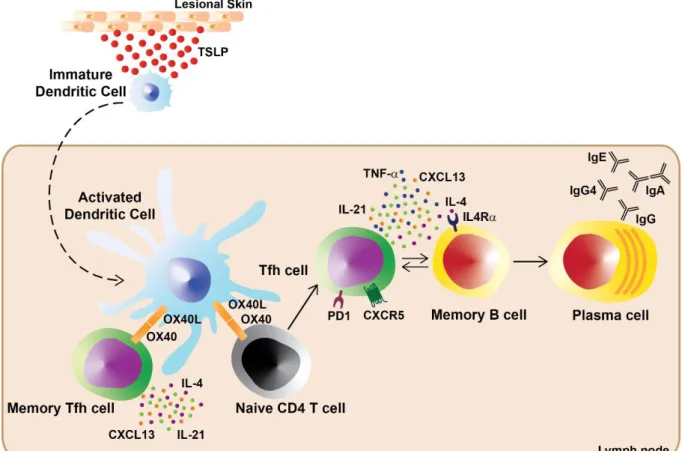
The human immune system is constituted of a sophisticated network of cells communicating through molecules expressed at their surface, or secreted in their microenvironment. When the organism is invaded by a pathogen, a complex response is set up, which is specific of the threat encountered. In this process, dendritic cells, which are located in the skin, will be one of the first cells to sense the pathogen. They will capture antigens in their microenvironment and get activated. Then, they will migrate to secondary lymphoid organs and present the antigens to naive CD4 T cells. Naive T cells able to recognize specific antigens will in turn get activated and adopt the proper T helper phenotype specific of the pathogen. T helper cells are characterized by their production of cytokines, which allow the recruitment and activation of many other cell types of both innate and adaptive immune system, in order to mount the appropriate immune response. If this complex process is not controlled correctly, unregulated T helper responses will arise and possibly become pathogenic. Indeed, T helper cells have been described to be involved in many diseases, which shows the necessity of regulating T helper responses, but also suggests the potential for therapies targeting specifically T helper pathways.
I focused my PhD work on studying T helper cell subset diversity and specific regulation: first in the context of TSLP-activated dendritic cells, then, with the purpose of understanding dendritic cell impact on T helper cell differentiation and finally in a pathologic setting, by monitoring T helper cell populations in atopic dermatitis patients.
In the introduction, I start by presenting T helper cells, the different subsets that have been identified as well as their features and functions. Then, I continue by describing dendritic cells, which are the main drivers of T helper cell polarization, and how their different characteristics influence Th cell differentiation. Finally, I present the link between T helper cells and diseases, with the specific example of atopic dermatitis.
My results are divided in three projects. The first results are in the form of a publication, demonstrating TSLP-activated dendritic cells ability to induce T follicular helper cells through OX40L. The second results are in the form of an accepted manuscript, showing a mathematical model able to predict the behavior of 18 T helper cell parameters in response to 36 dendritic cell-derived signals. This model allowed us to identify a context-dependent role for 12p70 in the presence of 1 in the differential induction of IL-17F without IL-17A. The last results are in the form of a manuscript in preparation describing the evolution of eight T helper and T follicular helper cell populations in peripheral blood from atopic
6
dermatitis patients along the course of their treatment with Dupilumab, an immunotherapy targeting the IL-4 receptor alpha subunit. This study led us to show that decrease of the Th17 cell percentage measured during Dupilumab treatment correlated with improvement of the EASI clinical score.
In the general discussion and perspectives, I review these three projects in light of the current literature, discuss their limitations and potential perspectives.
In the appendices are included: 1) an ongoing work on OX40L impact on T cell polarization, 2) a publication from a collaboration with biophysicians on signal integration by dendritic cells, 3) a publication I was involved in showing plasmacytoid dendritic cells activation through TLR1/2 and 4) a summary of my PhD work in French.
7
LIST OF ABBREVIATIONS
AD Atopic Dermatitis
AHR Aryl hydrocarbon receptor CD Cluster of Differentiation CLA cutaneous lymphocyte antigen CLR C-type lectin receptors
CyTOF Cytometry by time-of-flight DC Dendritic cell
EASI Eczema Area and Severity Index FACS Fluorescence Activated Cell Sorting FOXP3 Forkhead box P3
GATA3 GATA Binding Protein 3
GM-CSF Granulocyte Macrophage Colony Stimulating Factor ICOSL Inducible costimulator ligand
IFN Interferon Ig Immunoglobulin IL Interleukin
ILC Innate Lymphoid Cell iTreg induced regulatory T LPS Lipopolysaccharides
MDC Macrophage-derived chemokine MHC Major Histocompatibility Complex MoDC Monocyte-derived dendritic cell
ODN Oligodeoxynucleotides
PAMP Pathogen Associated Molecular Patterns PBMC Peripheral blood mononuclear cells
PD1 Programmed Cell Death 1 pDC plasmacytoid dendritic cell Poly(I:C) Polyinosinic-polycytidylic acid
PRR Pattern Recognition Receptors
RORγT Retinoic acid-related orphan receptor γT SAP SLAM-Associated Protein
8
STAT Signal Transducer and Activator of Transcription TARC Thymus and activation-regulated chemokine T-bet T-Box Expressed in T Cells
TCR T cell receptor Tfh T follicular helper
Tfr T follicular regulatory TGF-β transforming growth factor β
Th T helper
TLR Toll-like receptors TNF Tumor Necrosis Factor
9
LIST OF FIGURES AND TABLES
INTRODUCTION
Figure 1: Human T helper cell subsets ... 15
Figure 2: Tfh cell differentiation in secondary lymphoid organs ... 17
Figure 3: Peripheral blood Tfh cell subsets partially mirror Th cell subsets ... 18
Figure 4: Combination of five surface markers identifies nine subsets of memory Tfh cells in human peripheral blood ... 19
Figure 5: Th17 cell heterogeneity and plasticity ... 21
Figure 6: Antigen-specific T cell response initiation ... 24
Figure 7: Human dendritic cell subsets ... 25
Table 1: Table recapitulating some of the human PRRs, their location, ligands and expression according to human DC subsets ... 34
Figure 8: T helper polarization by dendritic cells depends on the type of pathogen they encounter ... 35
Figure 9: T helper polarization towards Th1 or Th2 subset by dendritic cell requires 3 signals ... 37
Figure 10: Dendritic cell/T cell communication molecules ... 39
Figure 11: Atopic Dermatitis pathogenesis in Acute (A) and Chronic (B) phases ... 44
Figure 12: Dupilumab mechanism of action ... 49
RESULTS Figure 13: TSLP-activated DC induce human Tfh cell differentiation through OX40L ... 56
APPENDICES Figure 14: OX40L blocking decreases IL-21 and increases IL-4 production in TSLP-DC/T coculture .... 171
Figure 15: rhOX40L increases IL-21 and decreases IL-4 and IFN-γ in a DC-free Th polarization system ... 172
Figure 16: OX40L+ DC induce more IL-21 and less IL-4 producing cells than OX40L- DC ... 174
11
13
1. T helper cell subset diversity and functional impact
CD4 T helper (Th) cells play a major role in the adaptive immune response which allows host defense against a wide variety of pathogens. Through the secretion of specific sets of cytokines, Th cells instruct other cell types to set up the proper immune response, specific of the pathogen encountered, allowing its clearance.
1.1. T helper cell subsets, phenotypes and functions
1.1.1. Th1/Th2 paradigm
In 1986, was published the first report describing two in vitro-derived Th clones: Th1 and Th2, obtained after mice immunization with a protein antigen [1]. In 1989, Mosmann and Coffmann summarized the latest advances on T helper cells and reported that Th1 cells were characterized by production of IL-2, IFN-γ, TNF-α and TNF-β, while Th2 cells produced IL-4, IL-5, IL-6 and IL-13 [2].
Later, identification of master regulators associated to each cytokine profile and responsible for their setup introduced the notion of lineages. The transcription factors identified in Th1 cells are T-bet [3], STAT1 and STAT4 [4], while Th2 cell development involved GATA3, STAT5 and STAT6 [5].
Additionally, a mutual exclusion between the two subsets has been described: GATA3 represses STAT4, thus inhibiting Th1 features [6] and T-bet and Runx3 activate IFN-γ gene and silence GATA3 and IL-4 [7, 8]. Additionally, a positive feedback loop occurs, GATA3 will induce IL-4, which in turn will instruct non-IL-4 producer-cells to produce non-IL-4, but also enhance non-IL-4 production from non-IL-4 producer-cells [5]. Conversely, the IFN-γ-STAT1-T-bet pathway strongly amplifies Th1 differentiation [9].
Further characterization of the two subsets lead to the identification of specific chemokine receptors, homing receptors which will lead Th cells to different location. Th1 specifically express CCR5, receptor for MIP-1α, MIP-1β and RANTES, and CXCR3, receptor for IP-10 and MIG, which will direct them to inflamed tissues [9, 10]. On the other hand, Th2 exhibit CCR3, an eotaxin receptor, CCR4, receptor for MDC and TARC, and CCR8, receptor for TARC and I-309 (Figure 1). MDC, TARC, I-309 and eotaxin will not only attract Th2 to the inflammation site, but also eosinophils, basophils and monocytes. IL-4 and IL-5 production by Th2 will activate these different cell types and ensure their survival [10, 11]. Besides, Th2 express specifically CRTH2, a receptor for Prostaglandin D2 [12].
14
Moreover, respective functional roles have been identified for each Th subset. For instance, Th1 are necessary for the clearance of intracellular viruses and bacteria. IFN-γ activates phagocytosis on macrophages increasing their ability to kill intracellular pathogens. Th1 also secrete IL-2, α and TNF-β which participate in antimicrobial responses [13]. On the opposite, Th2 have been linked to the control of extracellular parasites such as helminths. Th2 production of IL-4 induces isotype switching on B cells which produce IgG1 and IgE [2]. By producing IL-4 and IL-13, Th2 are also able to activate macrophages [14]. And through their production of IL-5, Th2 recruit eosinophils as well [15].
1.1.2. Additional T helper subsets
For more than two decades, the Th1/Th2 paradigm prevailed, with the idea that T cells could only adopt one of two fates, until the discovery of several additional Th cell subsets.
First, Th17 cells were described as Th cells producing IL-17A and developing through a different lineage than Th1 and Th2 cells [16]. Additional characterization of Th17 cells demonstrated that they also produce the cytokines IL-17F, IL-21, IL-22 and IL-26, as well as the chemokines CCL20 and CXCL8, express the transcription factors RORγT, RORα and STAT3, exhibit the specific surface marker CD161 and the chemokine receptor CCR6 [17] (Figure 1). Th17 cells play an important role in inducing protective immunity against bacteria and fungi at mucosal sites [18]. IL-17A, IL-17F and IL-22 produced by Th17 cells are strongly pro-inflammatory and will induce expression of antimicrobial peptides from epithelial cells and keratinocytes but also their permeability, proliferation and survival [19]. CCL20 and CXCL8 produced by Th17 cells will attract more Th17 cells, but also neutrophils on the site of infection [18].
22 was first described as a Th17 cytokine, until a skin homing memory Th cell population secreting IL-22 but neither IL-17 nor IFN-γ was identified and named ThIL-22 [20]. ThIL-22 specific transcription factor has been identified as well: AHR [21]. Th22 express the chemokine receptor CCR6 and the skin homing receptors CCR4 and CCR10 indicating their crucial roles in skin inflammation [22] (Figure 1). And just as for Th17 cells, IL-22 secreted by Th22 induces production of antimicrobial peptides by epithelial cells and keratinocytes.
Similarly, IL-9 was originally described as a Th2 cytokine [23], secreted in combination with IL-4, but later, Th cells secreting IL-9 independently of IL-4 were identified, and labelled Th9 [24]. Th9 cell specific transcription factor is PU.1, but like Th2 cells, Th9 cell differentiation also involves GATA3 and STAT6 [25]. Th9 cells express the major skin homing receptor cutaneous lymphocyte antigen (CLA), suggesting their role in skin immunity and cutaneous defense against extracellular pathogens [26] (Figure 1). IL-9
15
has been shown to be important for mast cell recruitment and activation in tissues. Activated mast cells will in turn produce proinflammatory cytokines such as TNF-α and IL-6, which are involved in anti-fungal response. IL-9 can also attract neutrophils, on the infection site, which will have an important role in eliminating fungi as well [26].
In parallel to these Th cell subsets, induced regulatory T (iTreg) cells have been described arising from naive CD4 T cells in secondary lymphoid organs or inflamed tissues. iTreg are a particular subset characterized by the production of IL-10 and TGF-β, expression of the surface markers CD25 (IL-2 receptor), GITR and CTLA4 and the transcription factor FoxP3 [27, 28] (Figure 1). Treg cells are critical for the prevention of autoimmune diseases by inhibiting activation and proliferation of T and B cells specific for self-antigens [29]. IL-10 is important for keeping a state of immune tolerance, while CTLA4 binding to CD80/CD86 expressed by dendritic cells will lead to decreased naive CD4 T cell activation [27].
All these new subsets significantly complexified the view of the T helper cells (Figure 1).
Figure 1: Human T helper cell subsets
Schematic of known human Th cell subsets: Th1, Th2, Th17, Th9, Th22 and iTreg with their respective transcription factors, cytokines and chemokine/homing receptors
16
1.2. T follicular helper cells: A T helper cell subset specialized in B cell help
1.2.1. General features of T follicular helper cells
In addition to the six T helper cell subsets, particular T follicular helper (Tfh) cells were described. Initially named “follicular B helper T cells” based on their characteristic localization in secondary lymphoid organs, Tfh cells were identified in 2000. Several groups observed a large proportion of CD4 T cells expressing high levels of the chemokine receptor CXCR5 in tonsils, and discovered they were able to support immunoglobulin (Ig) production from B cells [30-32].
Since then a lot of work has been done to fully characterize them. Tfh cells express high levels of several effector molecules, including the surface markers ICOS, CD40L, OX40, PD1, BTLA, the cytoplasmic adaptor protein SAP and produce large amounts of the cytokine IL-21 and of the chemokine CXCL13, which is CXCR5 ligand [33]. Tfh cell differentiation depends on the transcriptional repressor Bcl-6, antagonist of Blimp-1 which is a strong inhibitor of Tfh polarization [34].
Tfh cells main function is to provide help to B cells by delivering signals that enable B cell proliferation, differentiation and isotype switching. Tfh cells are also necessary for the proper formation of germinal centers, particular structures forming inside B cell zone of secondary lymphoid organs [35].
Tfh cell differentiation happens in the secondary lymphoid organs and requires 3 steps (Figure 2). First, in the T cell zone, DC activate antigen-specific naive CD4 T cells expressing CCR7, the T cell zone homing receptor. Activated pre-Tfh will downregulate CCR7 and upregulate CXCR5, homing receptor to the B cell follicle, positioning them to the T-B border. Then, pre-Tfh cells will encounter activated antigen-primed B cells. This interaction will lead either: 1) to the B cell differentiation into short-lived extrafollicular plasmablasts, contributing to early production of specific antibodies, or 2) to the migration of pre-Tfh cells and B cells to form the germinal centers. Finally, further interaction with antigen-specific B cells will drive the complete differentiation of germinal center Tfh cells. Once in the germinal center, B cells will go through the processes of affinity maturation and isotype switching, and differentiate either into high-affinity long-lived plasma cells or long-lived memory B cells [33, 36, 37].
Even if the majority of Tfh cells reside in germinal centers, in human a small subset of memory Tfh cells have been identified in peripheral blood [32]. They express CXCR5 but low levels of other prototypical Tfh markers: PD1, ICOS, OX40 and even do not express Bcl-6 protein [38].
17
Figure 2: Tfh cell differentiation in secondary lymphoid organs Schema from Ma, Deenick, Batten and Tangye [33]
DC activate antigen-specific naive CD4 T cells, which will migrate from the T cell zone towards the B cell follicle. At the T-B border, activated pre-Tfh cells will interact with activated antigen-specific B cells. This interaction will lead to B cell differentiation into short-lived plasmablasts or to the migration of the pre-Tfh cells and B cells and formation of germinal centers. Further interaction between B cells and pre-pre-Tfh cells will enable full differentiation of germinal center Tfh cells. Germinal center B cells will differentiate into long-lived plasma cells or long-lived memory B cells.
Additionally, T follicular regulatory (Tfr) cells have been described, controlling germinal center responses by inhibiting Tfh and B cells. Tfr cells exhibit the same markers than Tfh cells, they express CXCR5, PD1, ICOS, Bcl-6, but they also possess specific Treg markers such as FoxP3, CD25, CTLA4, GITR [39]. Besides, they produce large amounts of inhibitory cytokines such as IL-10 and TGF-β. Similar to Tfh cells, Tfr differentiation is a multistep process requiring interaction with DC and B cells. Tfh and Tfr cells are necessary for the balance between immune activation and tolerance [40].
1.2.2. Peripheral blood Tfh cell subsets partially mirror Th cell subsets
After the discovery of Tfh cells as a new Th cell subset, Tfh producing not only IL-21 but also other Th signature cytokines, have been described first in mice.
Three different teams, using the same IL-4 reporter mice infected with different parasites, discovered IL-4 producing cells exhibiting all Tfh specific markers in the lymph nodes [41-43].
18
Later, Morita et al. demonstrated that Tfh cells from human peripheral blood mirror Th cells, and can also be subdivided into distinct subsets. Looking at the expression of CXCR5, CCR6 and CXCR3 in the CD4 memory cell compartment, they could identify three functionally distinct Tfh cell subsets mirroring the three Th cell subsets: Th1, Th2 and Th17. Tfh1 cells characterized by expression of CXCR5 and CXCR3, expressed T-bet, produced IFN-γ in addition to IL-21 and were not able of B cell help. Tfh2 cells were identified as CXCR5+CXCR3-CCR6-, expressed GATA3, produced IL-21, IL-4, IL-5 and IL-13 and induced
high levels of IgG and IgE and low levels of IgM and IgA from B cells. Finally, Tfh17 cells identified as CXCR5+CCR6+, expressed RORγT, produced IL-21, IL-17 and IL-22 and induced high levels of IgA, IgM and
IgG production by B cells [45] (Figure 3).
This demonstrates a partial mirror between peripheral blood Tfh cell subsets and Th cell subsets. We can wonder if, as well as for Th1, Th2 and Th17, we could identify a mirror for Th9 and Th22 subsets in the CXCR5+ memory compartment of peripheral blood, maybe using more markers.
Figure 3: Peripheral blood Tfh cell subsets partially mirror Th cell subsets
Schematic summarizing results from Morita et al. [45]. Tfh cell subsets in human peripheral blood: Tfh1, Tfh2 and Tfh17 cells with their respective transcription factors, cytokines, chemokine receptors, B cell help capacity and isotype switch.
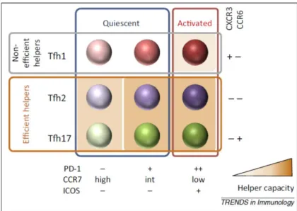
Looking at the expression of ICOS, PD1 and CCR7, three subsets of memory Tfh cells have been identified in human peripheral blood. ICOS+PD1+ subsets have been described as activated Tfh cells, while the
ICOS-PD1+ and PD1- subsets do not exhibit activation markers and have been defined quiescent. [46-48].
Added to the three subsets identified by Morita et al. [45], memory Tfh cell diversity reaches a total of nine distinct subsets [49] which strongly increases Tfh cell subset complexity (Figure 4).
19
Figure 4: Combination of five surface markers identifies nine subsets of memory Tfh cells in human peripheral blood
Figure from Schmitt, Bentebibel and Ueno [49]
The nine memory Tfh cell subsets identified in human peripheral blood. CXCR3 and CCR6 identify Tfh1, Tfh2 and Tfh17 cells and separate non-B cell helpers (Tfh1) from efficient B cell helpers (Tfh2 and Tfh17). ICOS expression delineate activation in each subset. Helper capacity is indicated by a color gradient.
1.2.1. Additional T follicular helper cell phenotypes
For a long time, B cell help function was attributed to Th2 cells because of their IL-4 production. Initially, IL-4 has been described as “B cell differentiation factor γ”, “B cell growth factor” or “B cell stimulatory factor-1” and it was known for inducing IgG1 and IgE switch from B cells [2].
However, the discovery of the T follicular helper cells questioned that view. Since then, Tfh cells have been described as the specialized B cell help providers through production of IL-21 and IL-4 and their capacity to enter the germinal center of secondary lymphoid organs [35]. Nevertheless, B cell helper capacities have been demonstrated from cells that do not display the prototypical Tfh phenotype.
First, in 2017 PD1hiCXCR5-CD4+ T cells were identified at very high frequency in synovial fluid and synovial
tissue of rheumatoid arthritis patients. Those T cells produced high levels of IL-21 and CXCL13 and when cocultured with memory B cells, they were capable of inducing B cell differentiation into plasma cells producing IgG [50].
In 2018, T cells from systemic lupus erythematosus exhibiting CD4+CXCR5-CXCR3+PD1hi were shown to
help B cells through the production of IL-10 and succinate (an intermediate of the tricarboxylic acid cycle), independently of IL-21 [51].
This demonstrated that there is not just one possible phenotype capable of providing B cell help. On the contrary multiple Th cell profiles seem to potentiate isotype switch and Ig production from B cells.
20
1.3. Limits of the current T helper cell classification
1.3.1. Th cell heterogeneity and plasticity
In the current Th cell classification, each subset is defined by a specific and strict set of cytokines associated to transcription factors. Th1 cells are known for their secretion of IFN-γ, TNF-α and IL-2 under control of T-bet, STAT1 and STAT4, Th2 cells produce IL-4, IL-5 and IL-13 regulated by GATA3, STAT5 and STAT6, etc. However, the system seems to be a lot more complex than that, and Th cells might be characterized by further plasticity than what was originally defined.
One example is the description of Th1/Th17 cells, in patients with Crohn’s disease, producing both IL-17 and IFN-γ and expressing at the same time RORγt and T-bet. In this study ThIL-17 clones cultured with IL-12 started producing IFN-γ in addition to IL-17 [52]. This shows that IL-17 and IFN-γ production are not exclusive.
Additionally, Cosmi et al. demonstrated that both Th17 and Th1/Th17 cells, if cultured with IL-12, could differentiate into “non-classic Th1”, downregulating RORγt expression and IL-17 production [53]. Th17, Th1/Th17 and non-classic Th1 cells were characterized by the expression of the CD161 marker, as opposed to classical Th1 which do not express it [54].
An additional intermediate profile of Th17/Th2 cells was described in peripheral blood of chronic asthma patients. These Th17/Th2 cells produced the Th17 cytokines IL-8, IL-17, IL-21 and IL-22, as well as Th2 cytokines IL-4, IL-5, IL-9, and IL-13. Th17/Th2 cells could be derived from Th17 cells cultured with IL-4 [55].
Furthermore, IL-9 production could be induced on memory Th17 cells, extracted from peripheral blood, when cultured with a cocktail of TGF-β, IL-1β, IL-6, IL-21 and IL-23 [56].
Additionally, Treg/Th17 co-expressing FoxP3, RORC and IL-17 have been described in human. And induction of IL-10 production by Th17 cells in response to IL-21 has been shown, promoting regulatory Th17 [57].
21 Figure 5: Th17 cell heterogeneity and plasticity Schema from Geginat [57]
Th17 cells can be induced to differentiate into Th17/Th9, regulatory Th17 (rTh17), Treg/Th17, Th17/Th2, Th1/17 or even non-conventional Th1.
These studies demonstrated the heterogeneity and plasticity of the Th17 cells (Figure 5). Similarly, few studies tend to prove that other Th cells might not be terminally differentiated either.
In atopic asthma patients, memory/effector Th2 cells producing the Th17 cytokines: IL-17A and IL-22, in combination with Th2 cytokines: IL-4, IL-5 and IL-13 have been identified, they also co-expressed both transcription factors RORγT and GATA3. This study further demonstrated, using a mouse model, that classical Th2 cells treated with IL-1β, IL-6, and IL-21 started producing IL-17 [58].
Moreover, Hegazy et al. described Th2/Th1 cells. They demonstrated both in vivo, in lymphocytic choriomeningitis virus infected mice, and in vitro, using type I and II interferon and IL-12, that Th2 cells could produce both IL-4 and IFN-γ and express both GATA3 and T-bet [59].
In a mouse model of house dust mite sensitization, Ballesteros-Tato et al. demonstrated that the first sensitization induced IL-4 committed Tfh cells, but no Th2 cells. Besides, they showed that following re-challenge with house dust mite, these IL-4 committed Tfh cells would differentiate into Th2 cells [60].
22
These studies question the relevance of the notion of Th lineages and their strict phenotypes. The Th cell polarization process seems substantially more flexible and plastic than what was initially described. Indeed, reprogramming of committed Th cells has been demonstrated in these studies, but also existence of mixed profiles showing combination of usually exclusive Th cell phenotypes. Therefore, we could imagine that all Th cytokine combinations are virtually possible. Polarized Th cells just need the proper stimulation from unique microenvironments to either change entirely their polarization or acquire an intermediate Th profile, in order to finely tune the immune response to specific threats.
1.3.2. Extensive diversity of the Th cell subsets
Recent studies essentially using mass cytometry, also known as cytometry by time-of-flight (CyTOF) and analyzing increasing number of parameters identified a lot more Th cell subsets than what was initially described.
Duhen et al. studied the expression of four chemokine receptors: CCR6, CXCR3, CCR4 and CCR10 on memory CD4+CD45RO+CD25hiCD127loFoxP3+ Treg cells sorted from human peripheral blood. They were
able to identify 4 distinct subsets: Th1-like Treg cells producing IFN-γ and expressing CXCR3, CCR6+CCR4+
Th17-like Treg cells producing IL-17, Th22-like cells secreting IL-22 and expressing CLA, CCR6, CCR4 and CCR10 and IL-4 producing Th2-like Treg cells expressing CCR4. Even though all populations possessed inhibitory functions, this suggests a mirror between human peripheral blood Th cells and Treg cells [61].
Mason et al. sorted CD4+CD25highCD127low Treg cells from peripheral blood mononuclear cells (PBMC)
from four healthy donors and analyzed them by CyTOF including 25 surface markers. They were able to identify 22 different subsets, among which they detected the five previously established Treg subsets [62]. This demonstrates an important phenotypical complexity and heterogeneity of the human peripheral blood Treg compartment.
Kunicki et al. used 23 markers, including surface markers and transcription factors, to study Th cells and Treg in PBMC from eight healthy donors by CyTOF. They analyzed their data by unsupervised clustering and visualized 15 Th cell subsets: three different populations in the Th1 subset, three populations among Th2 cells, one Th17 population, three Treg populations and five populations inside the Tfh subset. Moreover, many populations overlapped between subsets, for example Th1 and Tfh, Tfh and Th17, Th1 and Th17 or Th2 and Treg [63].
23
Additionally, Barcenilla et al. analyzed PBMC from nine healthy donors compared to nine patients with high risk of developing a type-1 diabetes. They used 33 markers, including transcription factors, chemokine receptors and activation markers, to study Th and Treg subsets by CyTOF. They identified 11 clusters of naive CD4 T cells, four clusters among the central memory CD4 T cells and five clusters in the effector memory CD4 T cells [64].
These new studies demonstrate an important heterogeneity among the Th cell subsets but also bring a lot more questions. As the original number of subsets defined appears obsolete, how many are they in vivo? Also, is it really relevant to consider Th cells as stringent subsets? Otherwise, since several populations seem to overlap, would it be more accurate to view Th cells as a continuum of profiles? Depending on the threat, particular combination of cytokines might arise to efficiently neutralize it. However, these last three studies only looked at surface markers and transcription factors, they do not analyze cytokines or functional properties of the different subsets they identified.
Wong et al. studied T cells in eight different human tissues: blood but also lymphoid and non-lymphoid tissues, by CyTOF using a panel of 41 markers including surface markers, chemokine receptors and cytokines. Using unsupervised clustering, they identified 75 clusters, indicating a wide heterogeneity, but they also identified tissue-specific profiles in particular when looking at the expression of chemokine receptors, which are not homogeneously expressed among tissues. They also analyzed all possible combinations of five Th specific cytokines: IFN-γ for Th1, IL-4 for Th2, IL-10 for Treg, IL-17A for Th17 and IL-22 for Th22, and calculated their frequencies within each tissue. Only 12 out of the 32 possible combinations were detectable among tissues. Within the 12 combinations, five corresponded to each cytokine produced alone, six corresponded to two cytokines produced and only one subset co-producing three cytokines: IFN-γ, IL17A and IL-22 was identified [65].
This study only includes five cytokines but it surprisingly demonstrates that not all cytokine combinations are relevant and that only specific ones are secreted by Th cells depending on the tissue considered. This same analysis would be very interesting to conduct including all Th cytokines. Especially, it would be informative to see if cytokines of a same subset are always co-produced together or if there is a tissue-specific signature for cytokine production. For example, are Th2 cytokines: 4, IL-5 and IL-13 always co-produced together by the same cells? Or are they produced by distinct cells present in the same microenvironment? In the end, an important work remains to be done to entirely capture Th cell diversity and complexity, as well as their relative physiopathological relevance.
24
2. Dendritic cells: the main drivers of T helper differentiation
Dendritic cells (DC) are responsible for the initiation of immune responses. Indeed, DC are professional antigen-presenting cells, thanks to their high expression of class II Major Histocompatibility Complex (MHC-II) molecules. At steady state, immature DC are resting in peripheral tissues and will get activated in case of infection through all the pattern recognition receptors (PRR) they express, which allow them to recognize Pathogen Associated Molecular Patterns (PAMP) from pathogens surrounding them. DC capture antigens from their microenvironment and process them into peptides in order to present them on their MHC-II molecules. Once activated, DC will migrate to secondary lymphoid organs in order to activate specific naive CD4 T cells. DC-T cell interaction involves recognition of the antigen-MHC-II complex by the T cell receptor (TCR) on the T cell. If a T cell recognizes its specific antigen, it will become activated and proliferate in order to launch the appropriate immune response [66-68] (Figure 6).
Figure 6: Antigen-specific T cell response initiation
Illustration from Summers deLuca and Gommerman [69]
At steady state, DC are resting in peripheral tissues. In case of infection, they uptake foreign antigens and get activated by PAMPs present in the microenvironment. As a consequence, they migrate to secondary lymphoid organs. Once there, they will present the antigens as processed peptides on their MHC-II molecules to antigen-specific naive CD4 T cells, which are able to recognize antigen-MHC-II complexes via their TCR. When activated, T cells will differentiate into effector T cells able to mount a proper adaptive immune response.
25
2.1. Role of the different dendritic cell subsets in the T helper cell diversity generation
2.1.1. Human dendritic cell subsets
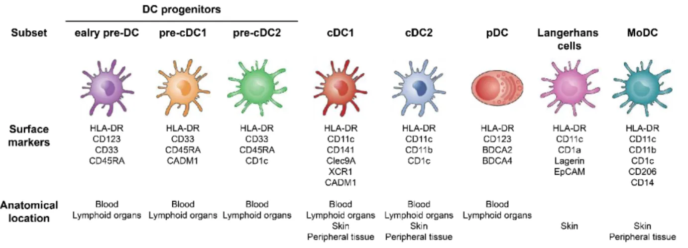
Several DC subsets have been identified, deriving from a common bone-marrow DC progenitor [70]. In addition to non-lymphoid tissue DC, which migrate from peripheral tissues to lymph nodes after antigen uptake and activation, some DC can be resident in lymphoid tissues in which they capture antigens from blood stream and lymph to present them directly to nearby T cells [71].
Figure 7: Human dendritic cell subsets
Human DC subset classification, including new findings from See et al. [70] and Dutertre et al. [72]. Under each DC subset, some of their specific surface markers and their anatomical location.
In human, depending on the location, several DC subsets have been described. First, plasmacytoid DC (pDC) are characterized by expression of BDCA-2, BDCA-4, CD123 and their major capacity to produce IFN-α upon activation and can be found in the blood and lymphoid organs [71]. Additionally, two subtypes of myeloid or conventional DC have been described, expressing CD11c: 1) cDC1 characterized by the surface markers CD141, CLEC9A, CADM1 and XCR1, 2) cDC2 expressing CD1c and CD11b, both subsets can be identified in the skin, the blood, lymphoid organs and peripheral tissues [73]. In the skin, one specific DC subset populate the epidermis: Langerhans cells expressing CD1a, Langerin and EpCAM [74] (Figure 7).
In addition, DC deriving from monocytes have been described first in the skin and referred to as dermal DC [74]. Then, a population of Inflammatory Dendritic Epidermal Cell distinct from Langerhans cells and phenotyped as HLA-DR+CD1a+CD1b+CD36+ were identified in the skin of atopic dermatitis patients [75].
26
Additionally, in a Leishmania infection model in mice, monocyte-derived DC (MoDC) were identified and originally termed inflammatory DC because of their involvement in inflammation [76]. Inflammatory DC were also identified in ascites from patients with breast tumors and described as deriving from monocytes [77]. Later, MoDC were further identified in peripheral tissue samples from healthy patients, strengthening their in vivo relevance [78]. Furthermore, due to the difficulties of studying human primary DC from blood or tissues, in vitro protocols to generate DC from blood monocytes have been created, using granulocyte macrophage colony stimulating factor (GM-CSF) and IL-4 and are widely utilized across the scientific community [79] (Figure 7).
New technologies, in particular single-cell RNA sequencing and CyTOF, brought new insights into the study of DC subsets.
Villani et al. revised DC classification by sequencing human blood cells from healthy donors. First, they demonstrated that cDC2 are actually constituted of two subsets with similar phenotypes: CD1c+_A
which are non-inflammatory and CD1c+_B displaying an inflammatory gene signature. Additionally, they
identified a cluster of CD141-CD1c- DC related to CD16 monocytes. These cells had previously been
described in the blood by MacDonald et al. as CD16+CD11c+CD14loHLA-DRlo DC [80], but they had been
poorly characterized since then. Finally, Villani et al. identified a new subset named “AS-DC” forming a continuum between cDC1c+ DC and pDC and sharing phenotypic markers with both subsets [81].
However, the results of the flow cytometry analysis to retrieve CD1c+_A and CD1c+_B cells at the protein
level are confusing, the two subtypes actually partly overlap. Plus, they did not observe real functional differences between the two subsets except when looking at cytokine production, CD1c+_A secreted
slightly higher levels of a few cytokines. If this slightly higher level of cytokines produced by the CD1c+_A
subset has any relevance in functional specialization remains to be determined.
Furthermore, another team also identified two subsets among cDC2 from blood and lymphoid organs based on CD5 expression by flow cytometry. They fully characterized CD5high and CD5low DC by looking
at their respective gene expression and functional properties [82]. These two populations were not identified by single-cell RNA sequencing and directly contradict results from Villani et al. [81].
Alcantara-Hernandez et al. used a CyTOF panel of 38 markers combined with unbiased analysis to characterize DC subsets from blood, skin, spleen and tonsils from 18 healthy donors. They retrieved cDC1 and cDC2 in all tissues, while pDC were present in the blood and lymphoid organs but not in the skin. They found Langerhans cells specifically in the skin. And also identified the AXL+ DC described by
27
CD1c+_B subsets identified by Villani et al. [81], nor the CD5high and CD5low subsets identified by Yin et
al. [82]. Furthermore, they identified 3 clusters among cDC2, based on the markers CD163 and CD172a, but they observed that their frequencies were dramatically variable between tissues, but also between individuals and also that the expression of the surface markers identifying them varied among the clusters. They concluded that rather than conserved subpopulations of cDC2, these clusters were an important interindividual heterogeneity of the cDC2 population [83]. As highlighted in the publication, there is a bias in the study by Villani et al. in the number of donors analyzed and the use of only one tissue. They do not discuss the results from Yin et al., however, CD5 is among the 38 surface markers used for their CyTOF analysis and is not retained as defining different cDC2 subsets. Another discrepancy appears between Alcantara-Hernandez et al. and Villani et al. studies. Alcantara-Hernandez et al. did not retrieve the CD16+ DC subset identified originally by MacDonald et al. [80] and described by Villani
et al. [81]. Besides, they did not identify the CD14+ DC which have been described, among other location,
in the skin [74]. However, they compared in vitro generated MoDC to the other DC subsets. MoDC clustered separately not only from DC but also from monocytes, which lead them to conclude that they are not representative of any DC subsets present in healthy individuals. However, the fact that they cluster away from other DC subsets is not surprising since they arise from different progenitors. Also, since in vitro derived, they are probably influenced during culture, which alter their phenotype compared to ex vivo. Nevertheless, in vitro derived-MoDC remain a good model to study DC functions and Th polarization.
Using combination of single-cell RNA-sequencing and CyTOF to study human DC in blood, spleen and bone marrow, See et al. described a continuous process of differentiation within the human DC lineage. A common DC progenitor CD34+ in the bone marrow give rise to pDC and pre-DC, sharing phenotypic
markers with pDC. Pre-DC can further differentiate into early-pre-DC and then give rise to pre-cDC1 and pre-cDC2. Pre-cDC1 and pre-cDC2 differentiate exclusively in cDC1 and cDC2 respectively. They also described how to specifically discriminate pre-DC from pDC: using CD33, CD2 and CX3CR1specifically expressed on pre-DC compared to pDC [70]. Since the markers and gating strategies are different, it is hard to formally conclude, but the AXL+ DC/AS-DC identified by Alcantara-Hernandez et al. [83] and
Villani et al. [81] might at least partially overlap with the pre-DC described by See et al. [70].
Very recently, Dutertre et al. confronted the results from Villani et al. [81]. Using Infinity Flow, RNA sequencing, single-cell RNA sequencing and CyTOF they analyzed each subset defined by Villani et al. [81] and contradicted some of their results. First, they demonstrated that the DC4 subset defined as CD141-CD1c- DC were not DC, but CD16+ non-conventional monocytes. Then, they showed that the
28
also pre-cDC2. Finally, they demonstrated that cDC2 heterogeneity was greater than the two CD1c+_A
and CD1c+_B subsets identified by Villani et al. [81]. Among the cDC2 they identified a subset of CD5+
DC with a gene signature corresponding to the CD1c+_A subset described by Villani et al. [81].
Corresponding to the CD1c+_B subset they actually found three distinct subsets: one CD5-CD163-CD14
-subset, one CD5-CD163+CD14- subset and one CD5-CD163+CD14+ subset. Dutertre et al. demonstrated
that all cDC2 subsets were functionally capable of inducing T cell proliferation and they showed an increasing capacity to induce IL-4 and IL-17 production from CD5+ cells and CD5-CD163-CD14- cells to
CD5-CD163+CD14- cells and finally CD5-CD163+CD14+ cells [72] (Figure 7). The top markers to
differentiate cDC2 subsets identified by Dutertre et al. were CD5, CD14, CD163 [72], while Alcantara-Hernandez et al. showed high variance in the expression of CD32, CD163, CD172a and BDCA1 between individuals and clusters [83]. Also, the number of cDC2 clusters is not the same between the two studies: four subsets for Dutertre et al. [72] compared to three clusters for Alcantara-Hernandez et al. [83].
In the end, much work remains to be done to harmonize these recent discoveries and fully comprehend the human DC system. In particular, in depth studies will be needed to understand cDC2 heterogeneity, and determine if they can be separated into distinct populations, if the different clusters are just interindividual heterogeneity as described by Alcantara-Hernandez et al. [83] or if they have to be considered as subsets with functional differences as suggested by Dutertre et al. [72].
2.1.2. T helper cell polarization induced by each subset
An important concept in the field of DC, is that DC subsets would intrinsically possess specific capacities to activate T cells and induce differential Th responses. This concept still remains to be fully demonstrated, but some studies already tried to demonstrate this point.
For instance, Klechevsky et al. showed that ex vivo human Langerhans cells induced more Th2 cytokines production from allogeneic naive CD4 T cells than dermal cDC2 and CD14+ DC [84]. Furio et al. confirmed
that human Langerhans cells were more potent than dermal cDC2 at inducing not only IL-4 but also IFN-γ production from allogeneic naive CD4 T cells, while dermal cDC2 induced more IL-10 producing-T cells [85].
Fujita et al. demonstrated that human Langerhans cells were more efficient than dermal cDC2 at polarizing naive CD4 T cells to produce IL-22, without IL-17, characteristic of Th22 cells [86]. Penel-Sotirakis et al. confirmed that Langerhans cells were the strongest inducers of IL-22 production without
29
IL-17 by T cells, compared to dermal cDC2 and CD14+ DC, but they also induced a higher production of
IL-21 [87].
Segura et al. studied human DC isolated from non-invaded lymph nodes or blood and compared their Th polarizing capacities. They demonstrated that Langerhans cells induced preferentially IL-5 and IL-13 production by allogeneic naive CD4 T cells. When comparing cDC1 and cDC2 from lymph nodes to cDC1 and cDC2 from blood they observed that all DC induced both Th1 and Th2 profiles from T cells, but blood DC induced more IFN-γ, while lymph nodes DC induced more IL-5 and IL-13 production [88]. This could demonstrate a functional specialization due to DC original location and microenvironment.
Furthermore, Durand et al. showed that ex vivo cDC2 from human tonsils are the most efficient to induce Tfh polarization, compared to cDC1 and pDC. Indeed, cDC2 induced significantly higher proportion of CXCR5+PD1+ cells and production of IL-21 and CXCL13 from allogeneic naive CD4 T cells,
compared to cDC1 and pDC [89]. This shows a functional specialization for tonsillar cDC2 to induce Tfh cell differentiation.
Yu et al. compared the Th polarization capacity of human blood cDC1 and cDC2 cocultured with allogeneic naive CD4 T cells. They demonstrated that cDC1 were more potent than cDC2 to induce a Th2 profile, characterized by IL-4 and IL-13 producing T cells, while cDC2 induced more IFN-γ producer-cells than cDC1 [90].
Nonetheless, despite these different studies, proving that freshly isolated human DC have intrinsic properties which give them capacities to induce specific Th cytokine patterns is a complicated task. Especially, since they are extracted from a specific microenvironment which could influence their Th polarization capacities. Also, since in vivo DC will migrate and activate T cells only when activated by external pathogens, studying their Th polarization capacities while immature and non-activated is not the most relevant.
2.2. Role of the dendritic cell activating signal
2.2.1. Immune sensing by dendritic cells
In order to recognize both exogenous and endogenous danger signals from their microenvironment, DC display a large repertoire of receptors.
30
Pattern recognition receptors (PRRs) allow them to recognize Pathogen-Associated Molecular Patterns (PAMPs) which are conserved pathogen motifs ranging from glycoproteins and polysaccharides, to double-stranded DNA and RNA and single-stranded RNA, but also Damage-Associated Molecular Patterns (DAMPs) which are endogenous danger signals released upon cellular stress or tissue damage such as histones, heat-shock proteins, ATP, actin for example [91, 92]. PRR binding to its specific PAMP or DAMP leads to the activation of an intracellular signaling cascade, resulting in DC activation, increase of maturation markers and production of proinflammatory cytokines and chemokines.
Toll-like receptors (TLRs) are membrane-bound PRRs. In human, the TLR family counts 10 members, from TLR1 to TLR10. TLR1, TLR2, TLR4, TRL5, TLR6 and TLR10 are expressed at the plasma membrane, where they directly encounter their ligands: bacterial and fungal PAMPs, while TLR3, TLR7, TLR8 and TLR9 are localized on the membrane of the endosomal compartment, where they detect nucleic acids from bacteria and viruses. Binding with their ligand facilitates TLRs dimerization. TLR2 has been shown to heterodimerize with TLR1, TLR6 and possibly TLR10, while the other members of the family are thought to homodimerize. TLR dimerization triggers activation of the intracellular signaling cascade, leading to DC activation [91, 93]. Human TLR ligands, locations and specific expression according to DC subsets are described in Table 1.
C-type lectin receptors (CLRs) are also membrane-bound proteins. The CLR superfamily includes more than 1000 proteins, which are divided into 17 subgroups according to their structures and domain composition. CLR play a role in the host defense against fungal infections by recognizing a wide range of carbohydrate structures, such as mannose, fucose, sialic acid and β-glucan [94, 95]. Details of the main CLR that have been described on human DC subsets with their ligands and locations are listed in Table 1.
Retinoic acid-inducible gene-I-like receptors (RLR) are cytosolic proteins and essential intracellular viral sensors which detect pathogens that bypassed detection in the extracellular and endosomal compartment. RLR are a family of RNA helicases which counts 3 members: RIG-I which senses ssRNA, MDA5 which recognizes dsRNA, and LGP2 which lacks the necessary domains to induce downstream signaling pathways and is thought to act as a cofactor of RLR signaling. RIG-I and MDA5 induce type-I IFN and proinflammatory cytokines production in response to viral infection [96, 97]. RLR expression on human DC subsets is listed in Table 1.
Nucleotide-binding oligomerization domain-like receptors (NLRs) are cytosolic sensors. The human NLR family contains 22 molecules which are structurally conserved and are able to recognize a wide range
31
of PAMPs from fungal zymosan to viral RNA and DAMPs such as products of cell death. NOD1 and NOD2 function as TLR, after recognition of their ligand and dimerization they lead to proinflammatory cytokines and chemokines production by DC. On the other hand, following ligand binding, the other NLR proteins form multi-protein oligomers, identified as “inflammasomes”, and responsible for proinflammatory responses [98, 99].
AIM2-like receptors (ALRs) are cytosolic proteins responding to bacterial- or viral-derived cytoplasmic double-stranded DNA. In human, the ALR family contains four members: AIM2, IFI16, PYHIN1 and MNDA. AIM2 and IFI16 have been shown to have the potential, like NLR proteins, to form inflammasomes. Ligand binding leads to proinflammatory cytokine production [100, 101]. AIM2 expression has been demonstrated in human pDC [102] and MoDC [103].
Formyl peptide receptors (FPRs) are seven transmembrane domains, Gi-protein-coupled receptors (GPCRs). The human family of FPRs contains three members: FPR1, FPR2 and FPR3. They recognize bacterial and mitochondrial peptides containing N-formylated methionine as well as endogenous non-formylated peptides and even lipids [104]. Recognition of their ligand leads to DC activation and production of reactive oxygen species [105]. FPR expression on human DC subsets is listed in Table 1.
Overall, not all DC subsets are able to recognize all pathogens, but across DC, all PRR are represented allowing recognition of every existing pathogen.
32
Class
of PRR PRR Location Ligand
PRR expression on human DC subsets
cDC1 cDC2 mDC (cDC1+cDC2) pDC Langerhans cells MoDC TLR TLR1/2 Heterodimer Plasma membrane Triacyl lipopeptides, PAM3CSK4 + [106] + [106-108] + [109] + [106-110] + [108] + [111-113] TLR2 Plasma membrane Peptidoglycan, Lipoproteins, Lipoteichoic acids, PAM2CSK4,
HKLM, HKSA, HKSP, HKCA + [106] - [114] + [106-108, 114, 115] + [109] - [106-109, 114, 115] + Upon PAM3 stimulation [110] + [108] + [111-113, 115] TLR3 Endosomal
membrane Double-stranded RNA, Poly(I:C)
+ [106, 114] + [106-108, 114, 115] + [109] - [106-110, 114, 115] + [108] + [111-113, 115] TLR4 Plasma membrane Lipopolysaccharide (LPS), Mannan - [106, 114] + [106-108, 114, 115] + [109, 116] - [106-110, 115, 114, 116] - [108] + [111-113, 115] TLR5 Plasma membrane Flagellin - [106, 114] + [106, 107, 114] + [109] - [106, 107, 109, 110, 114] Weak [108] + [111-113] TLR2/6 Heterodimer Plasma
membrane Diacyl lipopeptides, Zymosan + [106]
+ [106-108] + [109] + [106-110] + [108] + [112, 113] TLR7 Endosomal membrane Single-stranded RNA, Imiquimod, R848 - [106, 114] - [106, 114] + [107, 108] - [109] + [116] + [106-110, 114, 116] - [108] + [112, 113] TLR8 Endosomal
membrane Single-stranded RNA, R848
+ [106] - [114] + [106-108, 114] + [109] - [106-110, 114] - [108] + [112, 113] TLR9 Endosomal
membrane DNA with unmethylated CpG
- [106, 114] - [106-108, 114] - [109, 116] + [106-110, 114, 116] - [108] - [112, 113] TLR10 Plasma membrane Unknown + [106] + [106, 108] + [109] + [106, 108-110] + [108] + [113] - [112]
33
Class of
PRR PRR Location Ligand
PRR expression on human DC subsets
cDC1 cDC2 mDC (cDC1+cDC2) pDC Langerhans cells MoDC CLR Dectin-1/CLEC7A Plasma
membrane β-glucans, curdlan
+ [117] + [117, 118] weak [117] + [119] Dectin-2/CLEC6A Plasma
membrane High mannose, α-mannans
+ [117] + [117] + [117] Dectin-3/MCL/CLEC4D Plasma membrane mycobacterial Trehalose-6,6-dimycolate + [120] BDCA2/CLEC4C Plasma membrane Carbohydrates - [121] - [121] + [121] DC-SIGN/CLEC4L Plasma
membrane High mannose, fucose
+ [117] + [117] weak [117] + [119] Langerin/CD207 Plasma membrane β-glucan + [117] + [117] + [117] + [122] MRC1/CD206 Plasma membrane
mannose, fucose, or N-acetyl glucosamine from microbial
carbohydrates
+
[117] + [117] + [117]
MRC2/CD280 Plasma
membrane collagen ligands
+ [117] + [117] weak [117] DEC-205/CD205 Plasma membrane Unknown + [117] + [117] + [117] DCIR/CLEC4A Plasma
membrane Plasma membrane
+
[117] + [117] + [117]
MGL/CLEC10A/CD301 Plasma
membrane terminal GalNAc structures
+ [117] - [123] + [117, 123] weak [117] - [123] CLEC9A/CD370 Plasma
membrane actin filaments
+
[117] - [117] - [117]
Mincle/CLEC4E Plasma membrane
α-mannose, mycobacterial Trehalose-6,6-dimycolate
+ [119] MICL/CLEC12A Plasma
membrane Uric acid crystals
+
34
Class of
PRR PRR Location Ligand
PRR expression on human DC subsets
cDC1 cDC2 mDC (cDC1+cDC2) pDC Langerhans
cells MoDC
RLR
RIG-1 Cytosol Single-stranded RNA
+ Upon stimulation with poly(I:C) [125]
+ Upon stimulation with CpG-A [126, 127]
+ [128, 129] MDA5 Cytosol Double-stranded
RNA
+ Upon stimulation with poly(I:C) [125]
+ Upon stimulation with
CpG-A [126] + [129] LGP2 Cytosol Unknown FPR FPR1 Plasma membrane N-formyl-methionyl peptides + [105] + [105] FPR2 Plasma membrane N-formyl-methionyl peptides + [105] + [105] FPR3 Plasma membrane N-formyl-methionyl peptides + [105] + [105]
Table 1: Table recapitulating some of the human PRRs, their location, ligands and expression according to human DC subsets
Level of expression is annotated as + for positive constitutive expression, weak for weak constitutive expression, – for no expression detected and detailed if expression happens upon stimulation.
35
2.2.2. DC induce different Th profiles depending on their activating signal
The purpose of this variety of receptors expressed across DC subsets, is that DC will be able to detect any pathogen that will invade the organism. Nevertheless, different pathogens will lead to different Th cell polarization from the same DC (Figure 8).
Figure 8: T helper polarization by dendritic cells depends on the type of pathogen they encounter Illustration adapted from Kalinski and Moser [130]
Simplistic view of the Th polarization induced by DC in response to different pathogens. Depending on the pathogen immature DC will detect through their specific receptors, they will mature in a way to induce the most appropriate Th profile, in this example either Th1 or Th2.
For example, Agrawal et al. demonstrated that human MoDC stimulated with Escherichia coli LPS or flagellin, triggering TLR4 and TLR5 respectively, induced a Th1 response from allogeneic naive CD4 T cells, which highly produced IFN-γ. On the other hand, Pam3cys, TLR2 agonist, activated MoDC to induce Th2 polarization from T cells, with production of IL-5 and IL-13 [131]. In another study, they showed that human MoDC stimulated with Curdlan, a Dectin-1 (CLR family) agonist, or zymosan, a Dectin-1 and TLR2 agonist, induced IL-17 production by allogeneic naive CD4 T cells [132].
Thymic stromal lymphopoietin (TSLP) is a cytokine from the IL-7 family produced by keratinocytes in different allergic pathologies, like atopic dermatitis [133]. It has been demonstrated that human CD11c+
myeloid DC (pooled cDC1 and cDC2), stimulated with TSLP, polarized allogeneic naive CD4 T cells to produce the Th2 cytokines IL-4, IL-5 and IL-13 in combination with high levels of TNF-α, while
LPS-36
activated DC induced T cell production of IL-10 and IFN-γ [134]. Ito et al. confirmed the Th2 polarization induced by TSLP-activated DC in comparison to Poly(I:C), a TLR3 agonist, stimulated-DC which induced production of TNF-α, IL-10 and IFN-γ from naive CD4 T cells [135].
Also, human Langerhans cells stimulated with poly(I:C) polarized allogeneic naive CD4 T cells to produce IFN-γ in combination with IL-10 [136].
When stimulated with type B CpG oligodeoxynucleotides (ODN), a TLR9 agonist, human blood pDC were able to polarize allogeneic naive CD4 T cells into FoxP3+CD25+ T regs producing IL-6, IL-10, IFN-γ and
TGF-β [137]. On the other hand, when activated with curdlan, human blood pDC induced a Th2 profile on allogeneic naive CD4 T cells, with production of IL-4, IL-5 and IL-13 [138].
These different studies demonstrate that a same DC subset, activated via distinct PRR or cytokine receptor signaling, can induce different Th cell profiles, thus shaping the appropriate immune response to a specific pathogen.
2.3. Role of the diversity of communication molecules expressed by dendritic cells
2.3.1. Primary view: One signal induces one T helper cell profile
After antigen capture, DC process those antigens into peptides in order to load them onto their MHC class II molecules. They migrate to the secondary lymphoid organs, and present these peptides to antigen-specific T cells that recognize them through their TCR. This MHC-II/TCR interaction represents the first activating signal for T cells.
Once activated by antigens, DC upregulate their expression of costimulatory molecules CD80 and CD86, which bind to the CD28 molecules expressed by T cells. This is the second signal needed for Th polarization. In absence of this secondary signal, T cells become anergic, leading to tolerance.
Depending on the danger signal: pathogen or cytokine that activated DC, they will produce specific cytokines in order to launch an appropriate Th response. This is the third Th polarizing signal and this one really determines the Th polarization that will arise [139] (Figure 9).
37
Figure 9: T helper polarization towards Th1 or Th2 subset by dendritic cell requires 3 signals Illustration from Kapsenberg [139]
Signal 1 is the recognition by the T cell antigen-specific TCR of peptides loaded on the DC MHC-II molecules. Signal 2 comes from the binding of the co-stimulatory molecules CD80 and CD86 expressed at the surface of the DC with the CD28 molecules expressed at the surface of the T cell. Signal 3 is given by specific polarizing cytokines produced by the DC and signaling through corresponding receptors expressed by T cells, e.g. IL-12 induces Th1 polarization, while IL-4 promotes Th2 polarization.
Several DC molecules have been characterized as able to dictate a specific Th profile. For instance, IL-12 has been extensively described as a potent inducer of IFN-γ and Th1-polarization [140]. Nevertheless, the third signal can also be another DC surface molecule, rather than a cytokine.
TSLP-activated-DC are known for producing very few cytokines except TARC and CCL22 [134]. It has been demonstrated that in the context of TSLP-activated-DC, OX40L was responsible for the Th2 polarization, its blocking leading to a decrease of the IL-4, IL-5 and IL-13 production [135].
OX40L blocking during a coculture between influenza virus-activated cDC1 and allogeneic naive CD4 T cells also lead to significant decrease in IL-4 and IL-13 production [90]. These results confirmed OX40L role in Th2 polarization.
OX40L has also been linked to Tfh polarization. Addition of soluble recombinant human OX40L protein to a DC-free system of naive or memory CD4 T cell culture lead to the upregulation of multiple Tfh associated genes [141]. Furthermore, we established that blocking OX40L in a coculture between TSLP-activated DC and allogeneic naive CD4 T cells inhibited IL-21 and CXCL13 production [142], thus Tfh polarization, which confirmed results from Jacquemin et al. [141].
![Figure 2: Tfh cell differentiation in secondary lymphoid organs Schema from Ma, Deenick, Batten and Tangye [33]](https://thumb-eu.123doks.com/thumbv2/123doknet/2303106.25043/20.892.134.760.105.488/figure-differentiation-secondary-lymphoid-schema-deenick-batten-tangye.webp)


![Figure 8: T helper polarization by dendritic cells depends on the type of pathogen they encounter Illustration adapted from Kalinski and Moser [130]](https://thumb-eu.123doks.com/thumbv2/123doknet/2303106.25043/38.892.191.702.317.626/figure-polarization-dendritic-depends-pathogen-encounter-illustration-kalinski.webp)
![Figure 9: T helper polarization towards Th1 or Th2 subset by dendritic cell requires 3 signals Illustration from Kapsenberg [139]](https://thumb-eu.123doks.com/thumbv2/123doknet/2303106.25043/40.892.114.778.108.415/figure-helper-polarization-dendritic-requires-signals-illustration-kapsenberg.webp)