T
T
H
H
È
È
S
S
E
E
En vue de l'obtention du
D
DO
OC
C
TO
T
OR
RA
AT
T
DE
D
E
L
L’
’U
U
NI
N
IV
V
E
E
RS
R
SI
IT
TÉ
É
D
DE
E
T
T
OU
O
U
LO
L
OU
U
SE
S
E
Délivré par l'Université Toulouse III - Paul SabatierDiscipline ou spécialité : Microbiologie et Génétique moléculaire
JURY
Dr Arturo MUGA, Professeur, Universidad del Pais Vasco, Espagne
Dr Alessandra POLISSI, Professeur, Università degli Studi di Milano-Bicocca, Italie Dr Peter FALLER, Professeur, Laboratoire de Chimie de Coordination, Toulouse
Dr Jan Willem DE GIER, Professeur associé, Stockholm University, Suède Dr Marie-Pierre CASTANIE-CORNET, Maître de Conférence, Université de Toulouse
Dr Pierre GENEVAUX, Directeur de Recherche, CNRS, Toulouse
Ecole doctorale : Biologie, Santé, Biotechnologie (BSB)
Unité de recherche : Laboratoire de Microbiologie et Génétique Moléculaire (LMGM)
Directeur(s) de Thèse : Dr Pierre GENEVAUX (Directeur), Dr Marie-Pierre CASTANIE-CORNET
(co-Directeur)
Membre invité: Dr Joen LUIRINK (co-Superviseur)
Rapporteurs : Dr Arturo MUGA, Dr Alessandra POLISSI
Présentée et soutenue par Nicolas Bruel Le Mardi 2 avril 2013
Titre : Hsp33 controls Elongation Factor-Tu stability and allows Escherichia coli growth in
2 Contents ABBREVIATIONS ... 5 RÉSUMÉ ... 6 SUMMARY ... 7 INTRODUCTION ... 9
Part I: Protein folding and molecular chaperone functions ... 9
1- Protein folding within the cell ... 9
2- Role of the mRNA and the translation machinery ... 10
a- Rare codons, translational pausing and protein folding ... 10
b- tRNA concentration modulates nascent chain protein folding ... 11
c- Roles of EF-Tu ... 12
d- Chaperone function of the ribosome ... 13
e- Molecular chaperones, concept, functions and universality... 14
Part II: Chaperone-assisted de novo protein folding ... 15
1- The ribosome-bound Trigger Factor ... 15
a- Structure / interaction with the ribosome ... 15
b- TF substrate interaction and cellular functions ... 19
2- DnaKJE cycle and interactors ... 21
a- DnaKJE structure and cycle ... 21
b- Functions of DnaK ... 24
c- Interactors in the cell ... 24
3- The chaperonin GroESL ... 25
a- Structure of the GroESL complex and chaperonin cycle ... 25
b- Interactors in the cell ... 26
4- Interplay between TF/DnaKJE/GroESL during de novo protein folding ... 27
Part III: Interplays between TF, DnaKJE and GroESL and other chaperones during protein export ... 29
1- Sec Translocation system ... 30
2- Twin Arginin translocation system ... 31
Part IV: Chaperone-mediated response to protein misfolding and aggregation ... 33
1- Molecular chaperone networks and protein disaggregation ... 33
a- Presentation of other chaperones involved in protein disaggregation ... 33
3
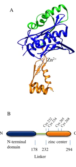
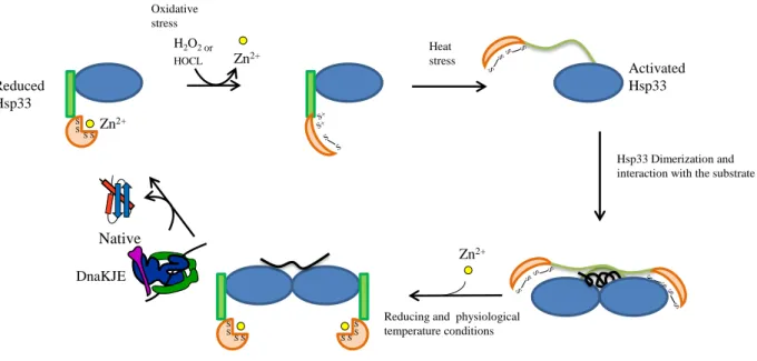
2- Hsp33 as a member of the chaperone network ... 37
a- Hsp33 is involved in the oxidative stress response ... 37
b- Structure of Hsp33 ... 38
c- Activation cycle of Hsp33 ... 39
d- Hsp33 deletion and functions in the cell ... 42
MATERIALS AND METHODS ... 44
1- Strains and plasmids ... 44
a- Bacterial Strains, phages, and culture conditions ... 44
b- Plasmid construction ... 45
2- In vivo experiments ... 46
a- Bacterial viability assay and genetic experiments ... 46
b- Isolation of protein aggregates and cell fractionation ... 46
c- In vivo pull-down assay ... 47
3- In vitro experiments ... 47
a- Western blot analysis ... 47
b- In vitro translation and cross-linking experiments ... 48
c- Pulse-chase and immunoprecipitation analyses ... 48
RESULTS ... 50
1- Hsp33 overproduction supports bacterial growth and prevents protein aggregation in the absence of TF and DnaK ... 50
2- Hsp33 function is critical in the absence of both TF and DnaK ... 54
3- Hsp33 specifically interacts with EF-Tu ... 58
4- Hsp33 triggers EF-Tu degradation by the stress protease Lon ... 63
5- EF-Tu inhibition helps bacterial growth in the absence of TF and DnaK ... 65
DISCUSSION ... 69
REFERENCES ... 74
5
ABBREVIATIONS
AAA+: ATPases Associated with a variety of cellular Activities DNA: DesoxyriboNucleic Acid
EF-Tu: Elongation Factor-Tu EF-Ts: Elongation Factor-Ts
FRET: Fluorescence resonance energy transfer HSP: Heat Shock Protein
IPTG: isopropyl β-D-1-thiogalactopyranoside JDP: J-domain protein
kDa: KiloDalton
NAC: Nascent polypeptide-Associated Complex NBD: Nucleotide Binding Domain
NEF: Nucleotide Exchange Factor NMR: Nuclear Magnetic Resonance OMP: Outer-Membrane β-barrel Protein PPIase: Peptidyl-Prolyl cis/trans Iisomerase RAC: Ribosome-Associated Complex REMP : Redox Enzyme Maturation Protein RNA: RiboNucleic Acid
SBD: Substrate Binding Domain sHSP: small Heat Shock Protein SRP: Sgnal Recognition Particle Tat: Twin-Arginine Translocon TF: Trigger Factor
6 RÉSUMÉ
Le repliement intracellulaire des protéines nouvellement synthétisées est assisté par des réseaux cellulaires de protéines chaperons. Chez Escherichia coli, la coopération entre les protéines chaperons Trigger Factor (TF) et DnaK est prédominante dans ce processus. En accord avec ceci, la délétion simultanée des gènes codants pour ces deux protéines chaperons conduit à une croissance bactérienne très réduite et à l’accumulationd’un grand nombre de protéines cytoplasmiques sous forme d’agrégats. Au cours de cette étude, nous avons utilisé ces phénotypes afin de mettre en évidence des interactions potentielles au sein du réseau de protéines chaperons in vivo. Nous avons montré que la perte des protéines chaperons TF et DnaK, et donc des voies de repliements dans lesquelles elles sont impliquées, pouvait être secourue de façon efficace par la surexpression du chaperon Hsp33, connu pour être activable en réponse à un stress oxydatif sévère. En outre, la délétion du gène hslO, codant pour Hsp33, n’était plus tolérée en l’absence de TF et DnaK. Cependant, en comparaison avec d’autres protéines chaperons comme GroESL ou SecB, la suppression de ces phénotypes par Hsp33 n’a pas pu être attribuée à un éventuel chevauchement de fonctions avec DnaK et TF. Au contraire, nos résultats montraient qu’ Hsp33 surexprimée fixait de façon spécifique le facteur d’élongation-Tu (EF-Tu) et favorisait sa dégradation par la protéase Lon. Cette action synergétique entre Hsp33 et Lon était responsable du rétablissement de la croissance bactérienne en l’absence de TF et DnaK, possiblement via le rétablissement du couplage entre la vitesse de traduction et les capacités de repliement des protéines nouvellement synthétisées du double mutant. Afin de soutenir cette hypothèse, nous avons ensuite montré que la surexpression de la toxine HipA qui inhibe EF-Tu, était aussi capable de supprimer le phénotype de thermosensibilité et de réduire significativement l’agrégation des protéines en l’absence de TF et DnaK.
7 SUMMARY
Intracellular de novo protein folding is assisted by cellularnetworks of molecular chaperones. In Escherichia coli, cooperationbetween the chaperones Trigger Factor (TF) and DnaK iscentral to this process. Accordingly, the simultaneous deletion of both chaperone-encoding genes leads to severe growth andprotein folding defects. Herein, we took advantage of suchdefective phenotypes to further elucidate the interactions ofchaperone networks in
vivo. We show that disruption of theTF/DnaK chaperone pathway is efficiently rescued by
over-expressionof the redox-regulated chaperone Hsp33. Consistentwith this observation, the deletion of hslO, the Hsp33 structuralgene, is no longer tolerated in the absence of the TF/DnaK pathway.However, in contrast with other chaperones like GroESL orSecB, suppression by Hsp33 was not attributed to its potentialoverlapping general chaperone function(s). Instead, we showthat over-expressed Hsp33 specifically binds to elongation factor-Tu (EF-Tu) and targets it for degradation by the proteaseLon. This synergistic action of Hsp33 and Lon was responsiblefor the rescue of bacterial growth in the absence ofTFand DnaK,by presumably restoring the coupling between translation andthe downstream folding capacity of the cell. In support of thishypothesis, we show that over-expression of the stress-responsivetoxin HipA, which inhibits EF-Tu, also rescues bacterialgrowth and protein folding in the absence of TF and DnaK.
8
9
INTRODUCTION
Part I: Protein folding and molecular chaperone functions
1- Protein folding within the cell
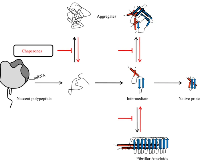
Proteins need to reach their native three-dimensional structure to become active. This folding process is determined by the primary amino-acid sequence (Anfinsen, 1973). Protein folding is not a random event but follows a specific energetic landscape through which protein gets some native-like structure as folding intermediate. These numerous pathways drive the protein to its native structure at the most stable energetic point (Dobson, 2004; Onuchic & Wolynes, 2004). Even if small single domain proteins, having less than 100 residues, can reach their native state without such intermediates (Jackson, 1998), folding of multi-domain proteins generally need parallel routes of “nucleation-condensation” of a small number of key residues, which allow the protein to condensate up to its native structure (Radford et al, 1992). Within the cell, such folding events coupled to the crowded cellular environment (i.e. proteins, nucleic-acids, complex sugars in high concentration ~400g/L (Ellis & Minton, 2003)) can lead to protein misfolding and aggregation, often associated with human diseases (Fig. 1) (Barral et al, 2004; Dobson, 2001; Thomas et al, 1995). To counteract such noxious pathways, intricate quality control systems have been maintained in all kingdoms of life (Hartl & Hayer-Hartl, 2002). Such quality controls initially take place during translation of nascent peptide chains by direct action on the ribosome and/or translational factors such as tRNAs, the elongation factor Tu (EF-Tu) and the mRNA itself (Caldas et al, 1998; Fedyunin et al, 2012; Komar, 2009; Voisset et al, 2008). Other factors known as “Molecular Chaperones” actively participate in this folding process by interacting directly with polypeptide chains in a co- and/or post-translational manner to prevent their aggregation (Fig. 1).
10
2- Role of the mRNA and the translation machinery
a- Rare codons, translational pausing and protein folding
The folding of nascent peptide chain is incorporated in the kinetics of mRNA translation during its elongation (Komar, 2009; Komar et al, 1999; Krasheninnikov et al, 1991; Wolin & Walter, 1988). In fact, mRNA sequences contain regions composed of rare codons slowing down the rate of translation and influencing the folding of the nascent peptide chain during its translation. Presence of these rare codons is due to the distribution of aminoacyl-tRNA isoacceptor species in a given organism (Chavancy & Garel, 1981; Garel, 1974; Ikemura, 1981). Such a variation in aminoacyl-tRNA isoacceptor concentration creates a strong codon bias within any given organism (Sharp et al, 1988). Pause sites were identified
Chaperones
Nascent polypeptide Intermediate Native protein
Aggregates
Fibrillar Amyloids
Fig. 1: Folding pathways of a newly synthesized protein. Proteins can pass through folding intermediates before reaching their native structure. Misfolding events such as aggregation may occur at each folding step. Molecular chaperones prevent this aggregation and favor the folding of nascent polypeptides until their native conformation.
11
by micrococcal nuclease protection assay of mRNA during translation (Hollingsworth et al, 1998; Wolin & Walter, 1988). Indeed, a frequently used codon, which corresponds to a high concentration in its aminoacyl-tRNA isoacceptor, is translated faster than one infrequently used. So that kind of punctuation into the mRNA during the translation facilitates, to some extent, the co-translational folding of the nascent polypeptide because of the positioning of these pause sites at specific domains in the encoded protein. Remarkably, a correlation between codon abundance and domain boundaries has been identified (Purvis et al, 1987). In addition, it has been found that α-helices are frequently associated with high-frequency codons, whereas β-strands and random coils are preferentially coded by rare codon regions (Thanaraj & Argos, 1996). Moreover, the location of these pause sites is highly conserved in some protein families including cytochromes C, globins, γ-B crystallins and chloramphenicol acetyltransferase (Krasheninnikov et al, 1989; Widmann et al, 2008). To study the relevance of such pause sites, silent mutations of mRNAs coding the Escherichia coli chloramphenicol acetyltransferase, Saccharomyces cerevisiae anthranilate synthase indole-3-glycerol phosphate synthase and Echinococcus granulosus fatty acid binding protein, replacing rare codons by common ones showed a decrease in their specific activities when compared to the wild type proteins (Cortazzo et al, 2002; Crombie et al, 1994; Komar et al, 1999). Furthermore, the proteins produced from these silently mutated mRNAs were misfolded. Although the kinetics of translation appear to play a significant role in the co-translational folding of nascent peptide chain, but there are additional factors, including tRNA concentration, Elongation Factor-Tu (EF-Tu), and ribosome, which participate directly or indirectly to the folding process.
b- tRNA concentration modulates nascent chain protein folding
tRNAs bring amino-acids to their cognate codons. Remarkably, it has been shown both in vitro and in vivo that the concentration of tRNAs acceptors can drastically modulate the rate of translation. Indeed, using an in vitro translational experiment, Anderson first showed that translation of a poly-U by tRNAPhe was inhibited at high concentration of tRNAPhe (tRNAPhe/70S ribosome ratio of 7) (Anderson, 1969). In addition, a different study pointed out that in the presence of a constant concentration of exogenous tryptophan, the rate of incorporation of this amino acid was considerably diminished when tRNATrp concentration was too high, following over-expression of tRNATrp from a plasmid. Such over-expression changed the ratio of uncharged tRNATrp / [Trp-tRNATrp] and the pool of uncharged tRNATrp was found to inhibit Trp incorporation into proteins (Rojiani et al, 1990). Remarkably, they
12
also showed that uncharged tRNATrp could inhibit incorporation of other amino acids, such as Lys and Cys (Rojiani et al, 1990). A third team studied the effect of low-abundant tRNAs upregulation on the folding and solubility of E. coli proteins (Fedyunin et al, 2012). In this case, upregulation of argU which encodes tRNA4Arg that pairs with arginine codons AGA and
AGG, ileY encoding for tRNA2Ile that pairs with AUA codon or leuW encoding tRNA3Leu that
pairs with CUA codon, modifying the rate of translation of these rare condons, was responsible of the aggregation of many proteins such as molecular chaperones which had been found to suffer from this misfolding effect (Fedyunin et al, 2012). This approach revealed the importance of the translational rate during co-translational folding of all the cellular proteome and the role of tRNA isoacceptors concentration in this translational rate.
c- Roles of EF-Tu
In E. coli, EF-Tu is the most abundant protein in the cell, representing 5 to 10% of the total proteins (Furano, 1975). EF-Tu has a GTPase activity and brings aminoacyl-tRNA to the A site of the ribosome in a GTP-dependent manner. GDP is replaced through interaction with the nucleotide exchange factor EF-Ts (Agirrezabala & Frank, 2009). EF-Tu and its eukaryotic homolog EF-1α have many other cellular functions. EF-Tu was found to be an essential subunit of the Qβ phage replicative complex (Blumenthal et al, 1972). EF-Tu can also stimulate RNA synthesis by interacting with the transcriptional apparatus (Travers et al, 1970). In Bacillus subtilis, EF-Tu plays an additional role in cell shape maintenance, interacting with the actin-like MreB (Defeu Soufo et al, 2010). This role was also identified for EF-1α, as it can bind actin filaments and microtubules and influence the assembly and the stability of these cytoskeletal polymers (Shiina et al, 1994; Yang et al, 1990). In addition, Caldas and coworkers also found that EF-Tu could reactivate chemically-unfolded citrate synthase and α-glucosidase with the same efficiency than DnaKJE in vitro, thus suggesting chaperone function (Caldas et al, 1998). The eukaryotic EF-1α chaperone activity was also identified using Phenylalanyl-tRNA synthase and seryl-tRNA synthase as substrates. In this case, it was shown that the mammalian EF-1α (called eEFiA) could refold and restore the enzymatic activity of these two aminoacyl-tRNA synthase (Lukash et al, 2004). In conclusion, EF-Tu does not only bring aminoacyl-tRNAs to the ribosome A site but participates in a plethora of cellular processes, most likely including protein folding. The last compound of the translational machinery to have a chaperone function protecting and actively participating in the co-translational folding of the nascent chain protein is the ribosome itself.
13
d- Chaperone function of the ribosome
Ribosomes represent 30% of the total cell mass with up to 105 and 106 ribosomes in a bacteria and a mammalian cell respectively (Bashan & Yonath, 2008). It is a ribonucleoprotein complex composed of 2 subunits. The small subunit (30S in bacteria; 40S in eukaryote) decodes the mRNA during translation and the large subunit (50S in bacteria; 60S in eukaryote) contains the peptidyl transferase center and the ribosomal exit tunnel through which nascent polypeptides will emerge and be released into the cytosol. The length of the exit tunnel is about 80 Å to 100 Å with a diameter comprised between 10 Å at its narrowest point and 20 Å at its widest point. (Ban et al, 2000; Harms et al, 2001; Nissen et al, 2000). These dimensions allow the ribosome to contain a nascent chain of 30 amino-acids in an extended conformation or 60 amino-acids as an α-helix (Malkin & Rich, 1967; Picking et al, 1992; Voss et al, 2006). During translation, nascent chain interacts with this exit tunnel composed of 23S rRNA and proteins L4, L22 and L23 in bacteria (Ban et al, 2000; Harms et al, 2001; Kosolapov & Deutsch, 2009; Lu & Deutsch, 2005; Lu et al, 2007; Nissen et al, 2000). Such interactions can regulate kinetics of translation and induce translational pausing, thus facilitating co-translational folding of the nascent peptide chain or its interaction with ribosome associated factors such as the molecular chaperone Trigger Factor in bacteria (TF) or the signal recognition particle (SRP) involved in the co-translational targeting of secretory or membrane proteins to the sec translocon (Eisner et al, 2006; Kramer et al, 2009; Marin, 2008). Moreover, the ribosomal exit tunnel directly participates to the folding of nascent chains by promoting its compaction or its helical conformation in the lower and upper part of the tunnel. The middle part of the tunnel is constricted by L4 and L22 proteins (Lu & Deutsch, 2005). This constriction site avoids protein folding in this area and, interacting directly with the nascent chain, may modulate the docking site shared by TF or SRP and then allow the recruitment of each of them to the ribosome exit tunnel (Gu et al, 2003; Houben et al, 2005; Ullers et al, 2003). The exit pore of the tunnel also plays a role in the co-translational folding of the nascent chain allowing the formation of tertiary structures such as β-hairspins (Kosolapov & Deutsch, 2009).
The ribosome itself also possesses some chaperone activity. Indeed, the 50S ribosomal subunit can interact post-translationally with unfolded proteins and efficiently participate to their folding (Kudlicki et al, 1994; Voisset et al, 2008). The interaction site of the ribosome with unfolded protein had been identified near from the peptidyl transferase center, in the domain V of the 23S rRNA (Chattopadhyay et al, 1996; Pal et al, 1997). The ribosome can
14
also participate in the co-translational folding of newly synthesized proteins in a different way. Indeed, Kaiser and coworkers (2011) studied the active folding activity of the ribosome exit tunnel following the folding of the T4 lysozyme and found that the ribosome could prevent misfolding of the nascent chain by modulating its folding rate (Kaiser et al, 2011). Furthermore, this exit pore is a docking site for many key proteins involved in the enzymatic processing of the nascent chain such as the methionine aminopeptidase and the peptide deformylase (Kramer et al, 2009), for targeting factors such as SRP and SecA, ribosome associated chaperones like Trigger Factor in bacteria or Ssb/Ssz/Zuotin chaperone triad, ribosome-associated complex (RAC) and nascent polypeptide-associated complex (NAC) for the eukaryote S. cerevisiae(Eisner et al, 2006; Ferbitz et al, 2004; Preissler & Deuerling, 2012).
e- Molecular chaperones, concept, functions and universality
Molecular chaperones facilitate protein folding and prevent and/or rescue noxious protein aggregation. They are involved in many cellular pathways such as: de novo protein folding, protein disaggregation, protein translocation through a biological membrane, oligomerisation and control of protein-protein interaction, as well as protein turn over (Hendrick & Hartl, 1993). Molecular chaperones are conserved in all the three kingdoms of life, represented by different protein families, and are known to function in a sequential manner on their substrates co-translationally and post-translationally (Frydman et al, 1994; Langer et al, 1992; Siegers et al, 1999).
A first group of molecular chaperones interacts with the ribosome and/or the nascent chain to facilitate co-translational folding. Bacteria use the chaperone Trigger Factor, interacting at the same time with the ribosome and the nascent chain (Hesterkamp et al, 1996). The Hsp70 chaperone DnaK from E. coli can also interact with nascent polypeptides to assist them during their co-translational folding (Teter et al, 1999). Although the eukaryotic chaperone system does not have Trigger Factor, they do have two different kinds of ribosome associated complexes acting at the ribosome exit pore to support co-translational folding (Preissler & Deuerling, 2012). Indeed, in Saccharomyces cerevisiae, the ribosome-associated complex (RAC) composed of the Hsp70/Hsp40 chaperone system Ssz/Zuotin and another Hsp70 named Ssb and the heterodimeric nascent chain-associated complex (NAC) may parallel Trigger Factor function in the folding of the nascent polypeptide chains (Preissler & Deuerling, 2012).Hsp70 may also bind substrates in a post-translational way to mediate their folding (Deuerling et al, 1999; Teter et al, 1999). The chaperonins represent the second group
15
of molecular chaperones, mostly participating in the post-translational folding of proteins. They are structured as double-ring complexes forming a central cavity in each ring, binding and encapsulating unfolded proteins in one ring, which protect the substrate from the crowded environment of the cell and help it to fold (Mayhew et al, 1996; Weissman et al, 1996). Other types of chaperones are more specifically involved in disaggregation, as it’s the case for Hsp90 and Hsp100, HtpG and ClpB in E. colirespectively.
In the following parts of the introduction, the structures, the mechanisms and the functions of the major E. coli chaperones involved in de novo protein folding, namely TF, DnaKJE and GroESL, will be presented. In addition, their functional cooperation with other chaperone systems during protein export, protein disaggregation and in response to severe oxidative stress will be developed.
Part II: Chaperone-assisted de novo protein folding
1- The ribosome-bound Trigger Factor
a- Structure / interaction with the ribosome
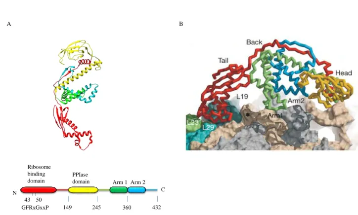
The ribosome-bound TF is the first molecular chaperone to interact with most of the newly synthesized polypeptides (Valent et al, 1995). It is a 48kDa protein constituted of 3 distinct domains elongated in a Dragon-like shaped structure (Ferbitz et al, 2004; Hesterkamp & Bukau, 1996; Zarnt et al, 1997) (Fig. 2A). The N-terminal domain of TF, composed of amino acids 1 to 149, is the ribosome binding domain, which also contributes to TF chaperone activity. This domain possesses some structural homology with the molecular chaperone Hsp33 except for the additional loop involved in the binding of TF to the ribosome via the TF signature motif “GFRxGxxP” (Genevaux et al, 2004; Hoffmann et al, 2010; Kramer et al, 2002; Kristensen & Gajhede, 2003). A long linker (aa 111 to 149) is found between the ribosome binding domain and the peptidyl-prolyl cis/trans isomerase domain (PPIase) of TF. The PPIase domain (aa 150 to 245) is related to the FK506 binding protein family of PPIase (Hesterkamp & Bukau, 1996; Scholz et al, 1997; Stoller et al, 1995). This domain is dispensable for chaperone function of TF in vivo, but it can enhance chaperone activity of TF as an auxiliary binding site of substrates (Genevaux et al, 2004; Hoffmann et al, 2006; Kramer et al, 2004a; Lakshmipathy et al, 2007; Merz et al, 2006). The C-terminal domain (aa 246 to 432), forming the body of the dragon, is found between the N-terminal domain (the tail of the
16
dragon) and the PPIase domain (the head of the dragon). It interacts with the long linker (aa 111 to 149), which stabilize its structure containing 2 protruding helical “arms” (Ferbitz et al, 2004; Martinez-Hackert & Hendrickson, 2007; Merz et al, 2006). This domain forms the main chaperone module of TF (Merz et al, 2006).
TF is conserved in bacteria and chloroplasts, but its PPIase domain may be absent in some species (Hoffmann et al, 2010; Kristensen & Gajhede, 2003). Some bacteria, such as
Desulfitobacterium hafniense, even possess several TF and TF-like molecular chaperones
with specific functions (Maillard et al, 2011). Indeed, among the 3 TF homologs present in this bacterium, the TF-like chaperone PceT, which lacks the TF N-terminal domain, is dedicated to the folding and the transport of the reductive dehalogenase PceA through the Twin arginine translocation system (Maillard et al, 2011).
149 245 360 432 N C Ribosome binding domain PPIase
domain Arm 1 Arm 2
A B
43 50 GFRxGxxP
Fig. 2: Structure of the E. coli TF and interaction with ribosome exit tunnel. (A) The upper part represents the three-dimensional structure ribbon diagram of TF (PBD 1W26). The bottom shows the domain arrangement of TF and the TF signature motif of the ribosome binding site (aa 43 to 50). (B) Interaction of TF with the exit-tunnel of the ribosome. TF binds L23 and L29 and covers the exit tunnel from the crowded environment. The star represents the exit pore of the tunnel. Ribosome binding site of TF is represented in red, the PPIase domain is in yellow, the Arm 1 is in green and the Arm 2 in blue. Figure adapted from (Ferbitz et al, 2004).
17
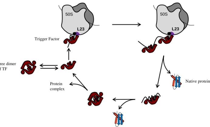
TF is an abundant cytosolic protein represented in 2- to 3- fold molar excess over ribosome (50µM in spite of 20µM respectively) (Crooke et al, 1988). It is constitutively expressed and dispensable under normal growth conditions (Martinez-Hackert & Hendrickson, 2009). TF cycles on and off the ribosome in 1:1 stoichiometry in an ATP-independent manner and creates a protective arch over the ribosomal exit tunnel (Fig. 2B) (Hoffmann et al, 2010; Kaiser et al, 2006; Rutkowska et al, 2008). During the TF binding/release cycle, it has been found that the presence of a nascent chain increases the affinity of TF for the ribosome 2 to 30 fold (Raine et al, 2006; Rutkowska et al, 2008). After release from the ribosome, TF may stay bound to the nascent chain of large multi-domain proteins allowing another free TF to bind the ribosome and in some cases to facilitate transfer of its substrate to the downstream chaperones DnaKJE and GroESL (Kaiser et al, 2006; Lakshmipathy et al, 2010). In addition, it has been found that TF from Thermotoga maritima TF (tmTF) can dimerize and encapsulate 2 tmS7 ribosomal proteins (Martinez-Hackert & Hendrickson, 2009). Such a dimerization of tmTF could create an Anfinsen-like cage which could protect S7 and participate to its incorporation into the ribosome (Martinez-Hackert & Hendrickson, 2009) (Fig. 3).
The N-terminal of TF interacts with the ribosome via its signature motif to the L23 ribosomal protein located at the exit port, the ribosomal RNA 23S and additional interaction occurs with the L29 ribosomal protein (Baram et al, 2005; Kramer et al, 2002; Schlunzen et al, 2005) (Fig. 2B). This TF-ribosome interaction is crucial for TF chaperone function during co-translational folding of nascent polypeptide chains (Lakshmipathy et al, 2007). In vitro experiments showed an early interaction with nascent polypeptide chains as short as 40 amino acids (Merz et al, 2008). Nevertheless, using ribosome profiling of purified ribosome – nascent chain – TF complexes and deep sequencing of mRNA fragments protected by these ribosomes, Oh and coworkers (2011) showed that, in vivo, TF seems to preferentially bind to nascent polypeptide chains of about 100 amino acids (Oh et al, 2011). This late interaction between TF and the nascent chain potentially allows earlier interaction with other ribosome associated nascent chain interacting factors, including the peptide deformylase, the methionine aminopeptidase and SRP (Ball & Kaesberg, 1973; Bingel-Erlenmeyer et al, 2008; Keenan et al, 2001).
Recent works identified nascent chains that are disliked by TF: a segment of poly-alanine folded into a helical conformation (Bhushan et al, 2010; Lu & Deutsch, 2005). Indeed, replacing 9 amino acids in the N-terminal of GFP by a poly-Alanine sequence near the exit
18
tunnel, the center of the tunnel or the peptidyl transferase center place this helical structure in different parts of the ribosome tunnel. Using this technique, Lin and coworkers showed that TF binding to the ribosome was disfavored when this helical structure was in an area near from the exit tunnel and interacted with L23. This interaction can induce conformational changes in the ribosomal binding site of TF and reduce its recruitment to the exit pore (Lin et al, 2012). To validate this approach, they used the signal anchor sequence of a cytoplasmic membrane protein and the signal sequence of the secretory protein pre-β-lactamase (Bornemann et al, 2008). These sequences are known to fold into the ribosome tunnel in a helical structure and to enhance the recruitment of SRP (Berndt et al, 2009; Bornemann et al, 2008; Halic et al, 2006; Woolhead et al, 2004). Both of them reduced the recruitment of TF at the ribosome exit tunnel after interaction with L23 inside the ribosomal tunnel (Lin et al, 2012). Therefore, the recruitment of TF to the exit pore is modulated by the nascent peptide chain itself, which modifies the ribosome binding site shared by TF and SRP (Lin et al, 2012).
Trigger Factor 50S L23 50S L23 Native protein Protein complex Free dimer of TF
Fig. 3: Chaperone cycle and assembly function of TF. TF interacts with nascent chains by direct association with the ribosome. More than one TF molecule may bind a nascent polypeptide of a multi-domain protein. This protein can be released in a native state or TF may stay bound to it in the cytosol to fulfill its folding. Martinez-Hackert and Hendrickson (2009) also identified a new role of TF in the assembly of protein complexes. TF could also form dimers and encapsulate two substrates mediating their folding and their interaction. This role is important for ribosome biogenesis.
19
b- TF substrate interaction and cellular functions
Several studies were developed to identify preferred interactors of TF. A screening of 2.842 membrane coupled peptides of 20 different proteins originating from bacteria and eukaryotes was used to identify TF binding motifs (Patzelt et al, 2001). The analysis revealed that TF preferentially interacts with motifs of 8 residues enriched in aromatic residues (Phe, Tyr, Trp and His) and basic residues (Arg and Lys) and impoverished in acidic residues (Asp and Glu). Even if TF possesses a PPIase activity, proline residues do not seem to participate in substrate binding. TF motifs occurs frequently, about every 32 residues in all protein tested. This frequent motif allows TF to interact virtually with all the proteins during translation (Patzelt et al, 2001). However, interactome studies pointed out some preferential substrates of TF. A comparison between co-purified TF substrates and aggregated proteins isolated in a tig null-mutant identified 178 potential TF substrates, a majority of them being ribosomal proteins or proteins from homo- or hetero-oligomers (Martinez-Hackert & Hendrickson, 2009). Recently, a quantitative ribosome profiling analysis of ribosomes whose nascent chains are bound to TF isolated outer-membrane β-barrel proteins (OMPs) as the strongest TF interactors (Oh et al, 2011).
Even if TF interacts with a large number of substrates, the mode of action may differ. TF can help peptide domains shorter than 150 residues to fold within its cradle (Hoffmann et al, 2006; Merz et al, 2008). In vitro experiments also showed that TF promotes folding of denatured proteins (Huang et al, 2000; Kramer et al, 2004a; Merz et al, 2006). In addition, TF can delay co-translational folding of multi-domain proteins like firefly luciferase and β-galactosidase as presented in the cycle of TF (Part II 1-a). This conclusion has been reached by measuring the time-dependent enzymatic activity of these two proteins with or without TF (Agashe et al, 2004). Two recent works have further studied such TF function. Indeed, Hoffmann and colleagues (2012) showed that TF was found to postpone the disulfide bond formation of ribosome arrested β-lactamase and barnase, to unfold a folded arrested barnase and to facilitate its degradation. In addition, modeling the co-translational folding of the N-terminal domain of β-galactosidase (216 residues), O’Brien and coworkers proposed that TF could act on the nascent peptide via three different mechanisms: TF might (i) decrease the rate of structural rearrangements, (ii) avoid tertiary structure formation, (iii) increase the effective length of the exit tunnel and entanglements between the nascent polypeptide chains (O'Brien et al, 2012). Such functions of TF could significantly help multi-domain proteins to fold correctly, thus avoiding protein misfolding.
20
Moreover, interaction of the ribosome-bound TF with its substrate can facilitate substrate degradation by proteases under certain conditions. Indeed, in vitro experiments showed that, in the absence of DnaK, TF could favor the degradation of the arrested complex barnase-arrested/ribosome (Hoffmann et al, 2012). In vivo works also revealed that TF favors the degradation of EF-Tu in the absence of DnaK following Hsp33 over-expression (Bruel et al, 2012). These results may pin point a new role of TF when DnaK is sequestered on aggregates during a stress.
TF may participate in OMPs stability as well (Crooke & Wickner, 1987; Oh et al, 2011; Ullers et al, 2007). Indeed, deletion of the tig gene encoding TF provokes a decrease in the steady state levels of OMPs compared with wild type cells (Oh et al, 2011) and some OMPs were isolated as strong TF interactors by ribosome profiling analysis (Oh et al, 2011). However, in vivo experiments revealed that TF deletion does not facilitate export of SecB-dependent presecretory proteins, including MBP, OmpA and SecA (Guthrie & Wickner, 1990; Lee & Bernstein, 2002; Ullers et al, 2007). Instead, the deletion of tig even accelerated the export of some presecretory proteins with Sec translocon (Lee & Bernstein, 2002). Another example of TF involved in protein secretion was identified in Streptococcus
pyogenes(Lyon & Caparon, 2003). TF is essential for the secretion and the maturation of the
cysteine protease SpeB. The PPIase activity of TF modifies SpeB then TF targets this substrate to the Sec translocon (Lyon & Caparon, 2003). Moreover, TF over-expression induces the aggregation of OmpF, partially reflecting the toxicity of TF (Genevaux et al, 2004). Finally, over-expression of TF is toxic and induces a strong filamentation phenotype similar to the filamentation observed after FtsZ depletion (Genevaux et al, 2004; Guthrie & Wickner, 1990; Ward & Lutkenhaus, 1985). These findings suggest that TF could be involved in cell division. In some strain background, TF is also known to be essential for viability at low temperature (10°C), perhaps due to its possible involvement in ribosome biogenesis and cell division (Kandror & Goldberg, 1997).
In conclusion, TF facilitates the folding of nascent chains by protecting them from the crowded cellular environment and delaying formation of unproductive folding intermediates. In addition, it is involved in ribosome biogenesis and in the stabilization of OMPs.
21 2- DnaKJE cycle and interactors
a- DnaKJE structure and cycle
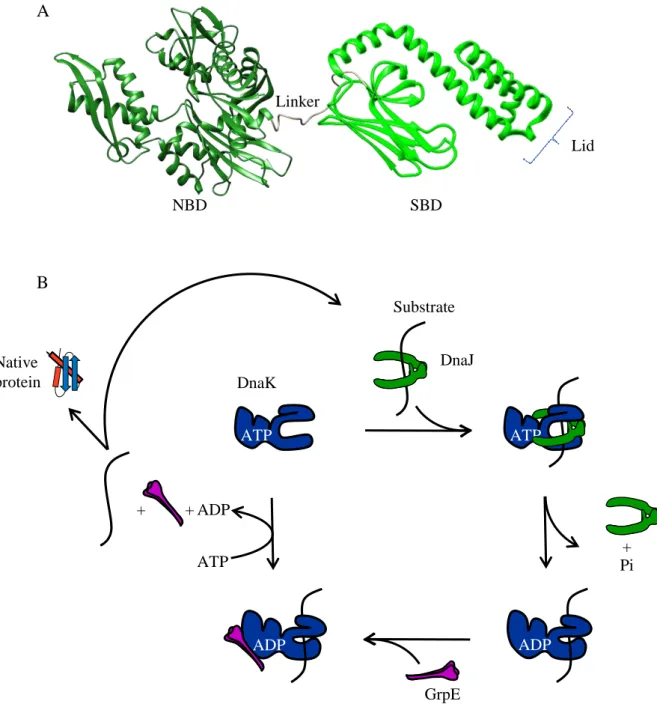
The Hsp70 (Heat shock protein of 70kDa) molecular chaperone family is the most conserved protein family in all organisms (Gupta, 1998). Thebest characterized Hsp70 family member is the bacterial protein DnaK from E. coli. DnaK is a heat shock induced protein present at about 10 000 copies per cell which double after a shift at 42°C (Genevaux et al, 2007). This molecular chaperone of 638 amino-acids in length possesses an N-terminal nucleotide binding domain (NBD) of 45kDa with ATPase activity (aa 1 to 380) and a C-terminal domain of 25kDa as substrate binding domain (SBD) subdivided into a β-sandwich subdomain and a α-helical domain forming a lid (aa 398 to 638) (Flaherty et al, 1994). Both domains are connected by a linker (aa 381 to 397) which is highly conserved and possesses a characteristic 388DVLLLD393 hydrophobic segment essential for allosteric communication
between the NBD and the SBD (Mayer & Bukau, 2005; Swain et al, 2007; Vogel et al, 2006a; Vogel et al, 2006b). In some eukaryotes the SBD contains an additional short motif interacting with other partner proteins, like Hop/p60/CHIP that modulate chaperone activity of Hsp70 (Mayer & Bukau, 2005; Young et al, 2004). The structure of a truncated DnaK (aa 1 to 605) bound to ADP and a substrate peptide (NRLLLTG) was revealed by NMR (Nuclear Magnetic Resonance) presented in Fig. 4A (Bertelsen et al, 2009). This study showed that the NBD and the SBD, in this conformation, are independent and mobile. This mobility is restricted in a cone of ± 35° (Bertelsen et al, 2009). A recent work studied the structure of DnaK bound to ATP. They showed that in this conformation, the α-helical lid and the β-sandwich substrate pocket of the DnaK SBD were docked to different positions of the NBD (Kityk et al, 2012). Then allosteric modifications induced by substrate binding and ATP hydrolysis will unlock the SBD of DnaK (Kityk et al, 2012). DnaK facilitates protein folding by cycles of binding/release of protein substrates. While ATP-bound DnaK has low affinity and fast exchange rates for substrates, ADP-bound DnaK possesses a high affinity for the substrate and a low exchange rate (Genevaux et al, 2007). The ATP hydrolysis by DnaK is essential for its chaperone activity but this molecular chaperone has a low rate of ATP hydrolysis (Karzai & McMacken, 1996; Laufen et al, 1999). Therefore, the DnaK chaperone function relies on its obligate cochaperone partners, namely the J-domain protein Hsp40/DnaJ and the nucleotide exchange factor GrpE (Schroder et al, 1993).
22
The DnaKJE cycle starts with the interaction of a substrate-bound DnaJ dimer with the ATP-bound DnaK. Indeed, DnaJ recruits the substrate and efficiently stimulates ATP hydrolysis by DnaK up to 1000-fold (Karzai & McMacken, 1996; Laufen et al, 1999; Liberek et al, 1991). ATP hydrolysis will close the SBD and enhance its affinity for the substrate. Then, a second cochaperone, namely the nucleotide exchange factor GrpE will bind to the NBD and facilitate ADP release. The subsequent binding of a new ATP opens the lid of the SBD and stimulates substrate release (Harrison et al, 1997). Once released from DnaK, the substrate can reach its native form or necessitates further DnaK cycles (Fig. 4B) or be transferred to other chaperones, like the chaperonin GroESL to complete its folding (see part II-4).
DnaK interacts with a broad range of substrates present in different conformations: folded, misfolded or aggregated. DnaK was found to be highly efficient when the substrates are misfolded but not aggregated. Indeed, a firefly luciferase variant from Photinus pyralis was unfolded by Freeze-thaw cycles and urea and incubated in the presence of the DnaKJE machinery (Sharma et al, 2010). Monitoring folding kinetics, the authors found that while ATP hydrolysis induces substrate unfolding, substrate release induces spontaneous refolding of the substrate (Sharma et al, 2010). Another study revealed that the lid of DnaK could stay opened or partially opened even after hydrolysis of ATP (Kityk et al, 2012; Schlecht et al, 2011). This conformation could allow DnaK to interact with aggregated proteins or amyloids and participate to their dissolution.
23 NBD SBD Linker Lid A B ATP Pi ADP DnaK DnaJ Substrate GrpE ADP ATP ADP + + + Native protein ATP
Fig. 4: The DnaKJE chaperone system. (A) Three dimensional structure of the E. coli DnaK. (aa 1 to 605) truncated structure of DnaK bound to ADP (Bertelsen et al, 2009) (PDB 2KHO). The N-terminal NBD (dark green) and the C-N-terminal SBD (green) are disjointed by a linker (gray). The Lid of the SBD is in a closed conformation. (B)DnaKJE chaperone cycle. The ATP bound DnaK has a low-affinity for substrate. The co-chaperone DnaJ mediates the recruitment of substrate to DnaK and stimulates its ATPase activity. Hydrolysis of ATP, leading to the ADP bound DnaK, induces allosteric modifications of DnaK allowing the sequestration of the substrate. Then, the Nucleotide Exchange Factor GrpE facilitates ADP/ATP exchange opening the lid of DnaK and the substrate release (Genevaux et al,2007).
24
b- Functions of DnaK
It is believed that DnaK interacts and assists the co- and/or post-translational folding of about 15% of the nascent peptides or newly synthesized proteins (Deuerling et al, 1999; Teter et al, 1999). DnaK also participates in the refolding of misfolded and aggregated proteins, translocation through biological membrane and oligomeric complex assembly/disassembly (Genevaux et al, 2007; Mayer & Bukau, 2005). DnaK acts on protein aggregates cooperating with other molecular chaperones: ClpB (Hsp100), Hsp31, or the small heat shock proteins (sHSPs) IbpA IbpB (Genevaux et al, 2007). DnaK is also involved in the oxidative stress response and controls the heat shock response by stimulating the σ32
degradation (Hoffmann et al, 2004; Straus et al, 1990). The central role of DnaK is revealed by deletion of its gene. Indeed, a dnaK null mutant in E. coli exhibits multiple phenotypes including cryosensitivity beyond 20°C, thermosensitivity above 35°C, filamentation, slow growth at the permissive temperature of 30°C, resistance to bacteriophages λ P1 and P2 infection, loss of motility, sensitivity to nutrient starvation and defective in plasmid maintenance (Genevaux et al, 2007). DnaK deletion induces a ribosome biogenesis defect indicating a role for DnaK in ribosome assembly (Al Refaii & Alix, 2009; Maki et al, 2002).
c- Interactors in the cell
Using a screening of 37 different proteins split in more than 4500 peptides and bound to a cellulose membrane, the recognition motif of DnaK has been defined as an extended five residue segment composed of hydrophobic amino acids, Leu, Ile, Val Phe and Tyr preferentially, framed by positively charged residues (Rudiger et al, 1997). These motifs occur often in proteins, every 50 to 100 amino acids (Rousseau et al, 2006). Such a variety of cellular functions and high frequency of binding motifs suggest that DnaK interacts with a large number of newly synthesized proteins. Indeed, a recent analysis of the DnaK interactome in E. coli revealed that DnaK interacts with more than six hundred proteins under physiological condition. 80% of these interactors are cytosolic proteins, but DnaK also interacts with inner membrane proteins (~11%), outer membrane proteins (~3%) and periplasmic proteins (~3%) (Calloni et al, 2012). The interaction with exported proteins is in agreement with the role of DnaK in protein export (Randall & Hardy, 2002). Analysis of the relative enrichment of substrates on DnaK pointed out that 40% of the total mass of DnaK substrates was highly enriched on DnaK. These enriched proteins were identified to be below average cellular abundance and with a low solubility property. These proteins are known to be
25
aggregation-prone (Tartaglia et al, 2010; Tartaglia et al, 2007). Interestingly, essential proteins are under the low enriched protein group of DnaK interactors. This is probably due to the fact that folding of these substrates is supported by the chaperonin GroESL.
3- The chaperonin GroESL
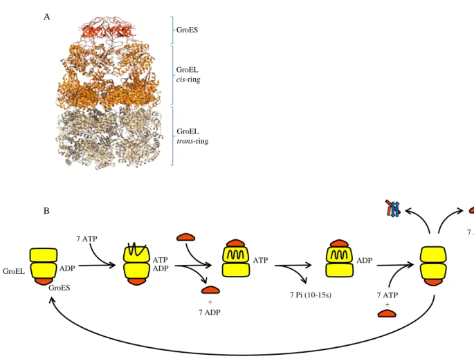
a- Structure of the GroESL complex and chaperonin cycle
The chaperonin GroESL is the only known chaperone system essential in E. coli(Fayet et al, 1989). GroESL is an asymmetric complex composed by two heptameric rings of 57kDa monomer of GroEL (Xu et al, 1997). A GroEL barrel is subdivided in three different domains: the ATPase equatorial domain, the intermediate domain responsible of GroEL rearrangement after substrate binding, and the apical domain presenting hydrophobic residues involved in substrate binding and forming the interaction site with the GroES cochaperone (Bukau & Horwich, 1998). GroES is structured in heptamer of 10kDa subunits and forms a dome closing one ring of GroEL. Rings are termed as follow: the cis side is composed of a GroESL complex encapsulating a substrate and the trans side is made by a free and open GroEL cylinder (Fig. 5A) (Hartl & Hayer-Hartl, 2009).
At the beginning of the cycle, 7 ATP molecules bind to the trans-ring of GroEL. Then the substrate is recruited to this GroEL ring. ATP molecules are known to induce rearrangements of the apical domain of GroEL mediating the capture and the compaction of the substrate to facilitate its encapsulation (Clare et al, 2012; Lin et al, 2008). Then a GroES dome encloses the substrate into the cage and creates the new cis-ring of the complex. This
cis-ring interaction between GroES and the apical domains of GroEL will enhance the
hydrophobicity of the cavity, inducing the release of the substrate into the cage formed by the GroEL cylinder. Then, rearrangements of the apical and the intermediate domains in the cis-ring will bury the hydrophobic residues and change the environment of the cage. The volume of the cage will also be enhanced and will be able to accommodate polypeptides up to 60kDa (Hartl & Hayer-Hartl, 2009). The new substrate enclosed into GroESL complex now folds into a secure and isolated space during 10s to 15s, time required for ATP hydrolysis. Then release of ADP and GroES will free the substrate. The cycle will repeat from the new trans-ring and the substrate will be free folded or will necessitate other cycles of folding into GroESL (Fig. 5B). This GroESL dependent folding may occur in a passive “Anfinsen cage” (Horwich et al, 2009; Motojima et al, 2012), with an active role via negatively charged residues in the cavity (Chakraborty et al, 2010; Lin et al, 2008; Tang et al, 2008; Tang et al,
26
2006) or by forced unfolding (Lin et al, 2008). Action of GroESL mediated folding may differ in relation to the substrate bound.
b- Interactors in the cell
In vivo, GroESL is known to interact with approximately 10% to 15% of the newly
synthesized cytosolic proteins (Houry et al, 1999). 250 GroESL substrates were identified by mass spectrometry grouped into 3 different classes using their necessity to be helped by chaperon systems. The Class I proteins have a relatively low propensity to aggregate and are chaperone independent. Proteins of Class II need DnaKJE or GroESL to fold properly. Class III proteins are obligate GroESL substrates These proteins range between 20kDa to 50kDa and are enriched in (αβ)8 TIM barrel (Kerner et al, 2005). 85 of GroESL substrates are strictly
GroES GroEL cis-ring GroEL trans-ring A B ADP 7 ATP + ATP GroEL ATP GroES ADP 7 ADP 7 Pi (10-15s) ADP 7 ATP + + 7 ADP
Fig. 5: The GroESL chaperonin system. (A) Three dimensional architecture of the GroESL-7ADP complex (Xu et al, 1997) (PDB 1AON). The GroES heptamer (dark orange) encapsulates the cavity of the cis-ring of GroEL (orange). The cis-ring of GroEL is in an extended conformation in comparison to the trans-ring (gray). (B) GroESL chaperonin cycle. Details concerning the cycle are given in the text.
27
GroESL-dependent for their folding from which 13 are essential proteins. These 85 substrates occupy 75% to 80% of GroESL capacity in the cell (Kerner et al, 2005).
Recently, Class III proteins were over-expressed and tested for their dependence to GroESL in GroESL-depleted cells (Fujiwara et al, 2010). They found that 60% of Class III proteins are in fact GroESL-obligated substrates, the other 40% can use other chaperone machineries for their folding under over-expressing conditions. These obligate GroESL-substrates were grouped as Class IV. Note that these proteins possess a positive bias in alanine and glycine content, making the proteins more aggregation-prone. Moreover, this group contains six essential proteins for E. coli: DapA, ASD, MetK, FtsE, HemB and KdsA (Fujiwara et al, 2010).
Interestingly, in the absence of both TF and DnaKJE, GroESL interacts with about 150 additional substrates (Kerner et al, 2005), thus reflecting interplay among these chaperone systems.
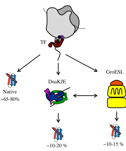
4- Interplay between TF/DnaKJE/GroESL during de novo protein folding
The molecular chaperones TF, DnaKJE and GroESL are known to work as a network participating together in the folding of nascent chains and newly synthesized proteins (Fig. 6). Both TF and DnaKJE most likely share more than 300 proteins as common substrates (Deuerling et al, 2003). Cooperation between these two chaperones had been shown in vitro using the multi-domain protein firefly luciferase, with TF and DnaKJE acting co-and post-translationally, respectively (Agashe et al, 2004). Analysis of the effect of TF, DnaKJE and GroESL on about 800 cytosolic proteins was carried out using a reconstituted cell-free translation system (Niwa et al, 2012). In this work, the authors showed that either DnaKJE or GroESL efficiently increase the solubility of more than 66% of the proteins tested. In this case, TF only had a minor effect on prevention of aggregation of these substrates. These results strongly support previous in vitro translation experiment (Agashe et al, 2004) (Fig. 6).
To go further inside the chaperone network, Calloni et al (2012) recently characterized interactors of DnaK following TF deletion or GroESL depletion. In the absence of TF, DnaK was found to interact with a higher number of substrates: 998, including 95% of the 674 DnaK interactors already characterized in the presence of TF. This extended substrate interaction of DnaK in the absence of TF correlates with the overlapping of function already observed between these two molecular chaperones (Deuerling et al, 1999; Teter et al, 1999),
28
i.e.in the absence of TF, DnaK interacts with 2 to 3 times more nascent chains than in the
presence of TF (Teter et al, 1999). Note that in this case, DnaK could also interact with shorter nascent polypeptides (<30kDa) (Deuerling et al, 1999; Teter et al, 1999).
As state above, E. coli tolerates the single deletion of either tig or dnaK(Deuerling et al, 1999; Genevaux et al, 2004; Teter et al, 1999) and upregulation of DnaKJE is observed in
tig null-mutant (Deuerling et al, 2003). Simultaneous deletions of both tig and dnaK genes
present synthetic lethality at temperatures ≥30°C with respect to the strain background used (Deuerling et al, 1999; Genevaux et al, 2004; Teter et al, 1999; Vorderwulbecke et al, 2004). This double mutant possessed a narrow temperature range of growth from 20°C to 30°C, presented a filamentous phenotype and accumulated aggregated proteins (Deuerling et al, 1999; Genevaux et al, 2004). When only 15 and 474 proteins aggregated in ∆tig and ∆dnaK single mutants respectively, this amount of aggregated proteins reached 1087 in the absence of both molecular chaperones (Calloni et al, 2012). A high number of ribosomal proteins were also found into these aggregates reflecting the strong defect in ribosomal biogenesis of the ∆tig ∆dnaK double mutant (Calloni et al, 2012). This is in agreement with the role of both TF and DnaKJE in ribosome biogenesis presented above.
GroESL is upregulated in the absence of DnaK and TF and correspondingly, GroESL depletion induces DnaKJE system and modifies DnaK interactors (Calloni et al, 2012). Indeed, in this case, 92 proteins had an increased interaction with DnaK; including 38 GroESL substrates of which 19 Class III obligated GroESL substrates. In contrast, 54 proteins had a reduced interaction with DnaK, including 11 Class II GroESL substrates (Calloni et al, 2012). This suggests that DnaK could substitute its substrates with GroESL-obligated ones, preventing their aggregation or mediating their degradation.
Remarkably, when both DnaK and TF are absent, GroESL interacts with 150 additional interactors (Kerner et al, 2005). Under these conditions, GroESL can substitute its substrates for DnaKJE and TF ones. This partial switch of substrates is most likely responsible of the partial aggregation of several Class III GroESL-obligatory substrates (Kerner et al, 2005). Supporting such interactome data, GroESL over-expression suppresses the growth defect of the ∆tig ∆dnaK double mutant (Genevaux et al, 2004; Vorderwulbecke et al, 2004).
These results indicate that TF, DnaKJE and GroESL are the main players of the intricate network of molecular chaperones (Fig. 6), in which DnaKJE appears to be a central
29
node, acting co- and/or post-transnationally downstream of TF and upstream of GroESL (Calloni et al, 2012).
Part III:Interplays between TF, DnaKJE and GroESL and other
chaperones during protein export
TF Native ~65-80% GroESL ~10-15 % ~10-20 % DnaKJE
Fig. 6: Interplay between TF, DnaKJE and GroESL during de novo protein folding: Nascent polypeptides interact with TF in a co-translational way. A majority of them (65% to 80%) may reach their native structures without further assistance. A subset of proteins may need the assistance of other molecular chaperones to get their native conformation. The DnaKJE system interacts with polypeptides both in a co- and a post-translational way, mediating the folding of about 10% to 20% of cytosolic proteins. This chaperone system cooperates with the chaperonin GroESL by substrate exchange. GroES mediates the folding of approximately 10% to 15% of cytosolic proteins (Hoffmann et al, 2010).
30
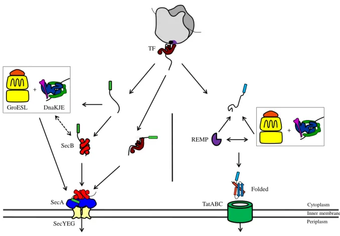
The major TF, DnaKJE and GroESL chaperones participate in the export of proteins by acting directly on substrates of the Sec and Tat (Twin-arginine translocation) general secretion pathways (Graubner et al, 2007; Li et al, 2010; Perez-Rodriguez et al, 2007; Phillips & Silhavy, 1990; Wild et al, 1992; Wild et al, 1996).
1- Sec Translocation system
The Sec translocon, through which substrates will cross the membrane, is composed of a hetero-oligomeric complex mainly formed by SecYEG located in the inner membrane (Van den Berg et al, 2004). SecA is a cytosolic protein promoting the translocation of the substrate through the Sec translocon in an ATP-dependent manner (Schiebel et al, 1991). In E. coli this system is supported by the molecular chaperone SecB. SecB facilitates export of OMPs and some periplasmic proteins which have to be kept unfolded to be exported via the Sec-translocon (Kumamoto & Beckwith, 1985). In agreement with such specific needs, the tetrameric SecB chaperone possesses an anti-folding activity (Collier et al, 1988; Randall & Hardy, 2002). It has been shown that TF, DnaKJE and GroESL play a role during protein export as well (Altman et al, 1991; Calloni et al, 2012; Lecker et al, 1989; Oh et al, 2011; Phillips & Silhavy, 1990; Qi et al, 2002; Ullers et al, 2007; Wild et al, 1992; Wild et al, 1996) (Fig. 7). In addition, functional overlaps and complementarities had been observed between SecB and theses chaperones during protein export. Indeed, both DnaKJE and GroESL facilitate the export of several SecB-dependent substrates and their over-expression efficiently rescue the cold-sensitive phenotype of a secB null mutant (Altman et al, 1991; Phillips & Silhavy, 1990; Qi et al, 2002; Ullers et al, 2007; Wild et al, 1992; Wild et al, 1996). Furthermore, the export of several OMPs following SecA depletion is abolished when DnaK functions are altered (Qi et al, 2002), suggesting that DnaK is able to maintain Sec-dependent OMPs in a translocation-competent state (Qi et al, 2002). In support of such overlaps, both
secB and dnaK mutations are synthetic lethal and a direct interaction between SecB and DnaK
has been recently identified in vivo(Calloni et al, 2012). Remarkably, SecB over-expression also suppresses the growth defect and the severe protein aggregation observed in the combine absence of both TF and DnaK (Ullers et al, 2004). This suppression was independent of SecB export function as judged by the used of the variant E77K impaired in SecA interaction (Fekkes et al, 1998; Kimsey et al, 1995).This indicates that in addition to its role in protein export, SecB can perform generic chaperone functions and facilitate cytosolic protein folding in the absence of TF and DnaK. Finally, such interplays are further supported by the fact that
31
SecB endogenous levels are significantly increased upon depletion of DnaKJE and GroESL, and reciprocally (Muller, 1996).
Although TF binds some OMPs and most likely facilitates their translocation (see Part II), it has been shown that TF can also antagonize the action of both SecB and DnaKJE during protein export, as judged by its ability to severely delay export of some Sec-dependent proteinswhen SecB or DnaKJE are absent (Lee & Bernstein, 2002; Ullers et al, 2007). A model presenting the different chaperone pathways involved in the Sec-dependent protein secretion is show in Fig. 7.
2- Twin Arginin translocation system
The second general secretion pathway in E. coli is the Tat system. The translocon is composed by three membrane proteins of which encoding genes are organized in an operon: genes encoding for TatA, TatB and TatC; another one, isolated, encodes for TatE and is a functional TatA duplication (Sargent et al, 1998). This translocon translocates proteins having the consensus N-terminal motif (S/T)-R-R-X-F-L-K previously folded into the cytoplasm (Berks, 1996). Majority of Tat substrates have their own specific chaperones called REMPs (Redox Enzyme Maturation Proteins) like DmsD, NarJ, NarD or TorD (Turner et al, 2004). They interact specifically with the twin-arginine leader peptide protecting the substrate from degradation, maintaining it in a translocation competent state and targeting it to the Tat translocon (Sargent, 2007; Turner et al, 2004). REMPs are not acting alone and participate in a chaperone cascade also orchestrated by TF, DnaKJE and GroESL (Perez-Rodriguez et al, 2007). Interactions between DmsD and these molecular chaperones were pointed out by several in vitro experiments (Li et al, 2010). TF, DnaKJE and GroESL were shown to interact directly with Tat substrates and to efficiently participate in their translocation (Fig. 7). TF was shown to interact with signal peptides of TorA and SufI until late translation but did not participate actively in their translocation (Jong et al, 2004). DnaK is essential for the translocation of over-expressed CueO, and efficiently binds to the leader peptide of DmsA (Graubner et al, 2007; Oresnik et al, 2001). Moreover, several Tat substrates appear to be in an unstable conformation in the absence of DnaK (Perez-Rodriguez et al, 2007). These data suggest that DnaK may work in cooperation with REMPs to stabilize and protect substrates from degradation. The GroESL interactome revealed the presence of the Tat-dependent amidase AmiA as a GroESL substrate, which required GroESL for its folding (Kerner et al, 2005; Rodrigue et al, 1996).
32 SecB TatABC Folded REMP + Inner membrane + SecA SecYEG Cytoplasm Periplasm DnaKJE GroESL TF
Fig. 7: Chaperone networks involved in protein export. Molecular chaperones play different roles in both systems. In Sec translocation TF, DnaKJE, GroESL, and SecB generally favor secretion by keeping substrates in an unfolded competent state for translocation. The N-terminal Signal sequence of the substrate is represented by a green rectangle. In Tat system, the three major chaperones work with REMPs to fold the substrate and target it to the Tat translocon. The N-terminal Signal sequence of the substrate is represented by a cyan rectangle.
33
Part IV: Chaperone-mediated response to protein misfolding and
aggregation
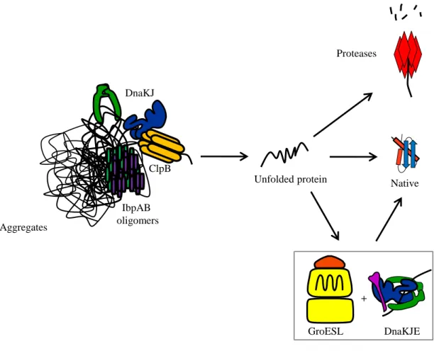
The TF, DnaKJE and GroESL chaperone network is not sufficient to rescue protein aggregation during heat-stress or oxidative protein unfolding. Other heat shock proteins (Hsps) are induced in these conditions to facilitate the action of DnaKJE and most likely GroESL. These chaperones include the Clp superfamily proteins (Hsp100), the small Hsps (sHsps) IbpA and IbpB, HtpG (the Hsp90 homolog) and the redox-regulated chaperone Hsp33 (Hartl, 1996; Hoffmann et al, 2004; Squires & Squires, 1992).
1- Molecular chaperone networks and protein disaggregation
a- Presentation of other chaperones involved in protein disaggregation
Members of the Clp family include protein complexes functioning as disaggregases and/or proteolytic machines with AAA+ ATPases activities (ATPases Associated with a variety of cellular Activities). They can be gathered into two groups depending on the number of Nucleotide Binding Domains (NBD) they possess. Class I AAA+ proteins comprising ClpA, ClpB, ClpC and ClpE monomers have two different NBDs separated by a coiled coil middle domain; Class II proteins with ClpX and ClpY monomers have only one NBD (Schirmer et al, 1996). Some AAA+ proteins possess a protease domain like FtsH and Lon. ClpB (Hsp100) possesses a disaggregase activity forming a barrel-shaped hexamer with an axial channel when it is bound to ATP (Akoev et al, 2004). This channel is surrounded by the two NBDs of ClpB (NBD1 and NBD2), the middle domain (M domain) of the monomer being outside of the cylinder (del Castillo et al, 2011; Lee et al, 2003). The N-terminal domain of each monomer forms a crown involved in the interaction with aggregated proteins (Barnett et al, 2005). Then ATP hydrolysis will mediate disaggregation by ClpB via translocation of the substrate through the channel (Weibezahn et al, 2004). The M domain, which is structured as a coiled coil with two wings, is essential for the disaggregase activity of ClpB (Kedzierska et al, 2003; Lee et al, 2003; Mogk et al, 2003b). It is involved in the stabilization of the hexameric structure of ClpB (del Castillo et al, 2011). Seyffer and co-workers (2012) showed that DnaK could bind the M domain and induce allosteric modifications in ClpB, which, together with substrate interaction, stimulate ClpB ATPase activity (Seyffer et al, 2012). The
E. coliclpB gene possesses an internal translation initiation site expressing two isoforms of
ClpB in vivo: the full length 95kDa ClpB and a truncated isoform of 80kDa without the N-terminal domain (Squires et al, 1991). It has been shown that these two isoforms cooperate to
34
favor solubilization of aggregated proteins and that each isoform alone is less effective in this task (Guenther et al, 2012; Zhang et al, 2012). The N-terminal domain is involved in the interaction with aggregates; this combination of isoforms could give more flexibility at this domain and then facilitate the interaction with substrates (Nagy et al, 2010). The deletion of
clpB in E. coli is known to present a growth defect at 44°C and is also more sensitive to heat
shock at 50°C than the wild type cell (Squires et al, 1991).
The E. coli IbpA and IbpB proteins (Inclusion body protein) are conserved small heat-shock proteins of 16 kDa that share about 50% amino acid sequence identity and that are known to associate with thermally aggregated proteins and inclusion bodies (Allen et al, 1992; Laskowska et al, 1996). These two sHsps are known to form molecular oligomeric structures (Kitagawa et al, 2002; Shearstone & Baneyx, 1999) and cooperate to disaggregate aggregated polypeptides. The ibpAB operon can be deleted with no effect on cell during normal growth conditions but causes a decreased viability during prolonged growth at 50°C (Kuczynska-Wisnik et al, 2002) and as a sensitivity phenotype to copper-induced stress under aerobic conditions (Matuszewska et al, 2008).
A third chaperone known to participate in disaggregation is the E. coli Hsp90 chaperone member named HtpG. It is an ATP-dependent chaperone functioning as a homodimer. HtpG monomer is made up of three domains: an N-terminal domain with ATPase activity, a middle domain and a C-terminal domain involved in Hsp90 dimerization (Krukenberg et al, 2011; Mayer, 2010; Pearl & Prodromou, 2006; Wandinger et al, 2008). HtpG is essential for the activity of the clustered regularly interspaced short palindromic repeats (CRISPR) system(Yosef et al, 2011) involved in the detection and the degradation of exogenous DNA (Barrangou et al, 2007; Brouns et al, 2008; Marraffini & Sontheimer, 2008) and RNA (Hale et al, 2009) in prokaryotes. As for clpB, htpG deletion only shows a growth defect above 44°C (Bardwell & Craig, 1988).
b- Chaperone networks involved in protein disaggregation
In vitro experiments showed that efficient interaction of ClpB with aggregates was
DnaKJE-dependentand thatClpB and DnaKJE systems are strong partners for protein disaggregation, acting as a bi-chaperone system by which the substrate is transferred from DnaKJE to ClpB (Acebron et al, 2009; Goloubinoff et al, 1999; Motohashi et al, 1999; Zolkiewski, 1999). This substrate transfer is mediated by a direct interaction between DnaK and the M-domain of ClpB (Seyffer et al, 2012). In addition it has been shown that DnaJ