HAL Id: hal-03004342
https://hal-cnrs.archives-ouvertes.fr/hal-03004342
Submitted on 20 Nov 2020HAL is a multi-disciplinary open access archive for the deposit and dissemination of sci-entific research documents, whether they are pub-lished or not. The documents may come from teaching and research institutions in France or abroad, or from public or private research centers.
L’archive ouverte pluridisciplinaire HAL, est destinée au dépôt et à la diffusion de documents scientifiques de niveau recherche, publiés ou non, émanant des établissements d’enseignement et de recherche français ou étrangers, des laboratoires publics ou privés.
Crystal structure of the F component of the
Panton-Valentine leucocidin
Jean-Denis Pedelacq, Gilles Prevost, Henri Monteil, Lionel Mourey,
Jean-Pierre Samama
To cite this version:
Jean-Denis Pedelacq, Gilles Prevost, Henri Monteil, Lionel Mourey, Jean-Pierre Samama. Crystal structure of the F component of the Panton-Valentine leucocidin. Journal of Medical Microbiology, Society for General Microbiology, 2000, 290, pp.395 - 401. �hal-03004342�
© Urban & Fischer Verlag
http://www.urbanfischer.de/journals/ijmm
IJMM
Crystal structure of the F component of the Panton-Valentine leucocidin
Jean-Denis Pedelacq
1,Gilles Prevost
2,Henri Monteil
2,Lionel Mourey
1,Jean-Pierre Samama
11 Groupe de Cristallographie Biologique, Institut de Pharmacologie et de Biologie Structurale du CNRS, 205 route de Narbonne, 31077 Toulouse cedex, France
2 Laboratoire de Toxinologie et d' Antibiologie Bacteriennes, Institut de Bacteriologie de la Faculte de Medecine, 3 rue Koeberle, 67000 Strasbourg, France
Abstract
Leucocidins and y-hemolysins are bi-component staphylococcal toxins that form lytic transmem-brane pores. Their cytotoxic activities involve the synergistic association of a class S and a class F component, produced as water-soluble monomers which assemble on the surface of specific cells. The structure of the F protein from Panton-Valentine leucocidin, solved at 2.0
A
resolution, and se-quence alignment suggest that it represents the fold of any secreted protein in this family of toxins. The comparison of this structure to that of the homoheptameric a-hemolysin provides some insights into the molecular events that may occur during pore formation.Key words: crystal structure - Panton-Valentine leucocidin -lytic transmembrane pores
Introduction
Staphylococcus aureus, one of the most prevalent
hu-man pathogens isolated in hospitals, produces a host of toxins that includes a-hemolysin (a-HL), y-hemolysins (Hlg), and leucocidins (Luk) (for a review, see (Tomita and Kamio, 1997)). These toxins are secreted as water-soluble protein monomers which assemble on the sur-face of host cells to form oligomeric transmembrane channels (Finck-Barban<;on et al., 1993; Sugawara et al., 1997). Hlg and Luk, but not a-HL, form the fam-ily of staphylococcal bi-component leucotoxins (Pre-vost et al., 1995). Their toxicity involves the synergis-tic action of a class S and a class F component (Woo-din, 1960) and is essentially directed against host de-fence cells, e. g. polymorphonuclear cells, monocytes, macrophages, and erythrocytes.
Severalleucocidins and y-hemolysins have been iso-lated from various S. au reus strains and from S.
inter-medius (Cooney et al., 1993; Rahman et al., 1993;
Ka-neko et al., 1997; Prevost, 1999). Among these toxins,
the Panton-Valentine leucocidin (PVL) was the first to be reported and characterized (Panton and Valentine, 1932; Woodin, 1960). PVL-producing strains are asso-ciated with primary cutaneous infections such as fu-runcles (Prevost, 1999), and the toxin was shown to be highly active on human and rabbit polymorphonucle-ar leucocytes (Finck-Bpolymorphonucle-arban<;on et al., 1993; Prevost et al., 1995).
The first event of the mode of action of PVL is the interaction of the S component (LukS-PV) (Colin et al., 1994) with a membrane-bound target, followed by the binding of LukF-PV (Woodin and Wieneke, 1968; Noda et al., 1981). These sequential interactions at the membrane surface induce a Ca2+ influx through the opening of Ca2+ channels (Staali et al., 1998), the acti-vation of the targeted cells (Konig et al., 1995), and the formation of pores which are specific for the traffic of monovalent cations (Finck-Barban<;on et al., 1993; Meunier et al., 1995; Staali et al., 1998). Biochemical and biophysical studies indicated that bi-component toxins form membrane-attached heteromers and
Corresponding author: Jean-Pierre Samama, Groupe de Cristallographie Biologique, Institut de Pharmacologie et de Biologie Structurale du CNRS, 205 route de Narbonne, F-31 077 Toulouse cede x, France, Phone: +33 5 61 17 54 44, Fax: +33 5 61 17 54 48, E-mail: [email protected]
396
J.
D. Pedelacq et al.argued for hexameric assemblies contalOlOg similar amounts of F (MW = 34 kDa) and 5 (MW = 32 kDa) components (5ugawara et al., 1997; Ferreras et al., 1998).
Our goal was to describe the molecular species at the first step of pore formation, i. e. as the monomeric form that approaches the membrane surface. We have crys-tallized the secreted LukF-PV (301 residues) and solved its crystallographic structure at 2.0 A resolution. This study shows the conservation of the core domains between the Luk, Hlg, and a-HL (50ng et al., 1996) proteins, and illustrates the fold of the stem domain of these proteins prior to membrane insertion.
Materials and methods
Methods used for the structure determination of LukF-PV have been described recently (Pedelacq et al., 1999). Briefly, the protein was purified from the reference S. aureus
ATCC49775 using Fast Performance Liquid Chromatogra-phy (Finck-Barbanc;:on et al., 1991) and crystallized at 4°C using the hanging drop diffusion method in the presence of PEG and divalent cations at neutral pH. The structure was determined using the Multiwavelength Anomalous Disper-sion (MAD) method on a single crystal soaked with an irid-ium derivative, and refined at 2.0 A resolution.
Results and discussion
Crystallographic studies
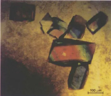
Two crystal forms of LukF-PV were obtained (Table 1) and in both cases, crystal growth required the design of appropriate seeding protocols. The orthorhombic crystals (Figure 1) diffracted to higher resolution and were used for structure determination. 50lving the
Table 1. Characteristics of the two crystal forms of LukF-PV.
Crystal form Plate (trigonal) Bar (orthorhombic) Crystallization PEG40008% PEG4000 21 % conditions Hepes, Tris 80 mM MES, Tris 200 mM (equilibrium) pH 7.5 pH 6.8-7.0
MgCI2 53 mM CdCll 7 mM
Space group P3121 P212121
Cell parameters (A) a = 50.6, b = 50.6, a=50.7, b=73.3,
c=247.7 c = 99.7
W molecules/a. u.1 1 1
Diffraction limit (Al2 3.2 1.8
1 asymmetric unit.
1 Determined using synchrotron radiation. Recently, a diffraction limit of 1.2
A
was attained with orthorhombic crystals on beam line ID14-EH4 at ESRF (Grenoble, France).
1100 ~m I Fig.1. Orthorhombic crystals of LukF-PV.
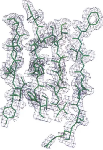
phase problem by the multiple isomorphous replace-ment (MIR) method was impaired by severe non iso-morphism upon heavy atom binding. We therefore per-formed a MAD experiment from a single crystal soaked in a solution of the hexachloroiridate salt (NH4hlrCI6• The data were collected to 2.0 A resolu-tion at the EMBL Hamburg outstaresolu-tion beam line X31. The intensities were measured at four wavelengths which were chosen according to the fluorescence spec-trum of the iridium derivative in the cryocooled crystal (Pedelacq et al., 1999). MAD phasing using seven heavy-atom sites, followed by solvent flattening, led to an initial electron density map of high quality (fig-ure 2) which allowed tracing of most of the LukF-PV molecule. 5tructure refinement was conducted between 24.4 and 2.0 A. The final crystallographic Rand Rffee
values were 0.20 and 0.24, respectively. Overall structure
LukF-PV is a global protein of ellipsoidal shape with overall dimensions 75A X 30A X 20A. Most residues are engaged in secondary structure elements. There are 22 ~ strands and three short helices which involve 64.4 % and 5.3 % of protein residues, respectively. These secondary structure elements delineate three structural domains: the ~-sandwich, the rim, and the folded stem (Figure 3). The ~-sandwich domain (resi-dues 1-61, 80-102, 154-169, 219-249, and 268-301) is made of two six-stranded antiparallel ~-sheets
(strands 51, 52, 53, 54, 56, and 55, and strands 5D, 5E, SA, SF, 5G, and SH) facing each other. The rim domain (residues 62-79, 170-218, 250-267) forms an anti-parallel four-stranded open-face sandwich (strands
SRI, SR2, SF, and SR5) topped by residues 182-218. This stretch can be described as two consecutive ome-ga loops followed by all the helical segments (HI, H2, H3) found in LukF-PV. The folded stem region, made of residues 106-148, includes three antiparallel ~
strands (STl, ST2, ST3) linked by one ~-turn and one right-handed crossover connection. The junction of the stem domain to the ~-sandwich domain is provided by two short antiparallel strands SB (l 03-1 05) and SC (149-153).
The structure of LukF from the Smith 5R strain of
s.
aureus, was reported recently (Olson et aI., 1999). Itdisplays a similar architecture to that of LukF-PV. Structural conservation among staphylococcal pore-forming toxins
The core of LukF-PV, made of the ~-sandwich and of the rim domains, is similar to that of the a-HL proto-mer (Song et aI., 1996; Gouaux et aI., 1997). Superpo-sition of the two protein structures can be achieved us-ing either the sandwich or the rim domains (Figure 4). These domains undergo rigid bodies movement and
Fig. 2. The initial electron density map, computed between 24.0 and 2.0
A
and contoured at 1.00, displayed around a portion of the final model of LukF-Pv.Fig. 3. Ribbon representation of LukF-PV with the ~-sandwich, rim and stem domains colored in cyan, violet and orange, respectively.
adopt different relative orientations in the water-soluble monomeric protein (LukF-PV) and the assem-bled toxin (a-HL).
Based on this superimposition, a sequence alignment of the two proteins was performed. It was extended to all class F ('" 70 % identity) and class S (59-75 % iden-tity) proteins. These two protein classes only present 26-30 % identity between each other. The multiple sequence alignment revealed the conservation of sever-al residues that play an important structursever-al role (Table 2). The side-chains of residues from a first group (Ile59, Tyr82, Tyr99, Pr0101, Tyr149, Leu153, Trp164, Leu216, Phe221, Pr0223, Phe225, and Tyr245) form a continuous hydrophobic patch which involves all the strands in the ~-sandwich domain, ex-cept SI , S2, S3, SG and SH. However, the strands S2, S3, and S4 are held together by hydrogen bond inter-actions involving the main chain oxygen atoms of res-idues 30 and 60, and the invariant Arg247. A second group of residues was identified. The side-chains of Phe76, Trp78 , Metl92, Phe193, Phe207, Asp250, Tyr252, and Asn265, located at the bottom of the rim domain, provide a number of hydrophobic contacts
398
J.
D. Pedelacq et al.and polar interactions. The conservation of the resi-dues from both groups and of their interactions might be regarded as folding determinants of the ~-sandwich
and the rim domains.
Transmembrane
Stem
~-Sandwich
Fig. 4. Superposition (j3-sandwich domains) of LukF-PV (colored as in Figure 3) and one proto mer of a-hemolysin (black). The rotation axis and the angular range required to fit the rim domains are depicted in red.Conformational changes in the stem region during assembly
Pore formation is commonly described as a four-step process involving the water-soluble secreted form, the membrane-bound monomer, an oligomeric pre-pore, and finally, the transmembrane pore (Walker et ai., 1992; Valeva et ai., 1997). The three-dimensional structures of LukF-PV and of the a-HL homoheptamer illustrate the molecular species at the first and at the last step of this process. The stem region, also known as the glycine-rich stem, forms two 55 A long anti-parallel ~-strands in each pore-forming a-HL proto-mer. These strands protrude from the protein core (Fig-ure 4) and constitute one building unit of the membrane-spanning 14-strand ~-barrel (Song et ai., 1996). In contrast, the stem region of the water-soluble LukF-PV forms a three-stranded antiparallel ~-sheet
and constitutes the third domain of the protein. It is packed onto the convex face of the ~-sandwich domain (Figure 3). The resulting stem/~-sandwich interface is mainly stabilized through van der Waals interactions between hydrophobic residues. In addition, this inter-face is tightened at each side by a set of polar interac-tions. At one edge, the side-chain of Asp43 (strand S3 of the ~-sandwich domain) is at hydrogen bond dis-tance to the main chain oxygen atom of residue 119 and to the phenolic group of Tyr116 (strand ST1 of the stem). At the other edge of the interface, hydrogen bonds are exchanged between the side-chains of Thr151 (strand SC) and Tyr144 and Gln146 (strand ST3 from the folded stem). The invariance of these res-idues in a-HL and class F, class S components, suggests that the fold of the stem domain should be similar in all the secreted monomeric water-soluble proteins.
A search for structural homology in the Protein Data Bank revealed a significant similarity of the stem do-main with toxins isolated from snake venom, despite very weak sequence homology. The highest structural similarity was displayed by erabutoxin A (PDB code
Table 2. Residues involved in the conservation of the core domains within the superfamily of staphylococcal pore-forming toxins.
~-sandwich Residue No 59 82 99 101 149 153 164 216 221 223 225 245 Conservation 1 1 C C C C C 1 1 1 1 1 1 Location 54 5A 55 S5 5C 5C 5E H3 56 56 56 5F Rim Residue No 76 78 192 193 207 250 252 265 Conservation 1 C I C I 1 I C C Location 5R2 5R2 2 2 2 5F 5F 5R5 1 I, invariant; C, conserved.
5EBX) and toxin y (1 TGX), a short a-neurotoxin and a cardiotoxin found in the venoms of sea snake Lati-cauda semifasciata and of the cobra Naja nigricollis,
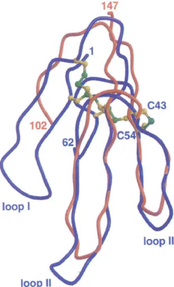
respectively. The molecular architecture of these 60-70 amino-acid proteins is known as the three finger fold (Rees and Bilwes, 1993). The three central ~-strands,
and loops II and III of these toxins, whose conforma-tion is constrained by four disulfide bridges, encom-pass the fold of the stem in LukF-PV (Figure 5). The disulfide bond found between Cys 43 and Cys 54 in erabutoxin A, anchors loop III to the core of the mole-cule. In LukF-PV, there is no structural constraint that could stabilize the corresponding long solvent-exposed loop of the glycine-rich stem (residues 127-137 between strands ST2 and ST3). The occurrence of five glycines in this peptide stretch (over the eight found between residues 102 and 147) may confer important conformational flexibility. Indeed, Ser129 and the three consecutive glycine residues display the highest temperature factors in the structure and no clear
elec-147
loop III
Fig. 5. The stem domain of LukF-PV (orange) superimposed on the three-dimensional structure of erabutoxin A (blue),
tron density could be assigned to residues 133-135. Flexibility of the polypeptide chain in this region should be invoked since SDS-PAGE electrophoresis of dissolved crystals only revealed a single protein band corresponding to the full-length protein.
Erabutoxin is a postsynaptic curare-mimetic protein which binds specifically to peripheral nicotinic acetyl-choline receptors, thus preventing the binding of ace-tylcholine. Cardiotoxins (Kumar et aI., 1997) are also called cytotoxins and cause heart failure. They have numerous pharmacological properties and exhibit a large spectrum of biological activities. Their targets, unlike those of neurotoxins, and mode of interaction with membranes are not well understood. However, it has been established that cardiotoxins elicit biological response from cells by primarily binding to the mem-brane surface owing to a pattern of hydrophobic and basic residues. The possible functional significance of the fold similarity between these toxins and the stem of LukF-PV, with respect to protein-membrane interac-tions, remains to be documented.
Functional implications
Pore formation with staphylococcal toxins requires un-folding of the stem region. The differences in solvent accessibilities of the residues of this domain in its folded and extended conformation, indicate that the side-chains which will be oriented toward the non polar part of the lipid bilayer are, for most of them, in-volved in the interface between the stem and the ~
sandwich domains. The protein-membrane interac-tions likely promote the drastic conformational transi-tions leading to unfolding of the stem domain and to the formation of significant protomer-protomer inter-faces in the oligomeric species (Song et aI., 1996).
A significant structural difference between LukF-PV and the a-HL protomer was observed in their N-terminal regions. In monomeric LukF-PV, this region constitutes the outermost external strand (S1) of the ~-sandwich domain. In contrast, the N-terminallatch (residues 1-16) of a-HL in the heptameric assembly has no defined secondary structure and forms numer-ous interactions with adjacent protomers within the lumen of the cap domain (Song et aI., 1996). It was suggested that the transition from the soluble mono-meric form of a-HL to the pore-forming heptamono-meric structure was related to a conformational change of the N-terminal region (Cheley et aI., 1997). Gouaux and coworkers proposed that the stem is still folded in the heptameric pre-pore intermediate, and that its inser-tion into the membrane may occur with the concomi-tant rearrangement of the N-terminal region in the cap domain (Olson et aI., 1999). According to the structure of LukF-PV, this process would imply that strand S1
400
J.
D. Pedelacq et al.dissociates from its antiparallel strand S2. We have ex-amined the possibility that the F and S components may form homo- or heteroheptameric pores similar to that of a-HL. Our modeling approaches revealed that the formation of these heptamers was unlikely (Pede-lacq et al., 1999), which would agree with the pro-posal that the oligomeric bi-component toxins contain equimolar proportions of F and S components in hexa-meric assemblies (Sugawara et al., 1997; Ferreras et al., 1998). Based on a canonical 12-stranded antiparallel
~-barrel (Sansom and Kerr, 1995), a heterohexamer of PVL was modeled without altering the position of strand Sl (Pedelacq et al., 1999). Its topological and geometrical features underlined protomer inter-actions involving the folded N-terminal strand. The inner diameter of 21
A
is in good agreement with the experimental data on y-hemolysin (Sugawara et al., 1997) and PVL (Colin et al., 1997).References
Cheley, S., Malghani, M. S., Song, L., Hobaugh, M., Gouaux, ]. E., Yang, ]., Bayley, H.: Spontaneous oligo-merization of a staphylococcal a-hemolysin conforma-tionally constrained by removal of residues that form the transmembrane ~-barrel. Protein Eng. 10, 1433-1443 (1997).
Colin, D. A., Mazurier, I., Sire, S., Finck-Barban<;:on, V.: Interaction of the two components of leukocidin from
Staphylococcus aureus with human polymorphonuclear leukocyte membranes: sequential binding and subsequent activation. Infect. Immun. 62, 3184-3188 (1994). Colin, D. A., Meunier, 0., Staali, L., Prevost, G., Monteil,
H.: Bi-component leukotoxins from Staphylococcus au-reus. In: Cold Spring Harbor Laboratory Symposium on Microbial Pathogenesis and Host Response (Maloy et aI., eds.), pp.150. Cold Spring Harbor Laboratory Press, Cold Spring Harbor, New York 1997.
Cooney, j., Kienle, Z., Foster, T.]., O'Toole, P. W.: The gamma-hemolysin locus of Staphylococcus au reus com-prises three linked genes, two of which are identical to the genes for the F and S components of leukocidin. Infect. Immun. 61, 768-771 (1993).
Ferreras, M., Hoper, E, Serra, M. D., Colin, D. A., Prevost, G., Menestrina, G.: The interaction of Staphylococcus aureus bi-component y-hemolysins and leucocidins with cells and lipid membranes. Biochim. Biophys. Acta 1414, 108-126 (1998).
Finck-Barban<;:on, V., Duportail, G., Meunier, 0., Colin, D. A.: Pore formation by a two-component leukocidin from
Staphylococcus aureus within the membrane of human polymorphonuclear leukocytes. Biochim. Biophys. Acta 1182,275-282 (1993).
Finck-Barban<;:on,
v.,
Prevost, G., Piemont, Y.: Improved pur-ification of leukocidin from Staphylococcus aureus and toxin distribution among hospital strains. Res. Microbiol. 142,75-85 (1991).Gouaux, E., Hobaugh, M., Song, L.: a-Hemolysin, y-hemo-lysin, and leukocidin from Staphylococcus aureus: distant in sequence but similar in structure. Protein Sci. 12, 2631- 2635 (1997).
Kaneko,
J.,
Muramoto, K., Kamio, Y.: Gene of LukF-PV-like component of Panton-Valentine leukocidin in Staphylo-coccus aureus P83 is linked with lukM. Biosci. Biotech. Biochem. 61, 541-544 (1997).Konig, B., Prevost, G., Piemont, Y., Konig, W.: Effects of
Staphylococcus aureus leukocidins on inflammatory me-diator release from human granulocytes. j. Infect. Dis. 171,607-613 (1995).
Kumar, T. K. S., ]ayaraman, G., Lee, C. S., Arunkumar, A.I., Sivaraman, T., Samuel, D., Yu,
c.:
Snake venom cardio-toxins-structure, dynamics, function and folding. ]. Bio-mol. Struct. Dyn. 15,431-463 (1997).Meunier, 0., Falkenrodt, A., Monteil, H., Colin, D. A.: Appli-cation of flow cywmetry in toxinology: pathophysiology of human polymorphonuclear leukocytes damaged by a pore-forming toxin from Staphylococcus au reus. Cytome-try 21, 241-247 (1995).
Noda, M., Kato, I., Matsuda, E, Hirayama, T.: Mode of ac-tion of staphylococcal leucocidin: relaac-tionship between binding of 12sI-labeled Sand F components of leucocidin to rabbit polymorphonuclear leukocytes and leucocidin activity. Infect. Immun. 34, 362-367 (1981).
Olson, R., Nariya, H., Yokota, K., Kamio, Y., Gouaux, E.: Crystal structure of staphylococcal LukF delineates con-formational changes accompanying formation of a trans-membrane channel. Nature Struct. BioI. 6, 134-140 (1999).
Panton, P. N., Valentine, E C. 0.: Staphylococcal toxin. Lan-cet 222,506-508 (1932).
Pedelacq,j. D., Maveyraud, L., Prevost, G., Baba-Moussa, L.,
Gonzalez, A., Courcelle, E., Shepard, W., Monteil, H., Samama, j. P., Mourey, L.: The structure of a Staphylococ-cus aureus leucocidin component (LukF-PV) reveals the fold of the water-soluble species of a family of transmem-brane pore-forming toxins. Structure 7, 277-287 (1999). Prevost, G.: The bi-component staphylococcal leucocidins
and y-hemolysins. In: The comprehensive sourcebook of bacterial protein toxins (J. E. Alouf,
J.
H. Freer, eds.), pp. 402-418. Academic Press, London 1999.Prevost, G., Cribier, B., Couppie, P., Petiau, P., Supersac, G., Finck-Barban<;:on,
v.,
Monteil, H., Pie mont, Y.: Panton-Valentine leucocidin and gamma-hemolysin from Staphy-lococcus au reus ATCC 49775 are encoded by distinct ge-netic loci and have different biological activities. Infect. Immun. 63,4121-4129 (1995).Rahman, A., Izaki, K., Kamio, Y.: Gamma-hemolysin genes in the same family with lukF and lukS genes in methicillin re-sistant Staphylococcus aureus. Biosci. Biotech. Biochem. 57,1234-1236 (1993).
Rees, B., Bilwes, A.: Three-dimensional structures of neuro-toxins and cardioneuro-toxins. Chern. Res. Toxicol. 6, 385-406 (1993).
Sansom, M. S., Kerr, I. D.: Transbilayer pores formed by beta-barrels: molecular modeling of pore structures and prop-erties. Biophys.j. 69,1334-1343 (1995).
Song, L., Hobaugh, M. R., Shustak,
c.,
Cheley, S., Bayley, H., Gouaux, j. E.: Structure of staphylococcalalpha-hemo-lysin, a heptameric transmembrane pore. Science 274, 1859-1866 (1996).
Staali, L., Monteil, H., Colin, D. A.: The staphylococcal pore-forming leukotoxins open Ca2+ channels in the
membrane of human polymorphonuclear neutrophils.
J.
Membr. BioI. 162,209-216 (1998).Sugawara, N., Tomita, T., Kamio, Y.: Assembly of
Staphylo-coccus aureus y-hemolysin into a pore-forming
ring-shaped complex on the surface of human erythrocytes. FEBS Lett. 410, 333-337 (1997).
Tomita, T., Kamio, Y.: Molecular biology of the pore-forming cytolysins from Staphylococcus aureus, u- and y-hemolysins and leukocidin. Biosci. Biotech. Biochem. 61, 565-572 (1997).
Valeva, A., Palmer, M., Bhakdi, S.: Staphylococcal alpha-toxin: formation of the heptameric pore is partially coop-erative and proceeds through multiple intermediate stages. Biochemistry 36,13298-13304 (1997).
Walker, B., Krishnasastry, M., Zorn, L., Bayley, H.: Assem-bly of the oligomeric membrane pore formed by staphy-lococcal alpha-hemolysin examined by truncation muta-genesis.
J.
BioI. Chern. 267, 21782-21786 (1992).Woodin, A. M.: Purification of the two components of leu-cocidin from Staphylococcus aureus. Biochem.
J.
75, 158-165 (1960).Woodin, A. M., Wieneke, A. A.: The cation-sensitive phos-phatases of the leucocyte cell membrane. Biochem. Biophys. Res. Commun. 33, 558-562 (1968).