2014 ESC/ESA Guidelines on non-cardiac surgery:
cardiovascular assessment and management
The Joint Task Force on non-cardiac surgery: cardiovascular
assessment and management of the European Society of Cardiology
(ESC) and the European Society of Anaesthesiology (ESA)
Authors/Task Force Members: Steen Dalby Kristensen
*
(Chairperson) (Denmark),
Juhani Knuuti
*
(Chairperson) (Finland), Antti Saraste (Finland), Stefan Anker
(Germany), Hans Erik Bøtker (Denmark), Stefan De Hert (Belgium), Ian Ford (UK),
Jose Ramo´n Gonzalez-Juanatey (Spain), Bulent Gorenek (Turkey),
Guy Robert Heyndrickx (Belgium), Andreas Hoeft (Germany), Kurt Huber (Austria),
Bernard Iung (France), Keld Per Kjeldsen (Denmark), Dan Longrois (France),
Thomas F. Lu¨scher (Switzerland), Luc Pierard (Belgium), Stuart Pocock (UK),
Susanna Price (UK), Marco Roffi (Switzerland), Per Anton Sirnes (Norway),
Miguel Sousa-Uva (Portugal), Vasilis Voudris (Greece), Christian Funck-Brentano
(France).
ESC Committee for Practice Guidelines: Jose Luis Zamorano (Chairperson) (Spain), Stephan Achenbach (Germany), Helmut Baumgartner (Germany), Jeroen J. Bax (Netherlands), He´ctor Bueno (Spain), Veronica Dean (France), Christi Deaton (UK), Cetin Erol (Turkey), Robert Fagard (Belgium), Roberto Ferrari (Italy), David Hasdai (Israel), Arno W. Hoes (Netherlands), Paulus Kirchhof (Germany/UK), Juhani Knuuti (Finland), Philippe Kolh (Belgium), Patrizio Lancellotti (Belgium), Ales Linhart (Czech Republic), Petros Nihoyannopoulos (UK), Massimo F. Piepoli (Italy), Piotr Ponikowski (Poland), Per Anton Sirnes (Norway), Juan Luis Tamargo (Spain), Michal Tendera (Poland), Adam Torbicki (Poland), William Wijns (Belgium), Stephan Windecker (Switzerland).
ESA Clinical Guidelines Committee: Maurizio Solca (Chairperson) (Italy), Jean-Franc¸ois Brichant (Belgium), Stefan De Herta, (Belgium), Edoardo de Robertisb, (Italy), Dan Longroisc, (France), Sibylle Kozek Langenecker (Austria), Josef Wichelewski (Israel).
*Corresponding authors: Steen Dalby Kristensen, Dept. of Cardiology, Aarhus University Hospital Skejby, Brendstrupgardsvej, 8200 Aarhus Denmark. Tel: +45 78452030;
Fax:+45 78452260; Email:[email protected].
Juhani Knuuti, Turku University Hospital, Kiinamyllynkatu 4 – 8, P.O. Box 52, FI-20521 Turku Finland. Tel:+358 2 313 2842; Fax: +358 2 231 8191; Email:[email protected]
The content of these European Society of Cardiology (ESC) Guidelines has been published for personal and educational use only. No commercial use is authorized. No part of the ESC Guidelines may be translated or reproduced in any form without written permission from the ESC. Permission can be obtained upon submission of a written request to Oxford University Press, the publisher of the European Heart Journal and the party authorized to handle such permissions on behalf of the ESC.
Other ESC entities having participated in the development of this document:
ESC Associations: Acute Cardiovascular Care Association (ACCA); European Association for Cardiovascular Prevention & Rehabilitation (EACPR); European Association of Cardiovas-cular Imaging (EACVI); European Association of Percutaneous CardiovasCardiovas-cular Interventions (EAPCI); European Heart Rhythm Association (EHRA); Heart Failure Association (HFA). ESC Councils: Council for Cardiology Practice (CCP); Council on Cardiovascular Primary Care (CCPC).
ESC Working Groups: Cardiovascular Pharmacology and Drug Therapy; Cardiovascular Surgery; Hypertension and the Heart; Nuclear Cardiology and Cardiac Computed Tomography; Thrombosis; Valvular Heart Disease.
Disclaimer. The ESC Guidelines represent the views of the ESC and were produced after careful consideration of the scientific and medical knowledge and the evidence available at the time of their dating. The ESC is not responsible in the event of any contradiction, discrepancy and/or ambiguity between the ESC Guidelines and any other official recommendations or guidelines issued by the relevant public health authorities, in particular in relation to good use of healthcare or therapeutic strategies. Health professionals are encouraged to take the ESC Guidelines fully into account when exercising their clinical judgment as well as in the determination and the implementation of preventive, diagnostic or therapeutic medical strategies; however, the ESC Guidelines do not override, in any way whatsoever, the individual responsibility of health professionals to make appropriate and accurate decisions in consideration of the condition of each patient’s health and in consultation with that patient and, where appropriate and/or necessary, the patient’s caregiver. Nor do the ESC Guidelines exempt health profes-sionals from taking full and careful consideration of the relevant official updated recommendations or guidelines issued by competent public health authorities in order to manage each patient’s case in the light of the scientifically accepted data pursuant to their respective ethical and professional obligations. It is also the health professional’s responsibility to verify the applicable rules and regulations relating to drugs and medical devices at the time of prescription.
&The European Society of Cardiology 2014. All rights reserved. For permissions please email: [email protected]. doi:10.1093/eurheartj/ehu282
by guest on December 11, 2014
Document Reviewers: Massimo F. Piepoli (Review co-ordinator) (Italy), William Wijns (Review co-ordinator) (Belgium), Stefan Agewall (Norway), Claudio Ceconi (Italy), Antonio Coca (Spain), Ugo Corra` (Italy), Raffaele De Caterina (Italy), Carlo Di Mario (UK), Thor Edvardsen (Norway), Robert Fagard (Belgium), Giuseppe Germano (Italy), Fabio Guarracino (Italy), Arno Hoes (Netherlands), Torben Joergensen (Denmark), Peter Ju¨ni (Switzerland), Pedro Marques-Vidal (Switzerland), Christian Mueller (Switzerland), O¨ ztekin Oto (Turkey), Philippe Pibarot (Canada), Piotr Ponikowski (Poland), Olav FM Sellevold (Norway), Filippos Triposkiadis (Greece), Stephan Windecker (Switzerland), Patrick Wouters (Belgium).
ESC National Cardiac Societies document reviewers listed in appendix.
The disclosure forms of the authors and reviewers are available on the ESC websitewww.escardio.org/guidelines a
Scientific Committee Chairperson & ESA Board Representative;b
NASC Chairperson; andc
EBA/UEMS representative Online publish-ahead-of-print 1 August 2014
See page 2342 for the editorial comment on this article (doi:10.1093/eurheartj/ehu295)
-Keywords Guidelines † Non-cardiac surgery † Pre-operative cardiac risk assessment † Pre-operative cardiac testing † Pre-operative coronary artery revascularization † Perioperative cardiac management † Anti-thrombotic therapy † Beta-blockers † Valvular disease † Arrhythmias † Heart failure † Renal disease † Pulmonary disease † Cerebrovascular disease † Anaesthesiology † Post-operative cardiac surveillance
Table of Contents
Abbreviations and acronyms . . . .2385
1. Preamble . . . .2386
2. Introduction . . . .2387
2.1 The magnitude of the problem . . . .2387
2.2 Change in demographics . . . .2387
2.3 Purpose and organization . . . .2387
3. Pre-operative evaluation . . . .2389
3.1 Surgical risk for cardiac events . . . .2389
3.2 Type of surgery . . . .2389
3.2.1 Endovascular vs. open vascular procedures . . . .2389
3.2.2 Open vs. laparoscopic or thoracoscopic procedures. .2390 3.3 Functional capacity. . . .2390
3.4 Risk indices . . . .2391
3.5 Biomarkers . . . .2392
3.6 Non-invasive testing . . . .2392
3.6.1 Non-invasive testing of cardiac disease . . . .2393
3.6.2 Non-invasive testing of ischaemic heart disease. . . .2393
3.7 Invasive coronary angiography . . . .2395
4. Risk-reduction strategies . . . .2395
4.1 Pharmacological . . . .2395
4.1.1 Beta-blockers . . . .2395
4.1.2 Statins . . . .2398
4.1.3 Nitrates . . . .2398
4.1.4 Angiotensin-converting enzyme inhibitors and angiotensin-receptor blockers . . . .2398
4.1.5 Calcium channel blockers . . . .2399
4.1.6 Alpha2receptor agonists . . . .2399
4.1.7 Diuretics . . . .2399
4.2 Perioperative management in patients on anti-platelet agents . . . .2400
4.2.1 Aspirin . . . .2400
4.2.2 Dual anti-platelet therapy . . . .2400
4.2.3 Reversal of anti-platelet therapy . . . .2401
4.3 Perioperative management in patients on anticoagulants . .2401
4.3.1 Vitamin K antagonists . . . .2401
4.3.2 Non-vitamin K antagonist oral anticoagulants . . . . .2402
4.3.3 Reversal of anticoagulant therapy . . . .2402
4.4 Revascularization. . . .2403
4.4.1 Prophylactic revascularization in patients with asymptomatic or stable ischaemic heart disease . . . .2404
4.4.2 Type of prophylactic revascularization in patients with stable ischaemic heart disease . . . .2405
4.4.3 Revascularization in patients with non-ST-elevation acute coronary syndrome . . . .2405
5. Specific diseases . . . .2406
5.1 Chronic heart failure . . . .2406
5.2 Arterial hypertension . . . .2408
5.3 Valvular heart disease . . . .2408
5.3.1 Patient evaluation . . . .2408
5.3.2 Aortic stenosis . . . .2408
5.3.3 Mitral stenosis. . . .2409
5.3.4 Primary aortic regurgitation and mitral regurgitation 2409 5.3.5 Secondary mitral regurgitation . . . .2409
5.3.6 Patients with prosthetic valve(s) . . . .2409
5.3.7 Prophylaxis of infective endocarditis. . . .2409
5.4 Arrhythmias . . . .2410
5.4.1 New-onset ventricular arrhythmias in the pre-operative period . . . .2410
5.4.2 Management of supraventricular arrhythmias and atrial fibrillation in the pre-operative period. . . .2410
5.4.3 Perioperative bradyarrhythmias. . . .2411
5.4.4 Perioperative management of patients with pacemaker/implantable cardioverter defibrillator . . . .2411
5.5 Renal disease . . . .2411
5.6 Cerebrovascular disease . . . .2413
5.7 Peripheral artery disease . . . .2414
5.8 Pulmonary disease . . . .2415
5.9 Congenital heart disease . . . .2416
6. Perioperative monitoring . . . .2416
6.1 Electrocardiography . . . .2416
6.2 Transoesophageal echocardiography . . . .2417
6.3 Right heart catheterization. . . .2418
by guest on December 11, 2014
6.4 Disturbed glucose metabolism . . . .2418
6.5 Anaemia . . . .2419
7. Anaesthesia. . . .2419
7.1 Intra-operative anaesthetic management . . . .2420
7.2 Neuraxial techniques . . . .2420
7.3 Perioperative goal-directed therapy . . . .2420
7.4 Risk stratification after surgery . . . .2421
7.5 Early diagnosis of post-operative complications . . . .2421
7.6 Post-operative pain management. . . .2421
8. Gaps in evidence . . . .2422
9. Summary . . . .2422
10. Appendix . . . .2425
References . . . .2425
Abbreviations and acronyms
AAA abdominal aortic aneurysm
ACEI angiotensin converting enzyme inhibitor
ACS acute coronary syndromes
AF atrial fibrillation
AKI acute kidney injury
AKIN Acute Kidney Injury Network
ARB angiotensin receptor blocker
ASA American Society of Anesthesiologists
b.i.d. bis in diem (twice daily)
BBSA Beta-Blocker in Spinal Anesthesia
BMS bare-metal stent
BNP B-type natriuretic peptide
bpm beats per minute
CABG coronary artery bypass graft
CAD coronary artery disease
CARP Coronary Artery Revascularization Prophylaxis
CAS carotid artery stenting
CASS Coronary Artery Surgery Study
CEA carotid endarterectomy
CHA2DS2-VASc cardiac failure, hypertension, age≥75 (doubled), dia-betes, stroke (doubled)-vascular disease, age 65 – 74 and sex category (female)
CI confidence interval
CI-AKI contrast-induced acute kidney injury
CKD chronic kidney disease
CKD-EPI Chronic Kidney Disease Epidemiology Collaboration
Cmax maximum concentration
CMR cardiovascular magnetic resonance
COPD chronic obstructive pulmonary disease
CPG Committee for Practice Guidelines
CPX/CPET cardiopulmonary exercise test
CRP C-reactive protein
CRT cardiac resynchronization therapy
CRT-D cardiac resynchronization therapy defibrillator
CT computed tomography
cTnI cardiac troponin I
cTnT cardiac troponin T
CVD cardiovascular disease
CYP3a4 cytochrome P3a4 enzyme
DAPT dual anti-platelet therapy
DECREASE Dutch Echocardiographic Cardiac Risk Evaluation Apply-ing Stress Echocardiography
DES drug-eluting stent
DIPOM DIabetic Post-Operative Mortality and Morbidity
DSE dobutamine stress echocardiography
ECG
electrocardiography/electrocardiographically/electro-cardiogram
eGFR estimated glomerular filtration rate
ESA European Society of Anaesthesiology
ESC European Society of Cardiology
EVAR endovascular abdominal aortic aneurysm repair
FEV1 Forced expiratory volume in 1 second
HbA1c glycosylated haemoglobin
HF-PEF heart failure with preserved left ventricular ejection frac-tion
HF-REF heart failure with reduced left ventricular ejection frac-tion
ICD implantable cardioverter defibrillator
ICU intensive care unit
IHD ischaemic heart disease
INR international normalized ratio
IOCM iso-osmolar contrast medium
KDIGO Kidney Disease: Improving Global Outcomes
LMWH low molecular weight heparin
LOCM low-osmolar contrast medium
LV left ventricular
LVEF left ventricular ejection fraction
MaVS Metoprolol after Vascular Surgery
MDRD Modification of Diet in Renal Disease
MET metabolic equivalent
MRI magnetic resonance imaging
NHS National Health Service
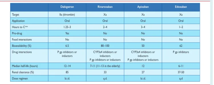
NOAC non-vitamin K oral anticoagulant
NSQIP National Surgical Quality Improvement Program
NSTE-ACS non-ST-elevation acute coronary syndromes
NT-proBNP N-terminal pro-BNP
O2 oxygen
OHS obesity hypoventilation syndrome
OR odds ratio
P gp platelet glycoprotein
PAC pulmonary artery catheter
PAD peripheral artery disease
PAH pulmonary artery hypertension
PCC prothrombin complex concentrate
PCI percutaneous coronary intervention
POBBLE Peri-Operative Beta-BLockadE
POISE Peri-Operative ISchemic Evaluation
POISE-2 Peri-Operative ISchemic Evaluation 2
q.d. quaque die (once daily)
RIFLE Risk, Injury, Failure, Loss, End-stage renal disease
SPECT single photon emission computed tomography
SVT supraventricular tachycardia
SYNTAX Synergy between Percutaneous Coronary Intervention
with TAXUS and Cardiac Surgery
TAVI transcatheter aortic valve implantation
TdP torsades de pointes
TIA transient ischaemic attack
TOE transoesophageal echocardiography
TOD transoesophageal doppler
TTE transthoracic echocardiography
UFH unfractionated heparin
VATS video-assisted thoracic surgery
VHD valvular heart disease
VISION Vascular Events In Noncardiac Surgery Patients Cohort
Evaluation
VKA vitamin K antagonist
VPB ventricular premature beat
VT ventricular tachycardia
by guest on December 11, 2014
1. Preamble
Guidelines summarize and evaluate all available evidence, at the time of the writing process, on a particular issue with the aim of assisting health professionals in selecting the best management strategies for an individual patient with a given condition, taking into account the impact on outcome, as well as the risk – benefit ratio of particular diagnostic or therapeutic means. Guidelines and recommendations should help health professionals to make decisions in their daily prac-tice; however, the final decisions concerning an individual patient must be made by the responsible health professional(s), in consult-ation with the patient and caregiver as appropriate.
A great number of guidelines have been issued in recent years by the European Society of Cardiology (ESC) and the European Society of Anaesthesiology (ESA), as well as by other societies and organisations. Because of their impact on clinical practice, quality criteria for the de-velopment of guidelines have been established in order to make all decisions transparent to the user. The recommendations for for-mulating and issuing ESC/ESA Guidelines can be found on the ESC web site (http://www.escardio.org/guidelines-surveys/esc-guidelines/ about/Pages/rules-writing.aspx). These ESC/ESA guidelines represent the official position of these two societies on this given topic and are regularly updated.
Members of this Task Force were selected by the ESC and ESA to represent professionals involved with the medical care of patients with this pathology. Selected experts in the field undertook a com-prehensive review of the published evidence for management (including diagnosis, treatment, prevention and rehabilitation) of a given condition, according to the ESC Committee for Practice Guidelines (CPG) and ESA Guidelines Committee policy. A critical evaluation of diagnostic and therapeutic procedures was per-formed, including assessment of the risk – benefit ratio. Estimates of expected health outcomes for larger populations were included, where data exist. The level of evidence and the strength of
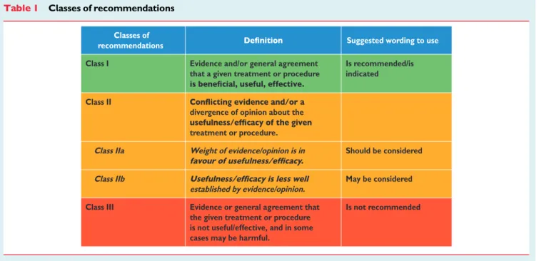
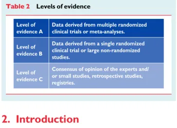
recommendation of particular management options were weighed and graded according to pre-defined scales, as outlined in Tables1and 2.
The experts of the writing and reviewing panels completed ’declara-tions of interest’ forms which might be perceived as real or potential sources of conflicts of interest. These forms were compiled into one file and can be found on the ESC web site (http://www.escardio.org/ guidelines). Any changes in declarations of interest that arise during the writing period must be notified to the ESC/ESA and updated. The Task Force received its entire financial support from the ESC and ESA, without any involvement from the healthcare industry.
The ESC CPG supervises and co-ordinates the preparation of new guidelines produced by Task Forces, expert groups or consensus panels. The Committee is also responsible for the endorsement process of these guidelines. The ESC and Joint Guidelines undergo extensive review by the CPG and partner Guidelines Committee and external experts. After appropriate revisions it is approved by all the experts involved in the Task Force. The finalized document is approved by the CPG/ESA for simultaneous publication in the European Heart Journal and joint partner journal, in this instance the European Journal of Anaesthesiology. It was developed after careful consideration of the scientific and medical knowledge and the evidence available at the time of their dating.
The task of developing ESC/ESA guidelines covers not only the integration of the most recent research, but also the creation of edu-cational tools and implementation programmes for the recommen-dations. To implement the guidelines, condensed pocket versions, summary slides, booklets with essential messages, summary cards for non-specialists, electronic versions for digital applications (smart phones etc.) are produced. These versions are abridged and thus, if needed, one should always refer to the full-text version, which is freely available on the ESC and ESA web sites. The national societies of the ESC and of the ESA are encouraged to endorse, trans-late and implement the ESC guidelines. Implementation programmes
Table 1 Classes of recommendations
Classes of
recommendations Suggested wording to use
Class I Evidence and/or general agreement
that a given treatment or procedure
Is recommended/is indicated
Class II
divergence of opinion about the treatment or procedure.
Class IIa Weight of evidence/opinion is in Should be considered
Class IIb
established by evidence/opinion.
May be considered
Class III Evidence or general agreement that
the given treatment or procedure is not useful/effective, and in some cases may be harmful.
Is not recommended
by guest on December 11, 2014
are needed because it has been shown that the outcome of disease may be favourably influenced by the thorough application of clinical recommendations.
Surveys and registries are needed to verify that real-life daily prac-tice is in keeping with what is recommended in the guidelines, thus completing the loop between clinical research, writing of guidelines, disseminating them and implementing them into clinical practice.
Health professionals are encouraged to take the ESC/ESA guide-lines fully into account when exercising their clinical judgment, as well as in the determination and the implementation of preventive, diagnostic or therapeutic medical strategies; however, the ESC/ESA guidelines do not, in any way whatsoever, override the individual re-sponsibility of health professionals to make appropriate and accurate decisions in consideration of the condition of each patient’s health and in consultation with that patient and, where appropriate and/or necessary, the patient’s caregiver. It is also the health profes-sional’s responsibility to verify the rules and regulations applicable to drugs and devices at the time of prescription.
2. Introduction
2.1 The magnitude of the problem
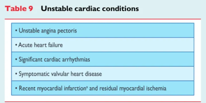
The present Guidelines focus on the cardiovascular management of patients in whom heart disease is a potential source of complications during non-cardiac surgery. The risk of perioperative complications depends on the condition of the patient before surgery, the preva-lence of comorbidities, and the urgency, magnitude, type, and dur-ation of the surgical procedure.
More specifically, cardiac complications can arise in patients with documented or asymptomatic ischaemic heart disease (IHD), left ventricular (LV) dysfunction, valvular heart disease (VHD), and arrhythmias, who undergo surgical procedures that are associated with prolonged haemodynamic and cardiac stress. In the case of peri-operative myocardial ischaemia, two mechanisms are important: (i) a mismatch in the supply – demand ratio of blood flow, in response to metabolic demand due to a coronary artery stenosis that may become flow-limiting by perioperative haemodynamic fluctuations and (ii) acute coronary syndromes (ACS) due to stress-induced rupture of a vulnerable atherosclerotic plaque in combination with vascular inflammation and altered vasomotion, as well as haemosta-sis. LV dysfunction and arrhythmias may occur for various reasons at all ages. Because the prevalence of not only IHD but also VHD and arrhythmias increases with age, perioperative cardiac mortality and
morbidity are predominantly an issue in the adult population under-going major non-cardiac surgery.
The magnitude of the problem in Europe can best be understood in terms of (i) the size of the adult non-cardiac surgical group and (ii) the average risk of cardiac complications in this cohort. Unfortunately, systematic data on the annual number and type of operations—and on patient outcomes—are only available at a national level in 23 European countries (41%).1 Additionally, data definitions vary, as do data quantity and quality. A recent modelling strategy, based on worldwide data available in 2004, estimated the number of major operations to be at the rate of 4% of the world population per year.1When applied to Europe, with an overall population of over 500 million, this figure translates into a crude estimate of 19 million major procedures annually. While the majority of these procedures are performed in patients with minimal cardiovascular risk, 30% of patients undergo extensive surgical procedures in the presence of cardiovascular comorbidity; hence, 5.7 million procedures annually are performed in European patients who present with increased risk of cardiovascular complications.
Worldwide, non-cardiac surgery is associated with an average overall complication rate of 7 – 11% and a mortality rate of 0.8 – 1.5%, depending on safety precautions.2 Up to 42% of these are caused by cardiac complications.3When applied to the population in the European Union member states, these figures translate into at least 167 000 cardiac complications annually due to non-cardiac surgical procedures, of which 19 000 are life-threatening.
2.2 Change in demographics
Within the next 20 years, the ageing of the population will have a major impact on perioperative patient management. It is estimated that elderly people require surgery four times as often than the rest of the population.4In Europe, it is estimated that the number of patients undergoing surgery will increase by 25% by 2020. Over the same time period, the elderly population will increase by 50%. The total number of surgical procedures may increase even faster because of the rising frequency of interventions with age.5 The results of the United States National Hospital Discharge Survey show that the number of surgical procedures will increase in almost all age groups and that the largest increase will occur in the middle-aged and elderly. Demographics of patients undergoing surgery show a trend towards an increasing number of elderly patients and comorbidities.6 Although mortality from cardiac disease is decreasing in the general population, the prevalence of IHD, heart failure, and cardiovascular risk factors—especially dia-betes—is increasing. Among the significant comorbidities in elderly patients presenting for general surgery, cardiovascular disease (CVD) is the most prevalent.7Age per se, however, seems to be re-sponsible for only a small increase in the risk of complications; greater risks are associated with urgency and significant cardiac, pul-monary, and renal disease; thus, these conditions should have greater impact on the evaluation of patient risk than age alone.
2.3 Purpose and organization
These Guidelines are intended for physicians and collaborators involved in the pre-operative, operative, and post-operative care of patients undergoing non-cardiac surgery.
Table 2 Levels of evidence
Level of evidence A
Data derived from multiple randomized clinical trials or meta-analyses. Level of
evidence B
Data derived from a single randomized clinical trial or large non-randomized studies.
Level of evidence C
Consensus of opinion of the experts and/ or small studies, retrospective studies, registries.
by guest on December 11, 2014
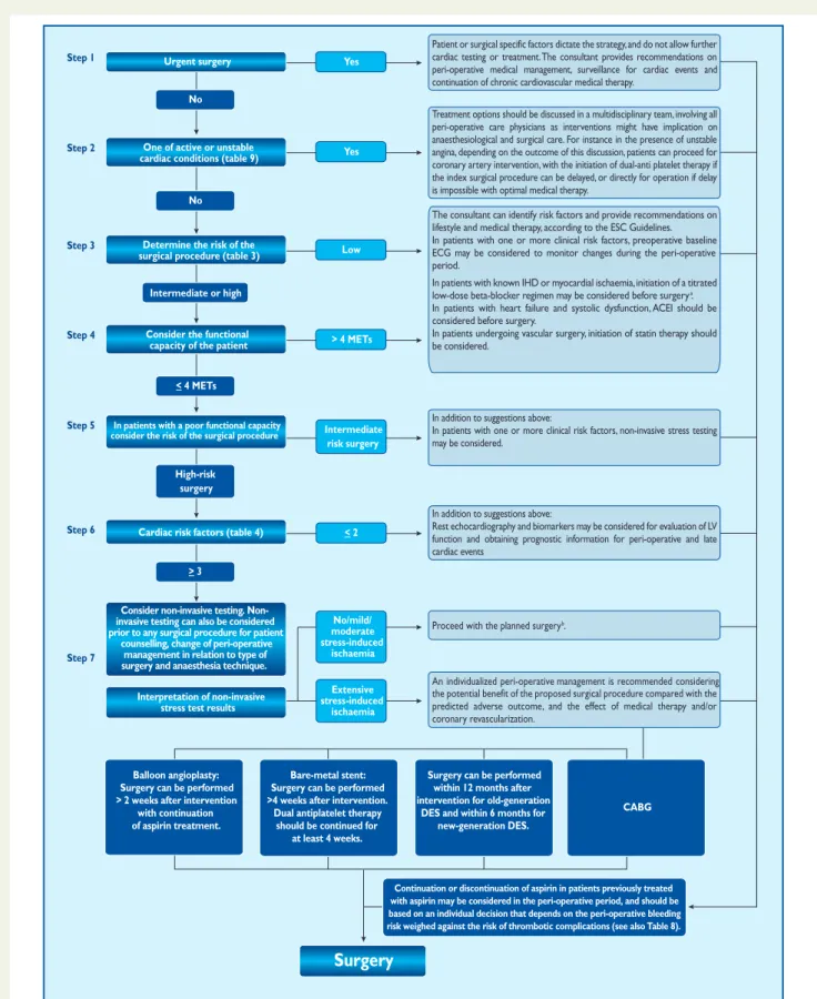
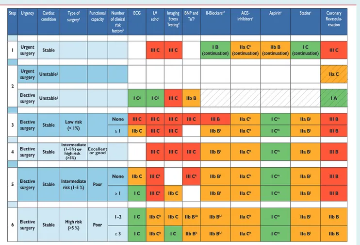
The objective is to endorse a standardized and evidence-based ap-proach to perioperative cardiac management. The Guidelines recom-mend a practical, stepwise evaluation of the patient that integrates clinical risk factors and test results with the estimated stress of the planned surgical procedure. This results in an individualized cardiac risk assessment, with the opportunity of initiating medical therapy, cor-onary interventions, and specific surgical and anaesthetic techniques in order to optimize the patient’s perioperative condition.
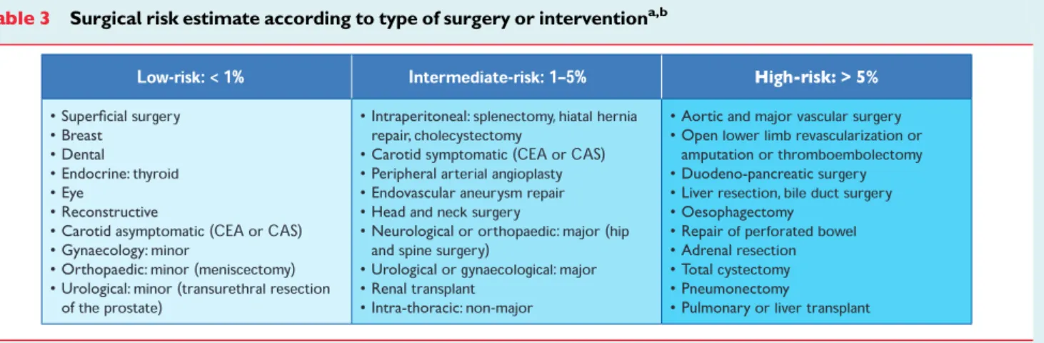
Compared with the non-surgical setting, data from randomized clinical trials—which provide the ideal evidence-base for the guide-lines—are sparse. Consequently, when no trials are available on a specific cardiac-management regimen in the surgical setting, data from the non-surgical setting are extrapolated and similar recom-mendations made, but with different levels of evidence. Anaesthesiol-ogists, who are experts on the specific demands of the proposed surgical procedure, will usually co-ordinate the pre-operative evalu-ation. The majority of patients with stable heart disease can undergo low and intermediate-risk surgery (Table3) without additional evalu-ation. Selected patients require evaluation by a team of integrated multidisciplinary specialists including anaesthesiologists, cardiolo-gists, and surgeons and, when appropriate, an extended team (e.g. internists, intensivists, pulmonologists or geriatricians).8
Selected patients include those identified by the anaesthesiologist because of suspected or known cardiac disease with sufficient complexity to carry a potential perioperative risk (e.g. congenital heart disease, unstable symptoms or low functional capacity), patients in whom pre-operative medical optimization is expected to reduce periopera-tive risk before low- and intermediate-risk surgery, and patients with known or high risk of cardiac disease who are undergoing high-risk surgery. Guidelines have the potential to improve post-operative outcomes and highlight the existence of a clear opportunity for im-proving the quality of care in this high-risk group of patients. In add-ition to promoting an improvement in immediate perioperative care, guidelines should provide long-term advice.
Because of the availability of new evidence and the international impact of the controversy over the DECREASE trials, the ESC/ESA and American College of Cardiology/American Heart Association
both began the process of revising their respective guidelines concur-rently. The respective writing committees independently performed their literature review and analysis, and then developed their recom-mendations. Once peer review of both guidelines was completed, the writing committees chose to discuss their respective recommenda-tions regarding beta-blocker therapy and other relevant issues. Any differences in recommendations were discussed and clearly articu-lated in the text; however, the writing committees aligned a few recommendations to avoid confusion within the clinical community, except where international practice variation was prevalent.
Following the development and introduction of perioperative cardiac guidelines, their effect on outcome should be monitored. The objective evaluation of changes in outcome will form an essential part of future perioperative guideline development.
Recommendations on pre-operative evaluation
Recommendations Classa Levelb Ref.c
Selected patients with cardiac disease undergoing low-and intermediate-risk non-cardiac surgery may be referred by the anaesthesiologist for cardiological evaluation and medical optimization.
IIb C
A multidisciplinary expert team should be considered for pre-operative evaluation of patients with known or high risk of cardiac disease undergoing high-risk non-cardiac surgery. IIa C 8 a Class of recommendation. b Level of evidence. c
Reference(s) supporting recommendations.
Table 3 Surgical risk estimate according to type of surgery or interventiona,b
CAS ¼ carotid artery stenting; CEA ¼ carotid endarterectomy.
a
Surgical risk estimate is a broad approximation of 30-day risk of cardiovascular death and myocardial infarction that takes into account only the specific surgical intervention, without considering the patient’s comorbidities.
b
Adapted from Glance et al.11
by guest on December 11, 2014
3. Pre-operative evaluation
3.1 Surgical risk for cardiac events
Cardiac complications after non-cardiac surgery depend on patient-related risk factors, on the type of surgery, and on the cir-cumstances under which it takes place.9Surgical factors that influ-ence cardiac risk are related to the urgency, invasiveness, type, and duration of the procedure, as well as the change in body core temperature, blood loss, and fluid shifts.5Every operation elicits a stress response. This response is initiated by tissue injury and mediated by neuro-endocrine factors, and may induce sympatho-vagal imbalance. Fluid shifts in the perioperative period add to the surgical stress. This stress increases myocardial oxygen demand. Surgery also causes alterations in the balance between prothrom-botic and fibrinolytic factors, potentially resulting in increased cor-onary thrombogenicity. The extent of such changes is proportionate to the extent and duration of the intervention. These factors, together with patient position, temperature management, bleeding, and type of anaesthesia, may contribute to haemodynamic derangements, leading to myocardial ischaemia and heart failure. General, locoregional, and neuraxial anaesthesia differ in terms of the stress response evoked by surgery. Less invasive anaesthetic techniques may reduce early mortality in patients at intermediate-to-high cardiac risk and limit post-operative complications.10 Although patient-specific factors are more important than surgery-specific factors in predicting the cardiac risk for non-cardiac surgical procedures, the type of surgery cannot be ignored.9
With regard to cardiac risk, surgical interventions—which include open or endovascular procedures—can be broadly divided into low-risk, intermediate-risk, and high-risk groups, with estimated 30-day cardiac event rates (cardiac death and myocardial infarction) of ,1%, 1 – 5%, and .5%, respectively (Table3).
The need for, and value of, pre-operative cardiac evaluation will also depend on the urgency of surgery. In the case of emergency sur-gical procedures, such as those for ruptured abdominal aortic aneur-ysm (AAA), major trauma, or for a perforated viscus, cardiac evaluation will not alter the course or result of the intervention but may influence management in the immediate perioperative period. In non-emergency but urgent surgical conditions, such as bypass for acute limb ischaemia or treatment of bowel obstruction, the mor-bidity and mortality of the untreated underlying condition may out-weigh the potential cardiac risk related to the intervention. In these cases, cardiological evaluation may influence the perioperative mea-sures taken to reduce cardiac risk but will not influence the decision to perform the intervention. In some cases, the cardiac risk can also influence the type of operation and guide the choice to less-invasive interventions, such as peripheral arterial angioplasty instead of in-fra-inguinal bypass, or extra-anatomical reconstruction instead of an aortic procedure, even when these may yield less favourable results in the long term. Finally, in some situations, the cardiac evalu-ation (in as far as it can reliably predict perioperative cardiac compli-cations and late survival) should be taken into consideration when deciding whether to perform an intervention or manage conserva-tively. This is the case in certain prophylactic interventions, such as the treatment of small AAAs or asymptomatic carotid stenosis,
where the life expectancy of the patient and the risk of the oper-ation are important factors in evaluating the potential benefit of the surgical intervention.
3.2 Type of surgery
In general, endoscopic and endovascular techniques speed recovery, decrease hospital stay, and reduce the rate of complications.12 However, randomized clinical trials comparing laparoscopic with open techniques exclude older, sicker, and ’urgent’ patients, and results from an expert-based randomized trial (laparoscopic vs. open cholecystectomy) have shown no significant differences in conversion rate, pain, complications, length of hospital stay, or re-admissions.13
The wide variety of surgical procedures, in a myriad of different contexts, makes difficult the assignation of a specific risk of a major adverse cardiac event to each procedure. When alternative methods to classical open surgery are considered, either through endovascular or less-invasive endoscopic procedures, the potential trade-offs between early benefits due to reduced morbidity and mid- to long-term efficacy need to be taken into account.
3.2.1 Endovascular vs. open vascular procedures
Vascular interventions are of specific interest, not only because they carry the highest risk of cardiac complications, but also because of the many studies that have shown that this risk can be influenced by adequate perioperative measures in these patients.14 Open aortic and infra-inguinal procedures must both be regarded as high-risk procedures. Although it is a less-extensive intervention, infra-inguinal revascularization entails a cardiac risk similar to—or even higher than—that of aortic procedures. This can be explained by the higher incidence of diabetes, renal dysfunction, IHD, and advanced age in this patient group. This also explains why the risk related to peripheral artery angioplasties, which are minimally inva-sive procedures, is not negligible.
Endovascular AAA repair (EVAR) has been associated with lower operative mortality and morbidity than open repair but this advantage reduces with time, due to more frequent graft-related complications and re-interventions in patients who underwent EVAR, resulting in similar long-term AAA-related mortality and total mortality.15–17
A meta-analysis of studies, comparing open surgical with percutaneous transluminal methods for the treatment of femoro-popliteal arterial disease, showed that bypass surgery is associated with higher 30-day morbidity [odds ratio (OR) 2.93; 95% confidence interval (CI) 1.34 – 6.41] and lower technical failure than endovascular treatment, with no differences in 30-day mor-tality; however, there were higher amputation-free and overall survival rates in the bypass group at 4 years.18Therefore, multiple factors must be taken into consideration when deciding which type of procedure serves the patient best. An endovascular-first ap-proach may be advisable in patients with significant comorbidity, whereas a bypass procedure may be offered as a first-line interven-tional treatment for fit patients with a longer life expectancy.19 Carotid artery stenting has appeared as an attractive, less-invasive alternative to CEA; however, although CAS reduces the rate of
by guest on December 11, 2014
periprocedural myocardial infarction and cranial nerve palsy, the combined 30-day rate of stroke or death is higher than CEA, particularly in symptomatic and older patients, driven by a differ-ence in the risk of periprocedural non-disabling stroke.20,21 The benefit of carotid revascularization is particularly high in patients with recent (,3 months) transient ischaemic attack (TIA) or stroke and a .60% carotid artery bifurcation stenosis.22 In neurologically asymptomatic patients, carotid revascularization benefit is questionable, compared with modern medical therapy, except in patients with a .80% carotid stenosis and an estimated life expectancy of .5 years.21 The choice between CEA and CAS must integrate operator experience and results, anatomical characteristics of the arch vessels, neck features, and comorbidities.21–23
3.2.2 Open vs. laparoscopic or thoracoscopic procedures
Laparoscopic procedures, compared with open procedures, have the advantage of causing less tissue trauma and intestinal paralysis, resulting in less incisional pain, better post-operative pulmonary function, significantly fewer wall complications, and diminished post-operative fluid shifts related to bowel paralysis.24However, the pneu-moperitoneum required for these procedures results in elevated intra-abdominal pressure and a reduction in venous return. Typical physiological sequelae are secondary to increased intra-abdominal pressure and absorption of the gaseous medium used for insufflation. While healthy individuals on controlled ventilation typically tolerate pneumoperitoneum, debilitated patients with cardiopulmonary compromise and obese patients may experience adverse conse-quences.25Pneumoperitoneum and Trendelenburg position result in increased mean arterial pressure, central venous pressure, mean pulmonary artery, pulmonary capillary wedge pressure, and systemic vascular resistance impairing cardiac function.26,27Therefore, com-pared with open surgery, cardiac risk in patients with heart failure is not reduced in patients undergoing laparoscopy, and both should be evaluated in the same way. This is especially true in patients under-going interventions for morbid obesity, but also in other types of surgery, considering the risk of conversion to an open proced-ure.28,29Superior short-term outcomes of laparoscopic vs. open procedures have been reported, depending on type of surgery, oper-ator experience and hospital volume, but few studies provide direct measures of cardiac complications.30–32Benefit from laparoscopic procedures is probably greater in elderly patients, with reduced length of hospital stay, intra-operative blood loss, incidence of post-operative pneumonia, time to return of normal bowel function, in-cidence of post-operative cardiac complications, and wound infec-tions.33Few data are available for video-assisted thoracic surgery (VATS), with no large, randomized trial comparing VATS with open thoracic lung resection. In one study involving propensity-score-matched patients, VATS lobectomy was associated with no significant difference in mortality, but with significantly lower rates of overall perioperative morbidity, pneumonia, and atrial arrhythmia.34
Recommendations on the selection of surgical approach and its impact on risk
Recommendations Classa Levelb Ref.c
It is recommended that patients should undergo pre-operative risk assessment independently of an open or laparoscopic surgical approach.d
I C 26,27, 35
In patients with AAA 55 mm, anatomically suited for EVAR, either open or endovascular aortic repair is recommended if surgical risk is acceptable.
I A 15–17
In patients with asymptomatic AAA who are unfit for open repair, EVAR, along with best medical treatment, may be considered.
IIb B 15,35
In patients with lower extremity artery disease requiring revascularization, the best management strategy should be determined by an expert team considering anatomy,
comorbidities, local availability, and expertise.
IIa B 18
AAA ¼ abdominal aortic aneurysm; EVAR ¼ endovascular aortic reconstruction.
a
Class of recommendation.
b
Level of evidence.
c
Reference(s) supporting recommendations.
d
Since laparoscopic procedures demonstrate a cardiac stress similar to that of open procedures.
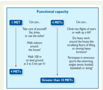
3.3 Functional capacity
Determination of functional capacity is a pivotal step in pre-operative cardiac risk assessment and is measured in metabolic equivalents (METs). One MET equals the basal metabolic rate. Ex-ercise testing provides an objective assessment of functional cap-acity. Without testing, functional capacity can be estimated from the ability to perform the activities of daily living. One MET repre-sents metabolic demand at rest; climbing two flights of stairs demands 4 METs, and strenuous sports, such as swimming, .10 METS (Figure1).
The inability to climb two flights of stairs or run a short distance (,4 METs) indicates poor functional capacity and is associated with an increased incidence of post-operative cardiac events. After thoracic surgery, a poor functional capacity has been associated with an increased mortality (relative risk 18.7; 95% CI 5.9 – 59); however, in comparison with thoracic surgery, a poor functional status was not associated with an increased mortality after other non-cardiac surgery (relative risk 0.47; 95% CI 0.09 – 2.5).38This may
by guest on December 11, 2014
reflect the importance of pulmonary function—strongly related to functional capacity—as a major predictor of survival after thoracic surgery. These findings were confirmed in a study of 5939 patients scheduled for non-cardiac surgery, in which the pre-operative func-tional capacity measured in METs showed a relatively weak associ-ation with post-operative cardiac events or death.39Notably, when functional capacity is high, the prognosis is excellent, even in the pres-ence of stable IHD or risk factors;40otherwise, when functional cap-acity is poor or unknown, the presence and number of risk factors in relation to the risk of surgery will determine pre-operative risk strati-fication and perioperative management.
3.4 Risk indices
For two main reasons, effective strategies aimed at reducing the risk of perioperative cardiac complications should involve cardiac evaluation, using medical history before the surgical procedure,. Firstly, patients with an anticipated low cardiac risk—after thorough evaluation—can be operated on safely without further delay. It is unlikely that risk-reduction strategies will further reduce the perioperative risk. Secondly, risk reduction by pharmacological treat-ment is most cost-effective in patients with a suspected increased cardiac risk. Additional non-invasive cardiac imaging techniques are tools to identify patients at higher risk; however, imaging techniques should be reserved for those patients in whom test results would in-fluence and change management. Clearly, the intensity of the pre-operative cardiac evaluation must be tailored to the patient’s clinical condition and the urgency of the circumstances requiring surgery. When emergency surgery is needed, the evaluation must necessarily be limited; however, most clinical circumstances allow the application of a more extensive, systematic approach, with cardiac risk evaluation that is initially based on clinical characteristics and type of surgery
and then extended, if indicated, to resting electrocardiography (ECG), laboratory measurements, or other non-invasive assessments. Several risk indices have been developed during the past 30 years, based on multivariate analyses of observational data, which represent the relationship between clinical characteristics and peri-operative cardiac mortality and morbidity. The indices developed by Goldman et al. (1977),41 Detsky et al. (1986),42and Lee et al. (1999)43have become well-known.
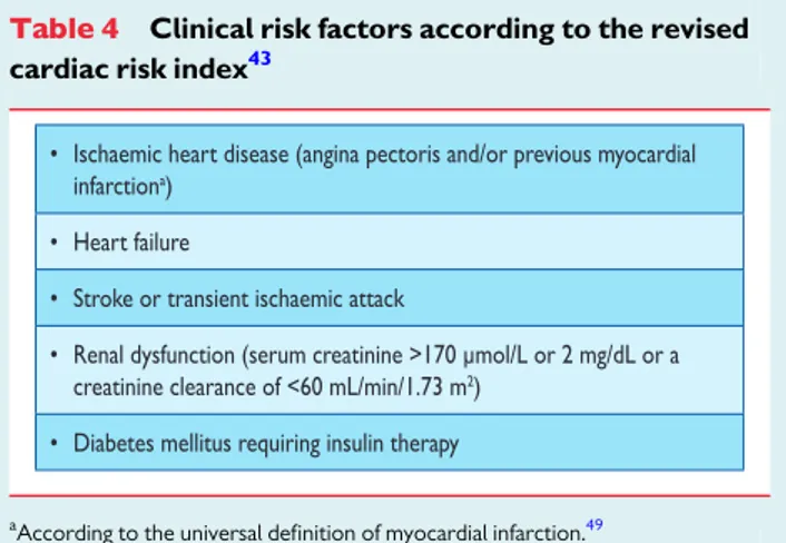
Although only a rough estimation, the older risk-stratification systems may represent useful clinical tools for physicians in respect of the need for cardiac evaluation, drug treatment, and assessment of risk for cardiac events. The Lee index or ‘revised cardiac risk’ index, a modified version of the original Goldman index, was designed to predict post-operative myocardial infarction, pulmonary oedema, ventricular fibrillation or cardiac arrest, and complete heart block. This risk index comprises six variables: type of surgery, history of IHD, history of heart failure, history of cerebrovascular disease, pre-operative treatment with insulin, and pre-operative creatinine .170 mmol/L (.2 mg/dL), and used to be considered by many clin-icians and researchers to be the best currently available cardiac-risk prediction index in non-cardiac surgery.
All of the above-mentioned risk indices were, however, developed years ago and many changes have since occurred in the treatment of IHD and in the anaesthetic, operative and perioperative management of non-cardiac surgical patients. A new predictive model was recently developed to assess the risk of intra-operative/post-operative myo-cardial infarction or cardiac arrest, using the American College of Sur-geons National Surgical Quality Improvement Program (NSQIP) database.44This NSQIP MICA model was built on the 2007 data set, based on patients from 180 hospitals, and was validated with the 2008 data set, both containing .200 000 patients and having pre-dictability. The primary endpoint was intra-operative/post-operative myocardial infarction or cardiac arrest up to 30 days after surgery. Five predictors of perioperative myocardial infarction/cardiac arrest were identified: type of surgery, functional status, elevated cre-atinine (.130 mmol/L or .1.5 mg/dL), American Society of Anesthesiologists (ASA) class (Class I, patient is completely healthy; Class II, patient has mild systemic disease; Class III, patient has severe systemic disease that is not incapacitating; Class IV, patient has incapacitating disease that is a constant threat to life; and Class V, a moribund patient who is not expected to live for 24 hours, with or without the surgery), and age. This model is presented as an interactive risk calculator (http://www.surgicalriskcalculator. com/miorcardiacarrest) so that the risk can be calculated at the bedside or clinic in a simple and accurate way. Unlike other risk scores, the NSQIP model did not establish a scoring system but pro-vides a model-based estimate of the probability of myocardial infarc-tion/cardiac arrest for an individual patient. The risk calculator performed better than the Lee risk index, with some reduction in performance in vascular patients, although it was still superior; however, some perioperative cardiac complications of interest to clinicians, such as pulmonary oedema and complete heart block, were not considered in the NSQIP model because those variables were not included in the NSQIP database. By contrast, the Lee index allows estimation of the risk of perioperative pulmonary oedema and of complete heart block, in addition to death and Functional capacity
Can you... Take care of yourself?
Eat, dress, or use the toilet?
Walk indoors around the house? Walk 100 m on level ground at 3 to 5 km per h? Can you... Climb two flights of stairs
or walk up a hill? Do heavy work around the house like scrubbing floors of lifting
or moving heavy furniture? Participate in strenuous
sports like swimming, singles tennis, football, basketball, or skiing? 1 MET
4 METs
4 METs
Greater than 10 METs
Figure 1 Estimated energy requirements for various activities. Based on Hlatky et al. and Fletcher et al.36,37km per h ¼ kilometres per hour; MET ¼ metabolic equivalent.
by guest on December 11, 2014
myocardial infarction ( http://www.mdcalc.com/revised-cardiac-risk-index-for-pre-operative-risk/). A recent systematic review of 24 studies covering .790 000 patients found that the Lee index discri-minated moderately well patients at low vs. high risk for cardiac events after mixed non-cardiac surgery, but its performance was hampered when predicting cardiac events after vascular non-cardiac surgery or predicting death.45Therefore, the NSQIP and Lee risk index models provide complementary prognostic perspectives and can help the clinician in the decision-making process.
Risk models do not dictate management decisions but should be regarded as one piece of the puzzle to be evaluated, in concert with the more traditional information at the physician’s disposal.
3.5 Biomarkers
A biological marker, or ’biomarker’, is a characteristic that can be ob-jectively measured and which is an indicator of biological processes. In the perioperative setting, biomarkers can be divided into markers focusing on myocardial ischaemia and damage, inflammation, and LV function. Cardiac troponins T and I (cTnT and cTnI, respectively) are the preferred markers for the diagnosis of myocardial infarction because they demonstrate sensitivity and tissue specificity better than other available biomarkers.46The prognostic information is in-dependent of—and complementary to—other important cardiac indicators of risk, such as ST deviation and LV function. It seems that cTnI and cTnT are of similar value for risk assessment in ACS in the presence and absence of renal failure. Existing evidence sug-gests that even small increases in cTnT in the perioperative period reflect clinically relevant myocardial injury with worsened cardiac prognosis and outcome.47–49The development of new biomarkers, including high-sensitivity troponins, will probably further enhance the assessment of myocardial damage.48Assessment of cardiac tropo-nins in high-risk patients, both before and 48 – 72 hours after major surgery, may therefore be considered.3It should be noted that tropo-nin elevation may also be observed in many other conditions; the diagnosis of non-ST-segment elevation myocardial infarction should never be made solely on the basis of biomarkers.
Inflammatory markers might pre-operatively identify those patients with an increased risk of unstable coronary plaque; however, in the surgical setting, no data are currently available on how inflammatory markers would alter risk-reduction strategies.
B-type natriuretic peptide (BNP) and N-terminal pro-BNP (NT-proBNP) are produced in cardiac myocytes in response to increases in myocardial wall stress. This may occur at any stage of heart failure, independently of the presence or absence of myocardial ischaemia. Plasma BNP and NT-proBNP have emerged as important prognostic indicators across many cardiac diseases in non-surgical settings.50Pre-operative BNP and NT-proBNP levels have additional prognostic value for long-term mortality and for cardiac events after major non-cardiac vascular surgery.51–53
Data from prospective, controlled trials on the use of pre-operative biomarkers are sparse. Based on the existing data, assess-ment of serum biomarkers for patients undergoing non-cardiac surgery cannot be proposed for routine use, but may be considered in high-risk patients (METs≤4 or with a revised cardiac risk index value .1 for vascular surgery and .2 for non-vascular surgery).
Recommendations on cardiac risk stratification
Recommendations Classa Levelb Ref.c
Clinical risk indices are recommended to be used for peri-operative risk stratification.
I B 43,44
The NSQIP model or the Lee risk index are recommended for cardiac peri-operative risk stratification.
I B 43,44,54
Assessment of cardiac troponins in high-risk patients, both before and 48–72 hours after major surgery, may be considered.
IIb B 3,48,49
NT-proBNP and BNP measurements may be considered for obtaining independent prognostic information for peri-operative and late cardiac events in high-risk patients.
IIb B 52,53,55
Universal pre-operative routine biomarker sampling for risk stratification and to prevent cardiac events is not recommended.
III C
BNP ¼ B-type natriuretic peptide; NT-proBNP ¼ N-terminal pro-brain natriuretic peptide.
NSQIP ¼ National Surgical Quality Improvement Program.
a
Class of recommendation.
b
Level of evidence.
c
Reference(s) supporting recommendations.
3.6 Non-invasive testing
Pre-operative non-invasive testing aims to provide information on three cardiac risk markers: LV dysfunction, myocardial ischaemia, and heart valve abnormalities, all of which are major determinants of adverse post-operative outcome. LV function is assessed at rest, and various imaging methods are available. For detection of myocar-dial ischaemia, exercise ECG and non-invasive imaging techniques may be used. Routine chest X-ray before non-cardiac surgery is not recommended without specific indications. The overall theme is that the diagnostic algorithm for risk stratification of myocardial is-chaemia and LV function should be similar to that proposed for patients in the non-surgical setting with known or suspected IHD.56Non-invasive testing should be considered not only for cor-onary artery revascularization but also for patient counselling, change of perioperative management in relation to type of surgery, anaesthetic technique, and long-term prognosis.
by guest on December 11, 2014
3.6.1 Non-invasive testing of cardiac disease 3.6.1.1 Electrocardiography
The 12-lead ECG is commonly performed as part of pre-operative cardiovascular risk assessment in patients undergoing non-cardiac surgery. In IHD patients, the pre-operative ECG offers important prognostic information and is predictive of long-term outcome, inde-pendent of clinical findings and perioperative ischaemia.57However, the ECG may be normal or non-specific in patients with myocardial ischaemia or even with infarction.
Recommendations on routine pre-operative ECG
Recommendations Classa Levelb Ref.c
Pre-operative ECG is recommended for patients who have risk factor(s)d
and are scheduled for intermediate- or high-risk surgery.
I C 57
Pre-operative ECG may be considered for patients who have risk factor(s) and are scheduled for low-risk surgery.
IIb C
Pre-operative ECG may be considered for patients who have no risk factors, are above 65 years of age, and are scheduled for intermediate-risk surgery.
IIb C
Routine pre-operative ECG is not recommended for patients who have no risk factors and are scheduled for low-risk surgery.
III B 71 ECG ¼ electrocardiography. a Class of recommendation. b Level of evidence. c
Reference(s) supporting recommendations.
d
Clinical risk factors in Table 4.
3.6.1.2 Assessment of left ventricular function
Resting LV function can be evaluated before non-cardiac surgery by radionuclide ventriculography, gated single photon emission
Recommendations on resting echocardiography in asymptomatic patients without signs of cardiac disease or electrocardiographic abnormalities
Recommendations Classa Levelb
Rest echocardiography may be considered in patients undergoing high-risk surgery.
IIb C
Routine echocardiography is not recommended in patients undergoing intermediate- or low-risk surgery. III C a Class of recommendation. b Level of evidence.
computed tomography (SPECT), echocardiography, magnetic res-onance imaging (MRI) or multislice computed tomography (CT), all with similar accuracy. Echocardiography is the most readily available and versatile tool for evaluating ventricular function. Routine echo-cardiography is not recommended for the pre-operative evaluation of ventricular function but may be performed in asymptomatic patients with high surgical risk.58Pre-operative LV systolic dysfunc-tion, moderate-to-severe mitral regurgitadysfunc-tion, and increased aortic valve gradients are associated with major cardiac events.59 The limited predictive value of LV function assessment for perioperative outcome may be related to the failure to detect severe underlying IHD.
3.6.2 Non-invasive testing of ischaemic heart disease Physical exercise, using a treadmill or bicycle ergometer, provides an estimate of functional capacity, evaluates blood pressure and heart rate response, and detects myocardial ischaemia through ST-segment changes. The accuracy of exercise ECG varies signifi-cantly among studies.56 Risk stratification with an exercise test is not suitable for patients with limited exercise capacity, owing to their inability to reach their target heart rate. Also, pre-existing ST-segment abnormalities at rest—especially in precordial leads V5 and V6—hamper reliable ST-segment analysis. A gradient of se-verity in the test result relates to the perioperative outcome: the onset of a myocardial ischaemic response at low exercise workloads is associated with a significantly increased risk of perioperative and long-term cardiac events. In contrast, the onset of myocardial ischae-mia at high workloads is associated with only a minor risk increase, but higher than a totally normal test. Pharmacological stress testing with either nuclear perfusion imaging or echocardiography is more suitable in patients with limited exercise tolerance.
The role of myocardial perfusion imaging for pre-operative risk stratifications is well established. In patients with limited exercise cap-acity, pharmacological stress (dipyridamole, adenosine, or dobuta-mine) is an alternative stressor. Studies are performed both during stress and at rest, to determine the presence of reversible defects, reflecting jeopardized ischaemic myocardium or fixed defects, reflecting scar or non-viable tissue.
The prognostic value of the extent of ischaemic myocardium, using semi-quantitative dipyridamole myocardial perfusion imaging, has been investigated in a meta-analysis of patients undergoing vascular surgery.60Study endpoints were perioperative cardiac death and myocardial infarction. The authors included nine studies, totalling 1179 patients undergoing vascular surgery, with a 7% 30-day event rate. In this analysis, reversible ischaemia in ,20% of the LV myocar-dium did not alter the likelihood of perioperative cardiac events, com-pared with those without ischaemia. Patients with more extensive reversible defects from 20– 50% were at increased risk.
A second meta-analysis pooled the results of 10 studies evaluating dipyridamole thallium-201 imaging in candidates for vascular surgery over a 9-year period from 1985 to 1994.61The 30-day cardiac death or non-fatal myocardial infarction rates were 1% in patients with normal test results, 7% in patients with fixed defects, and 9% in patients with reversible defects on thallium-201 imaging. Moreover, three of the 10 studies analysed used semi-quantitative scoring, dem-onstrating a higher incidence of cardiac events in patients with two or more reversible defects.
by guest on December 11, 2014
Overall, the positive predictive value of reversible defects for peri-operative death or myocardial infarction has decreased in more recent studies. This is probably related to changes in perioperative management and surgical procedures; however, because of the high sensitivity of nuclear imaging studies for detecting IHD, patients with a normal scan have an excellent prognosis.
Stress echocardiography using exercise or pharmacological (dobutamine, dipyridamole) stress has been widely used for pre-operative cardiac risk evaluation. The test combines information on LV function at rest, heart valve abnormalities, and the presence and extent of stress-inducible ischaemia.62 In one study, 530 patients were enrolled to evaluate the incremental value of dobutamine stress echocardiography (DSE) for the assessment of cardiac risk before non-vascular surgery.63 Multivariate predictors of post-operative events in patients with ischaemia were found to be a history of heart failure (OR 4.7; 95% CI 1.6 – 14.0) and ischaemic threshold ,60% of age-predicted maximal heart rate (OR 7.0; 95% CI 2.8 – 17.6). DSE has some limitations: it should not, for example, be used in patients with severe arrhythmias, significant hypertension, large thrombus-laden aortic aneurysms, or hypotension.
In general, stress echocardiography has a high negative predictive value and a negative test is associated with a very low incidence of cardiac events in patients undergoing surgery; however, the positive predictive value is relatively low (between 25% and 45%); this means that the postsurgical probability of a cardiac event is low, despite wall motion abnormality detection during stress echocardiography.
A negative DSE, performed before scheduled aortic surgery, does not, however, rule out post-operative myocardial necrosis.64Failure to achieve target heart rate is not uncommon, despite an aggressive DSE regimen. A negative DSE without resting wall motion abnormal-ities has excellent negative predictive value, regardless of the heart rate achieved. Patients with resting wall motion abnormalities are at increased risk for perioperative events, even if ischaemia cannot be induced.65
In a meta-analysis of 15 studies comparing dipyridamole thallium-201 imaging and DSE for risk stratification before vascular surgery, it was demonstrated that the prognostic value of stress imaging abnormalities for perioperative ischaemic events is similar with both pharmacological stressors, but that the accuracy varies with IHD prevalence.61In patients with a low prevalence of IHD, the diagnostic accuracy is reduced, com-pared with those with a high incidence of IHD.
Cardiovascular magnetic resonance (CMR) imaging can be used for detection of ischaemia; both perfusion and wall motion can be
detected during stress and at rest.66 Its accuracy in assessment of ischaemia is high, with a sensitivity of 83% and a specificity of 86% when wall motion is used (14 studies; 754 patients). When per-fusion is assessed (24 studies; 1516 patients), its sensitivity was 91% and specificity 81%. When evaluated prospectively in a multicentre study, the sensitivity was 67% and the specificity was 61%.67There are limited data on CMR in the pre-operative setting; in one study dobutamine stress CMR was used in 102 patients undergoing major non-cardiac surgery; in multivariate analysis, myocardial is-chaemia was the strongest predictor of perioperative cardiac events (death, myocardial infarction, and heart failure).68Currently no data are available in the setting of pre-operative risk stratification. Computed tomography can be used to detect coronary calcium, which reflects coronary atherosclerosis, and CT angiog-raphy is useful for excluding coronary artery disease (CAD) in patients who are at low risk of atherosclerosis.69Currently, no data are available in the setting of pre-operative risk stratification. All the various imaging tests have their intrinsic risks and these need to be taken into account when they are used.70
Recommendations on imaging stress testing before surgery in asymptomatic patients
Recommendations Classa Levelb
Imaging stress testing is recommended before high-risk surgery in patients with more than two clinical risk factors and poor functional capacity (<4 METs).c
I C
Imaging stress testing may be considered before high- or intermediate-risk surgery in patients with one or two clinical risk factors and poor functional capacity (<4 METs).c
IIb C
Imaging stress testing is not
recommended before low-risk surgery, regardless of the patient’s clinical risk.
III C
MET ¼ metabolic equivalent
a
Class of recommendation.
b
Level of evidence.
c
Clinical risk factors in Table 4.
How can these data contribute to a practical algorithm? Testing should only be performed if its results might influence perioperative management. Patients with extensive stress-induced ischaemia re-present a high-risk population in whom standard medical therapy appears insufficient to prevent a perioperative cardiac event. Pre-operative testing is recommended in the case of high-risk surgery in patients with poor functional capacity (,4 METS) and more than two of the clinical risk factors listed in Table4, but may also be considered in patients with fewer than three of these risk factors. Im-portantly, pre-operative testing might delay surgery. A similar recom-mendation is made for intermediate-risk surgery patients, although no data from randomized trials are available. Considering the low event rate of patients scheduled for low-risk surgery, it is unlikely that test results will alter perioperative management in stable cardiac patients.
Table 4 Clinical risk factors according to the revised cardiac risk index43
a
According to the universal definition of myocardial infarction.49
by guest on December 11, 2014
3.7 Invasive coronary angiography
Coronary angiography is a well-established, invasive, diagnostic procedure but is rarely indicated for assessing the risk of patients undergoing non-cardiac surgery. There is a lack of information from randomized clinical trials, relating to its usefulness in patients scheduled for non-cardiac surgery. Also, adopting an invasive cor-onary angiography assessment may cause an unnecessary and un-predictable delay in an already planned surgical intervention, as well as adding an independent procedural risk to the overall risk. Despite the fact that CAD may be present in a significant number of patients requiring non-cardiac surgery, indications for pre-operative coronary angiography and revascularization are similar to angiography indications in the non-surgical setting.56,72–75 Pre-operative treatment of myocardial ischaemia, either medically or with intervention, is recommended whenever non-cardiac surgery can be delayed.
Recommendations on pre-operative coronary angiography
Recommendations Classa
Levelb
Ref.c
Indications for pre-operative coronary angiography and revascularization are similar to those for the non-surgical setting.
I C 56
Urgent angiography is recommended in patients with acute ST-segment elevation myocardial infarction requiring non-urgent, non-cardiac surgery.
I A 75
Urgent or early invasive strategy is recommended in patients with NSTE-ACS requiring urgent, non-cardiac surgery according to risk assessment.
I B 73
Pre-operative angiography is recommended in patients with proven myocardial ischaemia and unstabilized chest pain (Canadian Cardiovascular Society Class III–IV) with adequate medical therapy requiring non-urgent, non-cardiac surgery.
I C 56,72
Pre-operative angiography may be considered in stable cardiac patients undergoing non-urgent carotid endarterectomy surgery.
IIb B 76
Pre-operative angiography is not recommended in cardiac-stable patients undergoing low-risk surgery.
III C
NSTE-ACS ¼ non-ST-segment elevation acute coronary syndromes.
a
Class of recommendation.
b
Level of evidence.
c
Reference(s) supporting recommendations.
4. Risk-reduction strategies
4.1 Pharmacological
The stress of surgery and anaesthesia may trigger ischaemia through an increase in myocardial oxygen demand, a reduction in myocardial oxygen supply, or both. Besides specific risk-reduction strategies adapted to patient characteristics and type of surgery, pre-operative evaluation can check and optimize the control of cardiovascular risk factors.
4.1.1 Beta-blockers
Concerns were raised over a number of studies of the Dutch Echo-cardiographic Cardiac Risk Evaluation Applying Stress Echocardiog-raphy (DECREASE) family,77and the results of these studies were not included in the present Guidelines.
The main rationale for perioperative beta-blocker use is to de-crease myocardial oxygen consumption by reducing heart rate, leading to a longer diastolic filling period and decreased myocardial contractility. Additional cardioprotective factors have been sug-gested; however, the answer to whether or not this translates into clinical benefit requires randomized trials analysing the incidence of cardiovascular events. Six randomized trials evaluating the effect of perioperative beta-blockade on clinical endpoints have been pub-lished in English in peer-reviewed journals (Table5).78–83
Two trials targeted patients at high risk for perioperative compli-cations relating to the type of surgery, the presence of IHD, or risk factors for perioperative cardiac complications.79,83 Three other trials did not require clinical risk factors, except for diabetes in one case.80–82 The Peri-Operative ISchemic Evaluation (POISE) trial covered a wide spectrum of risk of perioperative cardiac complica-tions.78One trial randomized 200 patients with at least two IHD risk factors or with known IHD, who were scheduled for non-cardiac surgery under general anaesthesia, including 40% for major vascular surgery.83Atenolol was associated with a significant decrease in overall mortality at 6 months, which was sustained for up to 2 years; however, seven in-hospital deaths, five in the atenolol group and two in the placebo group, were not taken into account. The Peri-Operative Beta-BLockadE (POBBLE) trial randomized 103 low-risk patients undergoing elective infrarenal vascular surgery to metopro-lol tartrate or placebo,82resulting in a similar incidence of death, myocardial infarction or stroke at 30 days (13% and 15%, respectively; P ¼ 0.78). Patients at low cardiac risk and those with a history of myo-cardial infarction within the past 2 years were excluded. The Metoprolol after Vascular Surgery (MaVS) trial randomized 497 patients undergoing abdominal or infra-inguinal vascular surgery to metoprolol succinate or placebo.80 The combined incidence of death, myocardial infarction, heart failure, arrhythmias, or stroke at 30 days was similar (10.2% and 12.0%, respectively; P ¼ 0.57). The revised cardiac risk index was≤2 in 90% of patients and ≤1 in 60%.
The Diabetes Post-Operative Mortality and Morbidity (DIPOM) trial randomized 921 patients with diabetes, age .39 years, and dur-ation of surgery of .1 hour (39% low-risk surgery) to receive meto-prolol succinate or placebo.81The combined incidence of death, myocardial infarction, unstable angina, or heart failure at 30 days was again similar (6% and 5%, respectively; P ¼ 0.66); however, only 54% of patients had a history of IHD or an additional cardiac risk factor, and underwent high- or intermediate-risk surgery.
by guest on December 11, 2014