Université de Montréal
The potential role of the multivalent ionic compound PolyP in the
assembly of the liquid nature in the cell
par
Lara Michel Matta
Département de Biochimie et Médecine Moléculaire Faculté de Médecine
Mémoire présenté à la Faculté de Médecine
en vue de l’obtention du grade de Maître ès Sciences (M.Sc.) En Biochimie
Option cheminement libre
November 2016
i
Résumé
Les protéines de type prion, contenant des Séquences en acides aminés de Faible Complexité (SFC), ont tendance à s’agréger et à former des compartiments non-membranaires dans la cellule. Ces derniers ont des propriétés physiques communes à celles des liquides, telles que la capacité de mouiller les surfaces, de s’écouler et de fusionner avec d’autres corps liquides. Dans cette étude, nous avons démontré que la protéine Hrp1 forme, in vitro, des
gouttes de différentes tailles via une transition de phase liquide à liquide, et ce, uniquement lorsqu’elle est exposée à un milieu chargé négativement. Exclusivement dans ce même milieu, nous avons aussi observé que le domaine SFC de Hrp1 s’assemble et forme une matière de type gel. Sur la base de ces observations, nous avons émis l’hypothèse que la tendance des systèmes moléculaires à former des compartiments liquides in vivo peut être influencée par la présence, dans le cytosol, de polyélectrolytes chargés négativement tels que l'ADN, l'ARN et les polyphosphates (PolyP). En utilisant la levure comme modèle cellulaire et des techniques de microscopie à fluorescence, nous nous sommes focalisés sur l’étude du rôle des PolyP dans l'assemblage des P-bodies. Les P-bodies ont été choisis comme système moléculaire de référence in vivo, étant des corps qui, après une transition de phase, se trouvent dans le cytosol sous forme de gouttes. Nous avons démontré que la déplétion du phosphate et la délétion du gène vtc4, responsable de la synthèse des PolyP dans la levure, n’ont pas d’influence dans la formation des P-bodies. Nous avons aussi remarqué que les PolyP et la protéine Edc3, une des composantes principales des P-bodies, ne sont pas co-localisés dans la cellule. Cette étude préliminaire nous suggère un manque de corrélation entre la formation des P-bodies et la présence de PolyP dans la cellule. Cependant, pour confirmer nos observations, des expériences complémentaires doivent être envisagées, en considérant d’autres composantes des P-bodies, tel que Lsm4, ou en analysant, in vivo, les effets des PolyP sur d’autres systèmes moléculaires de nature liquide.
Mots-clés : les protéines de type prion, séquence de faible complexité, les gouttes liquides, matière de type gel, PolyP, transition de phase liquide à liquide.
ii
Abstract
Prion-like proteins containing Low Complexity Sequences (LCSs) have the propensity to aggregate and form membrane-less compartments in the cell. These proteins form droplets that have liquid features such as wetting, dripping and fusion. In this study, we demonstrated that the prion domain-containing protein Hrp1 forms droplets of different sizes in the presence of negatively charged polymers via liquid-liquid phase separation, whereas under the same conditions, the prion-like domain PolyQ/N of Hrp1 forms a gel-like material. Based on these findings, we hypothesize that droplets in vivo could be modulated by negatively charged polyelectrolytes found in the cell such as DNA, RNA and polyphosphate (PolyP). My goal was to examine the role of the polyanionic nature of PolyP on the assembly of P-bodies using
Saccharomyces cerevisiae as a cellular model and fluorescence microscopy. We chose to
study processing (P)- bodies, based on previous findings that these cellular subcompartments are formed by liquid-liquid phase separation of component proteins in the cytoplasm. We found that depleting phosphate from the media and deleting vtc4 gene, which is responsible for PolyP synthesis, did not have any effect on P-body formation. In addition, we demonstrated that PolyP and the protein Edc3, a core component of P-bodies, do not co-localize. Our data suggest that PolyP does not affect P-body formation. However, further and complementary studies have to be performed to confirm that PolyP have no effects on other membrane-less organelles.
Keywords: Prion like proteins, Low Complexity Sequence, droplet, gel-like material, PolyP, liquid-liquid phase
iii
List of Contents
Résumé ... i!
Abstract ... ii!
List of Contents ... iii!
List of Tables ... v!
List of Figures ... vi!
Abbreviation ... vii!
Acknowledgment ... ix!
1! Introduction ... 1!
1.1! Proteins containing low complexity sequence domains can undergo liquid-liquid phase separation to form membrane-less organelles ... 2!
1.2! The Prion like protein Hrp1 forms visible aggregates ... 12!
1.3! The P-body is a liquid droplet ... 13!
1.4! The role of the multivalent ionic compound polyphosphate in the cell ... 16!
1.5! Research project ... 19!
1.5.1! Problematic and hypothesis ... 19!
1.5.2! Objectives ... 21!
1.5.3! The techniques used in this study ... 21!
2! Materials and Methods ... 21!
2.1! Media ... 22!
2.2! Strains and Plasmids ... 22!
2.3! The designed primers for different experiments ... 23!
2.4! Extraction of yeast genomic DNA ... 24!
2.5! Overexpression and purification of Hrp1 and its variants ... 25!
2.6! Circular Dichroism Spectroscopy ... 26!
2.7! Transmission Electron Microscope analysis ... 26!
2.8! Design a cassette for homologous recombination to replace the gene to be deleted with an antibiotic as a selection marker ... 26!
iv
2.10! Observation of P-bodies in mid-log glucose starved cells ... 28!
2.11! Detection of PolyP with DAPI staining using the fluorometer ... 28!
2.12! P-body analysis and quantification of number and size ... 29!
2.13! Statistical analysis ... 30!
3! Results ... 31!
3.1! Polyanionic molecules induce liquid-liquid phase separation of the RNA binding Protein Hrp1 in vitro ... 32!
3.2! Hrp1 is largely in an alpha helical conformation ... 36!
3.3! Phosphate depletion has no effect on P-body formation ... 38!
3.4! Deletion of vtc4 increases P-body numbers ... 42!
3.5! The PolyP does not associate with the P-body core component Edc3 ... 46!
4! Discussion ... 48!
5! Conclusion and Perspectives ... 54!
6! References ... 56!
7! Appendix ... 63!
7.1! Supplementary figures ... 64!
v
List of Tables
Table&2.1. Bacterial&and&yeast&strains&...&22!
Table&2.2.&Plasmids&for&protein&expressions&and&gene&deletion&...&23!
vi
List of Figures
Figure&1.1.&The&features&of&prion&like&proteins&...&4! Figure&1.2.&The&morphological&and&physical&differences&between&gelClike&and&liquidClike&materials&.&8! Figure&1.3&Different&types&of&interactions&that&trigger&liquidCliquid&phase&separation&...&10! Figure&1.4&Model&for&PCbody&assembly&...&15! Figure&1.5&Pathway&of&Inositol&pyrophosphate&and&polyphosphate&metabolism&...&18! Figure&3.1.&Hrp1&purification&approach&...&33! Figure&3.2.&Induction&of&Hrp1&droplets&is&a&concentrationCdependent&and&sensitive&to&polyanionic& molecules&...&35! Figure&3.3.&Hrp1&is&predominantly&helical&...&37! Figure&3.4.&Phosphate&depletion&has&no&effect&on&PCbody&number&and&size&...&41! Figure&3.5.&Deletion&of&vtc4&gene&leads&to&an&increase&in&the&number&of&PCbodies&per&cell&...&45! Figure&3.6.&PolyP&does¬&associate&with&PCbodies&...&47! Fig.S&7.1&Scheme&for&Hrp1&purification&and&its&variants&...&65! Fig.S&7.2.SDSCPAGE&of&RRM&and&PrD&after&affinity&chromatography&and&Size&exclusion& chromatography&...&66! Fig.S&7.3.&Screening&for&droplet&formation&...&67! Fig.S&7.4.&Dhh1&protein&forms&droplets&under&NaCl&physiological&conditions&...&68! &&vii
Abbreviation
BSA Bovine Serum Albumine
BNI1 Formin encoding mRNA
CD Circular Dichroism
FG Phenylalanine Glycine repeats
FRAP Fluorescence After Photobleaching FUS Fusion in Sacroma protein
G Glycine
HMM Hidden Markov Model algorithm
HLM Helical Leucine Motif
hnRNPA1 Heterogeneous nuclear Ribonucleoprotein A1 IP (1-7) Inositol pyrophosphate (1-7)
K Lysine
LCS/D Low complexity Sequence/Domain LLPS Liquid-Liquid phase separation MEG1/3 Maternal-Effect Germline 1/3
MW Molecular weight
NPC Nuclear pore complex
Nsr1 Nuclear Signal Recognition 1
OD Optical Density
P Proline
PKK1 polyphosphate kinases 1 PolyP polyphosphate
PolyQ/N Poly (Glutamine/Asparagine) Ppx1, Ppn1 Exopolyphosphatases
PrD Prion forning Domain
PRM Proline Rich motif
viii
RGG Arginine Glycine Box
RNA Ribose Nucleic Acid
RRM RNA Recognition Motif
RT Room Temperature
S Serine
SD Synthetic Dextrose Media
SDS Sodium Dodecyl Sulfate
SEC Size exclusion chromatography
SGD Sacharomyces Genome Database
SH3 SRC homology 3
thT Thioflavin dye
TEM Transmission Electron Microscopy
TOP1 Topoisomerase 1
5’ UTR 5’ Untranslated Region 3’ UTR 3’ Untranslated Region
Vtc (1-4) Vacuolar Transporter Chaperone (1-4)
ix
Acknowledgment
First, I would like to thank my supervisor Prof. Stephen W. Michnick, for giving me the chance to pursue my science training at the UdeM in his laboratory, despite the difficult start. Moreover, I thank him for being very patient with me and for giving me the time to reflect on, dream about, and spread my ideas. I would like also to thank my PhD committee Dr. Marlene Oeffinger and Dr. Alexis Vallée-Bélisle for their helpful comments during my last committee meeting. I am also grateful to my current thesis committee members, Professor Marlene Oeffinger and Professor Pascal Chartrand
In addition, I would like to express my gratitude to Dr. Cornelia Zorca, for her guidance throughout my academic years at UdeM, for all the scientific discussions that we had, and for encouraging me to ask focused questions with defined objectives.
I would like to thank my colleague in Prof. Michnick Laboratory, Dr. Durga Sivanesan, for the scientific discussions that we had during the long working hour in the laboratory, and for looking over my master thesis. I will always remember the fun Sushi nights. I would like also to thank Diala Abd Rabbo for spiritual and personal life coaching. I thank also Bram, Sinan, Luz, Emmanuelle, and our invited researcher Dr. Alessandra Nurisso for the funny talks and the good time that we spent at McCarold’s. I also thank the angel and the engine of our laboratory Jacqueline Moreno Kovarzyk for calming me down when I began to jump in the laboratory with excitement.
Not to forget the engine of the department and the lovely lady, Madame Sylvie Beauchemin for making my life easier throughout my journey on campus.
Finally, I would like to thank the chemistry folk for letting me borrow materials without hesitation to perform my experiments, especially Jean Richard Bullet and Adeline Lafon from Prof. Françoise Winnick’s laboratory and Julien from Prof. William D. Lubell’s laboratory.
1.1 Proteins containing low complexity sequence domains can
undergo liquid-liquid phase separation to form
membrane-less organelles
The cytoplasm of a eukaryotic cell is a complex crowded fluid where biochemical reactions and biochemical events take place in organelles that are maintained by membrane bound as well as membrane-less compartments. The assembly and the transport of molecules into and out of membrane delimited organelles, such as Golgi apparatus and the mitochondria, have been deciphered1. In contrast, the chemical, and physical properties, and the organization of the heterogeneous matter that contributes to the assembly of the membrane-less organelles are still enigmatic1,2. Both the nucleus and the cytoplasm contain membrane-less organelles. For instance, P-bodies and stress granules are found in the cytoplasm, while the nucleus contains the nucleolus, Cajal bodies and nuclear speckles.
Recently, the composition and specificity of the macromolecules within membrane-less organelles such as RNA-granules have been identified. RNA granules are involved in mRNA metabolism such as storage, splicing and modification. They consist mainly of an ensemble of mRNAs and RNA binding proteins. Interestingly, RNA binding proteins within these granules have RNA Recognition Motifs (RRMs) and Low Complexity sequences (LCSs)1,3,4, meaning
repetitive sequences composed of few amino acids, which could be the same amino acids or small stretches of few amino acids. These stretches are intrinsically disordered; i.e. they do not form well-defined structures. The composition of one class of LCSs is identical to that of the prion-forming domains (PrD) or the so-called prion like domain such as Poly Q/N (Glutamine/Asparagine). These domains consist of about 60 amino acids and are rich in uncharged polar residues such as Asparagine (Q), Glutamine (N), Serine (S), Proline (P), Glycine (G) and tyrosine (Y)1,5–9. Despite the fact that these stretches are intrinsically disordered, they have the propensity to spontaneously self-assemble and to form highly ordered structures, amyloid fibrils. These are rich in β-sheets secondary structure and are connected through an internal network of stable hydrogen bonds. The self-templating amyloids contribute to phenotypic changes and this could be infectious, transmissible from one organism to another, which gives the feature of a prion1,10. The prions can be harmful,
when they cause neurodegenerative diseases such as Parkinson’s, or beneficial as yeast prions that might serve for diversifying the microbial population via a mechanism called bet hedging4.
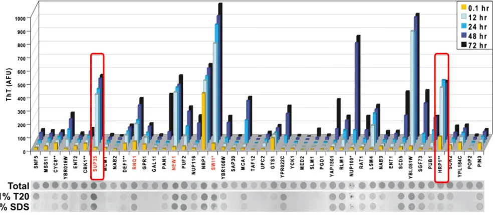
Alberti et al. (2009) performed a proteomic screening in Saccharomyces cerevisiae, searching for prion candidates by using a Hidden Markov Model (HMM) algorithm11. As a training model, Sup35 and Ure2 yeast prions were used. The former is a translation termination factor, while the latter is involved in Nitrogen metabolism. Both of these prions are enriched in PolyQ and PolyN stretches, respectively. These protein candidates were able to bind to thioflavin T dye (thT dye) over time, a property or feature known for the amyloid proteins. However, unlike amyloids, they depolymerize at very low detergent concentration (2% SDS)11, implying that these protein assemblies are dynamic (Figure 1.1). They can switch from a soluble phase, the protein solution, which has the viscosity of water, to a more rigid state, the amyloid-like structure. Moreover, the implication of PolyQ/N in the formation of RNA granules has been elucidated. RNA binding proteins within these assemblies form fibrillar structure with amyloid like properties. PolyQ/N domains are intrinsically disordered and aggregation-prone12,13. Lindquist et al. (2011) revealed via mutational experiments that the replacement of Q’s with N’s stimulates the formation of benign self-templating amyloids, whereas the substitution of N’s with Q’s triggers the formation of toxic, non-amyloid aggregates14. PolyQ stretches in Htt-exon1 (Huntington disease) protein causes
neurodegenerative disorders above a threshold of 35 Qs. In contrast, ataxin-3 at a PolyQ length well above 50 Qs remains soluble and non-pathogenic13. The question that emerges is how the arrangement of the linear information (the primary sequence) could influence the behaviour and the nature of the protein. What is the stimulus, the switch or the fundamental principles that lead to such differences?
Figure 1.1. The features of prion like proteins
The prion protein sup35 binds to Tht dye over time and do not dissolve upon treatment with low concentration of detergent. Whereas, the prion like protein Hrp1, similarly to sup 35 binds to Tht dye, but dissolve under low concentration of detergent SDS (adapted from 11)
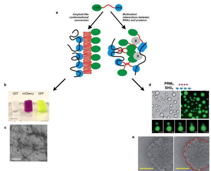
LCSs have been shown to undergo phase separation, where a liquid state, droplets, or gel-like material, hydrogel, formed (Figure 1.2.a). Hydrogels contain mainly water that tends to swell the gel and removing the water from the inside requires external forces. However, it has not yet been deciphered what are the arrangements and rearrangements of the primary sequences that trigger either the gel-like or the droplet states. Besides, it is still enigmatic why the cell chooses the liquid matter or the gel-like matter to convey information, signals or to organize heterogeneous matter in different organelles with different physical properties.
Han et al. (2012) revealed that the polymerization of LCSs triggers the formation of RNA granules, which consist of prion-like proteins, and consequently serve to localize mRNAs and in return can control spatial and temporal nature of translation. However, the structural transitions that prion-like proteins undergo and the regulation of these transitions are unknown3,4. Han and co-workers revealed that the LCSs of the prion-like proteins undergo phase transitions, resulting in the formation of hydrogels. Their observation was confirmed using the N-terminal of FUS protein (fusion in sarcoma) as a model. The N-terminal domain of the RNA binding protein FUS consists of 27 repeats of the amino acid sequence Gly.Tyr.Ser. At high protein concentrations (~ 50 mg /ml), following incubation for one week at 4 °C, a FUS hydrogel was formed. Hydrogel formation takes place via homotypic or heterotypic interactions of the prion-like proteins, meaning, promiscuous interactions between prion-like proteins that have the same or distinct identities (Figure 1.2.b). The X-ray diffraction pattern of hydrogels shows a characteristic cross-β structure, where X-ray reflections were observed at 4.6 A° and 10 A°, and transmission electron microscopy (TEM) shows a well-defined filament` (Figure 1.2.c). Similarly, the multivalent cohesive FG repeats (Phenylalanine-Glycine repeats) of the nuclear pore complex (NPC) was found to form a gel-like material at high concentrations namely 200 mg/ml with incubation for 24 hours at room temperature (RT). More importantly, this study found that the NQ sequence of the FG repeats triggers protein–protein interactions and that the FG-hydrogel is stabilized by β-structures15,16. However, the question remains, does the intrinsically disordered region, namely the LCS PolyQ/N, form a hydrogel β-structure in the cytoplasm? Do macromolecular complexes of LCSs-containing proteins form gels in the cell?
In contrast to limited evidence of RNA binding proteins forming gels in vitro liquid droplets composed of these proteins have been demonstrated to form via liquid-liquid phase separation (Figure 1.2.d). The droplet is recognizable as separate and soluble aggregates of materials having different physical properties from those of the surrounding bulk; the cytosol. The phase separation enables the component of a biological system to rapidly concentrate in one place and the entry of other proteins or regulator could trigger the disassembly.
An interesting example of a droplet is the germ cell P-granules of Caenorhabditis
elegans (C. elegans), which localize to the posterior of one cell-embryos and display
liquid-like properties including wetting, dripping and fusion. The condensation and the dissolution of these assemblies, called P-granules, are triggered by a Mex5 protein gradient in the cytoplasm. Increasing the concentration of Mex5 within P-granule droplets led to dissolution of the droplets. FRAP analysis (Fluorescence Recovery After Photobleaching) revealed that the components forming these droplets are very dynamic, where exchange of material takes place between the bulk and the droplets on timescales of seconds to minutes17–19. Another interesting example is the storage of mTORC protein within stress granules, which inhibits its kinase. The activation of the DYRK3 kinase disassembles the stress granule and releases mTORC for signalling20.
The collective behaviour of the prion-like proteins forming droplets is still enigmatic. A recent study demonstrated that multivalency is essential for droplet formation21. Multiple
weak interactions, namely protein-protein and protein-mRNA interactions, lead to phase separation and droplets formation. This study used multimodular domains such as the SRC homology 3 (SH3) and Proline Rich Motif (PRM), where a tandem repeat of SH3m and PRMn
(m,n :1-5) were separately engineered. The high affinity of SH31-PRM1 monovalencyblocks
the phase transition, but by increasing the valency SH34-PRM4 after few hours at high protein
concentrations, phase separation occurred and droplets formed (Figure 1.2.c). Moreover, droplets were seen after two days by mixing the Polypyrimidine Tract Binding Protein (PTB) with RNA oligonucleotides. More importantly, the droplet was observed in vitro and in vivo as well and the cryo-electron microscopy analyses of the droplets display a well-defined ring shape (Figure 1.2.e)21. These observations suggest that the affinity of the interacting proteins and the valency are the tipping points for phase separation. The morphology, the structure and
physical properties of the liquid state formed via multiple weak interactions are completely different from that of the hydrogel.
Figure 1.2. The morphological and physical differences between gel-like and liquid-like materials
a) Prion-like proteins containing LCSs and RRMs tend to polymerize and form amyloid like structure or to undergo multiple weak interactions and undergo phae separation to form droplets. b) The LC domain of FUS protein labeled with different fluorophore, incubated for one week at 4 °C. c) Multiple week interactions between multimodular domains undergo phase transition and form droplets with various sizes. d) Cryo-electron microscope exhibits a ring morphology (Adapted from 3, 21, 22)
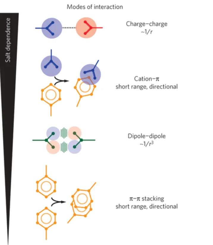
In addition, several recently published studies revealed that the LCSs alone or in concert with RNA undergo liquid-liquid phase separation (LLPS), where different physical states with different morphologies are formed. LLPS is modulated by many variables including increased protein concentrations22, posttranslational modifications23,24, decreased temperature25, environmental crowding26–28, decreased salt counter ions29 and RNA22,30. Numerous studies have highlighted the contribution of long-range (the electrostatic interactions), and the short-range interactions, as driving force for phase separation. In long-short-range interactions, charged motifs interact with each other, as in the case of the P-granule disordered protein LAF130 and the RNA binding protein DDX425. In addition, DDX4 undergoes short-range interactions, where the blocks of aromatic residues, namely Phenylalanine (F) and Tyrosine (Y), interact with the positively charged Arginine (R). Furthermore, the FUS protein undergoes directional short-range π-π stacking interactions, between F residues, as well as dipole-dipole interactions between the G residues (Figure 1.3)31,32.
Figure 1.3 Different types of interactions that trigger liquid-liquid phase separation Long-range interactions, the electrostatic interactions, and short-range interactions, namely cation-π, π-π and dipole-dipole, were found the essence of liquid-liquid phase separation (Adapted from 31)
For decades, RNA was considered as a decoder for genetic information. However, recently the polyanionic nature of RNA was found either to trigger or to prevent the formation of liquid droplets and to have an impact on the physical properties of these droplets. Under which circumstances RNA could perform either role is still enigmatic. For instance, RNA decreases the viscosity of the P-granule disordered protein LAF130. On the other hand, the viscosity of the Whi3 protein in Ashbya gossypii is tunable by RNA depending on the transcripts involved and on their concentration. The protein Whi3 contains an expanded stretch of Q residues and binds to two distinct mRNAs, CLN3 and BNI1. Depending on the mRNA identity, Whi3 protein forms two distinct organizations with different viscosities, fusion event and exchange rate with the bulk22. In addition to the previously mentioned function of RNA, it was also found that RNA delays the “aging” event of Whi3 proteins, in which the protein undergoes a conformation change, which is reflected by the transition from liquid to gel states22. The gel-like state is usually associated with the pathogenic state, the fibrous conformation. However, the liquid state or the phase separation that the protein undergoes is not an obligatory intermediate for fibrillization. For instance, hnRNPA1 can form fibrils without going through the liquid state33–35.
Ultimately, the liquid states have different component rearrangements, where the components of the liquid state can easily reshuffle, while the degree of freedom of the solid-state component is low and the component reshuffling is slow. For instance, water undergoes phase transition to form a liquid state with different physical properties to the solid state. At present, the arrangement of the primary sequences that triggers either gel-like or droplet state is still enigmatic. In particular, the structure-function relationship of macromolecule assemblies that can undergo phase transition is poorly understood. For instance, the cytoplasmic bodies in yeast namely stress granules and P-bodies possess different physical states. The former was found to have a gel-like state while the latter possess liquid states36. In this study, our first aim was to understand whether prion-like proteins containing the LCS PolyQ/N undergo phase transitions and to determine chemical and physical factors that stimulate or trigger phase separation (liquid-solid transition or liquid-liquid phase separation). We chose as a tool, the prion like protein Hrp1.
1.2 The Prion like protein Hrp1 forms visible aggregates
The protein Hrp1 is an mRNA binding protein, containing two RRMs and a PolyQ/N stretch at its N-terminus and an RGG Box at its C-terminus. It is involved in RNA metabolism37. More precisely, it is implicated in 3’ end processing event, such as cleavage and polyadenylation38. It shuttles between the nucleus and the cytoplasm, and contributes to the export of mRNA39. Hrp1 is posttranslationally modified by methylation of the Arginine residues within the RGG box, which affects protein-RNA or protein-protein interactions or the stability of the protein40.
Hrp1 was found by Alberti et al. (2009), to be a prion-like protein candidate containing the prion-forming domain PolyQ/N. It forms dynamic visible aggregates, which dissolve upon treatment with low concentrations of detergent, namely 2% SDS and binds to thioflavin T dye over time, suggesting that it ages to form fibrils. (Figure 1.1, Hrp1 is highlighted in red)11. Moreover, Hrp1 is implicated in the assembly of stress granules in yeast and is considered as a core component of these cytoplasmic structures4,41. In yeast, non-translated mRNAs could be recruited to these RNP-granules. P-bodies act as mRNA storage and contain mRNA degradation machinery, while stress granules consist mainly of the translation initiation components, such as, eIF4E, eIF4G, eIF4A, eIF4B, PolyA binding protein, eIF3, eIF2 and the 40S ribosomal subunit42. Stress granules are formed by inhibition of translation initiation, coexist with P-bodies and are known to undergo phase separation. A recent paper demonstrated the architecture and the organization of stress granules assembly. Stress granules consist of a stable core, formed by a network of protein-protein interactions that is surrounded by a dynamic shell. ATP influences the dynamic and the assembly of the granule. CCT, RVB and MCM ATPases modulate different steps in granule assembly and disassembly43,44. Stress
granules are known to be associated with neurodegenerative diseases, where an increase in the formation of stress granules is observed due to mutations occurring within proteins that cause the disease. For instance, the TDP43 protein associates with stress granules and mutations in TDP43 alter the dynamic of these assemblies45,46. Unlike P-bodies, stress granules possess a solid-like state. However, stress granules aggregate but do not display an amyloid features36. The solid-like state of stress granules and the liquid-like state of P-bodies are discernible using the compound 1,6 hexandiol. Unlike P bodies, stress granules do not dissolve in cells treated
with 1,6 hexandiol, suggesting that stress granules have solid-like nature. How and why hexandiol triggers the dissolution of P-bodies and has no influence on stress granules is not yet clear36. It is possible that 1,6 hexandiol disrupts weak interactions in liquid state but has no effect on strong interactions in the solid state. A better understanding of the assembly and the composition of P-bodies and stress granules are necessary to address these issues.
1.3 The P-body is a liquid droplet
Non-translated mRNAs are recruited into mRNP granules that are known as P-bodies or stress granules. More importantly, mRNA degradation competes with translation initiation both of which are triggered by mRNA decapping and shortening of the polyA tail. Moreover, the cap binding protein eIF4E inhibits the decapping factors Dcp2 and Dcp1 in vitro and in
vivo, which are P-body components41,42,47–49.
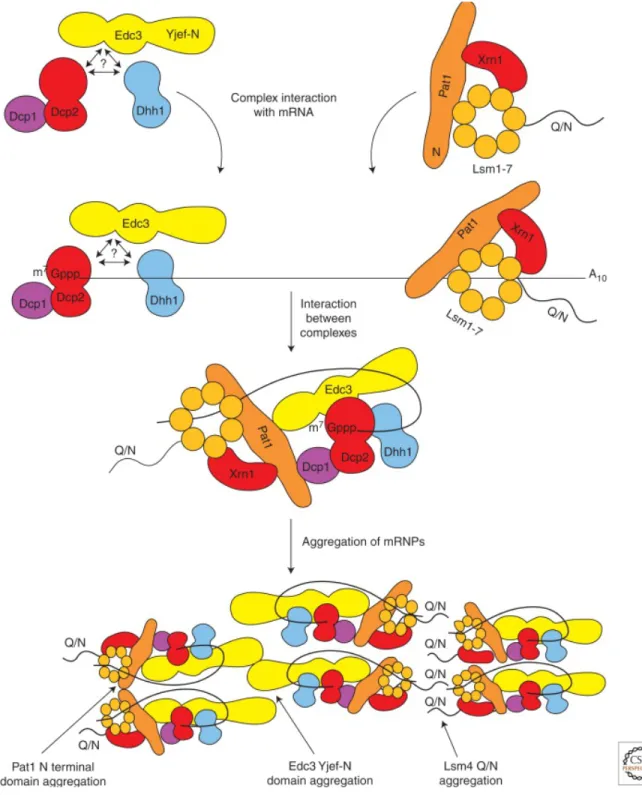
As processing bodies, P-bodies are cytoplasmic granules that harbour the RNA degradation machinery and mediate translation repression. They contain mainly mRNA and RNA binding proteins with Low complexity domains PolyQ/N, known as prion-like domains. In addition, proline-rich motifs are located either within the LCS stretches or downstream. The proline-rich motifs provide flexibility to multimodular domains (LCS) to undergo multiple promiscuous interactions with their partners42,50. P-bodies consist of sub-complexes of proteins that are responsible for mRNA deadenylation, translation repression, decapping and degradation. mRNA decay occurs by two different pathways. First, shortening the PolyA tail of the mRNA to 12 Adenine nucleotides is mediated by the deadenylase CCR4/Pop2/Not complex, and then the exosome mediates the mRNA degradation in the 3’ to 5’ direction. Alternatively, the shortened mRNA is decapped by the enzymes Dcp2 and Dcp1 and then degraded from 5’ to 3’ by the exonuclease, Xrn1. The core components of P-body, which are also known as enhancers for Dcp2 and Dcp1enymes, such as Dhh1, Pat1p, Edc3 and the Lsm1-7 complex, affect the decapping factors. For instance, Edc3 and Pat1p binds to Dcp2 decapping factor and increase its activity42.
Notably, P-bodies are multicomponent membrane-less organelle, where hundred of proteins are involved in the assembly, however few molecules called the scaffold proteins maintain the structure of these assemblies via protein-protein interactions. However, the composition and the assembly of these collectives are still largely not yet clear. P-bodies in yeast containing
non-translating mRNA are dynamic structure and function during cellular stress. For their formation they depend on the amount of the non-translated mRNA and their size and number alter in response to stress such as glucose deprivation, osmotic stress, exposure to UV and the stage of cell growth. Glucose deprivation leads to rapid inhibition of protein synthesis and inhibition of translation initiation, which results in increase of P-body51,52. It has been suggested that mRNA triggers the assembly of P-bodies, where protein complexes are formed independently in the cytoplasm and then recruited sequentially to the mRNA forming mRNP particles. Consequently, aggregation of mRNP particles via protein-protein interactions forms a larger collective, the so-called P-body. For example, Dcp1/Dcp2/Edc3 or alternatively Dcp1/Dcp2/Dhh1/Scd6 and Lsm1-7/Xrn1/pat1 are recruited to the mRNA in an unknown order. Subsequently the 5’ end of the mRNA binds Edc3, Dcp2 and Dhh1 and interacts with Pat1 that binds to the 3’ end of mRNA and forms a closed loop. In addition, the aggregation of mRNP particles in P-bodies may be driven by the interaction of the self-interaction domain (Yjef-N) in Edc3 and the C-terminus of Lsm4 that contains a PolyQ/N stretch (Figure 1.4)42. The organization of these prion-like proteins within P-bodies is not well characterized. However, Fromm et al. (2014) were able to reconstitute in vitro the liquid phase of P-body core component, such as, Dcp2, Dcp1, Edc3 and pdc1 in Schizosaccharomyces pombe. Dcp1 activates the decapping enzyme Dcp2 and the remaining proteins are known to form the scaffold of the processing body. The core is built via a network of promiscuous interactions between the 10 HLM (helical leucine Motif) in Dcp2, Pdc1 and the Edc3-lsm domain. Disruption of these interactions prevents the coalescence of the liquid phase in vitro53. Lastly,
a study made by Banani et al. (2016), shed light on the fundamental rules that determine the composition of the phase separated P-bodies in the cell. RNP particles consist of a scaffold and recruits clients depending on the composition, the stoichiometry and the free sites of the scaffold. The clients are molecules with low valency and are much more dynamic than that of the scaffold54.
Finally, mRNA plays a crucial role in the assembly of P-granules. However, the cell contains other electrolytes including DNA, polyamine and polyphosphates that are known to impact the aggregation and disaggregation of numerous protein assemblies. Here, we addressed whether polyphosphate play a role in the assembly of P-bodies in the cell.
Figure 1.4 Model for P-body assembly
mRNA recruits protein sub-complexes such as the decapping machinery, the transalation repression complexes in an unknown order. The core component Edc3 binds to Pat1 and form a loop. And form an mRNP particle. Aggregation of mRNP takes place via LC sequence interactions between Edc3 and Lsm4 (Adapted from 42)
1.4 The role of the multivalent ionic compound polyphosphate in
the cell
Polyphosphate (PolyP) are a linear chain of negatively charged phosphorous moieties connected through energy rich phosphoanhydride bonds55. They were first discovered in the cytoplasm of the bacterium Spirillum volutans as metachromatic granules and were given the name volutin, which were stained pink by a basic blue dye56. PolyP are also found in different living organisms such as bacteria, fungi, plants and animals57. The chain of PolyP could range from 3 to thousands of residues. Acid-soluble PolyP with a short chain is found at the cell surface, in the periplasm and in the plasma membrane. In bacteria, PolyP are generated by polyphosphate Kinases (PKK1). PKK1 uses ATP as a substrate to catalyze the polymerization of the PolyP chain. It transfers the gamma phosphate group from ATP to generate the negatively charged polymer PolyP. PKK1 is found in various organisms including archaea, fungi and yeast but not in mammals58,59.
PolyP are implicated in distinct biological processes, however, how these are related and the mechanism by which PolyP modulate these biological events are not yet known. For instance, PolyP serve not only as an energy source, but also act as phosphate storage molecules60,61, as a chelator for metal ion such as Ca2+ and Mn2+ 62, as a buffer against alkali ions63 and as a regulator of cellular stress64 . They also play various roles in many cellular events, such as in gene regulation, in signalling and in proliferation. In E.coli, PolyP bind to the Lon protease and form a complex that triggers the degradation of ribosomal proteins. Lon is a protease that is known to bind to DNA and is implicated in the regulation of several cellular events65. In addition, PolyP interact with basic positively charged proteins such as histones64. For instance, in the bacterium Caulobacter crescentus, PolyP interact with the positively charged polyphosphate kinase1 (PPk1). Moreover, in E.coli PolyP act as chemical chaperone, where they bind to numerous proteins and protect them against stress. Cellular stress induces aggregation and unfolding of proteins, but once the stress is released, PolyP promote refolding66.
It was recently shown in vitro that PolyP accelerate the formation of amyloid fibril proteins such as CsgA and α-synuclein in prokaryotes and eukaryotes, respectively. PolyP bind to the amyloid fibrils and alters their seeding capacity, their nucleation propensity and stabilizes the amyloid proteins in their β-sheet conformation. By contrast, PolyP increase the formation of biofilms in pathogenic bacteria and reduce the cytotoxic effect of amyloids in neuronal cells67.
In yeast PolyP are synthesized by the vacuolar transporter chaperone (VTC) and accumulates in the extracellular space and in the vacuole68,69. Yeast possesses 4 VTCs, a small transmembrane protein called VTC1 and VTC2, 3 and 4 that have transmembrane domains and a cytoplasmic segment. VTC4 is essential for the accumulation of PolyP in the cell. It produces PolyP from ATP. PolyP in yeast are catalyzed by two exopolyphosphatases, Ppx1 and Ppn1. The latter catalyzes long chains of PolyP and generates a small stretches, while the former catalyzes the end of PolyP and releases orthophosphates (Figure 1.5)59,60,70. PolyP in yeast were found to regulate the level of phosphate in the cytoplasm71. Shirama et al. (1996) showed that the phosphate in the cytosol remains constant after 4 h of phosphate starvation, but the PolyP in the vacuole decrease during this time. However, the amount of inorganic phosphate in the cytosol begins to be depleted after 6 h of starvation72–74. Moreover, PolyP in
yeast sequester amines such as Arginine75, in the vacuole and acts as cytoplasmic pH
regulator.
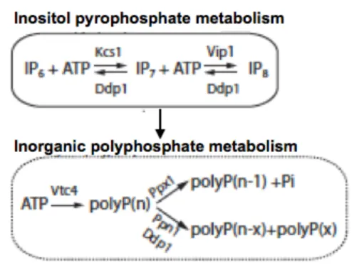
Importantly, PolyP and inositol pyrophosphate (IP7) are metabolically linked. IP7 is
generated by Kcs1, and deletion of Kcs1 decreases the amount of Polyp in the cell. However, PolyP have no effect on IP776. The products of inositol pyrophosphate pathway such as IP4,
IP6, IP7 and IP8 are involved in numerous cellular events such as gene expression, maintaining
the telomere length, chromatin remodelling, RNA export and endocytic trafficking77–88. IP7
and PolyP modify proteins non-enzymatically via post-translational modifications86,89. IP7
pyro-phosphorylates a Pre-phosphorylated serine, while the poly-phosphorylation takes place on a lysine. For instance, the nuclear signal recognition 1 protein (Nsr1) that is involved in rRNA biogenesis and its interacting partner topoisomerase1 (Top1) in yeast are poly-phosphorylated. The phosphorylation takes place on a lysine residue in the N-Terminal Polyacidic cluster, enriched in Serine (S) and Lysine (K)76. Protein modification via
phosphorylation regulates cellular events and signalling pathways, and consequently modulates protein-protein interactions and localization. Regarding the liquid state, it was shown that post-translational modifications such as phosphorylation, could triggers condensation or dissolution of membrane-less organelles23,24. Rosen and co-workers demonstrated that increasing the phosphorylation sites of a peptide, that represent an artificial model of the transmembrane protein nephrin, leads to phase separation21. However, the effect of polyphosphate and Kcs1 on RNA granules assembly is unknown. Hence, I investigated whether PolyP play a role in mediating phase transitions in the cytoplasm.
Figure 1.5 Pathway of inositol pyrophosphate and polyphosphate metabolism
1.5 Research project
1.5.1 Problematic and hypothesis
Prion-like proteins containing low complexity domain PolyQ/N are found to form visible aggregates that unlike the traditional known amyloid aggregates, they dissolve under low concentration of detergent11. A study suggests that the LCS has the propensity to aggregate, to polymerize and to form a solid-like state, the hydrogel. The solid-like state was formed under extremely non-physiological condition via homo and heterotypic interactions. Another study proposed an alternative scenario demonstrating that LCSs are able to undergo multiple weak promiscuous interactions resulting the formation of the droplet, via liquid-liquid phase separation mechanism21. Regarding the secondary structure of the solid-like state and the liquid-like state, the former was found to form a β-secondary structure with filamentous morphology, while the latter was reported to form coiled-coils with a ring shape.
The study established by Alberti et al. (2009), aiming to identify prion-like domain in
S. cerevisiae, discovered 200 protein candidates with prion-like domain containing
LCS-PolyQ/N that form visible aggregates in vitro. Among these proteins are mRNA binding proteins that dissolve in the presence of the non-denaturing condition 0.1%T20 and 2% SDS. Moreover, the effect of the thioflavin (Tht) dye is absent or delayed well over 24 hours. One example is the mRNA binding proteins Pop2 and Ccr4, which are components of P-body. In contrast, the mRNA binding protein Hrp1 was found to bind to Tht dye after 0.1 hour, to be retained in 0.1% T20 but to dissolve in low detergent concentration (2% SDS). The properties and the nature of these aggregates were not yet identified. At first, the study established by mcknight et al. (2012) claims that LCS forms hydrogel. In contrast, Rosen et al. (2012) claims that LCS forms droplets. I initiate my study by hypothesizing that mRNA binding proteins containing LCS form droplets in the cell. As a prove of concept, I chose the mRNA binding protein Hrp1 as an artifitial model, first to shed the light on the LCS and their function as a force to generate droplet, and second to solve the conundrum concerning the nature and the organization of the collectives containing LCS in vitro and in vivo.
In vivo, the assembly of these collectives is triggered by the crowded nature of the cytoplasm26, by the post-translational modifications such as phosphorylation or methylation25, by the protein gradient concentration within the cell90 and more importantly by the negatively charged RNA29. Numerous studies have shown that the RNA could trigger or inhibit the assembly of the droplet in the cell. More importantly, the polyelectrolyte RNA was shown to dictate the biophysical properties of these assemblies. It is not surprising, that the polyelectrolyte RNA plays a crucial role in the assembly of the liquid matter in the cell and is most likely the driving force for droplet condensation. The multivalent ion salts, the negatively charged/positively charged polyelectrolytes are used in the industry to coalesce and flocculate colloids in order to clarify wastewater. Moreover, they are also used to change the biophysical properties of the polymer-based colloids or so-called smart materials that have applications in drug delivery, sensor design and smart emulsifiers91.
Similarly, the cell uses biopolymers to organize membrane-less organelles. The cell contains a varietiy of biopolymers such as RNA, DNA, polyamines and polyphosphates that are known to influence the aggregation and disaggregation of numerous protein assemblies. For instance, the polyamine, spermidine, is known to condense the DNA and to trigger the aggregation of distinct proteins such as polyglutamine (PolyQ) protein, α synuclein, and the F-actin92,93. It was reported that spermidine triggers the aggregation of F-actin, while the nucleoside phosphates disassemble the aggregation. The negatively charged phosphates could act as an antagonist to the positively charged amines in inducing protein condensation94.
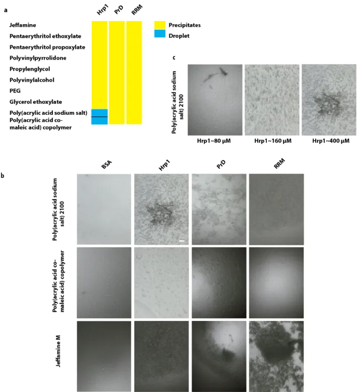
Based on an initial study, I demonstrated that the prion like protein Hrp1 containing LCS PolyQ/N forms droplets in the presence of negatively charged polyelectrolyte such as polyacrylic acid. In addition, ample of studies have shown the formation of protein droplets containing LCS in vitro in the presence of the negatively charged polyelectrolyte, the RNA. Hence, I hypothesized, that the negatively charged polymer under which Hrp1 forms droplets could mimic the negatively charged polyelectrolyte such as RNA, DNA and PolyP in the cell. Based on these observations, I proposed to explore the potential and the universal role of the highly abundant polyelectrolyte PolyP as a regulator in the condensation/dissolution of membrane-less compartment forming liquid droplet in the cell initiating with the P-body, knowing that P-bodies consists of mRNA binding proteins containing LCS and posses liquid nature in vivo.
1.5.2 Objectives
To obtain insights into whether prion-like proteins undergo liquid-solid or liquid-liquid phase separation, whereby a gel-like material or a droplet develops respectively, I chose the prion- like protein Hrp1 as a model. Specifically I investigated:
• Whether the purified Hrp1 undergoes phase separation under different chemical conditions that trigger the formation of droplets using a high-throughput screen. • Whether the prion-like domain (PrD) or the RRM of Hrp1 is responsible for the phase
separation under the same conditions.
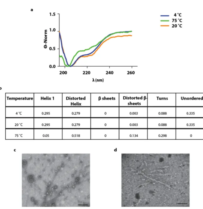
We found that the PrD alone formed a gel-like material that has a cross-β secondary structure. In contrast, another study, suggests that PrDs form droplets via multiple weak interactions that are mediated by coiled-coil secondary structure. Hence, I investigated the secondary structure and the morphology of Hrp1 full-length protein and its PrD domain using Circular Dichroism (CD) and Transmission electron microscopy (TEM).
Recent studies have shown that the negatively charged RNA triggers formation of droplets by phase-separation. Notably, I found that negatively charged polyelectrolytes, which promotes the formation of Hrp1 droplets, mimics RNA and I hypothesized that the highly abundant polyelectrolyte PolyP regulate P-body formation via phase separation and investigated:
• Whether depletion of phosphate has an effect on P body formation
• Whether depletion of PolyP from the cell would prevent P-body formation • Whether PolyP colocalize with P-bodies in the cytoplasm
1.5.3 The techniques used in this study
In this Study different techniques were used to investigate the potential role of PolyP in P-body formation. First to investigate whether prion-like proteins undergo phase separation, the
protein of interest Hrp1 was purified via affinity chromatography using NI-NTA column and Size Exclusion Chromatography (SEC). I then tested for phase separation under a variety of conditions, including the presence of polyelectrolytes. Subsequently, using light microscopy, I monitored the formation of droplets. To obtain insights on the organization and the conformation of the heterogeneous matter within the droplets, Circular Dichroism (CD) and Transmission electron microscopy (TEM) were performed on the purified proteins Hrp1 and its PrD domain.
After elucidating the conditions under which Hrp1 protein forms droplets. I verified whether PolyP have an effect on the condensation and dissolution of P-bodies. Specifically, I determined whether PolyP affect the number and size of P-bodies labeled with mCherry Edc3 using fluorescence microscopy.
2.1 Media
For protein overexpression, E.coli Bl21 (De23) was cultured in terrific Broth media (TB) containing 12 g/l of tryptone, 24 g/l yeast extract and 4 ml glycerol in addition to a salt solution separately autoclaved consisting of 0.17 M KH2PO4 and 0.72 M K2HPO4 .
For the phosphate and glucose deprivation experiment, synthetic media was mainly used. The composition of synthetic media was 0.17 % yeast Nitrogen base, 0.5 % ammonium sulphate, 2 % glucose and amino acid.
For phosphate depleted synthetic media, the phosphate was precipitated using 1 M MgSO4 with high concentrated ammonium hydroxide and kept at RT for 30 min followed by a
filtration.
2.2 Strains and Plasmids
In this study, bacteria and yeast strains as well were used. DH5α and BL21 (De3) were used for cloning and for protein expression of Hrp1 and its variants, respectively. On the other hand, the yeast strains BY4741-mcherry Edc3 and BY4741-mcherry Edc3-vtc4Δ were used for P body analysis.
Table 2.1. Bacterial and yeast strains
Strain Genotype Reference
DH5α F– Φ80lacZΔM15 Δ(lacZYA-argF) U169
recA1 endA1 hsdR17 (rK–, mK+) phoA supE44 λ– thi-1 gyrA96 relA1
Common laboratory strain
E.coli Bl21(De3) B F– ompT gal dcm lon hsdSB(rB–mB–) λ(DE3
[lacI lacUV5-T7p07 ind1 sam7 nin5]) [malB+]K-12(λS)
Common laboratory strain
BY4741 MATa his3Δ1 leu2Δ0 met15Δ0 ura3Δ0 Common
laboratory strain BY4741-mcherry Edc3
MATa his3Δ1 leu2Δ0 met15Δ0 ura3Δ0:hphR Jacqueline Kowaryk
BY4741-mcherryEdc3- vtc4Δ
MATa his3Δ1 leu2Δ0 met15Δ0 ura3Δ0:nat-1R
In this study
The used plasmids were either for protein expressions in vitro namely Hrp1 and its variants or to delete the gene that is responsible for PolyP synthesis vtc4. For this purpose, pet24d(+) and PAG25 as well were utilized, respectively.
Table 2.2. Plasmids for protein expressions and gene deletion
Plasmids Genotype Reference
Pet24d (+) Protein overexpression Dr. Christian Baron
PAG25 Cassette containing the
selection marker CloNat to delete the gene of interest via homologous recombination
Common laboratory plasmid
2.3 The designed primers for different experiments
Primers were designed to amplify the genes of interest namely Hrp1 and its variants from yeast genomic DNA and Hrp1 plasmid construct, respectively. Hrp1 and RRM fragment were amplified using PCR, whereas the PrD was amplified via SOE-PCR.
Moreover, primers were designed to delete vtc4 gene using PAG25 as a template and to perform a diagnostic PCR in order to confirm the successfulness of the deletion.
The red highlighted sequences are the restriction enzyme sites.
Table 2.3. PCR primers for in vitro and in vivo studies
Primers Forward (5’ ! 3’) Reverse (5’ ! 3’)
Hrp1 CGGCGTACGAGCTCTGACGAA
GAAGAT
CGCGGATCCTTACCTATTATATGGAT
GGTAGCCATT
AGAT CTTT
PrD –Fragment2 AAAGCGGATTTGTCTGCTGAGCCA
AGACAT CGCGGATCCTTACCTATTATATGGAT GGTAGCCATT Hrp1-RRM CGGCGTACGAAAGAAAGTTGCAA GATG CGCGGATCCTTATCTCTTGATTTCGAT Vtc4 GCTAACAATCAAATCGGCCAATAA AAGAGCATAACAAGGCAGGAACA GCTAGATCTGTTTAGCTTGCCTTGT CCCCG TTATTACTTAATTATACAGTAAAAAA AACACGCTGTGTATTTCGACACTGGA TGGCGGCGTTAG Vtc4-diagnostics primers CCAGATGCGAAGTTAAGTGC CGCCTGGTGAAGGTGTGC
2.4 Extraction of yeast genomic DNA
The yeast By4741 was grown overnight in 10 ml YPD media (1 % yeast extract, 2 % peptone, 2 % dextrose) at 30°C and 250 rpm to saturation. The cells were harvested by centrifugation at 4000 rpm for 5 min. The supernatant was removed, the pellet was re-suspended in 500 µl distilled water and centrifuged at 13 000 rpm for 30 sec.
For cell disruption, 200 µl of Lysis buffer (2 % Triton X-100, 1 % SDS, 100 mM NaCl, 10 mM Tris pH 8 and 1 mM EDTA), 200 µl of Phenol chloroform and washed glass beads were added to the pellet. The latter was lysed by vortexing about 3 to 5 times for 1 min each and kept for 1 min on ice in between the vortexing events. Subsequently, 200 µl of TE buffer (10 mM Tris pH 8 and 1 mM EDTA) was added and the lysate was clarified via centrifugation at 13000 rpm for 5 min. To precipitate the DNA, 1 ml of 100 % Ethanol was added to the supernatant and inverted allowing mixing. Next, it was centrifuged for 2 min at 13000 rpm, re-suspended in 400 µl TE buffer containing 30 µg of RNaseA and incubated at 37°C for 30 min. Furthermore, 10 µl of 4 M Ammonium acetate and 1 ml of Ethanol was added to the sample and then placed at – 20°C for 30 min to allow precipitation. Ultimately, the sample was centrifuged, air-dried and re-suspended in 30 µl TE buffer.
The extracted genomic DNA was verified or detected by electrophoresis on 1 % agarose gel in TAE buffer (40 mM Tris-acetate, 1 mM EDTA, pH 8.2).
2.5 Overexpression and purification of Hrp1 and its variants
The gene encoding for Hrp1 Protein was amplified via PCR using extracted yeast genomic DNA. The amplified fragment was inserted in Pet24d (+) plasmid containing His-Tev Tag. In contrast, RRM and PrD were amplified via PCR and SOE-PCR respectively, using Hrp1 construct as a template. Similar to Hrp1, these fragments were also inserted in Pet24d (+).
The proteins of interest were all overexpressed in E.coli BL21 (De3) with 1 mM IPTG for 4 hours (hrs) at 37 °C and 300 rpm. The cells were harvested via centrifugation at 15000 rpm for 20 min and re-suspended in binding buffer containing 5-10 mM imidazole (20 mM Tris pH8, 500 mM NaCl and protein inhibitor cocktail). Subsequently, the cells were disrupted using the French-Press for two cycles followed by a centrifugation at 15000 rpm for 30 min. The supernatant was filtered and then loaded on 4 ml Ni-NTA packed resin. The column was washed with 5-column volume (CV) of binding buffer and the bound proteins were eluted stepwise from the resign using the elution buffer consisting of 500 mM imidazole (20 mM Tris pH8, 500mM NaCl).
The protein fractions were pooled together and dialyzed against the TEV protease enzyme buffer (10 mM Tris pH 8, 125 mM NaCl and 5 mM DTT). The dialysis was performed for 4 hours using the dialysis membrane and by changing the buffer hourly. Subsequently, the protein was treated with Tev-protease enzyme at RT for overnight. For the digestion reaction, 3 ug of protein were treated with 1 Unit (U) of enzyme. In order to verify whether the His-tag was cleaved, a 10 % of SDS-PAGE was performed.
The cleaved protein was dialyzed against equilibration buffer (20 mM Tris pH 7.5, 200 mM NaCl and 0.5 mM EDTA) and loaded on Size Exclusion Chromatography (SEC) to separate the protein of interest from the contaminants. The purified protein was concentrated with Amicon Ultra Centrifugal filters and stored at -80°C for further manipulation.
The purity of the purified proteins was verified via SDS-PAGE and the concentration was accessed using The BSA assay.
2.6 Circular Dichroism Spectroscopy
For CD spectroscopy analysis, Hrp1 and PrD were dialyzed against a buffer (50 mM NaCl, 20 mM phosphate buffer pH 7.5), followed by protein quantification using the BSA assay. The proteins of interest were diluted to the desired final concentration (2.5 µM) and the CD spectra were acquired using Jasco J810. The ellipticity of the proteins was measured in 0.2 cm quartz cuvette from 195 nm to 260 nm at different temperatures (4°C, 20°C and 75°C). The molar ellipticity [Θ] was calculated using the following formula;
! deg ∗ !"!∗ !"#$!! = !!"# !"#$
10 ∗ ! !"#! ∗ ! !"
Where Θ is the observed ellipticity obtained from the instrument, c is the concentration of the protein and l is the quartz cuvette diameter.
The obtained spectra were smoothed and corrected against the buffer and the secondary structure of the protein was determined using the ContinLL algorithm obtained from the Dichroweb website, which allows an online analysis of CD spectra.
2.7 Transmission Electron Microscope analysis
For TEM analysis, different protein concentrations were prepared from (1mg/l - 10 mg/ml), where 5 µl of the samples were loaded on a carbon-coated grid for 5 min at RT. Subsequently, the excess of liquid was removed using filter paper and then negatively stained with 2 % Uranyl acetate. Finally, the imaging was performed using Philips Tecnai 12 (Eindhoven, The Netherland), operated at 80 KV.
2.8 Design a cassette for homologous recombination to replace the
gene to be deleted with an antibiotic as a selection marker
The cassette for homologous recombination was prepared by amplifying the antibiotic selection marker Clonnat via PCR using the plasmid PAG25 as a template. The designed primers for the PCR were flanked with the 5’UTR and 3’ UTR of the gene to be deleted.
2.9 Homologous Recombination
Homologous recombination was carried out by first preparing competent cell of the strain, where the designed cassette will be transformed. Hence, a single colony of the strain of interest was inoculated in 5 ml YPD and incubated overnight at 30°C and 250 rpm. The next day, the optical density (OD) of the culture was determined by spectrophotometer at a wavelength of 600 nm and diluted in 50 ml of YPD media to an OD600 of 0.1. It was kept to
grow at 30°C and 250 rpm until an OD600 of 0.4-0.6 is reached. The cells were collected by
centrifugation at 4000 rpm for 5 min and the pellet was re-suspended and washed twice with 25 ml of distilled water. Next, the pellet was re-suspended with 1 ml of distilled water and centrifuged at 4000 rpm for 1 min. The supernatant was removed and again 1 ml of distilled water was added and aliquots of 100 µl were prepared for the transformation.
The prepared cell aliquots were centrifuged at 13000 rpm for 1 min, the supernatant was discarded and the transformation mix ((PEG 3350 (50 % (W/v)), Lithium Acetate 1 M, single stranded carrier DNA (2 mg/ml) and sterile water) was added. 8 µl of the amplified cassette was added to the competent cells in the presence of the transformation mix and re-suspended vigorously by vortexing. Subsequently, the tubes were incubated at 42°C in a water bath for 40 min and then centrifuged at 13000 rpm for 1 min. The cells were then re-suspended by vortexing vigorously after addition of 1 ml YPD and incubated at 30 °C for 2-3 hours. Finally, the cells were plated on YPD media containing the appropriated antibiotic selection marker and kept at 30°C for 2-3 days.
To verify whether the homologous recombination was successful and consequently the gene of interest was deleted, a diagnostic PCR was performed on the colonies that grew on the plate with the appropriate selection marker. To carry out the diagnostic PCR, single colonies were picked, dissolved in 20 µl of sterile water and incubated at 99°C for 15 min and 30 sec. Subsequently, the aqueous layer was used as a template to run the PCR and the primers were designed in a manner that bind to the genome and to the selection marker that replaced the gene of interest.
2.10 Observation of P-bodies in mid-log glucose starved cells
In this study, the P body formation was studied in BY4741-mcherry Edc3 and in BY4741-mcherry Edc3-vtc4Δ, depending on the experiment to be performed.
To study the P body formation, the strains were grown in 5 ml complete synthetic media at 30°C and 250 rpm to saturation, and then diluted in 50 ml synthetic media to an OD600 of 0.1.
The latter was allowed to grow till an OD600 of 0.3-0.4. The 50 ml culture was split in two and
washed twice at 3000 rpm for 3 min with either a complete synthetic media that serve as a negative control for P body induction or with synthetic media lacking glucose that induces P body formation. Next, the cultures were re-suspended in synthetic media with and without glucose and kept for 15 min at 30°C and 250 rpm. At last, imaging was performed using Nikon TE200U. For imaging, wells of a multiwall Matriplate were coated with concanavalin A (ConA). The ConA enables the cells to gently adhere the surface of the well.
Con A was left for 15 min at RT in the well, removed and then washed with distilled water. Afterwards, ConA was activated by addition of 20 mM MnSO4 and 20 mM CaCl2 and
incubated for another 15 min. The activation of ConA through cooperative binding of the metal ions Ca2+ and Mn2+ enables cell fixation, whereby ConA binds to the cell surface. The
solution was then removed and the wells were washed with distilled water.
In order to detect the P-body, Edc3 the core component of the P-body was tagged with mCherry. It was visualized at an exposure time of 100 ms and by performing a Z stack with a step size of 0.6 µm using the Nikon TE 200U Microscope. For statistical analysis almost 100 cells were analyzed using the software ImageJ 64.
To note, for the co-localization experiment of P body and PolyP, the cells were starved for 30 min instead of 15 min due to the incubation time of DAPI73,95. Based on the fluorescence scanning experiment Dapi was visualized using YFP filter.
2.11 Detection of PolyP with DAPI staining using the fluorometer
The wild type BY4741-mcherry Edc3 and BY4741-mcherry Edc3-vtc4Δ were grown in 5 ml complete synthetic media at 30°C and 250 rpm. It was diluted in 50 ml synthetic media to an OD600 of 0.1 and kept to grow under the same condition.To detect PolyP in living cells, DAPI fluorophore was used, where the cells were incubated with 10 µg/ml of DAPI and kept at 30 °C for 30 min. Afterwards, the cells were gently washed twice at 3000 rpm for 2 min and re-suspended in synthetic media.
The fluorescence of PolyP were followed at different OD600 and the fluorescence
excitation and emission was acquired using the spectrofluorometer, SpectraMaxGEMINIXS. The fluorescence spectra were obtained by exciting the sample at specific wavelength and by scanning the emission between 400 nm and 600 nm. The bandwidth between the excitation and emission is about 50 nm. For the fluorescence measurement, it is noteworthy that the cells were consistently diluted to an OD600 of 0.2 to avoid saturation of light diffraction.
2.12 P-body analysis and quantification of number and size
The average number of of P-bodies per cell was analyzed using the Image J software, where a maximal projection was performed on the obtained Z stacks, followed by processing the image using the find maxima tool. The obtained maxima, which are the P-bodies, were re-directed to the selected or segmented cells and the foci number per cell were measured. This method determines the number of P-bodies in each segmented cell.
Another analysis was performed to quantify the number of the P-bodies, where Otsu thresholding was used. A maximal projection was carried out, the image was smoothed and the background was subtracted. Next, the image was converted to 8-bit and Otsu thresholding was performed. The LUT was inverted and the particles were analyzed using analyse particle tool. The measurements were re-directed to the original file. Finally, a table containing information concerning the area (pixel^2), the mean intensity and the sum of pixel is obtained. The area in nm2 was calculated given every Pixel is 0.43 nm for a total magnification of 150. Consequently, the radius was calculated assuming a spherical P-body area where;
A =!!!!
To note, the detection was performed using an objective of 100x with 1.5 magnifier resulting a total enlargement of 150.
2.13 Statistical analysis
The Phosphate and glucose deprivation experiment were done in triplicate. The statistical analysis was performed using the Student t-test and Mann-Whitney U-test for population analysis. A P value of less than 0.05 was considered to illustrate a statistical significance. The used softwares for the statistics were Excel, Graphpad prism and R.
3.1 Polyanionic molecules induce liquid-liquid phase separation of
the RNA binding Protein Hrp1 in vitro
To investigate whether prion-like proteins containing the LCD PolyQ/N form droplets, the RNA binding protein Hrp1 was selected as a model. Recently, Hrp1 was reported to form visible aggregates, to bind to ThT (Thioflavin) dye and to dissolve in the presence of 2 % SDS11. However, the nature and the physical properties of these aggregates were not
previously known. Hence, the purified Hrp1 was subjected to varieties of conditions and then screened for droplet formation. The scheme of the procedure is summarized in Supplementary Figures (Fig.S 7.1).
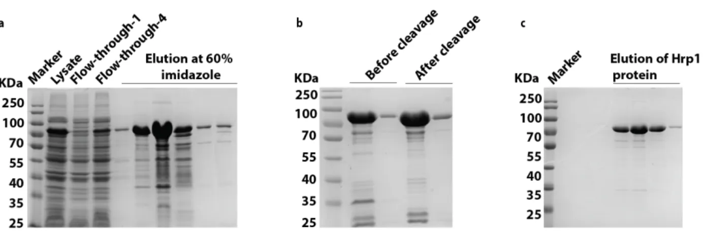
Hrp1 was purified via affinity chromatography using the NI-NTA column followed by Size Exclusion Chromatography (SEC) for further purification. Hrp1 was expressed in BL21 (De3) and eluted at 60% imidazole during NI-NTA purification (Figure 3.1.a). To rule out the possibility that droplet formation is hindered or induced by the His-Tev-Tag, the tag was cleaved with TEV-protease overnight at RT (Figure 3.1.b). Next, SEC enabled the separation of Hrp1 protein from the remaining contaminant proteins that have similar affinity to the NI-NTA column (Figure 3.1.c). Consequently, a pure protein was obtained. However, Hrp1 protein was found to migrate at higher molecular weight (MW) (~75 KDa) than expected (~59 KDa, as reported in SGD) (Saccharomyces Genome Database).
Figure 3.1. Hrp1 purification approach
a) Hrp1 protein expression and NI-NTA purification step. The lysate of Hrp1 was subjected to a pre-packed 4 ml NI-NTA column. The protein of interest was eluted with 60% imidazole, which is equivalent to 300 mM imidazole. b) Cleavage of His-tev tag from Hrp1 using Tev protease enzyme. Lanes 2 and 3 represent the pattern of two different concentrations of Hrp1 before cleavage and lanes 4 and 5 represent the pattern after cleavage. The difference of MW band migration before and after cleavage is approximately 1 KDa. c) Secondary purification of Hrp1 using SEC. Hrp1 was loaded on SEC to remove the contaminant proteins