(1)PLOS ONE
Moderate to severe acute pain disturbs motor cortex intracortical inhibition and
facilitation in orthopedic trauma patients: A TMS study
--Manuscript
Draft--Manuscript Number: PONE-D-19-32702R2
Article Type: Research Article
Full Title: Moderate to severe acute pain disturbs motor cortex intracortical inhibition and
facilitation in orthopedic trauma patients: A TMS study
Short Title: Acute pain in orthopedic trauma disturbs motor cortex intracortical inhibition and
facilitation
Corresponding Author: Louis De Beaumont
Universite de Montreal
Montréal, CANADA
Keywords: Pain intensity; primary motor cortex; cortical excitability; fracture; transcranial
magnetic stimulation.
Abstract: Objective: Primary motor (M1) cortical excitability alterations are involved in the
development and maintenance of chronic pain. Less is known about M1-cortical
excitability implications in the acute phase of an orthopedic trauma. This study aims to
assess acute M1-cortical excitability in patients with an isolated upper limb fracture
(IULF) in relation to pain intensity.
Methods: Eighty-four (56 IULF patients <14 days post-trauma and 28 healthy
controls). IULF patients were divided into two subgroups according to pain intensity
(mild versus moderate to severe pain). A single transcranial magnetic stimulation
(TMS) session was performed over M1 to compare groups on resting motor threshold
(rMT), short-intracortical inhibition (SICI), intracortical facilitation (ICF) and long-interval
cortical inhibition (LICI).
Results: Reduced SICI and ICF were found in IULF patients with moderate to severe
pain, whereas mild pain was not associated with M1 alterations. Age, sex, and time
since the accident had no influence on TMS measures.
Discussion: These findings show altered M1 in the context of acute moderate to
severe pain, suggesting early signs of altered GABAergic inhibitory and glutamatergic
facilitatory activities.
Order of Authors: Marianne Jodoin
Dominique M. Rouleau
Audrey Bellemare
Catherine Provost
Camille Larson-Dupuis
Émilie Sandman
Georges-Yves Laflamme
Benoit Benoit
Stéphane Leduc
Martine Levesque
Nadia Gosselin
Louis De Beaumont
Opposed Reviewers:
Response to Reviewers: Comment #1: In regard to contamination of SICI by SICF, I was not suggesting to use
AMT. The issue could have been accounted for by using a lower %RMT conditioning
stimulus. I understand why the authors would want to include the intensity commonly
conditioning stimulus would have been very feasible. At the very least, the possibility of
SICF contamination should be addressed to some degree in the discussion.
Response to Comment #1: We have addressed this comment in the limitation section.
Comment #2: The authors did not address why they elected to retain outcomes of all
post-hoc comparisons in the figures, despite the fact that they’re reported in the text
(see comment 9).
Response to Comment #2: Our apologies. We have made the necessary changes and
removed all results from the post-hoc statistics.
Comment #3: Typos on line 224 (RMT criteria still refer to 0.5mV MEP, which should
be 0.05mv) and 243 (LICI stimuli referred to as subthreshold, should be
suprathreshold).
Response to comment #3: Thank you for picking that up. We have made the
necessary changes.
Additional Information:
Question Response
Financial Disclosure
Enter a financial disclosure statement that
describes the sources of funding for the
work included in this submission. Review
the submission guidelines for detailed
requirements. View published research
articles from PLOS ONE for specific
examples.
This statement is required for submission
and will appear in the published article if
the submission is accepted. Please make
sure it is accurate.
Unfunded studies
Enter: The author(s) received no specific
funding for this work.
Funded studies
Enter a statement with the following details:
Initials of the authors who received each
award
•
Grant numbers awarded to each author
•
The full name of each funder
•
URL of each funder website
•
Did the sponsors or funders play any role in
the study design, data collection and
analysis, decision to publish, or preparation
of the manuscript?
•
NO - Include this sentence at the end of
your statement: The funders had no role in
study design, data collection and analysis,
decision to publish, or preparation of the
manuscript.
•
YES - Specify the role(s) played.
•
LDB received funding from the Fonds de Recherche du Québec en Santé for this work
Grant number: 35117
Website: http://www.frqs.gouv.qc.ca
The funders had no role in study design, data collection and analysis, decision to
publish, or preparation of the manuscript
* typeset
Competing Interests
Use the instructions below to enter a
competing interest statement for this
submission. On behalf of all authors,
disclose any competing interests that
could be perceived to bias this
work—acknowledging all financial support
and any other relevant financial or
non-financial competing interests.
This statement will appear in the
published article if the submission is
accepted. Please make sure it is
accurate. View published research articles
from PLOS ONE for specific examples.
NO authors have competing interests
Enter: The authors have declared that no
competing interests exist.
Authors with competing interests
Enter competing interest details beginning
with this statement:
I have read the journal's policy and the
authors of this manuscript have the following
competing interests: [insert competing
interests here]
* typeset
The authors have declared that no competing interests exist.
Ethics Statement
Enter an ethics statement for this
submission. This statement is required if
the study involved:
Human participants
•
Human specimens or tissue
•
Vertebrate animals or cephalopods
•
Vertebrate embryos or tissues
•
Field research
•
Write "N/A" if the submission does not
require an ethics statement.
This work was approved by the Hopital du Sacré-Coeur de Montréal' Ethics
Committee.
Approval number: 2017-1328
A written consent was obtained by all participating subjects prior to the start of the
study.
General guidance is provided below.
Consult the submission guidelines for
detailed instructions. Make sure that all
information entered here is included in the
Methods section of the manuscript.
Format for specific study types
Human Subject Research (involving human
participants and/or tissue)
Give the name of the institutional review
board or ethics committee that approved the
study
•
Include the approval number and/or a
statement indicating approval of this
research
•
Indicate the form of consent obtained
(written/oral) or the reason that consent was
not obtained (e.g. the data were analyzed
anonymously)
•
Animal Research (involving vertebrate
animals, embryos or tissues)
Provide the name of the Institutional Animal
Care and Use Committee (IACUC) or other
relevant ethics board that reviewed the
study protocol, and indicate whether they
approved this research or granted a formal
waiver of ethical approval
•
Include an approval number if one was
obtained
•
If the study involved non-human primates,
add additional details about animal welfare
and steps taken to ameliorate suffering
•
If anesthesia, euthanasia, or any kind of
animal sacrifice is part of the study, include
briefly which substances and/or methods
were applied
•
Field Research
Include the following details if this study
involves the collection of plant, animal, or
other materials from a natural setting:
Field permit number
•
Name of the institution or relevant body that
granted permission
•
Data Availability
Authors are required to make all data
underlying the findings described fully
available, without restriction, and from the
time of publication. PLOS allows rare
exceptions to address legal and ethical
concerns. See the PLOS Data Policy and
FAQ for detailed information.
A Data Availability Statement describing
where the data can be found is required at
submission. Your answers to this question
constitute the Data Availability Statement
and will be published in the article, if
accepted.
Important: Stating ‘data available on request
from the author’ is not sufficient. If your data
are only available upon request, select ‘No’ for
the first question and explain your exceptional
situation in the text box.
Do the authors confirm that all data
underlying the findings described in their
manuscript are fully available without
restriction?
Describe where the data may be found in
full sentences. If you are copying our
sample text, replace any instances of XXX
with the appropriate details.
If the data are held or will be held in a
public repository, include URLs,
accession numbers or DOIs. If this
information will only be available after
acceptance, indicate this by ticking the
box below. For example: All XXX files
are available from the XXX database
(accession number(s) XXX, XXX.).
•
If the data are all contained within the
manuscript and/or Supporting
Information files, enter the following:
All relevant data are within the
manuscript and its Supporting
Information files.
•
If neither of these applies but you are
able to provide details of access
elsewhere, with or without limitations,
please do so. For example:
Data cannot be shared publicly because
of [XXX]. Data are available from the
•
Committee (contact via XXX) for
researchers who meet the criteria for
access to confidential data.
The data underlying the results
presented in the study are available
from (include the name of the third party
and contact information or URL).
This text is appropriate if the data are
owned by a third party and authors do
not have permission to share the data.
•
* typeset
Additional data availability information: Tick here if the URLs/accession numbers/DOIs will be available only after acceptance
of the manuscript for publication so that we can ensure their inclusion before
To:
PLOS ONE
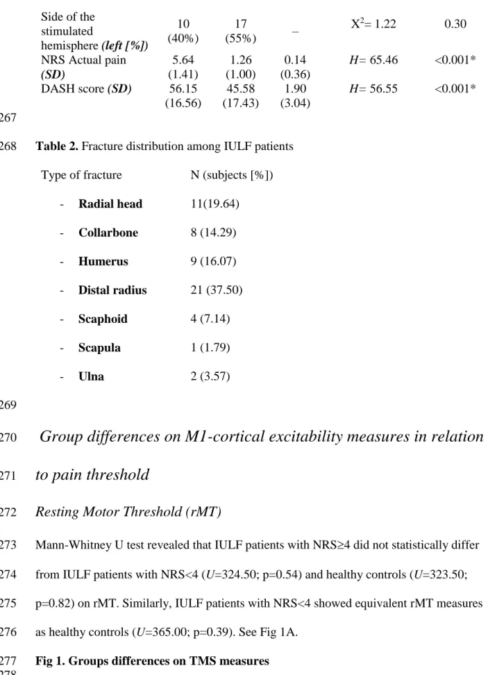
Subject:
Manuscript submission for publication
Montreal, November 22
nd
2019
Dear Editor,
We would like to submit this research article entitled “Clinically significant acute pain
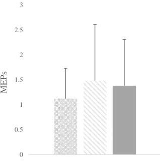
disturbs motor cortex intracortical inhibition and facilitation in orthopedic trauma patients: A
TMS study” for publication in PLOS ONE. This study adds to the current literature by showing
that clinically significant acute pain can alter GABAergic inhibitory and glutamatergic activities
mechanisms in orthopedic patients at an early stage post-injury. Other factors such as age, sex,
time elapsed since the injury, and the stimulated hemisphere had no impact on measures. Cortical
excitability alterations have been identified in orthopedic patients afflicted by chronic pain as
well as in healthy subjects with experimentally induced acute pain. These findings may
contribute to the ongoing effort of identifying early risk factors for chronic pain development.
Following, is a list of suggested reviewers: Catherine Mercier, Ph.D.
(
catherine.mercier@rea.ulaval.ca
); Sean Mackey, M.D., Ph.D. (
smackey@stanford.edu
); Shirley
Fecteau, Ph.D. (
shirley.fecteau@fmed.ulaval.ca
)
Suggested Academic Editor: David J Wright (
d.j.wright@mmu.ac.uk
)
All authors gave their final approval for the submitted version of our manuscript and
meet each of the authorship requirements as stated in the Uniform Requirements for Manuscripts
Submitted to Biomedical Journals (
www.icmje.org
). The authors also agree to be accountable for
all aspects of the work in ensuring that questions related to the accuracy or integrity of any part
of the work are appropriately investigated and resolved. The manuscript, including related data,
figures, and tables has not been previously published and is not under consideration elsewhere
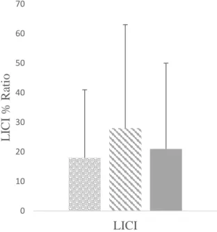
and was never previously submitted to PLOS. The authors report no conflicts of interest in
relation with this paper. The authors state that they have full control of all primary data and that
they agree to allow the journal to review their data.
We thank you for your consideration,
Corresponding author:
Louis De Beaumont. Department of Surgery, Université de Montréal, 2900 boul.
Edouard-Montpetit, Montreal, QC, Canada, H3T 1J4.
louis.de.beaumont@umontreal.ca
Moderate to severe acute pain disturbs motor cortex intracortical inhibition
1
and facilitation in orthopedic trauma patients: A TMS study
2
3
Short title: Acute pain in orthopedic trauma disturbs motor cortex intracortical inhibition
4
and facilitation
5
6
Marianne Jodoin
1,2
,
Dominique M. Rouleau
1,3
, Audrey Bellemare
1,2
, Catherine Provost
1
,
7
Camille Larson-Dupuis
1,2
, Émilie Sandman
1,3
, G-Yves Laflamme
1,3
, Benoit Benoit
1,3
,
8
Stéphane Leduc
1,3
, Martine Levesque
1,4
, Nadia Gosselin
1,2
, Louis De Beaumont
1,3*
.
9
Affiliations:
10
1.
Hôpital Sacré-Cœur de Montréal (HSCM), 5400 boul. Gouin Ouest, Montreal,
11
QC, Canada, H4J 1C5 (Where the work was performed)
12
2.
Département de psychologie de l’Université de Montréal, 2900 boul.
Edouard-Montpetit, Montreal, QC, Canada, H3T 1J4
3.
Département de chirurgie de l’Université de Montréal, 2900 boul.
Edouard-Montpetit, Montreal, QC, Canada, H3T 1J4
4.
Hôpital Fleury,
2180 Rue Fleury East, Montreal, QC, Canada, H2B 1K3
13
Corresponding author:
14
Louis De Beaumont:
louis.de.beaumont@umontreal.ca
15
16
17
18
19
20
21
22
23
24
25
26
Manuscript
Click here to access/download;Manuscript;Plos One_Revised
27
28
Abstract
29
Objective: Primary motor (M1) cortical excitability alterations are involved in the
30
development and maintenance of chronic pain. Less is known about M1-cortical
31
excitability implications in the acute phase of an orthopedic trauma. This study aims to
32
assess acute M1-cortical excitability in patients with an isolated upper limb fracture
33
(IULF) in relation to pain intensity.
34
Methods: Eighty-four (56 IULF patients <14 days post-trauma and 28 healthy controls).
35
IULF patients were divided into two subgroups according to pain intensity (mild versus
36
moderate to severe pain). A single transcranial magnetic stimulation (TMS) session was
37
performed over M1 to compare groups on resting motor threshold (rMT),
short-38
intracortical inhibition (SICI), intracortical facilitation (ICF), and long-interval cortical
39
inhibition (LICI).
40
Results: Reduced SICI and ICF were found in IULF patients with moderate to severe
41
pain, whereas mild pain was not associated with M1 alterations. Age, sex, and time since
42
the accident had no influence on TMS measures.
43
Discussion: These findings show altered M1 in the context of acute moderate to severe
44
pain, suggesting early signs of altered GABAergic inhibitory and glutamatergic
45
facilitatory activities.
46
47
48
49
Introduction
50
Orthopedic trauma (OT) patients are routinely afflicted by pain and it is
51
considered the most common and debilitating symptom reported among this population
52
[1, 2]. Optimal pain control is an OT care priority as pain interferes with trauma recovery
53
and affects outcome [3, 4].
54
A growing body of research is currently focused on developing alternative pain
55
management techniques to tackle the alarming drawbacks associated with current
56
standards of care. Among these alternatives, transcranial magnetic stimulation (TMS) has
57
gained attention in recent years for its dual role: 1) its ability to objectively assess pain
58
mechanisms; and 2) its potential applicability in pain management. In chronic pain
59
studies, the primary motor cortex (M1) commonly serves as the targeted brain region due
60
to its connections with the nociceptive system and the known effect of pain on motor
61
function [5, 6]. Despite some variability across TMS studies, there is extensive evidence
62
of an altered balance between inhibitory and facilitatory circuits of M1 in various chronic
63
pain conditions (i.e. fibromyalgia, neuropathic pain, complex regional pain syndrome,
64
phantom limb pain, chronic orofacial pain) [7, 8]. These results highlight maladaptive
65
plasticity within the motor system. M1-cortical excitability alterations have been
66
associated with the severity of the clinical symptoms such as pain intensity, hyperalgesia,
67
and allodynia [9, 10], pointing to the value of TMS as an objective tool that reflects
68
functional alterations. Moreover, cortical excitability restoration through repetitive TMS
69
(rTMS), a technique known to induce lasting modulation effects on brain activity through
70
a multiple day session paradigm, has shown some efficacy in reducing the magnitude of
71
pain, even in refractory chronic pain patients [11-16]. Overall, these results support the
72
role of cortical excitability on pain intensity in chronic pain patients and the potential
73
clinical utility of TMS in pain management among this population.
74
On the other hand, acute pain initiated by an OT, such as following a fracture, has
75
received little to no attention, despite being highly prevalent. With 15% to 20% of all
76
physician visits intended to address pain-related issues [17, 18], management of acute pain
77
following OT still remains medically challenging [19-22]. Knowing that acute and chronic
78
pain belong to the same continuum and that there is clear evidence of success in the use of
79
rTMS in treating chronic pain, this technique could serve as a potential treatment tool in
80
the early phase of fracture pain by tackling key elements of pain chronification. First,
81
however, a better understanding of the involvement of M1-cortical excitability in acute
82
pain is necessary.
83
From a physiological point of view, it remains unclear whether motor cortical
84
excitability impairments are expected in a context of acute pain following an OT. On one
85
hand, neuroimaging studies suggest that possible disturbances within M1 only arise once
86
chronic pain has developed, with acute and chronic pain exhibiting distinct and
non-87
overlapping brain activation patterns [23-27]. On the other hand, there is evidence
88
supporting alterations of M1-cortical excitability during acute pain states. Indeed,
89
Voscopoulos and Lema highlight early neuroplasticity involvement of GABA inhibitory
90
interneurons following a peripheral insult, which may contribute to later transition to
91
chronic pain [28]. In parallel, Pelletier and colleagues [29] suggested that pain intensity
92
may act as the driving factor leading to M1-cortical excitability alterations rather than the
93
state of chronic pain itself. This assumption was made by authors after obtaining similar
94
M1 deficiency patterns across chronic pain conditions of various origins. Other TMS
95
studies also showed that pain of moderate to severe intensity (score
4 on numerical rating
96
scale (NRS)) leads to greater motor cortex impairments [10]. The relationship between pain
97
intensity in the acute state and its impact on cortical excitability parameters appears a
98
relevant target of investigation.
99
So far, very few studies have looked into the association between acute pain and
100
M1-cortical excitability. These studies have mainly focused on experimental pain models
101
in healthy subjects. More specifically, acute experimental pain of low-to-moderate
102
intensity induces a generalized state of M1 inhibition, reflecting changes in both cortical
103
and spinal motoneuronal excitability in healthy participants [30-35]. Findings suggest that
104
acute experimental pain can modify cortical excitability of M1, but the result patterns
105
obtained are different from chronic pain states. In parallel, rTMS studies have been shown
106
effective in both alleviating acute experimental pain and modulating alterations in
M1-107
cortical excitability [36, 37]. Taken together, these findings show that M1 alterations can
108
occur in the context of acute pain and that rTMS over M1 can successfully modulate
109
nociceptive afferent information and restore M1 alterations, even for transient pain
110
sensation in healthy controls. However, due to the subjective nature of pain sensation along
111
with intrinsic differences in pain characteristics across conditions and individuals,
112
translation between experimental pain model and clinical pain following an OT is limited.
113
Therefore, if we are to consider the potential clinical utility of rTMS in alleviating acute
114
pain, studies need to be conducted in a clinical population.
115
This study therefore aims to assess acute M1-cortical excitability functioning
116
through well-established TMS paradigms according to pain intensity in patients who are in
117
the acute pain phase following an isolated upper limb fracture (IULF). We hypothesize that
118
M1-cortical excitability alterations will be found in patients with higher levels of pain
119
compared to healthy controls and to IULF patients with mild pain.
120
Materials and Methods
121
This work was approved by the Hôpital du Sacré-Coeur de Montréal' Ethics Committee
122
(Approval number: 2017-1328). A written consent was obtained by all participating
123
subjects prior to the start of the study.
A financial compensation was given to all subjects
124
for their participation.
125
Participants
126
Our sample included 1) patients who have suffered from an isolated upper limb fracture
127
(IULF) and 2) healthy controls. Patients with an IULF were initially recruited from
128
various orthopedic clinics affiliated to a Level 1 Trauma Hospital. To be included in the
129
study, patients had to be aged between 18 and 60 years old and have sustained an IULF
130
(one fractured bone from upper body extremities) within 14 days post-injury.
131
Recruitment of IULF patients took place on the day of the first medical appointment at
132
the orthopedic trauma clinic with the orthopedic surgeon. Testing was conducted within
133
24 hours post-medical consultation. All testing measures had to be completed prior to
134
surgical procedures (if any) given the known impact of surgery on increased
135
inflammatory response and pain perception [38]. Exclusion criteria consisted of a history
136
of traumatic brain injuries, a diagnosis of and/or a treatment for a psychiatric condition in
137
the last ten years, musculoskeletal deficits, neurological conditions (i.e. epilepsy), chronic
138
conditions (cancer, uncontrolled diabetes, cardiovascular illness, high blood pressure),
139
the use of central nervous system-active medication (hypnotics, antipsychotics,
140
antidepressant, acetylcholinesterase inhibitor, anticonvulsant), history of alcohol and/or
141
substance abuse, acute medical complications (concomitant traumatic brain injury,
142
neurological damage, etc.), and being intoxicated at the time of the accident and/or at the
143
emergency visit. Of note, IULF patients were not restrained from using analgesic
144
medication (acetaminophen, ibuprofen, opioids, etc.) during testing to assure comfort and
145
to avoid interfering with pain management.
146
147
The control group consisted of healthy right-handed adults recruited through various
148
social media platforms. As per usual practice in conducting M1 TMS studies, only
right-149
handed control participants were selected as stimulation over non-dominant M1 has been
150
associated with accentuated within-subject variability [39, 40]. They self-reported to be
151
free of all previously mentioned exclusion criteria.
152
Study participants were also screened for TMS tolerability and safety [41].
153
154
Assessment measures
155
Total assessment procedures (including consent) were conducted over a single, 90-minute
156
session. First, participants were invited to complete self-administered questionnaires to
157
gather demographic information and clinical outcome measures (pain intensity and
158
functional disability indices). More specifically, demographic data such as age, sex, and
159
level of education were documented and used to ensure homogeneity between groups.
160
161
Clinical outcome: Pain intensity and functional disability indices
162
To assess the perceived level of pain at the time of testing, the numerical rating scale
163
(NRS), a routinely used standardized generic unidimensional clinical pain questionnaire,
164
was administered [42, 43]. To complete the NRS, participants had to circle a number that
165
best fit their current level of pain on the 11-point pain intensity scale, with numbers
166
ranging from 0 (“no pain”) to 10 (“worst possible pain”). In order to test the hypothesized
167
impact of acute pain intensity on M1 cortical excitability, IULF patients were divided
168
into two distinct groups according to NRS score: 1) IULF patients who self-reported
169
moderate to severe pain intensity (NRS
4 out of 10); 2) IULF patients with mild pain
170
intensity (NRS <4). The cut-off pain intensity scores are based on previous pain studies
171
[10, 44, 45].
172
The disabilities of the Arm, Shoulder, and Hand (DASH) questionnaire was used as a tool
173
to assess an individual’s ability to perform common specific everyday activities relying
174
on upper extremity limbs [46, 47]. This questionnaire consists of 30 items, including 6
175
that are symptom-related and 24 that are function-related, where patients were asked to
176
rate the level of disability on each activity as experienced since their accident. Continuum
177
of scores on this questionnaire varies between 0 (no disability) and 100 (extreme
178
difficulty).
179
180
Comprehensive assessment of M1 cortical excitability using TMS.
181
To assess M1 cortical excitability, a TMS figure-of-eight stimulation coil (80mm wing
182
diameter), attached to a Bistim
2
Magstim transcranial magnetic stimulators (Magstim
183
Company, Whitland, Dyfed, UK), was used. The TMS-coil was positioned flat on the
184
scalp over M1 at a 45
angle from the mid-sagittal line, with its handle pointing
185
backwards. In the IULF group, the TMS coil was positioned over M1 contralaterally to
186
the injury, whereas in the control group, the TMS-coil was systematically positioned over
187
the dominant left hemisphere. Motor evoked potentials (MEP) recordings from the
188
abductor pollicis brevis (APB) was performed using three electrodes positioned over the
189
belly of the target muscle (active electrode (+)), between the distal and proximal
190
interphalangeal joints of the index (reference (-)), and on the forearm (ground). Optimal
191
stimulation site was determined based on the coil position which evoked highest
peak-to-192
peak MEP amplitudes from the target muscle. We used a 3D tracking system (Northern
193
Digital Instruments, Waterloo, Canada) to ensure accurate and consistent TMS coil
194
positioning on the targeted site.
195
196
Various well-established TMS protocols were conducted to investigate M1 excitatory and
197
inhibitory mechanisms using single and paired-pulse paradigms. Single pulse magnetic
198
stimulations were first used to establish the resting motor threshold (rMT), i.e. the
199
minimal stimulation intensity needed to elicit a MEP of at least 0.05mV in five out of ten
200
trials [48]. An interstimulus interval, varying from 8 to 10 seconds, was applied to control
201
for possible residual effects of TMS stimulation on M1 activity [49]. The sequence of
202
stimulation intensity was randomly generated by a computer. Short
intra-cortical-203
inhibition (SICI) and facilitation (ICF) were measured via a classic paired-pulse
204
paradigm [50, 51]. The latter protocol involves the application of two successive TMS
205
pulses, the first pulse set at 80% of the rMT intensity (subthreshold; conditioning
206
stimulus) and the second pulse set at 120% of the rMT (suprathreshold; test stimulus)
207
separated by an interstimulus interval (ISI) of a predetermined duration [50]. To test for
208
SICI, a measure attributed to GABA
A
interneurons and receptors activity [52], one
209
sequence of 10 paired-pulse stimulations was completed with an ISI set at 3ms. To test
210
for ICF, one sequence of 10 stimulations was performed with ISI set at 12ms. Measure of
211
ICF is thought to be mediated by excitatory glutamatergic interneurons and
N-methyl-D-212
aspartate (NMDA) receptors [52-56]. Results of SICI and ICF are expressed as
213
percentage ratios of MEP amplitudes. These ratios represent the mean MEP amplitude of
214
paired TMS over the mean MEP amplitude of the test stimuli baseline measurement (10
215
single magnetic pulses set at 120% rMT). Therefore, high SICI values reflect a lack of
216
intracortical inhibition, whereas a low value ICF corresponds to a lack of intracortical
217
facilitation. Finally, we measured long-interval cortical inhibition (LICI) through
paired-218
pulse TMS of identical suprathreshold intensity (i.e. 120% rMT) with an ISI of 100ms.
219
The first pulse corresponded to the conditioning stimulus whereas the second pulse was
220
the test stimulus. LICI is primarily known to be mediated by GABA
B
receptors [57, 58].
221
To calculate LICI, we used the percentage ratio between the mean peak-to-peak MEP
222
amplitude of the test stimulus response (TSR) and the mean peak-to-peak MEP amplitude
223
of the conditioning stimulus response (CSR) expressed as: mean (TSR)/mean(CSR).
224
Statistics
225
Statistical analyses were performed using IBM SPSS Statistics software version 25
226
(Armonk, NY, United States).
The Shapiro-Wilks test was used to determine the
227
normality of the data. Parametric and nonparametric tests were performed, where
228
appropriate, with a
-level fixed at 0.05. Descriptive analyses were used to characterize
229
and compare the three groups (1- IULF patients with NRS
4; 2- IULF patients with
230
NRS<4; 3- healthy controls) in our study sample. Results from descriptive analyses are
231
expressed as means, standard deviation (SD), and percentages. We used a Student’s t-test
232
or a Mann-Whitney U test to investigate group differences on TMS measures. An
233
analysis of variance (ANOVA) or the Kruskal-Wallis test were also used where
234
appropriate. Pearson and Spearman’s correlation analysis were also computed to assess
235
the relationship between functional disability outcomes and the other outcome measures
236
of interest (pain intensity and TMS measures). We corrected for multiple comparisons
237
using False Discovery Rate (FDR) where appropriate. Post-hoc analyses were conducted
238
to control for the effect of within-group variability of stimulated hemispheres across
239
IULF patients on TMS measures as it varied according to the injury location (left or
240
right). Therefore, we elected to create subgroups as follow: IULF patients stimulated over
241
the left hemisphere (IULF with left-M1) and IULF patients stimulated on the right
242
hemisphere (IULF with right-M1). Lastly, a post-hoc linear regression analysis was
243
computed to assess which independent variables between pain intensity
(NRS score from
244
0-10)
and the number of days between the accident and testing (independent variable)
245
best predict significant changes in M1-cortical excitability (dependent variable) in IULF
246
patients.
247
248
Results
249
Demographic information
250
A total of 84 subjects took part in the current study, of which 56 had suffered an IULF
251
(23 females; mean age: 39.41 years old) and 28 were healthy controls (17 females; mean
252
age: 34.93). Two subgroups of IULF patients were formed according to pain intensity:
253
Twenty-five IULF individuals met the criteria for moderate to severe pain (NRS
4),
254
whereas 31 IULF subjects were classified as having mild pain (NRS <4). Age (H=3.89;
255
p=0.14) and sex (F
(81)
=3.76; p=0.15) did not differ between groups, whereas the level of
256
education (F
(81)
=3.95; p=0.02) and the time elapsed between the accident and testing
257
(U=225.50; p=0.01) were statistically different across groups. More specifically, IULF
258
patients with NRS
4 were tested on average 4.48 (SD=3.50) days post-accident
259
compared to 7.55 (SD=4.45) days for IULF patients with NRS<4. Spearman’s
260
correlational analyses revealed a strong association between pain intensity and the extent
261
of functional disability as measured through the DASH questionnaire (r
s
=0.76; p<0.001).
262
Refer to tables 1-2 for additional descriptive information regarding study sample and
263
fracture distribution among IULF patients.
264
265
Table 1. Descriptive characteristics of study cohort by group
266
IULF
subgrou
p NRS
4
IULF
subgrou
p NRS
<4
Healthy
control
s
Results of analysis
p-value
N (subjects)
25
31
28
–
Age (years [SD])
42.36
(13.83)
37.03
(12.02)
34.93
(11.95)
H= 3.89
0.14
Sex (female [%])
12
(48%)
11
(35%)
17
(61%)
F= 3.76
0.15
Education (years
[SD])
13.44
(2.65)
14.74
(2.86)
15.54
(2.65)
F= 3.95
0.02*
Number of days
between trauma and
data
collection/assessmen
t (days [SD])
4.48
(3.50)
7.55
(4.45)
–
U= 225.50
0.01*
Side of the
stimulated
hemisphere (left [%])
10
(40%)
17
(55%)
–
X
2
= 1.22
0.30
NRS Actual pain
(SD)
5.64
(1.41)
1.26
(1.00)
0.14
(0.36)
H= 65.46
<0.001*
DASH score (SD)
56.15
(16.56)
45.58
(17.43)
1.90
(3.04)
H= 56.55
<0.001*
267
Table 2. Fracture distribution among IULF patients
268
Type of fracture
N (subjects [%])
- Radial head
11(19.64)
- Collarbone
8 (14.29)
- Humerus
9 (16.07)
- Distal radius
21 (37.50)
- Scaphoid
4 (7.14)
- Scapula
1 (1.79)
- Ulna
2 (3.57)
269
Group differences on M1-cortical excitability measures in relation
270
to pain threshold
271
Resting Motor Threshold (rMT)
272
Mann-Whitney U test revealed that IULF patients with NRS
4 did not statistically differ
273
from IULF patients with NRS<4 (U=324.50; p=0.54) and healthy controls (U=323.50;
274
p=0.82) on rMT. Similarly, IULF patients with NRS<4 showed equivalent rMT measures
275
as healthy controls (U=365.00; p=0.39). See Fig 1A.
276
Fig 1. Groups differences on TMS measures
277
278
MEPs test stimulus intensity
279
MEPs of the test stimulus used to measure SICI and ICF were equivalent between
280
groups. Indeed, IULF patients with NRS
4 did not statistically differ from IULF patients
281
with NRS<4 (U=336.00; p=0.40) and healthy controls (U=304.00; p=0.41). Moreover,
282
IULF patients with NRS<4 and healthy controls were comparable (U=431.00; p=0.96).
283
See Fig 1B.
284
Short intra-cortical inhibition (SICI)
285
Results showed that IULF patients with NRS
4 statistically differed from healthy
286
controls (U=202.00; p<0.01), with NRS
4 IULF patients exhibiting reduced
short-287
intracortical inhibition of M1. A tendency toward reduced short-intracortical inhibition
288
was found in IULF patients with NRS
4 compared to IULF patients with NRS <4, but
289
the difference failed to reach significance (U=282.50; p=0.08),. Lastly, IULF patients
290
with NRS<4 and healthy controls showed similar SICI (U=383.00; p=0.44). See Fig 1C.
291
We then conducted
a post-hoc linear regression to assess the contribution of both pain
292
intensity and delay between the accident and testing on SICI disinhibition. Data shows
293
that pain intensity at the time of testing significantly predicted SICI disinhibition and
294
explained 29% of the variance (
-coefficient = 0.29; p=0.05), whereas the delay between
295
the accident and testing poorly predicted SICI disinhibition (
-coefficient= 0.07; 0.63).
296
297
Intra-cortical facilitation (ICF)
298
IULF patients with NRS
4 exhibited a significantly reduced ICF (t
(54)
=2.44; p=0.02)
299
relative to IULF patients with NRS<4. IULF patients with NRS
4 (t
(51)
=-1.63; p=0.11)
300
and IULF with NRS<4 (t
(57)
=0.37; p=0.71) did not statistically differ from healthy
controls. See Fig 1D.
Results from a post-hoc linear regression showed that pain intensity
302
significantly predicted altered ICF (
-coefficient=-0.30; p=0.04), accounting for 30% of
303
the variance, whereas delay between the accident and testing (
-coefficient=-0.02;
304
p=0.87) poorly predicted altered ICF.
305
306
Long-interval cortical inhibition (LICI)
307
IULF patients with NRS
4 had similar LICI values compared to IULF patients with
308
NRS<4 (
U=339.00; p=0.42)
and healthy controls (
U=324.00; p=0.64)
. IULF patients
309
with NRS<4 and healthy controls were also equivalent on LICI (
U=405.00; p=0.66)
.
See
310
Fig 1E.
311
312
Post-hoc analyses controlling for the side of the stimulated
313
hemisphere in IULF patients
314
To investigate if the stimulated hemisphere had an impact on cortical excitability
315
measures, IULF patients were stratified into two distinct groups: IULF patients
316
stimulated on the left M1 and IULF patients stimulated on the right M1. Demographic
317
data such as age (U=296.00; p=0.12), sex (X
2(1)
=0.002; p=0.96), education level
318
(t
(54)
=1.17; p=0.25), and the timing of testing in relation to the accident (U=339.50;
319
p=0.39) were similar across groups (see table 3). Lastly, there was no between-group
320
difference in regard to pain intensity (U=297.50; p=0.12).
321
322
Table 3. Descriptive characteristics of IULF patients according to the stimulated
323
hemisphere
324
325
IULF subgroup
Left M1
IULF
subgroup
Right M1
Results of the
test analysis
p-value
N (subjects)
27
29
–
Age (years [SD])
36.44 (12.40)
42.17
(13.18)
U= 296.00
0.12
Sex (female [%])
11 (41%)
12 (43%)
X
2
= 0.002
0.96
Education (years
[SD])
14.59 (3.06)
13.70
(2.51)
t= 1.17
0.25
Number of days
between trauma and
data
collection/assessment
(days [SD])
5.67 (3.92)
6.66 (4.65)
U= 339.50
0.39
NRS Actual pain (SD)
2.81 (2.83)
3.59 (2.13)
U= 297.50
0.12
326
Group differences on M1-cortical excitability measures in relation to M1
327
stimulation side
328
None of the TMS measures differed across IULF patients according to the stimulated
329
hemisphere [rMT (U=359.00; p=0.93); SICI (U= 377.00; p=0.81); ICF (t
(54)
=-0.44;
330
p=0.6); LICI (U= 361.50; p=0.62)]. See Fig 2A-D.
331
332
Relationship between cortical excitability measures and functional disability
333
outcomes
334
The DASH questionnaire was used to investigate the relationship between functional
335
disability outcomes and cortical excitability parameters. Only IULF subjects were
336
included in this analysis, whereas healthy controls were excluded. Results show that the
337
DASH score was strongly associated with SICI (R
s
=0.37; p=0.006), whereas no
338
correlation was found with ICF (r= -0.11; p=0.46), LICI (R
s
=-0.06; p=0.67), and rMT
339
(R
s
= 0.18; p=0.22).
340
341
Fig 2A-D. Between IULF-group differences on TMS measures stratified according
342
to the stimulated hemisphere.
343
344
345
Discussion
346
347
This study provides new insights into the involvement of the primary motor cortex in the
348
early phase of recovery (<14 days post-trauma) following an IULF through various TMS
349
protocols assessing M1-cortical excitability. More precisely, results suggest a significant
350
decrease in intracortical inhibition and facilitation in IULF patients over the cortical
351
representation of the fractured bone. These neurophysiological alterations were only
352
observed in IULF patients with pain of moderate to severe intensity (NRS
4), whereas
353
IULF patients with mild pain did not differ from healthy controls. Furthermore, this study
354
highlights that the time elapsed between the accident and testing within the first 14 days
355
of the accident, as well as the stimulated hemisphere, do not influence any of the primary
356
motor cortex excitability measures. On the contrary, pain intensity emerges as the main
357
factor explaining acute abnormalities of M1 excitability in IULF patients relative to a
358
healthy cohort of similar age, sex distribution, and education level. To the best of our
359
knowledge, this is the first study to investigate M1-cortical excitability in acute pain
360
following an isolated upper limb fracture.
361
This study suggests a state of disinhibition through reduced SICI, a TMS measure
362
that is robustly associated to GABA
A
receptors activity [52], but only in patients with
363
moderate to severe pain intensity (NRS
4). Moreover, the extent of SICI disruption was
364
strongly associated with functional disability scores (DASH). Current findings highlight
365
possible resemblance across pain states, as SICI disturbances are also found in various
366
chronic pain conditions [7, 59-61]. A reduction of GABAergic inhibition has been shown
367
to play a prominent role in chronic pain development and in pain maintenance [62]. It is
368
therefore no surprise that GABA receptor agonists have proven effective as an analgesic
369
agent, but important side effects limit its long-term use [63, 64]. Identification of a state
370
of disinhibition at such an early stage of recovery in patients with a fracture is of
371
particular clinical relevance in this population since high initial pain is considered a risk
372
factor for chronic pain development [65]. These results may further our understanding as
373
to why high levels of pain in the acute phase is considered a risk factor for chronic pain.
374
Indeed, patients with moderate to severe pain (NRS
4) are affected by disrupted
375
GABAergic inhibition within the first few days post-trauma, which may hypothetically
376
contribute to CNS’ vulnerability to pain chronification.
377
Of note, current findings diverge from results found in experimental acute pain
378
studies. Experimentally induced pain in healthy controls shows an increase in M1
379
intracortical inhibition whereas the current study found a decrease in inhibition in IULF
380
patients presenting with moderate to severe acute pain (NRS
4). Increased SICI in acute
381
experimental pain has been suggested as an adaptation strategy to prevent CNS
382
reorganization [32]. Given the reverse pattern of M1 disinhibition in IULF patients, one
383
should investigate whether moderate to severe pain symptoms in the latter clinical
384
population may facilitate lasting CNS reorganization through sustained activation of
385
plasticity mechanisms. One reason for the discrepancies in SICI findings between
386
experimental and acute clinical pain could be that fracture pain involves multiple
387
physiological mechanisms that cannot be replicated in a human experimental setting. For
388
example, the physiological cascade following tissue injury and bone fracture alone,
389
including an acute inflammatory response, can modulate brain excitability [66] and
390
impair GABAergic and glutamatergic activities [67]. Future studies combining both
391
experimental paradigms in a healthy cohort and clinical pain in OT patients are warranted
392
if we are to investigate the mechanisms involved and to restrict results discrepancy due to
393
possible methodological variabilities.
394
Current results also reveal alterations of intracortical facilitation in IULF patients
395
with moderate to severe pain (NRS
4), a measure traditionally considered to be
396
mediated by glutamatergic facilitatory transmission [52-56]. The finding that both ICF
397
and SICI are reduced may appear counterintuitive from a physiological standpoint.
398
However, physiological underpinnings of TMS-induced ICF effects have been the subject
399
of ongoing debate, as some evidence suggest that the latter reflects an overlap between
400
inhibitory and excitatory mechanisms [54]. Along those lines, pharmacological studies
401
have shown that both NMDA receptors antagonists (such as dextromethorphan and
402
memantine) as well as GABA
A
agonists can modulate ICF. In parallel, some TMS and
403
chronic pain studies have shown reduced ICF, but this was mainly found in patients with
404
fibromyalgia [11, 61]. Additional factors relevant to the orthopedic population could also
405
account for current study findings. For example, other types of pain (muscle pain, bone
406
pain, etc.) and inflammatory response can influence the balance between inhibitory and
407
facilitatory mechanisms [66, 67]. Moreover, limb disuse may also affect brain plasticity
408
due to reduced sensorimotor input and output [68-70].
409
Current findings support work from Pelletier and colleagues [29] suggesting that
410
pain intensity, rather than pain state, appears to be linked to the extent of motor cortex
411
excitability alterations. As such, patients who reported moderate to severe pain (NRS
4)
412
showed accentuated SICI and ICF alterations as compared to patients with mild pain
413
levels who showed a similar M1 excitability profile to healthy controls. This is
414
particularly interesting as results from the current study showed that patients with higher
415
pain levels also reported greater functional disability. Therefore, study findings are not
416
only consistent with the notion that high initial pain is a good predictor for chronic pain,
417
but it also argues that altered cortical excitability of M1 could contribute to underlying
418
mechanisms of pain chronification following a fracture [71, 72].
419
Although a similar M1-cortical excitability profile may emerge between acute and
420
chronic injury phases, the involvement of the CNS may be different. One should bear in
421
mind that altered SICI and ICF in acute pain do not necessarily indicate permanent CNS
422
reorganization. Although speculative, acute changes in M1-cortical excitability could also
423
reflect the intensity of the nociceptive afferent originating from the periphery. It should
424
be noted that the group of patients reporting moderate to severe (NRS
4) pain levels
425
who also exhibited altered M1-cortical excitability were tested at a significantly shorter
426
delay following the accident relative to patients who reported mild levels of pain. One
427
cannot exclude the possibility that alterations of M1-cortical excitability within the first
428
few days of the injury could have subsided as pain intensity is expected to reduce with
429
additional time to recover. However, results from linear regressions, used to delimitate
430
the weight of the timing of testing in relation to the accident and pain intensity on altered
431
M1-cortical excitability, showed that pain intensity best predicted altered intracortical
432
inhibition and facilitation, whereas timing of testing had no impact within that short
14-433
day time frame. Longitudinal follow-ups are nonetheless needed to investigate
434
longitudinal changes of TMS-induced M1 excitability measurements in relation with pain
435
stages, particularly during the transition from acute to chronic pain.
436
LICI, another measure reflecting GABA
B
receptors inhibition, was found to be
437
unrelated to reported pain intensity following a peripheral injury. In a recent review,
438
authors only found scarce evidence of the involvement of LICI alterations in various
439
chronic pain conditions [7], either suggesting that GABA
B
receptors remain intact or that
440
the latter measure may be less sensitive to pain states. It would still appear relevant to
441
include other TMS paradigms known to measure GABA
A
and GABA
B
receptors, namely
442
short-afferent inhibition (SAI), long-afferent inhibition (LAI), and the cortical silent
443
period (CSP) in the context of future studies [54, 73]. This would allow us to deepen our
444
understanding of the involvement of acute pain on the GABAergic inhibitory system in
445
IULF patients.
446
Given the known durable effects of multisession rTMS protocols on M1-cortical
447
excitability and on pain reduction, rTMS appears as a highly relevant intervention avenue
448
for the IULF population. Acute rTMS application should be considered as an intervention
449
option as it may provide analgesic effects to suffering patients, in addition to possibly
450
tackling cortical excitability changes associated with pain chronification.
451
One limitation to the current study is the use of a single TMS session to
452
investigate M1-cortical excitability implications in the acute phase of an IULF in relation
453
to pain intensity. Longitudinal studies are needed among this population to further
454
explore the effects of early M1-cortical excitability dysregulations on recovery. This
455
would provide valuable insights as to whether acute altered M1-cortical excitability is a
456
predictor of pain chronification. Secondly, this study uses limited, but well established,
457
TMS parameters. Still, it should be considered that TMS parameters vary greatly across
458
studies (e.g. ISI, test and conditioned stimuli intensity), surely contributing to result
459
variability found in the literature. This poses a challenge for researchers to establish the
460
most sensitive and specific TMS parameters. In the context of the present study, it should
461
be considered that previous studies have highlighted possible contamination by
short-462
afferent cortical facilitation (SICF) in SICI according to the TMS parameters used [74,
463
75]. Although the present study uses parameters from previously published studies, SICF
464
contamination cannot be excluded. It would be important to account for these findings in
465
future studies. Moreover, the use of additional TMS paradigms (SAI, LAI, CSP) as well
466
as an objective measure of pain, such as conditioned pain modulation [76, 77], would be
467
highly relevant in the context of future studies to draw a thorough physiological profile of
468
ascending and descending tracks in IULF patients with moderate to severe pain (NRS
469
4). Thirdly, since the initial medical consultations varied across IULF individuals,
470
timing of testing accident was not equivalent within the IULF group. Although
post-471
hoc analyses showed that this factor did not influence TMS outcomes, future studies
472
should, to the extent possible, assess patients at a fixed day since the physiological
473
cascade following the injury is rapidly evolving. Fourthly, pain medication usage and
474
dosage at the time of testing were not restrained in IULF patients, possibly leading to
475
interindividual variability among the sample. Effects of analgesics medication on cortical
476
excitability measures cannot be excluded although very scarce evidence exists. One study
477
showed that acetaminophen can increase MEP, which facilitates the inhibition of
voltage-478
gated calcium and sodium currents [78]. In this case, and in relation with current study
479
results showing decreased intracortical inhibition, acetaminophen usage among study
480
sample could have masked cortical excitability deficiencies. As for opioid analgesics,
481
only one study mentioned that fentanyl does not alter MEP amplitudes [56], a drug that is
482
rarely used to treat acute pain. Fifthly, future studies should also account for additional
483
factors, such as the inflammatory cascade (pro-inflammatory cytokines levels) and
484
genetic predisposition, as they are known to impact pain intensity and M1-cortical
485
excitability measures [79-82]. Accounting for such factors would be beneficial to develop
486
tailored interventions for the IULF population. Sixthly, the stimulated hemisphere (right
487
or left M1) varied in IULF patients according to the injured side. This factor was
488
controlled for in IULF patients and no differences were found. On the other hand, all
489
healthy controls were right-handed and were stimulated on the left-M1, which
490
corresponds to the dominant hemisphere as per optimal TMS guidelines. Since no
491
differences were found among the clinical sample, we elected to follow the TMS
492
guidelines in the healthy sample. Finally,
evidence show that reduced use of limb (limb
493
immobilization) can indeed lead to brain changes (cortical thickness, cortical excitability,
494
etc.) in the motor cortex due to reduced sensory input/sensorimotor deprivation [68-70,
495
83]. We can by no mean exclude this factor entirely, but a few points should be
496
considered. First, IULF patients were tested very early post-injury, leaving less time for
497
measurable brain changes. Second, statistical analyses show that the number of days
498
between testing and the accident (possible indicator of reduced limb use) is not associated
499
with alterations in cortical excitability measures. Lastly, IULF patients who showed most
500
cortical excitability deficiencies were actually tested within shorter delays of accident
501
(NRS >4 group), leaving less time, compared to the other IULF group (NRS<4), for
502
cortical reorganization due to limb immobilization.
503
Conclusions
504
In conclusion, this is the first study to investigate M1 cortical excitability involvement in
505
an orthopedic trauma population suffering from acute pain. Current results show early
506
signs of altered GABAergic inhibitory and glutamatergic facilitatory activities in patients
507
with pain of moderate to severe intensity (NRS
4). These findings may bear major
508
clinical significance as this population is vulnerable to chronic pain development. Early
509
detection of at-risk patients could guide proactive intervention aiming to reduce the
510
likelihood of an unsuccessful recovery in this population, leading to a pathological
511
condition. This study also highlights that acute application of rTMS may reveal
512
promising in alleviating pain symptoms among this population and may have
513
implications in preventing chronic pain development.
514
![Table 1. Descriptive characteristics of study cohort by group 266 IULF subgrou p NRS 4 IULF subgroup NRS <4 Healthy controls Results of analysis p-value N (subjects) 25 31 28 – Age (years [SD]) 42.36 (13.83) 37.03 (12.02) 34.93 (11.](https://thumb-eu.123doks.com/thumbv2/123doknet/11530222.295314/19.918.146.753.558.1029/descriptive-characteristics-subgroup-healthy-controls-results-analysis-subjects.webp)


![Table 1. Descriptive characteristics of study cohort by group 267 IULF subgrou p NRS 4 IULF subgroup NRS <4 Healthy controls Results of analysis p-value N (subjects) 25 31 28 – Age (years [SD]) 42.36 (13.83) 37.03 (12.02) 34.93 (11.](https://thumb-eu.123doks.com/thumbv2/123doknet/11530222.295314/57.918.124.610.601.979/descriptive-characteristics-subgroup-healthy-controls-results-analysis-subjects.webp)

