HAL Id: hal-01248050
https://hal.archives-ouvertes.fr/hal-01248050
Submitted on 23 Dec 2015
HAL is a multi-disciplinary open access
archive for the deposit and dissemination of
sci-entific research documents, whether they are
pub-lished or not. The documents may come from
teaching and research institutions in France or
abroad, or from public or private research centers.
L’archive ouverte pluridisciplinaire HAL, est
destinée au dépôt et à la diffusion de documents
scientifiques de niveau recherche, publiés ou non,
émanant des établissements d’enseignement et de
recherche français ou étrangers, des laboratoires
publics ou privés.
bioactive molecules of pharmaceutical interest.
Virginie Mimouni, Lionel Ulmann, Virginie Pasquet, Marie Mathieu, Laurent
Picot, Gaël Bougaran, Jean-Paul Cadoret, Annick Morant-Manceau, Benoît
Schoefs
To cite this version:
Virginie Mimouni, Lionel Ulmann, Virginie Pasquet, Marie Mathieu, Laurent Picot, et al.. The
poten-tial of microalgae for the production of bioactive molecules of pharmaceutical interest.. Current
Phar-maceutical Biotechnology, Bentham Science Publishers, 2012, 13 (15), pp.2733-2750. �hal-01248050�
Title: The POTENTIAL of MICROALGAE for the PRODUCTION of BIOACTIVE MOLECULES of PHARMACEUTICAL INTEREST
Running title: BIOACTIVE MOLECULES FROM MICROALGAE 1
2
3
Title: THE POTENTIAL OF MICROALGAE FOR THE PRODUCTION OF BIOACTIVE MOLECULES OF PHARMACEUTICAL INTEREST
Virginie MIMOUNI1,2, Lionel ULMANN1,2, Virginie PASQUET2, Marie MATHIEU2, Laurent PICOT3, Gaël BOUGARAN4, Jean-Paul CADORET4, Annick MORANT-MANCEAU1, Benoît SCHOEFS1
1Mer Molécules Santé, LUNAM Université, University of Maine, EA 2160, Avenue Olivier Messiaen, 72085 Le Mans Cedex 9, France;
2IUT de Laval, Rue des Drs Calmette et Guérin, 53020 Laval Cedex 9, France;
3Université de la Rochelle, UMR CNRS 7266 LIENSs, La Rochelle, 17042, France;
4IFREMER Laboratoire PBA, Centre IFREMER de Nantes, Nantes, 44311, France
Corresponding author: Benoît SCHOEFS, Mer Molécules Santé, EA 2160, LUNAM University of Maine, EA 2160, Avenue Olivier Messiaen, 72085 Le Mans Cedex 9, France
Tel/Fax: + 33 2 43 83 37 72/39 17; benoit.schoefs@univ-lemans.fr 5
6
7 89
1011
12 13 14 15 1617
18 19ABSTRACT
Through the photosynthetic activity, microalgae process more than 25% of annual inorganic carbon dissolved in oceans into carbohydrates that ultimately, serve to feed the other levels of the trophic networks. Besides, microalgae synthesize bioactive molecules such as pigments and lipids that exhibit health properties. In addition, abiotic stresses, such as high irradiance, nutrient starvation, UV irradiation, trigger metabolic reorientations ending with the production of other bioactive compounds such as ω-3 fatty acids or carotenoids. Traditionally, these compounds are acquired through the dietary alimentation. The increasing, and often unsatisfied, demand for compounds from natural sources, combined with the decrease of the halieutic resources, forces the search for alternative resources for these bioactive components. Microalgae possess this strong potential. For instance, the diatom Odontella aurita is already commercialized as dietary complement and compete with fish oil for human nutrition. In this contribution, the microalga world is briefly presented. Then, the different types of biologically active molecules identified in microalgae are presented together with their potential use. Due to space limitation, only the biological activities of lipids and pigments are described in details. The contribution ends with a description of the possibilities to play with the environmental constrains to increase the productivity of biologically active molecules by microalgae and by a description of the progresses made in the field of alga culturing. 20 21
22
23
24
25
26
27
28
29
30
31
32
33
34
35
36KEYWORDS: bioactive compounds, algae, pigment, lipid, health benefit, abiotic stress, metabolic reorientation, diatom
37
38
INTRODUCTION
More than 70% of Earth is covered with water, in which the most dominant group of living organisms is that of algae. Algae belong to the plant phylum. They are mostly living in water while they have colonized every type of ecological niche. The preferences of individual algal species, which determine their geographical distribution, are based on their environmental tolerance and their responses to abiotic interaction. On the other hand, natural populations are morphologically, physiologically and biochemically diverse because of genetic variability and abiotic conditions [1].
Algae have a tremendous impact on the sustainability of the marine ecosystem as being the primary producers [2] and, therefore, a food source for other marine organisms. Their potential is not restricted to this point as through feeding of other organisms placed at higher levels in the food chain can take benefit from particular me-tabolites such as photoprotective compounds [3]. On the basis of their constituting number of cells, algae can be grouped as unicellular or pluricellular organisms, these terms being often taken as synonym for microalgae or phytoplankton and macroalgae, respectively. Algae represent a few percentage among the total number of species described so far (Fig. S1) even though the number of species is probably largely underestimated [4]. This is especially true for microalgae. The use of algae as fertilizers and food is established since the antiquity. Con-sidering the increasing need of food, bioenergy, pharmaceutical and cosmetic compounds, a particular attention has been paid for the last decade to sustainable resources that do not compete with usual food resources. Mi-croalgae are pretty good candidates for such a purpose and their long evolutionary and adaptive diversification has led to a large and diverse array of biochemical constituents. Amazingly, the development of industrial pro-cesses using algae remains weak (15 106 T produced/year) when compared to the field production (4 109 T pro-duced/year) [4], probably because of their typicalweak growth rate compared [5]. Therefore, the improvement of culturing performances constitutes the best way to make alga cost-competitive. This can be achieved through a deep knowledge of algal biochemistry and physiology and obviously through optimization of bioreactors. Never-theless, numerous new molecules are isolated, described at the atomic level and tested for their biological activi-ties, as testified by the increasing number of publications on this topic found in databases (total number of papers published between 1964 and 2011 = 705) (Fig. S2). This amount remains however very small when compared with the number of papers published about molecules originating from higher plants (> 13000) [1, 6-10]. Until recently, it was thought that the metabolism of algae is close to that of higher plants. However, the interpretation of sequenced genomes established the originality of the algal metabolism and will bring information about pri-mary and secondary metabolisms, and the presence of key molecules (e.g., [11]).
40 41
42
43
44
45
46
4748
49
50
51
52
53
54
55
56
57
58
59
60
61
62
63
64
65
66
67
68
69
In this contribution, the microalga world is first briefly overviewed. Then the different types of biologically active molecules identified in microalgae are presented together with their potential use. Due to space limitation, only the biological activities of lipids and pigments are discussed in details. The contribution ends with a description of the possibilities to play with the environmental constrains to increase the productivity of biologically active molecules by microalgae and of the progresses made in the field of alga culturing. The data presented in this manuscript are limited to the eukaryotic microalgae producing molecules with a biological activity. Molecules isolated from macroalga or dealing with other usages will not be covered here and the interested reader is invited to read the excellent papers published on these topics (e.g., [3,6-7,12-14]).
THE MICROALGA WORLD: A BRIEF OVERVIEW
Algae is a generic term used to designate eukaryotic organisms sharing photoautotrophy (most of the species) and the absence of land plant characteristics such as trachea. From the evolution point of view, alga is a polyphyletic group of taxons, all deriving from the internalization of a cyanobacterium-type organism into a eukaryotic heterotrophic cell. This explains why actual chloroplasts are surrounded by two envelopes [15-17]. On the basis of the chloroplast pigments, three lineages are currently considered as distinct evolutionary clusters of taxa [15-17]:
-
The blue lineage of primary endosymbionts in which chlorophyll a (Chl a) is the only Chl-type of molecule and the chloroplast still contains a peptidoglycan cell wall typical of cyanobacteria. These organisms being not eukaryotes, this lineage is not presented here.-
The red lineage of primary endosymbionts in which Chl a is also the only Chl-type of molecule. Belong to this lineage more than 6,000 species, mostly unicellular and marine, including many notable seaweeds, of red algae or Rhodophyta. Subcellular and phylogenetic analyses revealed that red algae are one of the oldest groups of algae [18-19]. The oldest fossil eukaryote so far identified is a red alga and was found in rocks dating to 1,200 million years ago [20].-
The green lineage of primary endosymbionts in which Chl a is associated to Chl b. Belongs to this lineage the green algae or Chlorophyta (more than 6,000 species), from which the higher plants emerged. Chlorophyta forms a paraphyletic group of unicellular, colonial, coccoid, caenobial and filamentous forms as well as seaweeds. 7071
72
73
74
75
76
77
78 79 8081
82
83
84
85
86
87
88
89
90
91
92
93
94
95
96
97
98To explain the presence of additional membranes around the chloroplasts, a secondary endosymbiotic act is usually invoked. The members of the red lineage of secondary endosymbionts constitute a very diverse group of organisms, the most important from the pharmaceutical point of view being the diatoms (Heterokonta) and the dinoflagellates (Alveolata).
Diatoms
With 250 orders and more than 105 species, the diatom taxon is one of the most diverse group of microalgae [21]. Diatoms are thought to contribute as much as 25% of the Earth primary productivity [22]. Diatoms have the unique property to have a siliceous cell wall and are characterized by a typical pigment composition: chlorophyll c as accessory Chl molecule and fucoxanthin as the main carotenoid [23-24]. Diatoms are used in aquaculture to feed mollusks whereas several intracellular metabolites such as lipids (eicosapentaenoic acid (EPA),
triacylglycerols) and amino acids are extracted and used by pharmaceutical and cosmetic industries [25-26]. Beside these compounds, diatoms may excrete toxins, pigments and antibiotics.
Dinoflagellates
It is a large group of photosynthetic organisms thought a large fraction are in fact mixotrophic cells i.e. combining photosynthesis with ingestion of prey [27]. Some species live in symbiosis with marine animals (called zooxanthellae). As diatoms, dinoflagellates use Chl c as an accessory pigment. Dinoflagellata are mostly known for red tides and the neurotoxins released during such a phenomenon.
MICROALGAE: NATURAL FACTORIES FOR BIOLOGICAL MOLECULES IMPORTANT FOR HEALTH
Toxins
Toxic compounds are mostly produced by dinoflagellates and diatoms, although not every specie produces this type of compound. For instance, the dinoflagellates Alexandrium sp., Karenia brevis (previously Gymnodinium breve) produces paralytic shellfish toxins saxitoxin (1) [28] and brevetoxin-B (2). This last toxin is responsible for neurologic disorders [29]. A single taxon can synthesize several toxins (Table S1). The blooms may cause hu-man irritation of eyes and throat in the coastal area. Occasionally, the consumption of contaminated shellfishs re-sults in human poisoning, the prominent symptoms being gastrointestinal disorders. Beside these toxins, the di-99
100
101
102
103 104 105106
107
108
109
110
111
112 113 114115
116
117
118 119120
121 122 123124
125
126
127
128
noflagellate Amphidinium klebii produces different groups of macrolides such as amphidinol-7 (3) [30] exhibit-ing extremely potent cytotoxicity against L1210 cells i.e. mouse lymphocytic leukemia cells and antifungus ac-tivity [29]. Goniodoma pseudogonyaulax excretes antimicrobial and antifungal substances such as goniodomin-A (4) [31-32]. In addition, goniodomoin goniodomin-A has been shown to inhibit angiogenesis [33]. Prorocentrum lima and Dinophysis sp. synthesize okadaic acid, a protein dephosphorylation inhibitor and Gambierdiscus toxicus pro-duces ciguatoxin and maitotoxin that cause diarrhetic disturbances (Table S1). Gambierdiscus toxicus also pro-duces fungus growth inhibitors, the gambieric acids [29] (Table S1).
Several diatom species have been reported to synthesize domoic acid (5) (Table S1) [34], a tricarboxylic acid an-tagonist of the neuroexcitatory glutamate insecticidal properties [25] that can be fatal after accumulating in shell-fish, some of which being able to retain high level of this compound [35]. Domoic acid was found to be very ef-fective in expelling ascaris and pinworms [29].
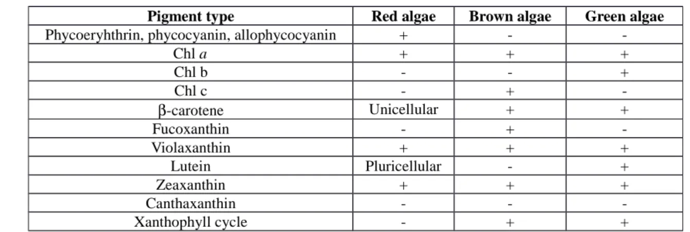
Pigments: As mentioned earlier, most of the algae are photoautotrophs. Consequently, their chloroplastsare rich in pigmented molecules such as tetrapyrroles and carotenoids. The molecules are able to absorb light thanks to their characteristic conjugated double bonds. Each photosynthetizing microalga contains at least the close tetrapyrrole Chl a (6). Except in red algae, in which Chl a is accompanied by the open tetrapyrroles
phycoerythrin, phycocyanin and allophycocyanin, green and brown algae contain another type of Chl molecule (Table 1). The set of light harvesting molecules is complemented with several carotenoids (Car) (Table 1). As it will be explained below in details, these molecules have a great health and therapeutic potential.
The diatom Haslea ostrearia synthesizes and excretes a hydrosoluble blue pigment, the so-called marrenine, responsible for the greening of oyster gills [7]. This pigment exhibits an antiproliferative effect on lung cancer model [36] and has potential antiviral and anticoagulant properties [37].
Amino acids: Beside the universal functions of amino acids in proteins, they are important for skin hydration, elasticity, photoprotection (see below) and are included in cosmetics [7]. Amino acids from diatoms exhibit dermatological properties [38].
Photoprotectants: The best known photoprotectants synthesized by microalgae are mycosporine-like amino acids (MAAs) (Fig. S3). MAAs act as sunscreens to reduce UV-induced damage and also as ROS scavengers [39]. Mycosporine-like amino acids have been found in more than 380 marine species, including microalgae 129
130
131
132
133
134
135
136137
138
139
140 141142
143
144
145
146
147
148149
150
151 152153
154
155 156157
158
[40]. A database referencing the studies in microalgae, cyanobacteria and macroalgae is available at the Univer-sity of Erlangen, Germany (
http://www.biologie.univ-erlangen.de/botanik1/html/eng/mar-database.htm
). A recent study reported the screening of 33 different species belonging to 13 classes ofmicroal-gae for MAAs [40]. The highest concentrations were found in dinoflagellates whereas diatoms contained only low amounts. MAAs have the potential to replace or supplement today’s available sunscreens and particularly those based on petrochemical products. More recently, other photoprotective molecules such as pyropheophytin a (Eicenia bicyclis: [41]), fucoxanthin (Fuco) (Hijikia fusiformis: [42]) have been isolated from brown macroal-gae [3,29]. Because these molecules are also present in microalmacroal-gae, they have been also considered here. Jeffrey et al. [43] have reported the occurrence of such compounds in 206 strains of 152 microalgae. In many microal-gae, the cell is made more resistant to UV by the accumulation in the cell wall of sporopollenin [44], a Car-poly-mer absorbing UV light.
Lipids: In animal cells, essential fatty acids and specifically polyunsaturated fatty acids (PUFAs) are incorporated into lipid membranes in which they increase the fluidity and exchanges between extra and
intracellular compartments. Numerous studies have demonstrated that dietary ω3 PUFAs have a protective effect against atherosclerotic heart disease [45-48]. The two principal ω3 fatty acids in marine oils, eicosapentaenoic acid (EPA; 20:5ω3) (7) and docosahexaenoic acid (DHA; 22:6 ω3) (8), have a wide range of biological effects. Both EPA and DHA are known to influence lipoprotein metabolism, platelet and endothelial function,
coagulation, and blood pressure. More specifically, EPA performs many vital functions in biological membranes, and is a precursor of several lipid regulators involved in the cellular metabolism. In addition, the effect of ω3 fatty acids may depend, to some extent at least, on the presence of underlying disorders such as dyslipidemia, hypertension, diabetes mellitus, and vascular diseases [48]. DHA is a major component of brain, eye retina and heart muscle, it has been considered as important for brain and eye development and also good cardiovascular health [49]. ω3 fatty acid supplementation in animals and humans results in substantial increases in the plasma and tissue levels of EPA and DHA, as well as variable incorporation of the phospholipid classes in various tissues. These differences may be important for the subsequent use and metabolism of EPA and DHA. Although both fatty acids are thought to be biologically active, most studies have focused on the relative importance and effects of EPA, primarily because of its predominance in marine oils and fish species. Because animal cells are unable to synthesize these molecules, they must be acquired through the diet. So far, the main source for PUFAs, free or methyl ester derivatives, fatty alcohols, fatty amines and glycerol is fishes. However, fish oil depends on 159
160
161
162
163
164
165
166
167
168
169
170 171172
173
174
175
176
177
178
179
180
181
182
183
184
185
186
187
188
fish quality and fish resources, which are declining and fish tends to accumulate poisonous subtances via the food chain. Therefore, alternate sources have to be exploited. Microalgae present an excellent potential for this purpose because (i) their fatty acid profile is simpler than that of fish oil, (ii) the production condition can be controlled and last but not least, (iii) the algal species can be selected according to the PUFA required (see below). In contrast to EPA, molecules from fish oil products are unstable and exhibit a poor taste, EPA esters from microalgae are of better quality and more stable [50].Importantly, selected PUFA can be favored through choosing culture conditions. Some species, such as Phaeodactylum tricornutum produce mainly EPA [51]. Among the lipids, arachidonic acid (Ara), an essential fatty acids, is produced by some algae such as Nitzschia conspicua [52]. Ara is also a precursor of prostaglandins and leukotrienes and, is also a component of mature human milk [53]. All these molecules can be used for different activities such as nutrition (human and animal), pharmaceutics, cosmetics, aquaculture and biodiesel production.
Polysaccharides
Best producers of polysaccharides of interest are brown and red seaweeds. Among the different types of polysaccharides synthesized by these algae and also synthesized by red microalgae such as Porphyridium sp., those that are highly sulfated present an antiviral activity [54-55].
Miscellaneous
In addition to their used in flavor and fragrance industries, monoterpenes have drawn increasing commercial attention because of their putative action as natural insecticides and antimicrobial agents [56]. Little is known about the production of these molecules in microalgae but their use as biotransformant has been reported [56]. Water extract of the marine diatom Haslea ostrearia exhibited anticoagulant activity [37].
Due to space limitation for this review and the availability of the data, only the lipids and pigments, as molecules with biological activities,are detailed in the next section.
LIPIDS AND PIGMENTS, TWO TYPES OF BIOLOGICALLY ACTIVE COMPONENTS SYNTHESIZED BY MICROALGAE Lipids 189
190
191
192
193
194
195
196
197
198
199
200 201 202203
204
205 206 207208
209
210
211212
213 214215
216 217Fishes and marine microalgae are the primary producers of ω3 PUFA. While microalgae synthesize ω3 PUFA, fishes usually obtain EPA via bioaccumulation in the food chain. So far, two of the questions that have been addressed are: (i) is it cheaper to produce ω3 fatty acids from algae is than from fishes? and (ii) are ω3 fatty acids obtained (EPA and DHA in particular) from microalgae as effective as those obtained from fish oil? Regarding the first question, it was shown that ω3 fatty acid production from microalgae would indeed be less expensive than the one from fishes. In addition, unlike fish oil, microalgal ω3 fatty acid extracts have no odour, are less susceptible to be contaminated by heavy metals, and do not contain cholesterol [57]. Finally, when microalgae are grown under controlled conditions, the composition of the fatty acids shows no seasonal variation [58]. As fish oil fails to meet the increasing demand for purified PUFA, alternative sources are being sought, especially from microalgae. Microalgae contain lipid levels between 20-50% (Table 2), but in stress conditions such as N-deprivation or an irradiance or temperature increase, some species of microalgae are able, to accumulate up to 80% of their dry weight in fat [59-60], including large quantities of high-quality ω3 PUFAs (Table 2). Thus, algae are gaining increasing attention because of their important values for human health as well as for aquaculture.
So far several algae are already used as dietary supplements. Chlorella sp., a freshwater unicellular green alga, is known to be a good source of proteins, lipid soluble vitamins, pigments, choline, and essential minerals in a bioavailable form. The administration of Chlorella affects some biochemical and physiological functions [71]. As algal sources of DHA come the brown alga Schizochytrium sp. (40% DHA, 17% docosapentaenoic acid (DPA)), the green alga Ulkenia sp. and the red alga Crypthecodinium cohnii (40-50% DHA) [72]. The production from the latter species is especially well described [73] and marketed by Martek company. DHA produced from microalgae is mainly used for child and adult dietary supplements [74]. Moreover, C. cohnii have effects in aquaculture [75]. It has already been showed that algal oils rich in DHA are nutritionally equivalent to fish oils in several tests [76-77], suggesting that algal oils could constitute a susbtitution to fish oils. In addition, new algal sources for the ω3 very long chain PUFAs (VLCPUFA) are being examined. These include the production of EPA from other strains such as marine diatoms. P. tricornutum, a marine diatom, has been widely used as a food organism in aquaculture and considered as a potential source for EPA production [77]. The sole marine microalga known to be rich in EPA used as a dietary supplement is the marine diatom O. aurita. It has been shown that extracts of this microalga have an anti-proliferative effect on cultures of bronchopulmonary and epithelial cells [78]. Different experimental models are used to conduct studies in relation with use of ω3 fatty acids from microalga sources. Using freeze-dried microalgae, animals and specifically murine models are often
218
219
220
221
222
223
224
225
226
227
228
229
230
231
232233
234
235
236
237
238
239
240
241
242
243
244
245
246
247
used as previously described by several authors. Normal or modified (chemically and genetically) strains of mice and rats have been already used to study the effects of Chlorella sp. on myelosupression induced by lead [79], on glycogenesis improvement in diabetic mices [71] and on dyslipidemia prevention in rats fed with high fat diet treatments [80]. The comparison of rats fed with freeze-dried O. aurita or with fish oil shows that the plasma triacylglycerol concentration in rats fed microalgae was lower than in the control group and also than in the fish oil group (Fig. 1). The plasma concentrations of HDL- and LDL-cholesterol were significantly higher by comparison with the control rats. For the rats fed with fish oil, LDL cholesterol was similar to the rats fed with control diet, while HDL cholesterol was higher than in the group of control rats. Nevertheless, the HDL/LDL cholesterol was statistically similar in both the control and microalga-fed groups of rats, whereas this ratio is greater in the rats fed with fish oil.
According to the use of microalga as an alternate to fish oil, differences in the enrichment of tissue in ω3 fatty acids and specifically in EPA were mentioned. Indeed, results reported in Fig. 2 show that the levels of EPA, obtained for each organ are significantly different from ones obtained in the two other groups (for all studied organs). In fact, whatever the organ considered (liver, heart or kidney), EPA levels were significantly higher in rats fed with the freeze-dried microalga diet than in those fed with fish oil or control diets. Moreover, significant higher amount of DPA was found in the liver and kidney total lipid of the rats fed with the diatom diet than in those from rats fed with fish oil or with the control diet. The n-6/n-3 ratio in liver, heart or kidney, were significantly different in the three experimental groups, the rats fed the control diet being systematically higher than in the two other groups. In addition, this ratio tends to be lower in the rats fed the freeze-dried microalga diet by comparison with those fed the fish oil one. These results showed that a freeze-dried O. aurita diet could be considered as an alternate source to fish oil in regulation of blood parameters involved in lipid metabolism and in the enrichment of tissue in ω3 fatty acids and specifically in EPA. This enrichment into EPA at the expense of Ara incorporation into total lipids of liver, heart and kidney could have beneficial effects in the cardiovascular disease prevention as described with fish oil. Moreover, when intact microalgae are used in diet, the effect of the ω3 fatty acid role could be potentiated with pigment content such as Fuco or other Cars. These results are in line with those published by Rao & Rao [81] and Micallef & Garg [82], who found a synergistic action between pigments, fatty acids and phytosterols on plasma lipid concentration decrease, on inflammatory response and thus on cardiovascular disease risk prevention. These molecules that are naturally contained in O. aurita make this organism a major actor in human nutrition as an alternate to fish oil.
248
249
250
251
252
253
254
255
256
257
258
259
260
261
262
263
264
265
266
267
268
269
270
271
272
273
274
275
276
277Pigments
Three major classes of photosynthetic pigments occur among microalgae: Chls and derivatives, Cars (carotenes and xanthophylls) and phycobilins, which together represent hundreds of molecule purification [83]. Considering their high structural diversity and the possibility to pharmacomodulate these molecules, the potential of microalga pigments to obtain molecules of therapeutical interest is very high. Because of their lability and difficult purification, the biological activity of most molecules remains unstudied [27,59]. A large number of studies designed to purify and identify bioactive molecules from microalgae have lead to the isolation of pigments. These purified pigments usually have a high activity on pharmacological and cellular effectors, at very low concentrations.
Antioxidant, anti-inflammatory and antimutagenic activities
Oxidative stress is a major cause of inflammatory events implicated in a large number of diseases, such as cancer, neurodegenerative and cardio-vascular diseases, or diabetes. The antioxidant and anti-inflammatory activities of microalga pigments is widely demonstrated and evidenced in numerous in vitro free radical scavenging assays and in vivo assays. The antiradical capacity of metal-free Chl-derivatives such as chlorins, pheophytins, and pyropheophytins is much weaker that the corresponding metallo-derivatives. Protoporphyrin methyl ester and its magnesium chelated derivative, as well as pheophorbide b and pheophytin b, were also identified as strong antioxidant molecules [84]. The ability of the porphyrin ring to transfer electrons explains the antioxidant activity of Chls and derivatives. The high antioxidant activity of pheophorbide b, compared to pheophorbide a, suggests that the presence of the aldehyde function may also be critical to this activity [85]. The antioxidant properties of Chls and Chl-derivatives disappear in the presence of light [86]. Metal-free and metallo-Chl derivatives have also antimutagenic activities, as demonstrated using a bacterial mutagenesis assay [87-88]. Microalgal carotenoids (e.g., zeaxanthin (Zea), astaxanthin (Asta) (9)) and epoxycarotenoids (e.g., neoxanthin) have strong antioxidant activities in vitro and in vivo in animal models. Particularly, Asta has a great potential to prevent cancer, diabetes and cardiovascular diseases [89-90]. The presence of the hydroxyl and keto endings on each ionone ring explains Asta unique features, such as the ability to be esterified [91], a higher antioxidant activity and a more polar configuration than other Cars [92]. Epidemiologic studies demonstrate an inverse relationship between cancer incidence and dietary Car intake or blood carotenoid levels, but intervention trials using a high dose of carotene supplements did not show protective effects against cancer or cardiovascular disease. Rather, the high risk population (smokers and asbestos workers) showed an increase in cancer cases in 278 279
280
281
282
283
284
285
286
287 288 289290
291
292
293
294
295
296
297
298
299
300
301
302
303
304
305
306
307
these trials [93]. Phycocyanin c and phycoerythrin also exhibit antioxidant and anti-inflammatory activities [94-96]. As a conclusion, most microalga pigments exerts strong in vitro antioxidant activity, but additional intervention trials are required to precise their absorption, metabolism and potential as natural antioxidant, anti-inflammatory and antimutagenic compounds in vivo.
Cytotoxicity
A large number of studies performed in cancer cells grown in vitro clearly demonstrate the antiproliferative, cytotoxic and pro-apoptotic activities of Chl derivatives, Cars, and phycobilins [27]. Moreover, several studies designed to purify antiproliferative molecules from marine microalgae have led to the isolation of carotene (Zea) [83,91] and epoxyCars (e.g., Fuco, violaxanthin (Viola) (10)) [78,92]. Fuco is the prototypical example of a microalgal cytotoxic pigment with an important therapeutic potential. Its strong antiproliferative, cytotoxic and pro-apoptotic activities , at concentrations inferior to 1 µM, have been widely studied and demonstrated on a large number of human cancer cell lines from various tissular origin (lung, breast, prostate, lymphoma, gastric, uterine, neuroblastoma,etc) [98-102]. The molecular mechanisms involved in the cytotoxic activity of Fuco are not completely understood, but various cellular targets of Fuco have been identified. Because of its hydrophobicity, Fuco easily crosses and integrates cell membranes. It inhibits mammalian DNA-dependent DNA polymerases [103], protects against ROS and UV-induced DNA injury [99,104-107], down regulates cyclins and CDK expression, disturbs major transduction pathways controlling cell survival and transcriptional activation of genes involved in resistance to apoptosis and anticancer drugs in cancer cells. (MAPK, NF-κB [99,101], p21WAF/Cip1 CDK inhibitor [108], Bcl-xL [109-110]). Fuco also enhances Gap junction intracellular communication, an important process in the control of cell growth, differentiation, apoptosis induction and diffusion of anticancer drugs [111]. Intestinal absorption and metabolism of dietary Fuco into its major metabolite fucoxanthinol was demonstrated in mices, but not in humans. Absorption studies in humans indicated that less than 1 nmol.L-1 is found in plasma after a 1 week diet containing Fuco- rich diet [112]. In the same way as Fuco, a large number of microalga pigments were identified as cytotoxic at very low concentrations in cancer cells. They belong to the epoxyCars class (e.g., Viola [96], halocynthiaxanthin [100,103,113-114], peridinin [114-117]), to Chl derivatives (e.g., Chl a, pheophytin a, pheophytin b, pheophorbide a) or to phycobilins (e.g., phycocyanin) [92]. Moreover, for some of them, their anticancer activity was confirmed after per os absorption. As an example, in the pathogen-free ddY strain mice, the development of skin tumors induced by 12-O-tetradecanoylphorbol-13-acetateis suppressed when 1 µmol peridinin is added in dietary water [118]. For most 308
309
310
311
312 313 314315
316
317
318
319
320
321
322
323
324
325
326
327
328
329
330
331
332
333
334
335
336
337
molecules, intestinal resorption, bioavailability and metabolism are unknown. Besides, their effect in noncancer cells and immune cells is mostly unexplored. Understanding their pharmacological activity in human cells may allow to obtain potent selective anticancer pharmaceutics.
Ref 95
Multi-drug resistance reversion in cancer cells
Microalgae pigments may have interest to restore drug sensitivity or reverse multi-drug resistance in cancer cells, as some of them inhibit or down-regulate drug efflux pumps. As examples, neoxanthin increases rhodamine 123 accumulation in multi-drug resistance (MDR) colon cancer cells [113], inhibits the P-glycoprotein(P-gp) efflux pump and reverses MDR in doxorubicin-resistant MCF-7 cells and hmdr1- transfected L1210, at 4 and 40 µ g.mL-1, respectively [119]. Viola and violeoxanthin are effective MDR modulators in Colo 320, at 4 and 40 µg.mL-1, respectively [120]. Mod erate P-gp inhibition by Viola was observed in hMDR1-transfected L1210 and MDA-MB-231 expressing the MRP1 pump (HTB26) at 20 µg.mL-1 [121-122]. In the same way, a significant reduction of P-glycoprotein expression R-HepG2 cells, at both transcriptional and translational levels, was observed when cells were treated with pheophorbide a [123].
Antiangiogenic activity
Fuco and its physiological metabolite fucoxanthinol have antiangiogenic effects, as demonstrated in the blood vessels and HUVEC tube formation assays. In SCID mice injected subcutaneously with 107 HUT-102 cells, fucoxanthinol did not affect tumor incidence, but significantly slowed tumor growth. It also significantly decreased microvessels outgrowth, in a dose-dependent manner, in an ex vivo angiogenesis assay.
Use as fluorescent probes
The physicochemical characteristics of phycobilins, Chl and Chl catabolites make them suitable for use as fluorescent probes for cellular and tissular analysis (e.g., cell sorting, cytofluorescence, flow cytometry, histofluorescence, binding assays, ROS detection, labeling of pathological or apoptotic cells, etc). Phycocyanin or phycoerythrin-coupled antibodies are common reagents available for research and medical use, in which phycobilins act as powerful and highly sensitive fluorescent probes (for reviews, see [96]).
338
339
340
341 342 343 344 345346
347
348
349
350
351
352
353
354 355 356357
358
359
360 361 362363
364
365
366
367Other preventive or therapeutical use
Microalgal pigments have demonstrated their lack of toxicity and biological activity in a wide range of biological applications, including prevention of acute and chronic coronary syndromes, atherosclerosis, rheumatoid arthritis, muscular dystrophy, cataract and neurological disorders. They are also recommended to protect the skin and eyes against UV radiation [124-125]. Lutein is one of the major xanthophylls found in green microalgae. It selectively accumulates in the macula of the human retina, protects the eyes from oxidative stress, and acts as a filter of the blue light involved in macular degeneration and age-related cataract [112,126-127]. Fuco anti-allergic activity was recently evidenced using a rodent mast cells model [127]. It could also have interest to limit the risk of obesity [127,129]). Because of their antioxidant and anti-inflammatory activity, most microalga pigments have neuroprotective effects in cultured rat cerebellar neurons, and hepatoprotective effects in hepatocytes grown in vitro (e.g., phycocyanin, phycoerythrin) [96]. Besides, some studies have demonstrated antiviral and antifungal activities for some pigments (e.g., allophycocyanin, phycocyanin) [96, 130].
Potential and obstacles to a possible pharmaceutical development of microalgae pigments and derivatives The lack of oral toxicity of microalgae pigments may be due to a weak intestinal resorption but also suggests a possible pharmaceutical development for these molecules (e.g, [24]). Most microalga pigments are labile molecules, sensitive to light and oxygen, and it is highly probable that their half-life in a physiological context is short [131]. This lability has interest when considering new applications, but is also a limit to their pharmaceutical development. It also explains the high price and low availability of pigments standards, necessary to set up intervention trials and clinical assays. Consequently, there is a lack of in vivo studies on absorption, metabolism and pharmacokinetics of microalga pigments [27]. Moreover, dozens of pigments and derivatives are unstudied because no standard is available. It is essential to carry on the development of economically viable industrial processes to obtain high amounts of pigments and derivatives (selection of over-producing species and strains, definition of physiological conditions giving the best production yields, optimization of eco-extraction and purification processes, development of chemical and chimioenzymatic synthesis). As an example, the average carotenoid concentration in dry microalgae is 0.1-2% (w:w). When grown under optimized conditions of salinity and light intensity, Dunaliella produces up to 14% β-carotene [72,130-132]. Purification from natural sources is much more expensive than chemical synthesis, but has the advantage of providing pigments in their natural isomer proportions (e.g., carotene) [72,130-132]
368 369
370
371
372
373
374
375
376
377
378
379
380 381 382383
384
385
386
387
388
389
390
391
392
393
394
395
396
397ABIOTIC STRESSES: A CONVENIENT WAY TO INDUCE THE METABOLIC REORIENTATION AND INCREASE THE PRODUCTION OF SELECTED BIOACTIVE COMPOUNDS.
As microalgae play a major role in CO2 uptake [2,22], numerous studies deal with effects of abiotic stresses on algal biology and metabolism. The main objectives of some of those studies were to predict how and what algae will cope with climatic change and increasing pollution. The commercial exploitation of the natural microalgal diversity for the production of carotenoids and PUFAs has already started up with few strains such as Chlorella vulgaris (Trebouxiophyceae), Dunaliella salina (Chlorophyceae), Haematococcus. pluvialis (Chlorophyceae) [133-134] and O. aurita (Bacillariophyceae). In this section, the impacts of abiotic stresses such as light, UV-ra-diation, nutrient starvation, temperature and metals on microalgal metabolism and on the production of biologi-cal active compounds is reviewed.
Light
More than terrestrial plants, microalgae display a diversity of light harvesting pigments (Table 1) allowing photosynthesis at different depths according to pigment content. Photosynthetic apparatus of most microalgae acclimates to light level and light quality by optimizing pigment content and composition [135-141]. Microalgae are confronted with variations of light by movements in the water column and emersion for coastal benthic species. To cope with high sunlight intensities, microalgae have developed different photoprotective
mechanisms. One of these, the xanthophyll cycle, consists in the reversible conversion of Viola, antheraxanthin and Zea in green algae and in the reversible conversion diadinoxanthin and diatoxanthin in brown algae [91 ,141-142]. Acclimation to low irradiance intensity or blue enriched light increases Car synthesis such as Fuco [140]. The photoprotection or the low photoacclimation leads carbon to Cars whereas in nonstressfull conditions, C serves mainly to growth (cell wall edification). In the marine diatom Haslea ostrearia, C fixation by β -carboxylation is almost equal to that in the C3 pathway whereas under low irradiation C3 fixation dominates [144]. Light intensity has also an impact on lipid synthesis, PUFAs: EPA, was significantly higher under low light, and saturating fatty acids and DHA levels were higher under high light in Pavlova lutheri [140]. EPA and DHA are now recognized as having a number of important neutraceutical and pharmaceutical applications.
UV-radiation
Microalgae experience high levels of UV-radiation in shallow areas, low turbide habitats or during low tides when they are deposited on intertidal flats. Several authors have shown that UV exposure increases carotenoid 398
399
400401
402
403
404
405
406
407
408 409 410411
412
413
414
415
416
417
418
419
420
421
422
423
424 425 426427
content [145-146] and, in some Bacillariophyceae, MMAs synthesis [147-148]. Guihéneuf et al. [149] have shown that a 8-day exposure to UV decreases the PUFA content, EPA by 20% and DHA by 16% in Pavlova lutheri but not in Odontella aurita in which the PUFA content remains unchanged. As other environmental stresses, UV radiation stimulates the intracellular ROS production [150-151] triggering antioxidative defence such as antioxidative enzyme activities and antioxidant compounds (glutathione, α-tocopherol, ascorbate, etc).
Nutrient starvation
The reorientation of the metabolism induced by nutrient starvation is illustrated by the accumulation of Asta in H. pluvialis under N-limitation and P- or S-starvation [133,152-153]. Asta accumulation is related to a massive increase in carbohydrate content up to 63% of the cell dry weight [154] and an increase of lipid content in the cytoplasm. In the Crytophyceae Rhodomonas sp., N-starvation triggers a rapid decline in N-containing compounds causing an almost complete loss of phycoerythrin [155]. Riyahi et al. [156] have shown that the production of β-carotene in Dunaliella salina was increased with nitrate 1 mM and salinity 30%, On the other hand, in the microalga Tradydiscus minutus (Eustigmatophyceae), N-starvation brings about a nearly 50% drop in triacylglycerols (TAGs) containing EPA, and also a decrease of TAGs containing Ara, while P-starvation has a sizable effect on those TAGs that contain two or three Ara [157]. Many microalgae promote a shift in lipid metabolism by producing substantial amount (20-50% of dry weight) of TAGs as lipid storage during the stationary phase when nitrate becomes depleted [158]. The amount of EPA partitioning into TAGs varies according to strains and also during the different phases of growth within a species.
Metals
Some metals such as Cu, Fe, Zn are essentials for cell metabolism since they are components of electron transporters involved in photosynthesis and respiration, some enzymes, etc. When metals are present in excess, they induce an oxidative stress [159-160] and antioxidant defense mechanisms already cited above. To cope with metals in excess, many microalgae excrete exopolysaccharides that adsorb metals in the medium and prevent them to enter inside the cells [161-162]. Polysaccharide depolymerization by different procedures allows the obtention a variety of oligomers with potential applications in therapeutics and in biotechnology [10]. However, in the presence of Cd, the xanthophyll cycle in Phaeodactylum tricornutum is inhibited [163]. The impact of metals depends on their speciation and the growth medium pH [164].
428
429
430
431
432
433 434 435436
437
438
439
440
441
442
443
444
445
446
447 448 449450
451
452
453
454
455
456
457Temperature
Microalgae can synthetize VLCPUFA as major fatty acid components [165]. Experiments in controlled conditions are necessary in order to select species producing those PUFAs, in what conditions, at what stage of their growth, and in what lipid class. Tonon et al. [158] have shown than fatty acids accumulate during the stationary phase of growth when nitrate concentration in the growth medium was low. EPA production is higher at 8°C than at 25°C in the red microalga P. purpureum [166]. In the marine diatom Nitzschia leavis cultivated at 15, 19 and 23°C, growth is enhanced at the highest temperature but the lowest temperature favours the
distribution of PUFAs in phospholipids and increases EPA content in TAGs [167]. As in terrestrial plants, the increase of PUFAs in membrane was suggested to be a strategy to maintain membrane fluidity under low temperature.
LARGE-SCALE CULTURE AND BIOMOLECULE PRODUCTION
Microalgae are a source for a variety of bioactive compounds. However, they remain largely unexplored and, until now, very few commercial achievements of microalgal biotechnology have emerged [168]. During the last decades, researchers and engineers have developed several cultivation technologies that are still in use today. Seen very often as obvious, the subsequent culture of a given microalgae can be more difficult than expected in the attempt to up-scaling. Numerous drawbacks and difficulties await the entrepreneur wishing to set up a commercial production. The choice of photobioreactors, light systems, control for pH, CO2 etc.. batch or continuous cultures, management of nutrients, water supply, water treatment onward and outward as well as the energy needed will constitute a strategic debate. Concerning the biological aspects, once the proper selected strain is chosen, the type of culture system, the feeding strategies (photoautotrophy, heterotrophy, mixotrophy reviewed hereafter), the confrontation with pathogens, contaminants and predators will enter in the game.
Photoautotrophic production
Photoautotrophic productions use light as the source of energy thank to photosynthesis and CO2 as the source of carbon. They are currently processed with either open ponds or closed systems, that can use sun light and artificial light. However, the major constraint that phototrophic production must address is how efficiently light is used. Indeed, productivity is tightly related to the surface to volume ratio of the cultivation system and many recent technological developments tended to improve this ratio.
458 459
460
461
462
463
464
465
466
467
468 469 470471
472
473
474
475
476
477
478
479
480 481 482483
484
485
486
Originally, open-ponds and raceways were used for microalgae production, but the quest for increased productivity and better control led to the development of closed photobioreactors. The latter are usually recognized as achieving higher biomass productivity than open systems [60,169-170]. Nevertheless, the maximum biomass productivity does not necessarily match the maximum productivity for a particular molecule, neither the maximal economic efficiency [171]. It is beyond the scope of this article to enter into the argumentation of the pro and contra open ponds versus photobioreactors. The solution might lie in between when the two technologies will be integrated in the same production line. Controlled production system like photobioreactors renders easier to explore the metabolic versatility of microalgae with different production strategies. Despite their high initial investment, photobioreactors provide a variety of attractive benefits for bioactive molecule production, when compared to open systems. First, they make possible monospecific and axenic cultures as well. They also are characterized by reproducible cultivation conditions and accurate control for abiotic factors such as temperature, pH, irradiance, evaporation and hydrodynamics. The production of a particular molecule can take advantage of these controls since abiotic factors can substantially impact the biochemical composition of microalgae, as discussed above.
Most of the commercial productions use photoautotrophic cultivation processes, with pigments, health food and aquaculture being the main markets. Several commercial companies produce Asta with Haematoccoccus : Mera Pharmaceuticals (Hawaii) reports a biomass production of about 6.6 T/year using closed tubular photobioreactors. Similar culture systems have been used by Algatechnologies (Israel) and Fuji Health Science (Hawaii). However, the production cost of Asta with Haematoccocus is still high because of physiological (slow growth rate) and technical (two-stage production process) constraints. Thus from the economic point of view, Asta produced with Haematoccocus can hardly compete with the synthetic pigment [92].
Dunaliella natural β-carotene is another widely distributed pigment from microalgae. Its global production through autotrophic cultivation is estimated at about 1.2 T/year [12]. Two cultivation processes are currently used for β-carotene production. Betaten (Adelaide Australia) or Aquacaroten (Subiaco, Australia) grow this microalgae in unmixed open ponds and Betaten reported a β-carotene production of about 13 T/year (about 400 ha of culture area). The associated production costs appear relatively low considering the optimal climate and, unlike other systems, no pumping is required [172]. Raceway ponds (intensive mode) are operated by the Nature Beta Technologies company (Eilat, Israel), reporting a β-carotene production of 3 tonnes per year. Several studies have been attempted to grow Dunaliella in closed photobioreactors, although up to date, none of these trials led to any significant production even at the pilot scale [173]. Several other little companies commercialize 487
488
489
490
491
492
493
494
495
496
497
498
499
500
501502
503
504
505
506
507
508
509
510
511
512
513
514
515
516
a variety of microalgae grown photoautotrophically for their high amount in EPA and DHA. For example, Isochrysis sp. is produced by Innovative Aquaculture Products Ltd (Lasqueti Island, Canada) and the diatom O. aurita is produced by BlueBiotech InT (Kollmar Germany) and Innovalg (Bouin, France). In the latter, O. aurita is grown photoautotrophically in open air 1,000 m2 raceways and co-cultured with the macroalgae Chondrus cripus, for increased productivity [65].
Heterotrophic production
Studies on microalgae heterotrophy were initiated in the 60s and demonstrated that some species could grow on organic carbon sources, such as sugars or organic acids, replacing the traditional support of light energy. The number of studies further increased in the 2000s with the growing interest for biofuel from microalgae. Among the microalgae species currently cultivated, only a few (e.g., Chlorella protothecoides, Crypthecodinium cohnii, Schizochytrium limacinum, Haematococcus pluvialis) have been successfully grown heterotrophically [174]. Conversely to photoautotrophy where productivity is related to the illuminated area of the culture, productivity for heterotrophic cultures relies on organic carbon concentration in the bulk volume of the culture. This facilitates the up-scaling for commercial production and usually results in higher productivity, with biomass production being one order of magnitude higher than for photoautotrophically grown cultures [175] and in reduced production, harvest and maintenance costs. For instance, high biomass concentration (45 g L−1) and volumetric productivity (20 g L−1 d−1) were achieved in heterotrophic cultures of Nitzschia alba [176].
Heterotrophic culture requires axenic conditions, a major drawback when compared to photoautotrophy. As pointed out by Bumback et al. [177], any, even minor, contamination introduced with the inoculum could easily outcompete the microalgal species for the organic carbon source. The prerequisite for axenicity and the additional care for its maintenance necessarily impact the production costs. Additionally, heterotrophic culture might not bring the same diversity and the same biochemical composition as reached with photoautotrophy. Yet, Perez-Garcia et al. [174] reported the possibility to produce lutein with Dunaliella sp. and Asta with Chlorella zofingiensis grown heterotrophically. Wang & Peng [178] reported the first growth-associated biosynthesis of Asta with Chlorella zofingiensis heterotrophic cultures using glucose as organic carbon source. This study suggested that maximal biomass and Asta production could be obtained simultaneously by a one stage culturing rather than the two stage process that was proposed for Haematococcus. Although commercial production of Asta with heterotrophic Chlorella zofingiensis culture has not yet been reported, this species may be a promising alternative to Haematococcus for the mass production of Asta. Besides, commercial production of 517
518
519
520
521
522 523 524525
526
527
528
529
530
531
532
533
534
535536
537
538
539
540
541
542
543
544
545
546
heterotrophically grown Chlorella in fermentor is common in Japan and Korea, mainly for aquaculture and health food applications [179]. Martek (USA) also successfully produces DHA health food with heterotrophic Crypthecodinium cohnii cultivation [180].
Mixotrophic production
If mixotrophy is defined so as to include osmotrophy, most of microalgae can be considered as mixotrophic. Many microalgae can grow on dissolved organic carbon [181] and, under inorganic nitrogen stress, use organic sources of nitrogen [182].
When microalgae are grown with CO2 as the sole carbon source, light provides the energy necessary for biomass production. However, under photoautotrophic conditions, growth is often limited by light availability and, during the night, the productivity is further reduced by respiration. Mixotrophic microalgae can concurrently drive phototrophy and heterotrophy to utilize organic energy and both inorganic and organic carbon substrates, thus leading to a synergetic effect of the two processes that enhances the culture productivity. Yang et al. [183] demonstrated that biomass yield on the supplied energy was four folds higher for true mixotrophically grown Chlorella pyrenoidosa than for the photoautotrophic culture. They also highlighted that cyclic autotrophic/ heterotrophic cultivations, could lead to even more efficient utilization of energy for biomass production than the true mixotrophy. Moreover, mixotrophy can overcome light limitation occurring at high densities. This mechanism has been demonstrated to be important for Scenedesmus obliquus [184] and is suggested to be widely spread among mixotrophic microalgae in general.
Hence, high productivity is one of the major benefits associated with mixotrophy. For some microalgae, the growth performance under mixotrophic conditions can even exceed that achieved with heterotrophic cultures. Indeed, Pulz & Gross [12] pointed out that the maximum specific growth rate of Chlorella vulgaris and
Haematococcus pluvialis growing mixotrophically was the sum of the photosynthetic and heterotrophic specific growth rates. Besides, Stadnichuck et al. [185] reported higher Chl a, carotenoids, phycocyanin and allophycocyanin content in Galdieria partita grown mixotrophically than in heterotrophically cultures. Mixotrophy can therefore overcome some of the drawbacks experienced with heterotrophic cultures [186] and might be an efficient means for enhanced production of light-induced pigments in microalgae. However, as for heterotrophic cultures, mixotrophic cultures require axenic conditions to prevent bacteria from outcompeting microalgae for organic substrates. Research will be needed to cope with the risk of favouring the prokaryotic part in the culture. To date, the processing of mixotrophic cultivation implies the availability and maintenance of 547
548
549
550 551 552553
554
555556
557
558
559
560
561
562
563
564
565
566567
568
569
570
571
572
573
574
575
576
axenic strains, the investment for sterilizable photobioreactors and higher operation costs. However, the higher productivity achieved with mixotrophy cultures could balance these drawbacks.
It is well documented that some economically important microalgae can be grown mixotrophically (Haematococcus pluvialis, Scenedesmus acutus, Chlorella vulgaris, Nannochloropsis sp.). However, despite the indisputable assets of mixotrophy, only one company reported the use of mixotrophic processes for industrial Asta production. Indeed, BioReal (Sweden) was the first company in the world to produce and commercialize from 15 to 30 T/year of Asta-rich biomass using mixotrophy culture in indoor closed photobioreactors [172].
CONCLUSIONS AND FUTURE DIRECTIONS
Microalgae represent a subset of single cell microorganisms that generally grow autotrophically using carbon dioxide as the sole carbon source and light as energy. They are ubiquitous in nature, occupying every type of ecological niche. Microalgae represent a major untapped resource of genetic potential for valuable bioactive agents and fine biochemicals. Screening studies should reveal the existence of new molecules potentially interesting for their biological activities. From the basic point of view, the mechanisms of action of the already marketed products should be better established. For instance, it has been reported that, beyond ω-3 and antioxidants, fish oil also contain peptides having bioactive activity. Many of them have an interest for health and pharmaceutical industries. In their natural environment, algae are subjected simultaneously to different abiotic factors with daily and seasonal variations that may be stressful, such as tidal movements, temperature, light levels or UV radiations. To cope with stress, the synthesis of molecules of interest such as antioxidants, PUFAs and glycerol is increased in tolerant microalgae. More basic research on this point should be performed to elucidate the metabolic and regulation circuits involved in these productions. This will help to discover what are the interactions between several abiotic factors and mechanisms involved in the biochemical responses. In silico research, biochemical characterization of microalgal products and in the same way the research of biological activities of algal extracts seem promising for biotechnology applications. Many molecules produced by microalgae show a high structural diversity and should be considered as potent bioactive molecules able to significantly modulate human cell functions, in a physiological or pathological context, at very low concentrations. Additional studies of their biological activity in vivo are required to precise their absorption, metabolism and interest as potential natural anticancer or cardioprotective agents. The development of efficient purification processes will stimulate their study and pharmaceutical development.
577
578
579580
581
582
583
584 585 586587
588
589
590
591
592
593
594
595
596
597
598
599
600
601
602
603
604
605
The cultivation means to produce bioactive compounds are various. Important are the source of energy and the biomass yield. The selection for high producing strains, the optimization of culture modes and harvesting and the management of molecule expression in cultures are crucial steps for the future. Whatever the species and molecules produced, the harvesting system is an expensive and limiting step that has to be adapted to preserve together the algae integrity but also the one of the molecule. Ideally, microalgae producers look for strains with a high valuable-product productivity. However, until now, the main commercial productions rely on a few wild-type strains and the selection for original strains with a high potential for biotechnology remains a challenge for the industry. Pioneer studies for strain selection were initiated in the 90s. The combination of mutagenesis to a selection procedure resulted in substantially increased production for pigments [187], PUFAs [188] or triacylglycerides [189]. These techniques offer an appealing alternative to GMOs.
Transgenic microalgae can be also used as bioreactor for production of therapeutic and industrial recombinant proteins [190-191]. To date, a variety of recombinant proteins have been expressed from nucleus and chloroplast of Chlamydomonas reinhardtii. These include pharmaceutical proteins, antibodies, vaccines, and others that showed a biological activity comparable to the same proteins produced by traditional commercial techniques [192]. Our groups were quickly intrigued by the potential of microalgae as a means to produce therapeutic proteins [193]. A private company was born from this research: Algenics, which is, to date, the first European privately-held biotechnology company focusing on innovative uses of microalgae to produce recombinant biotherapeutics (
http://www.algenics.com
). Concerning the use of microalgae as a platform of recombinant proteins, the recent success led to several patents [194-197] with the successful production of erythropoietin inPhaeodactylum tricornutum (unpublished work). The production costs for microalgal therapeutic proteins are very attractive (i.e., the cost for recombinant antibody is estimated to 0.002 US$ and 150 US$ per gram from microalgae and mammalian cell culture respectively [198]). Moreover, this cost could fall provided that recombinant protein production is coupled with recovery of valuable natural product. However, to the best of our knowledge, no microalgal therapeutic proteins have been yet commercially used.
Microalgae can also be used in biotransformation experiments. In such experiments, immobilized microalgae are incubated with particular substrates to use the in situ enzymes to produce products. Such a method has been used to study the potential of green microalgae such as Chlamydomonas sp. and Oocystis sp. to produce new monoterpenes. The molecular engineering described above combined with biotransformation principle opens many new avenues for algal biotechnology.
606
607
608
609
610
611
612
613
614
615
616617
618
619
620
621
622
623
624
625
626
627
628
629
630631
632
633
634
635ABBREVIATIONS: Asta: astaxanthin, Car: carotenoids, Chl: chlorophyll, DHA: docosahexaenoic acid, EPA: all-Z-eicosa-5,8,11,14,17-pentaenoic acid, Fuco: fucoxanthin, MAAs: mycosporine-lide amino acids, gp: P-glycoprotein, PUFA: polyunsaturated fatty acids, MDR : multi-drug resistance, TAGS: triacylglycerols, Viola: violaxanthin, VLC: very long chain, Zea: zeaxanthin
ACKNOWLEDGMENTS: This research was supported by European grants from the “Fond Européen de Développement Régional FEDER”, the European research program GIAVAP and VOLUBILIS and the ’Contrat de Projet Etat-Region CPER’‘ (Project ’Extraction of anticancer pigments from marine microalgae” XPIG). The authors thank the French cancer league (Ligue Nationale contre le Cancer), the French Ministery for Education and Scientific Research, the University of Le Mans for financial supports.
SUPPLEMENTARY MATERIAL
Fig. S1. Algae represent less than 10% of the total number of identified species.
The original data [S1] were not including the unicellular species. In order to take into account these organisms, we have substituted the number of original species by the number of species found in the AlgaeBase database
(
http://www.algaebase.org/
) although this number is probably largely underestimated.[S1] The World Conservation Union. IUCN Red List of Threatened Species. Summary Statistics for Globally Threatened Species (1996–2010).
http://www.iucnreditlist.org/documents/summarystatistics/2010_1RL_Strats_Table_1.pdf. Accessed 31/01/2012.
Fig S2. Number of publications describing a compound from algae having a biological actvity. The numbers of publications were taken from the Web of knowledge database
(http://www.webofknowledge.com/). Search performed in December 2011.
Fig. S3. Exemple of mycosporine-like amino acids.
REFERENCES
[1] Stengel, D.B.; Connan, S.; Popper, Z.A. Algal chemodiversity and bioactivity: Sources of natural variability and implications for commercial application. Biotechnol. Adv., 2011, 29, 483-501.
636