Université de Montréal
Glucolipotoxicity and the Controt of Pancreatic 0-Ceit Apoptosis
By
Wisal El-Assaad
Programme de Biologie Moléculaire faculté des Etudes Supérieures
Mémoire presented to the Faculté des Études Supérieures to obtain a Master’s degree in Science (M.Sc.)
in Molecular Biology
April, 2003
Jç
£41
de Montré al
Direction des bibliothèques
AVIS
L’auteur a autorisé l’Université de Montréal à reproduire et diffuser, en totalité ou en partie, pat quelque moyen que ce soit et sur quelque support que ce soit, et exclusivement à des fins non lucratives d’enseignement et de recherche, des copies de ce mémoire ou de cette thèse.
L’auteur et les coauteurs le cas échéant conservent la propriété du droit d’auteur et des droits moraux qui protègent ce document. Ni la thèse ou le mémoire, ni des extraits substantiels de ce document, ne doivent être imprimés ou autrement reproduits sans l’autorisation de l’auteur.
Afin de se conformer à la Loi canadienne sur la protection des renseignements personnels, quelques formulaires secondaires, coordonnées ou signatures intégrées au texte ont pu être enlevés de ce document. Bien que cela ait pu affecter la pagination, il n’y a aucun contenu manquant.
NOTICE
The author cf this thesis or dissertation has granted a nonexciusive license allowing Université de Montréal to reproduce and publish the document, in part or in whole, and in any format, solely for noncommercial educational and research purposes.
The author and co-authors if applicable retain copyright ownership and moral rights in this document. Neither the whole thesis or dissertation, nor substantial extracts from it, may be printed or otherwise reproduced without the author’s permission.
In compliance with the Canadian Privacy Act some supporting forms, contact information or signatures may have been removed from the document. While this may affect the document page count, it does not represent any loss of content from the document.
Université de Montréal faculté des Etudes Supérieures
This mémoire is entitled:
Gtucolipotoxicity and the Control of Pancreatic 6-Celi Apoptosis
Presented by: Wisal EI-Assaad
To be evaluated by the followmg members ofthejuiy: Dr. Richard Bertrand : President ofjuiy
1k. Marc Prentki : Research Director Dr. Christme Des Rosiers: Member ofjury
SUMMARY
Type 2 diabetes is a heterogeneous condition characterized by elevated plasma glucose levels caused by a combination of insulin resistance and defective insulin secretion. As fortype 1 diabetes, type 2 diabetes is increasingly being considered to be a disease of reduced -cell mass. The paffiogenic mechanisms causmg f3-celt loss in type 2 diabetes, however, are poorly understood. Many studies have indicated that elevated circulatmg free fafty acids (ffA) contribute to the undertymg pathophysiotogy of the disease and that fFA represent a crucial link between msulm resistance and f-cell dysfunction. We previously proposed the “glucolipotoxicity” hypothesis in which elevated free fatty acids (FFA) together with hyperglycemia are synergistic in causing istet [3-celi damage because high glucose inhibits fat oxidation and consequently lipid detoxification. The effects of l-2 days culture of rat INS $32/13 [3-ceils was investigated in medium containing glucose (5,11, 20 mM) in the presence or absence of various FfA (0.25-0.4 mM) on pancreatic B-cetl death. A marked synergistic effect of elevated concentrations of glucose and saturated FfA (palmitate and stearate) on inducing (3-celi death was fowid in 1NS $32/13 ceils. In comparison, linoleate (polyunsaturated) synergized only modestly with high glucose, whereas oteate (monounsaturated) was flot toxic. In INS $32/13 celis, the combination of elevated glucose and saturated fFA causcd an increase in DNA condensation and the activity of caspase-3 indicating apoptosis. The pan-caspase inhibitor z-VAD-fMK reduced ceit death induceil by high glucose but not elevated glucose together with FFA, due to a switch from apoptosis to necrosis. Treating ceils with the acyl-CoA synthase inhibitor friacsin C curtailed glucolipotoxicity. In çontrast, the fat oxidation inhibitor etomoxir enhanced palmitate-induced ccli deaffi such that, at Iow glucose wiffi palmitate, the same level of cd death occurred as in the condition of etevated glucose and palmitate wiffiout the inhibitor. The data indicate that fFA must be metabolized to LC-CoA to exert toxicity, the effect ofwhich can be enhanced
by inhibiting fatty acid oxidation. Ceramide levels were increased after treatment with palmitate but only at elevated levels of glucose. Treatmg ceils with fumonisin BI or myriocin, both inhibitors of de novo ceramide synthesis, did flot protect ceils from palmitate-induced ccli death suggestmg that the action of FFA to promote B-ceIl apoptosis does flot occur via the de novo synthesis of ceramide. The resuits support the glucolipotoxicity bypothesis of B-cell failure proposing that elevated FfA are particularly toxic in the context of hyperglycemia. In addition, the veiy divergent effects of saturated versus mononsaturated fFA on 8-celi apoptosis bas potential dietaiy implications. Finalty, sfrategies aimed at enhancing fat oxidation to counteract the mhibitoiy action of glucose might prove useful to protect 8-ceils from gtucolipotoxicity.
Key Words: Type 2 diabetes, glucolipotoxicity, apoptosis, long chain acyl CoA, pancreatic 13-cells, -oxidation.
RESUME
Le diabète de type 2 est une pathologie multifactorielle caractérisée par une glycémie élevée résultant d’une résistance à I’ insuline des tissus périphériques et d’une diminution de la sécrétion d’insuline en réponse au glucose des cellules-[3 pancréatiques. Comme pour le diabète de type I, plusieurs études ont démontré une réduction du nombre de cellules-f3 lors des autopsies chez des personnes ayant souffert de diabète de type 2. Les mécanismes pathogéniques responsables de cette perte de cellules-(3 sont encore mal compris. Plusieurs études ont indiqué que te taux élevé en acides gras libres (AGL) circulants, souvent observé chez les diabétiques, contribue à la pathophysiologie de cette maladie et que les AGL représentent un lien primordial entre la résistance à l’insuline et la dysfonction des cellules-(3. Nous avons proposé l’hypothèse de la «glucolipotoxicité» dans laquelle une synergie entre un haut taux d’AGL et une hyperglycémie induirait les
cellules-(3 pancréatiques à entrer en apoptose. L’induction de I’apoptose serait initiée
par l’action inhibitrice du glucose à haute concentration sur la (3-oxydation des AGL et par conséquent empêcherait la détoxification lipidique.
Pour étudier cette hypothèse, nous avons utilisé le modèle de cellules-fI de rat INS 832/13 et recherché les effets sur la mort cellulaire d’un traitement de 1 à
2 jours avec du glucose (5, 11 ou 20 mM) en présence ou absence de
différents AGL (0,25-0,4 mM) couplés à la BSA Un effet synergique du glucose à concentration élevée et des AGL saturés (palmitate et stéarate) a été trouvé sur l’apoptose des cellules INS $32/13. Le linoléate (acide gras polyinsaturé) présentait une faible synergie avec le haut glucose alors que l’oléate (acide gras monoinsaturé) n’était pas toxique. Dans les cellules INS 832/13, la co-incubation des cellules en présence de concentrations élevées en glucose et AGL saturés a provoqué des augmentations de la condensation de l’ADN et de l’activité de la caspase-3, deux phénomènes caractéristiques du processus de mort cellulaire par apoptose. Le z-VAD-finlç un inhibiteur non
spécifique des caspases, a réduit la mort cellulaire induite par le haut glucose mais pas celle provoquée par le co-traitement avec des taux élevés de glucose et d’AGL Dans ce dernier cas, le z-VAD-fmlç a induit un changement de type de mort cellulaire, l’apoptose faisant place à la nécrose. Le traitement des cellules 1NS $32/13 avec la kiacsin C, un inhibiteur de l’acyl-CoA synthase, a empêché la glucolipotoxicité. Par contre, l’etomoxir, un inhibiteur de la -oxydation, a accru la mort cellulaire induite par le palmitate. En effet, nous avons observé les même taux de mort cellulaire à bas glucose en présence de palmitate et etomoxir qu’à haut glucose plus palmitate sans etomoxir. Ces résultats indiquent que les AGL doivent être métabolisés en acyl-CoA à longues chaînes pour exercer leur toxicité et que celle-ci peut être accrue suite
à l’inhibition de la f3-oxydation. Etant donné que plusieurs études ont
démontré l’importance des céramides dans l’initiation de l’apoptose et que les céramides peuvent être produits à partir d’AGL (synthèse de novo), nous avons vérifié si les céramides contribuaient à l’initiation de l’apoptose induite par le palinitate et le haut glucose. Nous avons mesuré des taux élevés en céramides après incubation des cellules avec du palmitate à haut glucose seulement. Le traitement des cellules 1NS $32/13 avec la fumonisin BI ou la myriocin, deux inhibiteurs de la synthèse de novo des céramides, n’a pas protégé les cellules de la mort cellulaire induite par le pahnitate suggérant que l’effet pro-apoptotique des AGL n’a pas lieu via la synthèse de novo des céramides.
Dans leur ensemble, nos résultats appuient l’hypothèse de la glucolipotoxicité (toxicité de taux élevés d’AGL dans un contexte d’hyperglycémie) comme étant responsable de la perte en ceI1utes- pancréatiques. finalement, les effets divergents des acides gras saturés versus les acides gras insaturés sur l’apoptose des cellules-13 pourraient avoir des implications diététiques.
Finalement, des stratégies visant à accroire la f3-oxydation pour compenser l’action inhibitrice du glucose pourraient se montrer utiles pour protéger les
Mots clés: diabètes de type 2, glucolipotoxicité, apoptose, acyl-CoA à longues chaînes, ce1luIes-3 pancréatiques, r-oxydation.
TABLE 0F CONTENTS
Summary iii
Resume V
Table of Contents viii
List of figures xi
List ofAbbreviations xiii
Dedication xv
CHAPTER 1: INTRODUCTION I
1. Histoncal Information 1
2. Classification ofDiabetes 2
2.1 Type I Diabetes Mellitus 2
2.2 Type 2 Diabetes Mellitus 3
2.3 MODY (Maturity Onset Diabetes ofthe Young) 3
2.4 Statistics 4
2.5 Burden ofDiabetes for Affected Individuals 4
3.Glucose Homeostasis 5
3.1 Post Prandial (fed State) 5
3.2 Dunng Fastmg 6
4.Lipid Homeostasis 7
4.1 Post-Prandial (fed State) 7
4.2 Dunng fastmg 8
4.3 Role of Malonyl-CoA Regulation ofCPTIActivity in Lipid Homeostasis $
4.3.1 Malonyl-CoA 9
4.3.2CPTI 9
5. Pancreas 10
5.1 Anatomy ofthe pancreas 10
5.2 Exocrine Pancreas 11
6. Insutin .12
6.1 Insulm Biosynthesis. 13
6.2 Insulm function 13
7. Insulin Secretion 16
7.1 Pancreatic -Ce1l Secretagogues 16
7.2 Mechanism oflnsuim Secretion 17
7.2.1 Calcium and KA Channel Dependent Pathway 17 7.2.2 WATpChannel Independent Pathways ofinsulin Secretion 19
7.2.2.1 Anaplerotic /Malonyl-CoA/Lipid Signaling Pathway 19 7.3 Role offFA in Insuim Secretion 22 8. Pathogenesis of Type 2 Diabetes 23
8.1 Peripheral Insulin Resistance 25 8.2 Elevated Endogenous Glucose Production 26
8.3 Impaired Insuim Secretion 26
2.3.1 Dysfunction 26
8.3.2 Loss of -Cell Mass 27
8.4 Role ofElevated FfA in the Pathogenesis of Type 2 Diabetes 27 8.4.1 Insulin Resistance and Elevated FfA 28 8.4.2 Endogenous Glucose Production and Elevated ffA 29
8.4.3 Insulin Secretion 29
9. Islet p-CeII Failure in Type 2 Diabetes 30
9.1 Glucotoxicity 30
9.2 Lipotoxicity 31
9.3 Glucolipotoxicity: Glucose and FFA Synergize in Mediating -Cel1 Toxicity....33
10. Mmsof the Thesis 36
CHAPTER 2: MATERIALS AND METHODS 3$
1.Materials 3$
3. Preparation ofBSA-Bound Fatty Acids .39
4. Quantification ofCell Death 39
5. Annexin Staining 40
6. Western Blotting 40
7. In vitro Caspase-3 Activity 41
8. Ceramide Measurement 41
9. Statistical Analysis 43
CHAFTER 3: RESUETS 44
1. Optimization of Culture Conditions for -Cell Toxicity Experiments 44 2. Various Fatty Acids have Differential Effects on -Cell Death which, for Some are
Highly Glucose Dependent 45
3. Saturated Fatty Acids are Particularly Efficient in Synergizing with Glucose to
induce -Cell Apoptosis 4$
4. Palmitate at Elevated Glucose Induces Caspase-3 Activation and PARP
cleavage 51
5. Metabolism of Palmitate but not its Mitochondrial -Oxidation is Required for
Glucolipotoxicity 55
6. Accumulation ofCeramide after Saturated fatty Acid treatment 57
CHAPTER 4: DISCUSSION 60
LIST 0F FIGURES
Figure 1: Figure 2: Figure 3: Figure 4: Figure 5: Figure 6: Figure 7: Figure 8: Figure 9: Figure 10: Figure 11: Figure 12: Figure 13: Figure 14: Figure 15: Figure 16: Figure 17: Figure 18: Figure 19: Figure 20: Figure 21: Glucose homeostasis 6Anatomy ofthe pancreas 10
Islet histologic image 11
Insuim action in penpheral tissues 14 Stimulators and iiihibitors of insulin secretion 17 Model illustrating glucose induced insulin release in the pancreatic -cell: Calcium and KArpchannel dependent pathway 1$
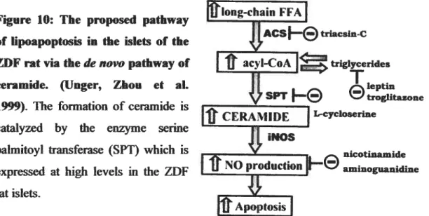
The regulation ofcytosolic LC-CoA levels 20 Model illustrating signal transduction pathway in pancreatic -cell 21 Pathogenesis of type 2 diabetes 24 The proposed pathway of lipoapoptosis in the islets of the ZDF rat via de novo
pathway ofceramide 33
Possible mechanism of -cel1 glucolipotoxicity 35 Optimizationof culture conditions 44
Dose response cun’e offFA 45
Effect of various free fatty acids at different glucose concentrations on t-cell
death 47
Time dependence ofthe apoptotic action of elevated glucose on the fEl-cell....47 High glucose and different ffA cause DNA condensation to various
extents 4$
Quantification of the effect of various free fatty acids at different glucose concentrations on 8-ceil apoptosis and necrosis 49 Stearate at elevated glucose levels causes phosphatidylserine flip 50 The action of vanous free fatty acids on caspase-3 activation is glucose
dependent 52
PARPcleavage induced by stearate is glucose dependent 52 z-VAD-FMK blocks activation of caspase-3 induced by fFA at elevated
Figure 22: Inhibition ofpalmitate-induced caspase activation is coupled with a switch to
necrosis 54
Figure 23: Palmitate must be metabolized to synergize with elevated glucose to mduce B
celldeath 55
figure 24: The fatty acid 8-oxidation inhibitor etomoxir induces palmitate toxicity at Iow glucose and enhances palmitate toxicity at high glucose 56 Figure 25: Elevated glucose plus saturated fatty acids cause ceramide accumulation in
1NS $32/13 celis 57
Figure 26: The ceramide synthase inhibitors fumonisin BI (FB1) and myriocin do flot block ffie rise in cellular ceramide caused by the combined presence of elevated glucose and palmitate 5$ Figure 27: The ceramide synffiase inhibitors fumonisin BI ami myriocin do flot block palmitate induced ccli death at elevated glucose levels and palmitate 59 Figure 28: Planned experimental approaches to further investigate the role of lipid
LIST 0F ABBREVIATIONS
ACC Acetyl-CoA carboxylase Ac-CoA : Acetyl-CoA
Ac-DEVD-AFC: N-acetyl-Asp-Glu-Val-Asp-amino trifluoromethyl coumann ACO AcyL-CoA oxidase
ACS : Acyt-CoA synthase
AGE : Advanced glycation end product
MCAR t 5-Aminoimidazole-4-carboxamide-1 --D-ribofuranoside
AMPK : AMP-activated protein kinase BSA : Bovine serum albumin
CL : Citrate lyase
CPT 1 Carnitine palmitoyltransferase-I CS : Citrate synthase
DAG : Diacylglycerol
FAS t fatty acid synthase
FBI t fumonisin Bi
FDP t fructose I ,6-bisphosphate
FFA t free fatty acid
F6P : fructose 6-phosphate
GK Glucokinase
GLP 1 t Glucagon-like peptide-1
GLUT : Glucose transporter
GPAT Glycerol-3-phosphate acyl transferase G6P : Gtucose 6-phosphate
G6Pase Glucose 6-phosphatase HSL Hormone sensitive lipase INOS : Inducibte mtric oxide synthase K-ATP : Potassium ATP-dependent channel KRBH : Krebs-Ringer bicarbonate hepes LC-CoA : Long chain fafty acyl-CoA
LPA 2 Lysophosphatidic acid
MCD : Malonyl-CoA decarboxylase
MODY Maturity-onset diabetes ofthe young NEFA 2 Non-esterified fatty acid
MDDM : Non-insuhu-dependent diabetes mellitus
PA : Phosphatidic acid
PC t Pyruvate carboxylase
PARP : Poly (ADP-ribose) polymerase
PDH Pyruvate dehydrogenase
PDX-1 : «Pancreatic and duodenal homeobox protein-1» PfK-1 t 6-Phosphofructose-1-kinase PI : Propidium iodide P13K Phosphatidyimositol-3 kinase PK : Pyruvate kinase PKC t Protein kinase C PL : Phosphotipids
PPAR Peroxisome proliferator activated receptor SPT : Serine palmitoyltransferase
TG : Tnglycende
ZDF : Zucker diabetic fatty rat (falfa)
The process of tearning and lite production of knowtedge invotve a large ,uimber of individuats and institutions. The development of evely work is guided by lite spin! and
inspiration of ail those who confributed in one way or another to every aspect of Lite work The input ofail those is substantially important.
The enthusiasm and support ofDr. Marc Prentki is behind the completion oftitis worlc my deepest thanks and gratitude go to him.
Ail members ofLite Prentki tab have been invaluable to titis work I would like to express my thanks for them alt.
Erik Joly for alt his hetp, guidance and discussions; Christopher Notan for his heip in discussions andintueproofreading oflite thesis.
My thanks are also extended to Johane Morin for ail her help in the lab, (o Serge Hardy Raphad Roduit, Jean Rideau, Ewa Przybytowskz Marie-Line Peyot, Viviane Augusto and Ahi Zutterfor ail her coi-e.
I am also grateful to Dr. Richard Bertrandfor his time in discussions.
My thanks and appreciation also go to Dr. Ghassan Dbaibo, my former
supervisor for ail his support andpatience in nsy earlyyears ofresearck finalty, niy thanks go10my mam for alt her prayers, to my dad and Ko my sisters
and brothersfor alt their love, help and support. Titis thesis is dedicated to alt of
them.
1. ffiSTORICAL INFORMATION
It is eighty ycars after flic discovery of msulm and we are stiil faced with many challenges conceming diabetes. Despite tremendous advances in our knowledge, we do flot understand the accelerated rate of diabetes appearance in our present time. An improved understanding of flic pathophysiological basis of diabetes is stii required.
Diabetes Mellitus is a medical condition known to physicians for thousands of years. References to this disease can be found in many ancient writings. It was first descnbed on an Egyptian papyrus which was discovered in Egypt in 1862, which is said to have been written between 3000 and 1500 BC. The first use of the term “Diabetes Mellitus” dates back to the second century AD. “Diabetes” stems from the Greek word “siphon” for ‘pipe-like’ or flic passing through of water. ‘Mellitus’ is Latin for ‘honey’ or ‘sweet’ (Halliweli and Guftendge, 1999).
In the middle of the 1 9’ century, evidence from autopsies startcd to suggest a link between the pancreas and Diabetes Mellitus. In 1869, a German scientist called Paul Langerhans discovered the existence oftwo systems ofcells in flic pancreas: the acinar ceils, secreting the pancreatic juice into the digestive system, and islets floating between flic acini with, at that tïme, an unknown function. Several years later, these ceils became known as flic islets of Langerhans. In I $89 Minkowski and Von Menng gave the first direct evidence ofthe hnk between diabetes and flic pancreas. They depancreatised a dog causing a state of polyuria indistinguishable from diabetes. This was followed by work by many researchers tili flic summer of 1920, when Banting and Best discovered insulin. On May 3, 1922, the discovery of insulin was officially announced to the medical community. It took another six years for
Steiner to establish diat insulin is a protem, and until 1955 for the pnmary structure of insulin to be elucidated by Sanger and co-workers. (Histoncal information was obtained from: www.diabetesforum.net www.med.uni giessen.de; www.diabetes.ca).
2. CLASSIFICATION 0F DIABETES
Diabetes mellitus is in reality, a group of disorders that have in cominon hyperglycemia resultmg from defects in insulin production, insulin action or both. A hallmark in understanding die etiology of a disease lies in a proper classification ami differentiating its varlous fonus depending on varlous factors. Targeted research, treatment and prevention of diabetes mellitus depend on an appropnate classification. ‘[bus, die growth of studies on die epidemiology and public health aspects was necessaly for die field to move on. This required a new classification of the disease based on die huge scientific interest. The recent update of die classification of diabetes mellitus took place in 1997 by die World Heaiffi Orgamzation (WHO) ami die American Diabetes Association (ADA), which was a revision of die 1985 WHO Study Group (Zimmet 1999). According to titis classification, die etiological types ofdiabetes are:
2.1 Type 1 Diabetes Meflitus
Type 1 (formerly called insulin-dependent diabetes mellitus, IDDM) is an autoimmune disease in which die (-ce11s of die pancreas are destroyed. In tins type, insulin is required for survival to prevent ketoacidosis, coma and death. ‘[bis is classically a disease of cbildren and young aduits, but recent studies indicate that type I diabetes can occur at any age. Genetic determinants such as HLA type seem important for die onset in many patients, with environmental factors, possibly viral infection a close second. Type I may account forS to 10 % of ail diagnosed cases ofdiabetes.
2.2 Type 2 Diabetes Mellitus
Type 2, (fonnerly catled non-insuim dependent diabetes mellitus NIDDM), is die most common fonu of diabetes. It is characterized by disorders of msulm resistance and rnsulm secretion, either of which can be the predommant feature. Bodi disorders are usually present at die time die disease is clinically manifested. It classically occurs in aduits, especially die elderly, but recently it is being diagnosed wiffi increasing frequency in chiidren and adolescents. It is in most instances a polygenic disease, but also very life style dependent. Its prevalence, like obesity which is a major nsk factor for ils development, is increasing rapidly particularly in developing counties. Type 2 diabetes wiIl be discussed in much greater depth later in die introduction.
23 MODY (Matunty Onset Diabetes of the Young)
MODY is a familial condition widi autosomal dominant inheritance. It is a monogemc form of diabetes charactenzed by early age of onset (< 25 years), and pancreatic f}-ceII dysfimction. ‘iliere are 6 types of MODY, four of which are caused by a mutation affectingatranscription factor. MODY 1, 3, 4 and 5
are caused by mutations affecting die transcription factors hepatocyte nuclear
factor HNf-4cz (MODYI), HNF-la (MODY3), pancreatic and duodenal homeobox protein-1 (PDX-1) (MODY4), HNF-1 (MODY5) and NeuroD/2 (MODY6). HNF regulates die expression of metabolic genes and glucose transporters whereas PDX-1 is implicated in die development of die pancreas and die regulation of die insulin gene. MODY2 is caused by a mutation affectmg die glucokinase gene, die enzyme which catalyzes die rate limiting step ofglycolysis (Froguel and Vefflo 1999; Wobser, Dussmann et al. 2002).
2.4 Statistics
Many swveys have conftrmed that increasmg urbanization and indusmalization is associated with an increased prevalence of type 2 diabetes. The world Heaiffi Organization (WHO) estimates diat, in 1997, 143 million people (6.2 % of die population) were afflicted wiffi diabetes worldwide. It is predicted that these numbers will likely double by die year 2020 (O’Rahilly 1997). At home, diabetes is considered as a leading cause of death by disease in Canada and die number of Canadians afflicted with die disease bas reached around 2 million people (www.diabetes.ca). Diabetes is one of the most common afflictions of die aged, but its prevaience, pafticularly of type 2 diabetes is rapidly increasing in younger individuals usually in association wiffi obesity (Bramblett, Huang et al. 2000).
2.5 Burden ofDiabetes for Affected Individuals
The long terni complications of diabetes are divided lute die microvascular complications, affecting die eye, kidney and nervous system, and die macrovascular complications with accelerated atherosclerosis and increased nsk of amputation, myocardial infarction and stroke. Diabetes is considered to be die leading cause cf ail diese complications, in addition to biochemical imbalances like ketoacidosis. The basic mecharnsms of each of diese complications is a combrnation cf die adverse effects cf hyperglycemia on tissues, stimulation of vanous growth factors, and the secondaiy effects of conditions frequently associated wiffi diabetes such as hypertension and hyperlipidemia. People wiffi diabetes are also more susceptible to many other illnesses. In 1999, diabetes was considered to be die sixth leading cause cf deaffi w die U.S., representing aroimd 19 % of ail deaths (www.ADA.com). In addition to die healdi issues, diabetes represents another burden, an economic one, on die individual and on die govemment.
3. GLUCOSE HOMEOSTASIS
The blood glucose level is a balance between glucose absorption from the gut, glucose production by the liver and glucose utilization by msulm-mdependent tissues (such as ffie brain, kidney, and erytbrocytes) and insuim-dependent tissues (such as fat and muscle). This balance is regufated by many neurohormonal factors including hormones of the endocrine pancreas (insulin from the -cells and glucagon from the Œ-cells) which have an important central role (Kahn 1994). Blood glucose level is normally at 5 mM. Insulin secretion from -cells and glucagon secretion from Œ-cells respond very precisely to small changes in glucose concentration in the physiologic range, ffiereby keeping glucose levels within the range of 3.7-7.0 mM in normal individuats (van Haeften 2002).
3.1 Post Prandial (Fed State)
Glucose, with other nufrients, is absorbed from the intestine into the portai circulation thus causing a rise in plasma glucose aiid other nutrient levels. The islet responds to this perturbation in nutrient levels by insulin secretion from the p-cells (Figure lA). Glucose is the primary nutrient secretagogue for insulin, but its effectiveness as a secretagogue can be enhanced by other nutrients such as amino acids, fatty acids and non-nutrient stimuli (incretins) which are elevated after a mixed meal such as GLP-1 and gastric inhibitory peptide (GIP) (van Haeften 2002). The resultant increase in circulating insulin levels stimulate glucose uptake into insulin-sensitive tissues such as skeletal muscle, heart and adipose tissue and inhibits endogenous glucose production from the liver and kidney. Glucose utilization by insulin-insensitive tissues, such as the brain, change minimally after a meal. Thus, insulin causes an increase in glucose clearance from blood and reduced glucose entiy into blood with the end effect of returrnng the post-prandial glucose nse back to pre-meal levels.
3.2 During fasting
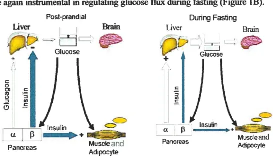
During fasting, ffie bram in particular requires a constant supply of glucose. This of course can flot corne ftom exogenous sources as glucose entry from the gut will be zero. It must corne from an endogenous supply. Islet hormones are again instrumental in regulating glucose flux dunng fasting (Figure lB).
Pst-pand:aI During Fasting
Liver &ain Liver Brain
Ï
GkccseV
+‘rsuh
(L
Pancreas Miselead
Adpcoite
Figure 1. Glucose homeostasis (A) postprandially and (B) during fasling. A. After a meal, the intestine will absorb glucose and there wilI be an increase in plasma glucose levels (> 5.5 mM). The rise in
glucose will stimulate the P-cells of the pancreas to secrete insulin. Glucose uptake by insulin-insensitive tissues such as brain will continue. Elevated insulin levels will inhibit endogenous glucose production from the liver and kidney and will promote storage of glucose in the liver, muscle and adipocytes. Thus normoglycemia is restored. B. During fasting, the brain requires a constant supply of glucose which has to corne from endogenous sources. Glucose levels fail which results in reduced insulin secretion and increased glucagon secretion. Glucose uptake by insulin sensitive tissues is consequently reduced and endogenous glucose production by glucagon stimulation of gtycogenolysis and gluconeogenesis is increased. Hepatic glycogenolysis is stimulated ami glucose entry to the muscle and adipocytes is decreased. Glucose levels are maintained in the range of 4-5mM (modified from Kahn, 1994).
With falling glucose levels, insulin secretion also falis, such that insulin effects on stirnulating glucose entry into muscle and fat will be minimal and
CL
Mjsce and Pa,çreas
Adipoite
Faliing glucose levels also result in the secretion cf glucagon from islet a celis. Glucagon stimulates endogenous glucose production by promotmg glycogenolysis and gluconeogenesis. Thus, endogenous glucose production will increase, and glucose entry mto the muscle and the adipocytes will decrease (Defronzo 1988). This state resuits in maintenance of nonnoglycemia (4-5 mM) with provision ofthe essential glucose supply to the brain.
Dysregulation of glucose homeostasis, whether resulting in elevated blood glucose (hyperglycemia) or low blood glucose levels (hypoglycemia), will have defrimental effects on the individual. Hyperglycemia is the hallmark of diabetes mellitus.
4. L1Pifi HOMEOSTASIS
Lipid, as for glucose homeostasis, is markedly altered between the fed and fasted states. free fatty acids (FFA) constitute an important energy source in most body tissues, representing the primary oxidative fuel for liver, resting skeletal muscle, renal cortex and myocardium (Coppack, Jensen et al. 1994). FFA also have an important physiological role during pregnancy. During late pregnancy, lipolytic hormones stimulate fat breakdown, leading to elevations in plasma FFA concentrations, which induces peripheral insulin resistance and causes a switch in fuel metabolism from carbohydrate to fat oxidation, thus maximizing the availability of glucose for the developing foetus (Boden
1996).
4.1 Post-Prandiat (Fed State)
In peripherat tissues, post-prandial nses in insulin promote the accumulation of triglyceride (TG) storage by the following: Inhibition of hormone sensitive lipase (HSL) with suppression of lipolysis of tissue TG stores; activation of
Iipoprotein lipase with hydrolysis of TG withm chylomicrons and VLDL and transfer of released FFA mto ceils for subsequent esterification and storage, and activation of lipogenesis pathways.
In die tiver, fatty acid oxidation is inhibited whereas fatty acid estertification ami synthesis of VLDL is promoted. Lipogenesis from glucose is also promoted (McGarry and Brown 1997).
4.2 During Fasting
In peripheral tissues, lipolysis of stored TG via HSL is promoted resulting in increased flux of FFA into the circulation ami increased availability of FFA for oxidation particularly in heart and skeletal muscle.
In the liver, fafty acid oxidation is increased with the production of ketone bodies (McGany and Brown 1997).
4.3 Rote of Matonyt-CoA Regulation of Carnitine Palmitoyl Transferase I Activity in Lipid Homeostasis
The relationship between malonyl-CoA, an intermediate in lipogenesis, and carnitine palmitoyl transferase I (CPT I), an outer mitochondnal membrane enzyme which is the rate limiting enzyme in mitochondrial -oxidation of fatty acids, was first reported in the liver. During states of high insulinllow glucagon ratio, i.e. with carbohydrate feeding the liver activeIy converts glucose carbons via malonyl-CoA to fatty acids and then to LC-CoA (McGarry and Brown 1997). If the newly formed LC-CoAs were to react with camitine under the influence of CPT I, it would be converted back to acetyl CoA through the process of -oxidation, thus generating a futile cycle. This does not happen. however, as the elevated pool of malonyl-CoA produced from anaplerosis and the acetyl-CoA carboxylase (ACC) reaction during active lipogenesis inhibits CPI I. The LC-CoA, therefore can react with glycerol phosphate to form TG which in the liver can be exported in the form
of VLDL. However, in periods of iow insulinlhigh giucagon, like starvation, the pathway of fatty acid synthesis cornes to a hait, the rnalonyl-CoA concentration falis, CPT 1 becornes demhibited and fafty acids reachmg the liver readily enter the -oxidation paffiway to form ketone bodies.
This interaction between malonyl-CoA and CPI I has been observed to be at work in a variety of nonhepatic tissues, particularly the heart and skeletal muscle and the -ce1I as well (Saddilç Gambie et al. 1993; Prentki and Corkey 1996; Saha, Vavvas et ai. 1997; Memil, Kurth et al. 1998). However, in the non-lipogenic tissues, the metabolic role ofmalonyl-CoA differs.
4.3.1 Matonyl-CoA
Malonyl-CoA is the product of ACC and in the liver as well as in other lipogenic tissues is the first committed intermediate in the pathway of fatty acid synthesis. In ail tissues, lipogenic and non-iipogenic, it acquires its significant regulatory role by inhibiting CPI I, the first step specific to the opposing process offatty acid oxidation (McGarry and foster 1980; McGarry, Woeltje et al. 1989). Malonyl-CoA inhibits a class of carnitine acyltransferases that catalytically have access to the cytosoiic pool(s) of long chain acyl-CoA esters.
4.3.2 CPTI
Ihe essential role of camitine in the oxidation of long-chain fatty acids by mammalian tissues first emerged in the mid 1950. The formation of acylcarnitines enables acyl moieties to cross intraceliular membranes, which are otherwise impenneable to acyl-CoA esters. This occurs through the action of specific carnitine/acylcamitme carriers, highly expressed in both mitochondrial membranes and peroxisomal membranes. Acyl-CoA first reacts with camitine under the influence of a CPI I on the outer aspect of the mitochondriai inner membrane, generating free CoA and acylcarnitine.
Acyicamitme then permeates the limer membrane (possibly via a specific camer mechanism) and reacts with a matrix pool of CoA in a reaction catalyzed by CPT 11 on the limer face of the limer membrane. The re-formed acyl-CoA then enters the pathway of -oxidation, while the released carnitine retums to the extramitochondnal compartment (McGarry and Brown 1997). In the liver, CPT I plays a pivotai role in the regufation of fafty acid oxidation (McGarry, Mannaerts et al. 1977; McGarry, Leatherman et al. 197$).
This metabolic branch point (fatty acid traffic through estenfication on oxidation) is controlled by malonyl-CoA (McGany and foster 1980; McGarry, Woeltje et al. 1989). Malonyl-CoA regulation of CPT I appears to play a cmcially important physiological role in the pancreatic 3-ce1l as is discussed below in the section on insulin secretion.
5. PANCREAS
5.1 Anatomy of the Pancreas
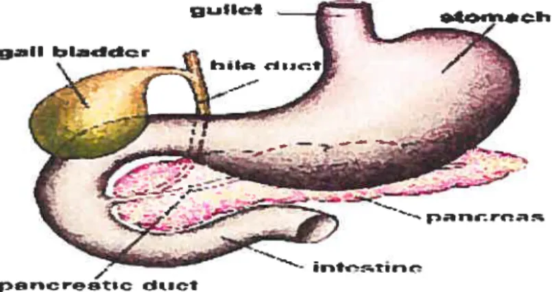
The pancreas is an elongated, tapered organ (about 22 cm long) tocated across the back ofthe abdominal cavity (Bramblett, Huang et al. 2000). It is in close proximity to the duodenum behind the stomach (figure 2).
figure 2. Anatomy of the pancreas. The right side of the organ (called the head) is the widest part ofthe organ and lies in die curve ofthe duodenum, the first division of the small intestine. The tapered Ieft side extends slightly upward (called the body of the pancreas) and ends near the spleen (called the tau).
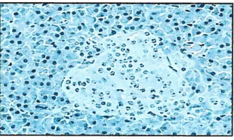
The structure of the pancreas is dominated by the fact that it is a dual function organ with both exocrine and endocrine celi types. The vast bu& of the pancreas is composed of exocrine tissue, and secretions from these cells flow into a series of ducts for ultimate delivery into the duodenum. Embedded within this exocrine tissue are roughty one million smalt clusters of cetis called the Islets of Langerhans. Islets of Langerhais are the endocrine component of the pancreas. Islets contain several ceil types (Figure 3) and are richly vascularized. They secrete insulin, glucagon and several other hormones (Bramblett, Huang et al. 2000).
w —. - Q ê • L — ‘4 ,‘ ê’ • • .•‘ ‘• •--- -Q ê - --4I _$•. ., - - Q Q. • ,-. *fr -p •• •• - -.-.-“ .. .-• ,,. -• . -— _rc. • -• — __; L4 . _i.• -- e •. -e — — -* - — • -s_•c_— ._ t — — - .- — — —. - • *&--_ -%..__ • .• ;,-•• - .‘- •.• --.u--- ‘.-ê. Q , q • -- •- •.- -—,-- --b e = •k -a-4 ,
figure3. Islet histotogic image.
A histologie image of an islet which is the large cluster of pale staining ceils in the middle. Ail of the surrounding tissue is exocrine tissue.
5.2 Exocrine Pancreas
The exocrine pancreas is composed of acini and ducts which comprises 90 % of the pancreas. The acini are composed of columnar to pyramidal epithelial ceils with minimal stroma. The pancreas produces 2 Iiters/day of bicarbonate nch fluid containing digestive enzymes. The pancreatic enzymes are tiypsin, chymotiypsin, aminopeptidases, elastases, amylases, lipases, phospholipases and nucleases.
53 Endocrine Pancreas
The endocrine pancreas consists of islets of Langerhans which are of endodermat origin and represents I % offfie pancreas. The size ofeacb islet is usually 0.1 to 0.2 mm. There are around one million islets per pancreas. The cellular composition of islets: beta
(f1)
cells (68 %), alpha (a) cells (20 %),delta (il) ceils (10%), PP ceils (2 %) and serotonin ceils (rare).
u-ceNs: secrete glucagon in response to low blood glucose levels which stimulates die release of glucose from stores thereby restoring blood glucose levets.
p-cells: secrete insulin in responseto high levels of glucose in die blood.
6-celis: secrete die hormone somatostatin (repfcsses the release of insulin and glucagon).
P? ceNs: secrete pancreatic potypeptide (PP). The rote ofPP is not absolutely clear, but it appears 10 be involved in the regulation of offier isfet hormones
and possïbly food intake.
6. INSULIN
In the fail of 1920, woddng at die University of Toronto, Fred Banting and Charles Best were able to make a pancreatic extract which had anti-diabetic characterisfics. This extract was calied insulin which ongmates from die Latin word “island” (Best 2002).
At diat time Sir Frederick Banting said:
“Insulinis flot a cure, more work needs Ko be done”.
Human insuhn is a peptide hormone contammg 51 amino acids. A single molecule consists of 2 polypeptide chains commonly labeled A (21 amino acids) and B (30 amino acids). The chains are linked by two disulfide bridges
(Kahn 1994). Insulin is secreted by the (3-celis of the pancreas and lias an important role in metabolism as discussed betow.
6.1 Insuhu Biosynthesis
The pancreatic p-cetl nonnally maintains a stable balance among insulin secretion, insulin biosynthesis and insulin degradation to keep optimal intracellular stores of die hormone. lnsuhn biosynffiesis follows die same pathway as offier peptide hormones (Vander, Shennan et al. 1990). Its biosynthesis starts in the nucleus of die (3-ceti of die pancreas as a senes of precursors beginning wiffi pre-proinsulin, die protein encoded in die insulin gene. These precursors direct die prohormone into die secretory paffiway ending fmalty in die secretoly granules where it is convefted into insulin and C-peptide. These products are stored and secreted togedier in a highly regulated manner in response to glucose and other stimuli (Steiner, Rouille et al. 1996).
6.2 Insulin Fanction
The actions of die anabolic hormone insulin are multifaceted affecting cellular metabolism (Kahn 1994; Bmmblett, Huang et al. 2000). Insulin plays an important rote in many aspects of ceil physiology, however, its most prominent mie is to stimulate glucose uptake in penpheral tissues after a meal. Insulin effects have been lmown for quite some time and these include:
• membrane transport of glucose, amino acids and certain ions; • incrcased storage of glycogen;
• formation oftriglycerides and other complex Iipids; • stimulation of DNA, RNA and protein synthesis.
These actions exert wide spectra of regulatory effects on die ceti (I3ramblett, Huang et al. 2000). They fali mto two categories: metabotic and growth
14
promoting effects. Metabolic effects are of short or long (with the mvolvement of genes) duration on the uptake, transport, mtennediary metabolism and storage of small food molecules-hexoses, amino acids and
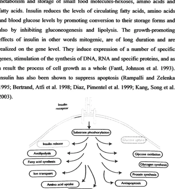
fatty acids. Insulin reduces the leveis of circulating fatty acids, amino acids and blood glucose levels by promotmg conversion to theif storage forms and also by inhibiting gluconeogenesis and lipolysis. The growth-promoting effects of msulm in other words mitogenic, are of long duration and are realized on the gene level. They mduce expression of a number cf specific genes, stimulation ofthe synthesis ofDNA, RNA and specific proteins, and as a resuit the process of ceil growth as a whoie (Fanti, Johnson et al. 1993). Insulm lias also been shown to suppress apoptosis (Rampalli and Zelenka 1995; Bertrand, Affi et al. 199$; Diaz, Pimentel et al. 1999; Kang, Song et al. 2003). Insulin rcCeptOf
1
Ct
phoshocyIatio subnease ±-(tty acid +, \ J\
Ctes
( Anijinocid uptkt
C
C
syndes) @ene expresso)Figure 4: Protem phosphorylation is required to mediate msulin action. After receptor autophosphoiylation, the f3-subunit becomes active as a tyrosine-specific kinase ami catalyzes phosphoiylation of several mtracellular proteins. This event provides the underpinning of the different actions cf insulin which inciude: 1- stimulation cf glucose turnover by favoring its transport across the plasma membrane; 2-promoting protein synthesis in almost ail tissues by affecting gene transcription, messenger RNA translation and amino acid uptake; 3-increasing DNA synthesis and prevention of apoptosis via its mitogenic effect; 4- stimulation of ion transport across the plasma membrane; 5- stimulation of lipid synthesis; and 6- prevention cf lipolysis by inhibiting hormone sensitive lipase (HSL) (Kahn 2000).
Once in the blood, insulm affects glucose homeostasis by stimulatmg the uptake of glucose mto skeletal muscle and, to a lesser extent, mto liver and adipose tissue and by inhibiting glucose production from liver or kidney. In muscle and adipocytes, glucose uptake is mediated by transiocation of the insulin-sensitive glucose transporter GLUT4 from mtracellular glucose storage sites to the plasma membrane. Insuim also switches glycogen metabolism from glycogenolysis to glycogen synthesis in muscle. With respect to endogenous glucose production, msulin, either directly or as a consequence of suppressing ffA levels by inhibiting adipocyte lipolysis, inhibits glycogenolysis and gluconeogenesis. The enzymes involved in the insulm-regulated processes of glucose metabolism appear to be regulated by (de)phosphorylation of serine andlor threonine residues.
Ail knowri actions of insulin are initiated at the plasma membrane by insulin receptors responding to ligand binding. Ligand binding promotes ATP binding and autophosphoiylation ofthe cytoplasmic tyrosine kinase domain of the receptor.
The insulin receptor primarily regulates nutritional metabolic pathways, whereas ail other receptor/tyrosine kinases mainly regulate celi growth and differentiation. The insulin receptor is a heterodimer consisting of two Πand
two [I subunits linked by disuiphide bridges. Transduction ofthe insulin signal includes the following events: (1) binding of hormone with the two extracellutar a-subunits (135 KDa) of the receptor which performs a ligand binding function; and leads to (2) activation of receptor tyrosine kinase activity and autophosphorylation of the two transmembrane -subunits (95 KDa); resuiting in (3) induction of a downstream cascade of tyrosine phosphoiylation of a wide spectnlm of effector proteins (Pertseva, Shpakov et al. 2003). The insulin receptor signal transduction, as for other receptors in the receptor tyrosine kinase family, does not form direct complexes with substrates andlor effector molecules after autophosphorylation. Instead, the insulin receptor, upon autophosphoiylation of at ieast the tri-tyrosine
subdomam, acquires exogenous kinase activity, phosphorylating its principal subsfrate: insulin receptor substrate 1 (IRS1). IRSI, in turn, binds effector molecules which are responsible for the actuat processes of glucose transport, metabolism and ceil growth (Kahn 1994; Pertseva, Shpakov et al. 2003).
7. INSULIN SECRETION
Insulin secretion from die pancreatic f-cell is a highly regulated process diat maintains blood glucose levels wiffiin a very narrow range. Glucose homeostasis is achieved by a complex interplay between nutrients, hormones and die autonomic neivous system.
7.1 Pancreatk -CeII Secretagogues
Insulin secretion is regulated by many factors such as nutrients and neuro hormonal agents (Figure 5). The calorigenic agents which stimulate insulin secretion include glucose, free fatty acids and amino acids such as glutamine and leucine. Important examples of neurobonuonal agents which stmiubte insulin secretion include glucagon like peptide-1 (GLP-1), die ‘gastric inhibitory peptide’ (GIP), glucagon, cholecystokinin (CCK) and acetylcholine. (Hedeskov 1980; Wollheim and Sharp 1981; Prenfld and Matschinsky 1987). Neurohormonal agents known to have an inhibitoiy effect on insulin secretion include somatostatin, galanine and -sympadio-mimetic agents.
Stimulators lahibitors
Glucose Somatostatin
Amino acids
Free fatty acids Galanme
Glucagon Adrenaline
GLP-I GW
LNradrena1ine
Vagiis nerve Spianchique nerve
(parasymp.) (symp.)
FigureS. Stimubtors and inhibitors of msulin secretion.
The calorigenic nufrients such as (glucose, amino acids and ftee fafty acids) and certain peptîdes like the incretin GLP-1, the gastrointestinal inhibitory polypeptide (GIP) and CCK stimulate the secretion of insulin, whereas somatostatin, galanine and adrenaline inhibit insulin secretion.
72 Mechanism of Insulin Secretion
Glucose, the primary stimulus for insulin secretion in the -ceIl, elicits a biphasic insulin secretion. The first phase is sharp, reaches its maximum at 3-5 mm, and lasts approximately 10 min. The second phase is more blunted and lasts as long as glucose levels remain elevated (Porte and Pupo 1969; Poitout and Robertson 1996). This biphasic response is believed to be due to two signaling pathways of glucose-stimulus-secretion coupling. One is the weII known KATpchannet dependent pathway by which glucose depolanzes the
3-ceil, actïvates voItage-dependent-Ca2 channels, and mises [Ca2’]1. The other pathway is the less known KATp channel independent pathway (Gembal,
Gilon et al. 1992; Sato, Aizawa et al. 1992; Straub, James et al. 199$).
7.2.1 Calcium andrATPChannel Dependent Pathway
Glucose enters the -cell through a facilitative glucose transporter, GLUT 2, which allows rapid equitibration between extra- and mtracellular glucose
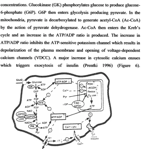
concentrations. Glucokinase (GK) phosphoiylates glucose to produce glucose-6-phosphate (G6P). G6P then enters glycolysis producing pyruvate. in the mitochondria, pyruvate is decarboxylated to generate acetyi-CoA (Ac-C0A) by the action cf pyruvate dehydrogenase. Ac-CoA then enters the Kreb’s cycle and an mcrease in the ATP/ADP ratio is produced. The increase in ATP/ADP ratio inhibits the ATP-sensitive potassium channel which resuits in depolarization cf the plasma membrane and opening of voltage-dependent calcium channels (VDCC). A major increase in cytosolic calcium ensues
Figure 6. Model iflustrating glucose indnced insulin release in the pancreatic -ceH: Calcium and JCATPchannel dependent pathway.
Glucose enters the ccli via the glucose transporter GLUT 2. It is metabolized and convefted te pyruvate via glycolysis which is subsequently oxidized in the Kreb’s cycle causing an increase in the ATP/ADP ratio. The increased ATP/ADP ratio closes the K ATP-dependent channel (resulting in depolarization of the membrane) which causes opening of the Ca2 voltage dependent channel and entry of Ca2 into the ceil. Ca2 activates exocytosis of vesicles containing insulin granules. [GK, Glucokinase; G6P, glucose 6-phosphate; F6P, fructose 6-6-phosphate; FDP, fructose I ,6-bis6-phosphate; PFK, phosphofructose-kinase; aGPDH, a-glycerol 3-phosphate dehydrogenase; GAPDH, glyceraldehydes 3-phosphate dehydrogenase; LDH, lactate dehydro-genase; PK, pynivate kinase; Pyr, pyruvate; PDH, pyruvate dehydrogenase; aKGDH, a-ketoglutarate dehydrogenase; ŒGP, (Z
glycerol 3-phosphate; fAD, flavin adenine dinucleotidej (Prentki 1996). which triggers exocytosis of insulin (Prentki 1996) (Figure 6).
7.2.2rATPChannel Independent Pathways oflnsulin Secretion
It is clear however, diat some additional components of glucose metabolism are needed for die full effect of glucose to be manifested. Prentki and Corkey Prentld, Corkey 1996) raised the intriguing possibility that one such
component involves an element of glucose-fatty acid cross talk, in other words, that die malonyl-CoAICPT I axis recognized in liver and later in the muscle and heart, may also be at work in die -cel1 in relation to insulin
secretion.
7.2.2.1 Anapkrotic /Malonyl-CoA/Lipid Signaling Patkway
Prentki ami Corkey (Corkey, Glennon et al. 1989; Prentki, Vischer et al. 1992; Prentki, Corkey 1996) proposed a model of -cell nutrient sensing and insulin secretion in which, in addition to the KArp dependent pathway, a parallel anaplerotic/lipid signaling pathway exists in which malonyl-CoA acts as a coupling factor. The cascade of events proposed in the model is as follows (Figure 7). Glucose is metabolised to pyruvate via glycolysis. Pyruvate is then metabolised by eiffier pyruvate dehydrogenase (PDH) or pyruvate carboxylase (PC). PDH transforms pyruvate to Ac-CoA, while PC, on the other hand, is an anaplerotic enzyme which channels pyruvate into mitochondnal oxaloacetate. The latter anaplerotic mechanism resuits in efflux of citrate from the
mitochondria to the cytosol. Cytosolic citrate is converted to Ac-CoA and
oxaloacetate via the action of citrate lyase (CL). Cytosolic Ac-CoA is then carboxylated by acetyt-CoA carboxylase (ACC) to form malonyl-CoA. Malonyl-CoA can enter fatty acid syndiesis via the action of the enzyme fatty acid synthase (FAS) which uses malonyl-CoA as a substrate. FAS, however, is not highly expressed in the -cells (Brun, Roche et al. 1996) such that in response to elevated glucose and anaplerosis, cytosolic malonyl-CoA level rises. Malonyl-CoA is an inhibitor of CPT I, die enzyme which catalyzes die rate limiting step for fatty acid entry into the mitochondria to undergo
f3-oxidation (Figure 7). Thus, CPT I inhibition by matonyl-CoA resuits in an increase in long chain acyl-CoA in the cytosol which potentiates insulin secretion either directly or indirectly by the production of complex lipid signaling molecules, activation of protein kinase C (PKC), or by protein acylation (Corkey, Deeney et al. 2000). In this model, malonyl CoA is believed to act as a regutatoiy molecule and long chain acyl-CoA and its metabolites as effector molecules in insulin secretion.
Glucose FFA
Figure 7. The regulation of cytosolic LC-CoA kvels.
Cytosolic LC-CoA is denved from either circulating FfAs or endogenous complex Iipids. The entry of LC-CoA into the mitochondria, where it is oxidized to acetyl-CoA, is confrolled by malonyl-CoA derwed from glucose or offier fuels, which regulates CPT I. AcCoA, acetyl-CoA; CL, citrate Iyase; CS, citrate synthase; G6P, glucose-6-phosphate; OAA, oxaloacetate; PC, pyruvate carboxylase; PDH, pyruvate dehydrogenase; PL, phospholipids; TG, triglyccride. Adaptedfrom Prentki M Corkey B 1996.
The evidence supporting the existence of an anaplerotic/malonyl-CoAllipid signaling in the (E-cell is summarized as follows: PC is abundant in the islet, yet the islet is neither a neoglucogemc nor a lipogenic tissue; citrate, an allosteric activator of ACC, nses markedly in response to glucose stimulation before secretion occurs (Corkey, Glennon et aI. 1989; Roche, farfari et al. 1998; farfari, Schuiz et aL 2000); only nuinents which cause an increase in
PL
I
EFAI
Gkicose 66P bi bi >-o u >. Pyruvate. Cytosol21
citrate levels cause insu tin secretion (Brun, Roche et al. 1996); ACC is abundant in the —celt; glucose increases ACC gene expression in INS ceils, and basat insulin secretïon correlates with the content of ACC protein (Brun, Roche et al. 1993); malonyl-CoA tises rapidly in responseto glucose and only nutrients that elevate malonyl-CoA cause secretion (Corkey, Glennon et al. 1989; Liang and Matschinsky 1991; Pren&i, Vischer et al. 1992). fAS is vety
low in the islets of Langerhans (Brun, Roche et al. 1996); potentiation of glucose-induced insulin secretion resuits aftec iiicubating the (3-cetis with fatty
acids for a short penod (Malaisse, Sener et al. 1979; Prenfld, Vischer et al. 1992). Insutin release is promoted after inhibiting CYT I by pharmacologicat
agents (Prentki, Viseher et al. 1992; Chen, Ogawa et al. 1994); the levets of
fatty acyl-CoA in response to different nufrients correlate with insulin secretion (Prentki, Vischer et aI. 1992). The abovefmdingstogether provide a
strong evidence for the concept diat an anaplerotic/malonyl-CoAIlipid
signaling pathway plays an instrumentalrole in P-cetI nutrient signaling. The followingfigure iltustrates the two patbways of insulin secretion in the l
ceti (Pren&i, Joly et al. 2002). figure 8. Model illustratrng
signa n uc on pa way ici
ia’i
in the pancreatic -œlL When t
transiently stimulated, die Ac
CoA/Ca2 and anaplerotic/lipid ..tAcCoAi.
signaling pathways synergize i Aœ(
to promote insutin secretion.
)
AcCoAc, cytosolic acetyl-CoA; t’ccl AcCoAm, mitochondrial acetyl- I
CoA; DAG, diacylglycerot; LPA, FAS
lysophosphatidic acid;
fr,
t1F
(TEoxidation of fatty acids; PA, s FaCŒAI
phosphatidic acid; PC, pyruvate ‘s QA
carboxylase; PDII, pyruvate s
dehydrogenase; MCD, malonyl- Ip&JpNDA
C’ Â À ..L. 1 S
Ot uiu.iXyiaSe,
Inglycerides; HSL, hormone
sensitive lipase (Prentki2002). IiidlflSetTdiOit
73 RaIe of FFA in Insulin Secretion
it is well documented diat acute FFA administration Ieads to augmentation of glucose-stimulated msulm secretion (GSIS). ffA enhance both basal and GSIS and are essentiat for stimulus-secretion couplmg in die pancreatic P-cett (Boden, Chen et al. 199$; Dobbins, Chester et al. 199$). Efforts to elucidate how fatty acids influence f-cell fimction led to a series of important fmdings which implicate an element of g1ucose-fitty acid cross-talk in stimulus secretion coupling within die -ceI1. 0f these fmdings that are of importance and relevance to this thesis are: 1- exposure of Synan-hamster insulinoma (HIT) celis or rat islets to glucose increases the cellutar malonyl-CoA content which is proportional to die stimulation of insulin release (Codcey, Glennon et al. 19$9; Liang and Matschinsky 1991; Prentki, Vischer et al. 1992); 2-glucose concentrations whicb stimulate insulin secretion also suppress die oxidation of long chain fatty acids (Tamarit-Rodriguez, Vara et aI. 1984); 3-inhibitors of CPT I (like 2-bromopahuitate (2-BrP), 2- bromostearate (2-BrS) and etomoxir) which reduce fatty acid oxidation (McGarry, Wockje et al. 1989; McGarry and Bmwn 1997) were found to stimulate insulin release from pcrifused islets (Bliss and Sharp 1992), perfiised rat pancreas and in HIT ceils (Prentki, Vïscher et al. 1992); 4- exogenous long chain fatty acids appreciably potentiate GSIS from rat islets (Vara, femandez-Martin et al. 198$) and HIT ceils (Pren&i, Vischer et al. 1992) which is concomitant with die nse in LC CoA esters.
Ail offfiese fmdings are consistent wiffi die malonyl-CoAILC-CoA bypoffiesis
of Prendd and Corkey (Prendd and Corkey 1996). The increase in malonyl
CoA concentrations under these conditions suppresses CFT I activity and therefore fatty acid oxidation. Tbis resuhs in an increase in die concentration
of cytosolic LC-CoAs which act as signating molecuies for insuiin secretion
in concert wiffi die risc in [C?], caused by die calcium and KArp channel dependent paffiway. Glucose inhibition of LC-CoA oxidafion will be associated wiffi much greater increase in LC-CoA levels where die exogenous
FFA supply is simukaneously elevated, therefore amplifying die effect of
elevated FFA.
This rote of FFA in insulin secretion raises die question as to wheffier ail FFA are equal in terms of their insulinotropic effects. To answer this question, studies were carned out in the perfused pancreas and it was found that the insulinotropic potency increased dramaticatly with increasing carbon chain and wiffi a decrease in die number of double bonds (McGany and Dobbins 1999). The reason for this difference between saturated and unsaturated fatty acids is flot clear. Wheffier dis is related to a difference in metabotism of the two types orto a difference in de physicochemical properties of the effector molecules still awaits an answer.
8. PATHOGENESIS 0F TYPE 2 DIÂBETES
Type 2 diabetes is a heterogeneous condition, die etiology of which is polygernc and life style dependent (Kahn 1994; Zimmet, Alberti et al. 2001). It is invariably charactenzed by progressive deterioration in insulin secretory fimction, diminished hepatic glucoregulation and penpheral insuhn resistance (Defronzo I 9$8; Porte 1991; Kahn 1994; Poitout and Robertson 1996; Prendd and Corkey 1996; Prentki, Joly et al. 2002; Kahn 2003). Most often it is associated with, and usually preceeded by central obesity and dyslipdemia (Carey, Jenkms et aI. 1996; Wei, Gaskill et aL. 1997; Kahn 2003). The majority of obese subjects, however, despite varying degrees of insulin resistance, do not develop diabetes (Polonsky, Sturis et al. 1996; Mokdad,
ford et al. 2003). Insulin resistance, wheder due to obesity or physiological
conditions such as in pregnancy (Freinkel 1980; Catalano, Tyzbir et al. 1991),
is usually compensated by islet 8-ceils adaptation wiffi boffi increased 8-ceil
number and/or volume (8-ceil mass) and augmented B-ecu function (Kioppel, Lohr et aI. 1985; Sorenson and Brelje 1997; Lingobr, Buettner et al. 2002; Liu, Jetton et al. 2002; Butler, Janson et al. 2003; Kahn 2003). It is only when
insulin resistance is accompanied by failure of the islet B-cell to compensate, that type 2 diabetes (Kahn 2003) and gestational diabetes (Buchanan 2001) develop. While the pathogenesis ofthe 6-celi failure is flot welI understood, it
isbecoming increasingly evident from animal models (Coleman and Hummel 1973; Pick, Clark et al. 199$) and human autopsy senes (Stefan, Orci et al. 1982; Kioppel, Lohr et al. 1985; Clark, Wells et al. 19$$; Butier, Janson et al. 2003) that, in addition to B-cell dysflmction, reduction in B-cell mass is involved.
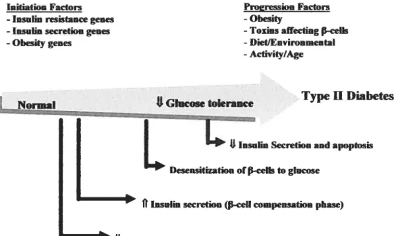
The following figure illustrates the pathogenesis ami the course of type 2 diabetes. (adapted from Kahn 2000).
InitiatioHFactors Proeression Factors
-Insulin resistance genes -Obesity
-lasulin secretion genes -Toxrnsaffecting P-celk
-Obesitygenes -Diet/Environmental
-Activity/Age
Type II Diabetes Normal *Glucose tokraace
F:
I
L..._...
L
Dnsiapoptosis
L
lInsuim secretion (-ceIIcompensation
phase)USeasitivityto insulin(Insulin resistance)
Figure 9. Pathogenesis oftype2 diabetes.
Many factors contribute to the etiology of type 2 diabetes including genetic and environmental factors. The genetic factors are likely to be multiple involving genes that affect insulin secretion, msuhn sensitivity and obesity. A decrease in the sensitivity to insulin by the insulin target tissues resuits initially in augmentation of insulin secretion by the f3-cells. This period is referred to as “f3-celI compensation phase”. With time, there is a graduai Ioss 0f f3-ceIl
compensation resulting in adecrease in glucose tolerance. Factors such as obesity, toxins and diet play a role in the progression ofthe disease and in desensitizing the -cells to glucose. Eventually, there is a failure of glucose induced insulin secretion, f3-cell death and thus the onset of the established disease of type 2 diabetes.
$1 Peripheral InsuhnResistance
Type 2 diabetes is mvariably characterized by peripheral msutm resistance which ptays a major rote in its development. Insulm resistance is defined as the impaired ability of msutm to promote penpheral glucose disposai. Insulin resistance is a hallmark of type 2 diabetes. Over 80 % of people with type 2 diabetes are obese, while virtually ail are insuim resistant (Boden 1997). Thus, it is hypodiesized that there is a tight coffelation between obesity and rnsuim resistance. Human and animal studies support this notion mdicating that weight loss/gain correlates closety with increasing/decreasing msulm sensitivity, respectively. Evidence indicates that rnsulm resistance is present years beftwe die onset of die disease and it is considered as die best predictor for die disease (ShuIman 2000). The penpheraI action of insuim primarily includes die promotion of glucose uptake into skeletal muscle and inhibition of lipotysis in adipose tissue which are both impaired with insuim resistance. It bas also been shown that muscie glycogen synthesis is decreased by 60 % in subjects with type 2 diabetes. Recent evidence have also mdicated diat insuim resistance is also present in die -ceii which might contribute to die
abnormalities in insulin secreflon observed in type 2 diabetes (Withers, Gutierrez et aI. 1998; Kuikami, Bmning et al. 1999). Many investigators have studied die mecharnsms involved in insulin resistance at die level of die insuhu receptor, die insulin-receptor signal transduction padiways, die insulin regulated intermediaiy metabolic paffiways such as glycogen synthesis and lipolysis and die transiocation of die insuhn sensitive glucose transporter GLUT 4. Their fmdings are beyond die scope of this thesis other dian to say
diat elevated FFA bas been implicated in causing peripheral insulin resistance and dis wilt be fiirther discussed below.
8.2 Elevated Endogenous Glucose Production
Studies cf hepatic glucose metabolism in type 2 diabetes mvariably show increasing basal glucose production and a failure of rnsulm to suppress glucose production (Defronzo 198$; Kahn 1994). Elevated glucose production is clearly important in die maintenance cf hyperglycemia in boffi the basal (fasted) state and the post-prandial state.
83 Impaired Insulin Secretion
Impaired insulin secretion in type 2 diabetes is due to defects in fI-ceil fimctiou and fi-celi mass. As discussed above, impaired insulin secretion is an essential component in die padiogenesis cf type 2 diabetes.
8.3.1 Dysfunction
Islet dysfunction, characterized by loss of boffi first and second phase cf glucose-induced insulin release, ptays an important role in die pathogenesis cf hyperglycemia (van Haeften 2002). Defects in die first phase insulin secretion are evident very early in die pathogenesis process with some studies showing dis in subjects at risk of diabetes and diose wiffi impaired glucose tolerance. Defects in second phase secretion occur later and continue to detenorate once type 2 diabetes lias developed. Dysfimction is evident pafticularly with respect to die ability of glucose to induce insulin secretion, as secretion to arginine is preserved at least early in die pathogenesis process. There is considerable evidence indicating a desensitization of die islet function during persistent hyperglycemia in vitro (Grodsky 1989) and in vivo (Leahy, Cooper et al.
8.3.2 Loss offi-Cet! Mass
Another factor which plays an important role in failure ofinsulin secretion in the etiology of type 2 diabetes is the reduction in the number of -cells. Regulation of -ceI1 mass appears to mvoive a balance between -ceIl replication and apoptosis and islet neogenesis from exocrine pancreatic ducts (Finegood, Scagila et al. 1995; Bonner-Weir 2000). Disruption of these processes causing either reduced -celI formation and/or increased rates of
f
ccli death could cause a decrease in -cel1 mass and, as a consequence, reduced msulin secretory capacity. There is controvcrsy wheffier -cell mass is decreased in type 2 diabetes (Stefan, Orci et al. 1982; Kloppet, Lobr et al. 1985; Clark, Welis et al. 198$; Guiot, Sempoux et al. 2001) and this is due to the scarcity of suitable pancreas specimens for histochemical analysis from individuals wiffi type 2 diabetes. However, recent reports in which clinical information was better characterized concluded that -ceIl mass ïs decreased in type 2 diabetes (Kioppel, Lohr et al. 1985; Clark, Wells et al. 198$; Sakuraba, Mizukami et al. 2002; Butler, Janson et al. 2003). Importantiy, in a recently reported autopsy series (Butier, Janson et al. 2003), with adequate subject number and pre-death clinical data, islet 8-ceil mass was shown to be reduced in both impaired glucose tolerance and type 2 diabetic subjects and was associated with evidence cf increased apoptosis. Thus, both type I and 2 diabetes are increasingly viewed as 8-celi mass defects.8.4 Role of Elevated FfA in the Pathogenesis of Type 2 Diabetes
Evidence is mounting to support a “lipocentric” approach to the understanding cf the metabolic derangements of type 2 diabetes rather than the traditional “glucocentric” one (McGarry 1992). In support cf this approach, it has long been known that, in addition te hyperglycemia, type 2 diabetic subjects aimost invariably marnfest a serious breakdown in Iipid dynamics, reflected by elevated levels cf circulating free fatty acids and triglycendes (1G) and
reduced tevels of HDL cholesterot. Elevated plasma FFA are common in type
2 diabetes (Reaven, Hollenbeck et al. 19$8) and in some studies have been
shown to be predictive for the transition of patients from impaired glucose toterance (IGT) to type 2 diabetes (Paolisso, Tataranni et al. 1995; Charles, Eschwege et al. 1997). High plasma ffA concentrations are also associated wiffi a number of cardiovascutar nsk factors Iinked to msulm resistance including hypertension, dyslipidemia ami abnonnal fibrinolysis (Reaven 19$$).
Moreover, evidence from in vitro studies mvolvmg long-term exposure of rat islets to high concentrations of fatty acids showed a paftem consistent with the characteristic features of -ceIt dysfimction in human type 2 diabetes (Sako and Gntl 1990; Unger 1995; McGarry ami Dobbms 1999). These wiIl be discussed later under the lipotoxicity section. In addition, observations drawn from expenments doue on obese type 2 diabetics ami on die Zucker diabetic fatty rat (ZDF) raised die possibility diat in individuals genetically predisposed to develop type 2 diabetes, long exposwe of die islets to elevated concentrations of circulating FFA and VLDL or both might have a deleterious effect on die -ceIl as welt as on die muscle and thus contribute to -cell dysfunction. k is not clear, however, whedier dis breakdown in lipid homeostasis is a resuh of die disease or is instrumental in its development.
8.4.1 Insulin Resistance and Etevated ffA
Studies have indicated diat elevated circulating ffA may conbibute to die underlying pathophysiology of type 2 diabetes, in particular, die development ofinsulin resistance boffi in die peripheiy and die liver (Boden 1997; Shulman 2000). Central obesity is strongly linked to elevated FFA levels wiffi central adipocytes possibly being first affected by insulin resistance as proposed by Bergman. (Bergman ami Mittehnan 199$). Widi die development of adipose tissue insulin resistance, insulin-mediated suppression of lipolysis is decreased
thus leadmg to increased circulating FfA ami ultimately insutin resistance in skeletal muscle and liver (Boden 1997; Shulman 2000).
As mentioned cartier, skeletal muscle is also a major contnbutor to rnsulin resistance in type 2 diabetes. A strong correlation is observed between increased plasma FfA, intramyocellular lipid accumulation and insulin resistance (Krssalç fa& Petersen et al. 1999; Perseghin, Scifo et al. 1999; Boden, Lebed et al. 2001). ffA contribute to insuim resistance by havmg an inhibitoiy effect early on in glucose utilization in die muscle at the level of glucose transport (Cime, Petersen et al. 1999; Dresner, Laurent et al. 1999).
8.4.2 Endogenou Glucose Production and Ekvated ffA
Endogenous glucose production, mainly contributed by die liver, is mcreased in type 2 diabetes (Boden and Shulman 2002). Plasma glucose levels have been closety corrclated wiffi die rate of hepatic glucose production. Littie is known about die mechanism of increased endogenous glucose production, however, a mie for elevated FFA bas been postulated. Fligh circulating FFA antagonize die effects of insuim to suppress endogenous glucose production.
ffA are known to stimulate endogenous glucose production by promotmg
gluconeogenesis as shown by animal and human studies (healdiy voluntcers and type 2 diabetic subjccts).
8.4.3 Insulin Secretion
The physiologicat nature of die giucose-fatty acid cross-talk in stimulus secretion coupling wiffiin die fi-ceil mentioned earlier, however, has a darker side. In order to maintam glucose homeostasis, die pancreatic fi-ceil constandy senses circulating nutrients and integrates diese signaIs to secrete insulin accordingly. Abnormally elevated nuffient levels, however, may cause detrimental effects possibly via die same signaling pathways implicated in
msutm secretion. There is ample evidence that proionged efevations of glucose “glucotoxicity” or FfA “tipotoxicity” are toxic to (E-cells and cause
boffi -cell dysfimction and apoptosis. Prentki and Corkey (Prendd and
Codcey 1996), however, hypothesized diat elevated glucose would mcrease the toxicity ofelevated fFA, a concept tenned “glucolipotoxicity”. Isiet î-cell glucotoxicity, lipotoxicity and glucolipotoxicity wili be discussed in detail below.
9. ISLET p-CEu FAILURE IN TYPE 2 DIABETES
9.1 Glncotoxicity
As mentioned earlier, the traditional approach to the understanding of type 2 diabetes pathogenesis was more “glucocentric”. This was based on a considerable body of evidence suggesting that chronic hyperglycemia impairs glucose-induced insuim secretion and msulin gene expression (LeRoith 2002; Poitout and Robertson 2002). This is due to a diminished activity oftwo major
fI—ceil transcription factors, pancreatic-duodenum homeobox-1 (PDX-1)
(Oison, Redmon et al. 1993; OIson, Shanna et al.