Université de Montréal
Faculté des études supérieures et postdoctorales
Cette thèse intitulée:
Transcriptional regulation of effector CD8+ T cell differentiation and molecular dysfunction during HIV-1 infection
Régulation transcriptionnelle de la différenciation et de la dysfonction des lymphocytes T CD8+ effecteurs au cours de l'infection par le virus de l'immunodéficience humaine de
type 1
Présentée par : Alessandra Noto
a été évaluée par un jury composé des personnes suivantes :
Dr Martin Guimond, président-rapporteur Dr Rafick Pierre Sekaly, directeur de recherche
Dr Laurent Sabbagh, membre du jury Dr Guido Silvestri, examinateur externe
Résumé
Les cellules T CD8+ jouent un rôle primordial dans le contrôle des infections virales en limitant la dissémination des cellules infectées. Lors de l’infection chronique par le virus HIV, les cellules T CD8+ HIV-spécifiques ne se différencient pas en cellules effectrices fonctionnelles capables de tuer les cellules infectées par le virus ; ces cellules ne sont plus capables de proliférer ou de produire l’ IL-2. Ces cellules expriment PD-1 et l’engagement de PD-1, par son ligand, aboutit a plusieurs de ces déficits fonctionnels des cellules T . Le rôle de PD-1 dans la régulation d'évènements transcriptionnels contrôlant la différentiation et l'obtention des fonction effectrices des cellules T CD8+ reste à démontrer.
Id2 joue un rôle central dans la différenciation des cellules T CD8+ effectrices. Nous avons émis l’hypothèse que le défaut de maturation observé chez les cellules T CD8+ PD-1 high HIV-spécifiques (CD8+PD-1hi) au cours de l’infection chronique par le virus HIV pouvait être lié à la diminution d’expression du régulateur Id2. Nous avons ainsi démontré que l'engagement de PD-1 contribuait à une diminution d'expression de Id2 et de ses cibles transcriptionnelles. La surexpression de Id2 de ces cellules a permis de restaurer l'expression de marqueurs tels que Granzyme B et Bcl-2 et diminuir l’expression du marqueur de maturation de CD27.
La famille des cytokines à chaine γ joue un rôle clef dans la survie et l’homéostasie des cellules T. Dans ce travail, nous avons démontré que l’IL-15 était unique grâce à ses capacités de stimulation de l’expression d’Id2 et ses propriétés favorisant la survie ainsi que la différenciation des cellules T CD8+ effectrices. l’IL-15 induit la prolifération de toutes les populations de cellules T mémoires provenant de donneurs sains. L’addition de cette
cytokine aux sous-populations cellulaires Ttm et Tem a permis leur différenciation en cellules effectrices capables de produire Granzyme B alors que la stimulation par l’IL-15 des cellules Tcm ne favorise pas leur différenciation. Un test de cytotoxicitié par cytométrie en flux nous a permis de confirmer que la stimulation de cellules T CD8+ HIV spécifiques par l’IL-15 favorisait l’expression de Id2 et restaurait les fonctions cytotoxiques des cellules T CD8+ HIV spécifiques.
En conclusion, nous avons pour la première fois dans cette thèse défini les mécanismes moléculaires impliqués dans la modulation de l’expression du régulateur transcriptionnel Id2 par l’IL-15. Nous avons également révélé comment l’engagement de PD-1 conduisait a une altération de l’expression et de la fonction d’Id2 et favorisait la diminution des fonctions effectrices des cellules T CD8-HIV spécifiques. Une perspective de traitement avec des agents tels que l’IL-15 ou le bloquage de PD-1, en combinaison avec les traitements conventionnels, pourrait contribuer à une meilleure stimulation des réponses immunes favorisant ainsi la réactivation des cellules T CD8+ et permettant la destruction de cellules T CD4+ infectées de manière latente.
Mots-clés : HIV, CD8 CTL, PD-1, Id2, IL-15, différenciation des cellules effectrices, cytotoxicité
Abstract
CD8+ T cells play a fundamental role in controlling viral replication and dissemination by killing virus-infected cells. However during chronic HIV infection HIV-specific CD8+ T cells fail to differentiate to functional cytotoxic effector cells and develop functional defects such as loss of IL-2 secretion, decreased proliferation and express high levels of PD-1. Persistent expression of PD-1 and triggering by its ligand results in immune dysfunction; it is not known how PD-1 signaling influences transcriptional events involved in T cell differentiation and effector function.
We found that the transcriptional regulator Id2 was downregulated in PD-1hi HIV-specific CD8+ T cells when compared to PD-1low CMV-specific CD8+ T cells from the same HIV-infected donors. Since Id2 has been shown to play a central role during differentiation of effector CD8+T cells, we hypothesized that skewed maturation of the PD-1hi HIV-specific CD8+ T during chronic HIV infection could result from decreased levels of Id2. We found that signals transduction pathways downstream of PD-1 ligation inhibited the expression of Id2; transfection of PD-1hi effector cells from HIV infected individuals with a Tat-Id2 construct could reverse an apoptotic fate associated with the exhausted phenotype. Finally, overexpression of Id2 restored expression of Granzyme-B and Bcl-2 and led to a decreased expression of the T cell maturation marker CD27.
Although the extrinsic signals and costimulation needed to activate cell proliferation and effector function are well known, signal-transduction pathways that regulate differentiation of memory cells to effector cells are beginning to be understood. Thechain family of cytokines is essential for the survival and homeostasis of T cells; they have pleiotropic
effects on the differentiation of effector and memory virus-specific CD8+ T cells. IL-15 was unique among -chain cytokines in upregulating the expression of Id2 and promoting the survival and differentiation of effector memory CD8+ T cells.
IL-15 induced proliferation of all memory subsets from healthy subjects but only induced differentiation, Granzyme-B production, and cytotoxic effector function in CD8+ Ttm and Tem cells. Stimulation of Tcm with IL-15 failed to induce their differentiation; this was associated with their decreased ex vivo levels of IL-15R when compared to Tem and Ttm subsets. Finally, we developed a single cell flow-cytometry cytotoxicity assay, and found that stimulation of CD8+T cells from HIV chronically infected subjects with peptide plus IL-15 induced the differentiation of tetramer+ CD8+ Ttm cells and restored Id2 expression and their cytotoxic activity .
Overall, we illustrate in this thesis, for the first time, the molecular mechanisms of effector T cell differentiation mediated by IL-15 and its downstream transcriptional regulator Id2; we reveal how PD-1 engagement leads to alteration of the Id2 pathway leading to decreased effector function of the HIV-specific CD8+ T cells. Immunotherapy with agents such as IL-15 or PD-1 blocking antibody that increase levels of Id2 expression , in combination with HAART, should trigger the functional re-activation of HIV-specific CD8+ T cells and the killing of latently HIV-infected CD4+ T cells.
TABLE OF CONTENTS CONTENTS Résumé p. i Abstract » iii Table of Contents » v Abbreviations » xiii Acknowledgements » ixx CHAPTER 1 INTRODUCTION » 1 1. Immunological Memory » 3
1.1 Generation of memory CD8+ T cells » 3
1.2. Central memory and Effector memory T cell subsets » 5
1.3 Models of memory T cells generation » 6
1.4 Role of cytokines in the homeostasis and
differentiation of effector CD8+ T cells » 6
1.5 Transcriptional factors that control effector and
memory CD8+ T cell differentiation » 8
1.1.5.1 T-bet and eomesodermin » 10
1.1.5.2 Blimp-1 and Bcl-6 » 10
2. Id proteins » 11
2.1 The properties of Id proteins » 11
2.2 Id proteins in cell cycle control » 13
2.3 Ids in Development and Differentiation » 14
2.4 Role Id2 and Id3 in memory CD8+ T cell differentiation » 15
3. CD8+ T cell exhaustion » 17
3.1 HIV infection » 17
3.1.1 Clinical Course of HIV infection » 18
3.1.2 Role of CD8+ T cells in HIV infection » 19
3.2 T cell exhaustion during HIV infection » 20
3.2.1 Programmed death 1 (PD1) » 20
3.2.1.1 Structure » 20
3.2.1.2 Role of PD-1 in CD8+ T cell exhaustion » 21
3.3 HIV Impairment of CD8+ T cell maturation » 22
HYPOTHESIS AND AIMS » 25 CHAPTER 2
Loss of ID2 during chronic HIV infection is mediated by PD-1 triggering and is responsible for the exhaustion
of HIV-specific CD8+ T cells » 29
ABSTRACT » 31
INTRODUCTION » 32
RESULTS » 34
1. Ex vivo Id2 levels are greatly reduced in HIV-specific CD8+ T cells » 34 2. Effector memory and transitional memory subsets from
chronically infected HIV+ individuals have decreased levels
of Id2 when compared to healthy donors » 35
3. High levels of PD-1 during HIV-infection negatively correlate
with Id2 expression » 37
4. PD-1 crosslinking decrease Id2 mRNA and protein levels » 37 5. Ectopic Id2 expression rescues the survival
of PD-1 hi effector memory CD8+ T cells: » 38
DISCUSSION » 39
METHODS » 42
Study population » 42
Peptides and Tetramers » 42
Phenotypic analysis » 42
Real Time PCR » 43
Gene array analysis of HIV vs CMV specific CD8+ T cells » 43
Pathway analysis of PD-1 hi and PD-1
negative cells after PD-1 crossliniking » 44
Construction and Purification of Tat-Id2 Protein » 44
Preparation of antibody-coupled beads » 45
Statistical analysis » 45
FIGURE LEGEND » 46
CHAPTER 3
Cell-Based Flow Cytometry Assay To Measure Cytotoxic Activity » 71
SHORT ABSTRACT » 73
LONG ABSTRACT » 73
INTRODUCTION » 73
Procedure » 75
1. Preparation of effector CD8+ T cells » 75
2. Preparation of target CD4+ T cells » 75
3. Co-culture » 777 REPRESENTATIVE RESULTS » 76 DISCUSSION » 79 FIGURE LEGEND » 81 REFERENCES » 87 CHAPTER 4
IL-15 rescues TCR signaling in anergic
HIV-specific CD8+ T cells and drives differentiation
and effector function by upregulating ID2 » 91
ABSTRACT » 93
INTRODUCTION » 94
RESULTS » 96
1. Upregulation of ID2 and BCL-2 in resting memory
CD8+ T cells by TCR triggering is necessary for proliferation. » 96 2. IL-15 synergizes with anti-CD3/CD28
to upregulate ID2 expression and drive effector cell
differentiation of activated CD8+ memory cells. » 97 3. Transcriptional control in CD8+ Tcm cells maintains
homeostasis in response to IL-15 » 97
4. IL-15 induces proliferation and upregulates CD45RA expression on all memory CD8+ T cell subsets
but induces differentiation in only Ttm and Tem subsets. » 98 5. IL-15 induces the differentiation and cytolytic function
DISCUSSION » 101
METHODS » 105
Phenotypic analysis » 105
CD8+ T cell Activation » 105
Real Time PCR » 106
Transfection and small interfering RNA assay » 106
Peptides and Tetramers » 106
Cytolytic assay » 107 Statistical analysis » 107 FIGURE LEGEND » 108 REFERENCES » 123 CHAPTER 5 DISCUSSION » 130 1. Summary of findings » 131
2. Role of transcriptional factors in the generation of effector cells » 133 3. Regulation of apoptosis by Id proteins during the immune response » 135 4. Role of cytokines in the generation of effector cells » 137 5. Transcriptional profiles and pathways associated with
progressive immune dysfunction » 139
6. Restoration of HIV-specific function by blocking PD-1/PD-L1 pathway » 141 7. Therapeutic potential of IL-15 in restoration of immune function » 143
7.1 In HIV therapy » 143
7.2 Il-15 as vaccines adjuvants » 145
CONCLUSION » 149
REFERENCES » 151
APPENDIX
LIST OF FIGURES
CHAPTER 1
Figure 1: Different stages of CD8+ T cell differentiation pag. 9 Figure 2: Schematic structure of E-proteins and Id- proteins » 12
Figure 3: Id and E-protein interaction » 12
Figure 4: ID proteins in cell cycle control » 14
CHAPTER 2
Figure 1: HIV-specific CD8+ T cells represent a distinct subset with a
unique transcriptional profile » 50
Figure 2:
Effector memory and transitional memory subsets from chronically infected HIV+ individuals have decreased levels of Id2 when compared to healthy donors
» 51
Figure 3: Id2 expression is downregulated in exhausted PD1+
population cells during HIV chronic infection » 52 Figure 4: PD-1 crosslinking leads to decreased Id2 mRNA and protein
levels and a consequent decrease in T cell function » 53 Figure 5: Ectopic Id2 expression rescues the survival of PD-1 hi
effector memory CD8+ T cells » 55
Supplementary Figure 1:
Ex vivo Id2 levels are greatly reduced in HIV-specific CD8+
T cells » 57
Supplementary Figure 2:
HIV-specific CD8+ T cells represent a distinct subset with a
unique transcriptional profile » 58
Supplementary
Figure 3: HIV-specific CD8+ T cells have a Ttm phenotype » 59 Supplementary
Figure 4: Gating strategy for memory CD8+ T cell subsets » 60 Supplementary
Figure 5: Gating strategy for sorted PD-1 high and PD-1 negative cells » 61 Supplementary
Figure 6 :
ID2-TAT construct localizes into the nucleus of CD8+ T
cells » 62
Supplementary Figure 7:
Id2 expression levels are decreased in PD-1 hi population
CHAPTER 3
Figure 1: Schematic representation of the cytotoxic assay co-culture » 82
Figure 2: Gating strategy of the cytotoxic assay » 83
Figure 3: Representative results of FACS staining » 84
Figure 4: Linear regression of specific lysis in function of the
effector/target ratio » 85
CHAPTER 4 Figure 1:
Id2 is upregulated on Effector cells and on activated memory CD8+ T cells. Id2 shut-down leads to significant decrease of Bcl-2 proteins and decrease survival and proliferation of Tem
» 112
Figure 2: The -chain cytokines IL-2 and IL-15 positively modulate
ID2 protein expression » 113
Figure 3: IL-15 increases survival, proliferation and CD45RA
expression from all memory CD8+ T cell subsets: » 114 Figure 4: IL-15 induces differentiation of memory CD8+ T cell
subsets 115
Figure 5:
IL-15 induces Granzyme production of Ttm and Tem cells and restores effector function of HIV-specific CD8+ T cells
116 Supplementary
Figure 1:
Upregulation of ID2 and BCL-2 in memory CD8+ T cells
by TCR triggering » 117
Supplementary Figure 2:
Activation of resting memory CD8 T cell with anti-CD3/CD28 induced significant proliferation and decreased cells undergoing apoptosis during in vitro culture
» 118 Supplementary
Figure 3:
The -chain cytokines IL-2 and IL-15 positively modulate
ID2 protein expression » 119
Supplementary Figure 4:
IL-15 increases differentiation of HIV-specific CD8+ T
cells: » 120
Supplementary
CHAPTER 5
Figure1: Model of CD8+ T cell exhaustion » 149
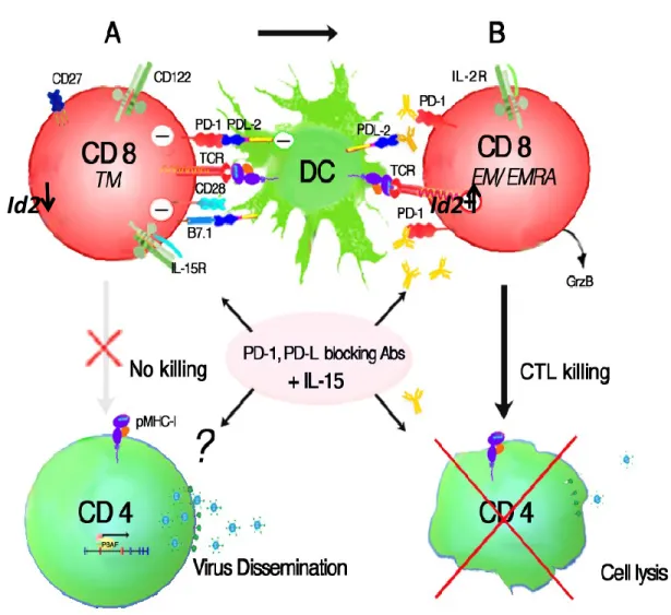
Figure 2: Restoration of exhausted antigen-specific CD8+ T cells by
ABBREVIATIONS
APC: Antigen Presenting CellAIDS: Acquired Immunodeficiency Syndrome Bcl-2: B-cell lymphoma 2
Bcl-6: B-cell lymphoma 6
Blimp-1: B lymphocyte-induced maturation protein 1 CBA: Cytometric Bead Array
CCR5: Chemokine (C-C motif) Receptor 5 CCR7: Chemokine (C-C motif) Receptor 7 CD3ζ: Cluster of Differentiation 3ζ CD4: Cluster of Differentiation 4 CD8: Cluster of Differentiation 8 CD27: Cluster of Differentiation 27 CD28: Cluster of Differentiation 28 CD38: Cluster of Differentiation 38 CD45RA: Cluster of Differentiation 45RA CD57: Cluster of Differentiation 57 CD70: Cluster of Differentiation 70 CD80: Cluster of Differentiation 80 CD86: Cluster of Differentiation 86 CD107a: Cluster of Differentiation 107a CD127: Cluster of Differentiation 127 CD160: Cluster of Differentiation 160
CDK: Cycline Dependent Kinases CHI: Chronically progressing subjects CMV: Cytomegalovirus
CTL: Cytotoxic T Lymphocyte
DC-SIGN: Dendritic Cell-Specific Intercellular adhesion molecule-3- EOMES: Eomesodermin
EC: Elite Controller Env: Enveloppe FASL: Fas Ligand Gag: Group Antigen
GALT: Gut-Associated Lymphoid Tissue GrzB: Granzyme B
gp120: Glycoprotein 120
HAART: Highly Active Antiretroviral Therapy HIV: Human Immunodeficiency Virus
HLH: Helix-Loop-Helix ID: Inhibitory of DNA binding IFN : Interferon
IL-2 : Interleukin 2 IL-15: Interleukin 15 IL-7: Interleukin 7
IS: Immunological Synapse
KLRG-1: Killer cell Lectin-like Receptor G1 LCMV: Lymphocytic Choriomeningitis Virus
LM: Listeria Monocytogenes
MHC: Major Histocompatibility Complex MPEC: Memory Precursor Effector Cells mRNA: messenger Ribonucleic acid Nef: Negative factor
NK : Natural Killer Cell
PAMPs : Pathogen-Associated Molecular Patterns PBMC : peripheral blood mononuclear cell
PCR: Polymerase Chain Reaction PD-1: Programmed cell death 1
PD-L1: Programmed cell death Ligand 1 PD-L2: Programmed cell death Ligand 2 pDC: plasmacytoid dendritic cell
Pol: Polymerase pRb: retinoblastoma
siRNA: small interfering RNA SLEC: Short-Lived Effector Cells ST: successfully HAART treated
STAT-3: signal transducer and activator of transcription 3 STAT-4: signal transducer and activator of transcription 4 STAT-5: signal transducer and activator of transcription 5 ST: Successfully Treated
T-bet: T-box transcription factor TCR: T cell receptor
Tcm: T central memory Tem: T effector memory
Temra: T terminally differentiated effector Ttm: T transitional memory
Tscm: T stem cell-like memory cells TNF: Tumor Necrosis Factor
TRAF: Tumor necrosis factor receptor associated protein VV: Vaccinia Virus
Ai mie genitori, Alfonso e Cristina,
per avermi sostenuto
in questo faticoso percorso della mia vita
e avermi fatto sentire sempre
il loro amore e la loro vicinanza
nonostante un oceano di mezzo.
Senza il loro supporto questo dottorato
ACKNOWLEDGEMENTS
Of utmost importance, I would like to confer my most deferential appreciation to my supervisor, Dr. Rafick-Pierre Sékaly, who has provided me with personal and financial resources for the timely completion of this work. Due to his extensive knowledge, patience, attention and scientific expertise, I have been able to grow professionally and learn about the different aspects of research methodology. The experiences I have gained under his tutelage have molded me into becoming the researcher that I had anticipated to be. His guidance and generosity extended beyond scientific matters through his personal goodwill as well as concern for my health and family. The long meetings during nights and weekends were always ameliorated by the provision of cookies, muffins, and croissants. I could not have asked for a better supervisor.
I would like to express my gratitude to Dr. Francesco A. Procopio for his valuable contributions through supervision and the numerous scientific discussions. Under his supervision, I was trained with a wide range of experimental techniques that allowed me to have a comprehensive perspective on planning and analyzing experiments. Not only did he mentor me during my PhD, but he also helped me maintain my sanity through friendly banter and his frequently humorous perspectives.
I also would like to thank Dr. Jeff Ahlers, Dr. Franck Dupuy, Dr. Hiroshi Takata, for their significant help in planning experiments and technical assistance; and Dr. Lydie Trautmann for her help in the optimization of the cytotoxic assay. I also thank Dr. Carmen Nichols, Dr. Joumana Zeidan, Dr. David Olagnier and Victor Joo and Dr. Remi Formentin for their editorial assistance.
The encouragement and love from my family and friends, are very important to me, they have supported me during my Ph.D. studies. Without the support of my parents, Matteo, Giorgia, Anna and Sabry this dissertation would have not been possible.
CHAPTER 1
1. Immunological Memory
Immunological memory is defined as the ability of the immune system to respond against a previously encountered pathogen. This characteristic was described for the first time by Thucidydes in the fifth century B.C., indeed he noticed that people who survived plague were not infected a second time. The property of the immunological memory are based on the fact that both antibody and T cell responses which mediate the adaptive immune response, are more efficient in responding the second time to the same antigen than in the first encounter. Immunological memory is the basis for generating protective immunity against many pathogens and represents the hallmark for vaccine strategies. While it is clear that B cell production of antibody is critical for the protective features of many vaccines, long-lived T cell immunity is also a critical component for good vaccine.
1.1 Generation of memory CD8+ T cells
Once a mature naïve T cell population exits the thymus after positive and negative selection, it circulates in the blood and in the periphery while expressing a wide repertoire of diverse T cell receptors (TCRs) that can recognize pathogens and induce efficacious immune responses. Naïve T cells are maintained by the signals provided by the continuous but transient interaction of their TCR with self–peptide major histocompatibility complexes (MHC) on antigen-presenting cells (APCs). Upon infection dendritic cells migrate to the lymphoid organs and present antigen to naïve T cells. This interaction is responsible for the formation of immunological synapses (ISs) between T cells and APC leading to more stable interactions. A naïve T cell activated by antigen stimulation undergoes proliferation, cytokine production, and differentiates into effector cells. Several studies have shown that the immune response requires three signals: 1) antigenic stimulation upon engagement of the TCR and CD8 as a coreceptor, 2) costimulatory signals, such as CD28, and 3) inflammatory cytokines such as IL-12 and IFNs (Curtsinger, Schmidt et al. 1999; Curtsinger, Johnson et al. 2003; Curtsinger, Lins et al. 2003; Kolumam, Thomas et al. 2005). The T cell-APC interactions lead to clonal expansion and effector and memory formation. The few naïve T cells specific for the cognate antigen are able to divide more
than 15 times (up to 50.000 fold expansion) in response to lymphocytic choriomeningitis virus (LCMV) and Listeria monocytogenes (LM), leading to their differentiation into effector cytotoxic T lymphocytes (CTLs) that will kill infected cells (effector phase). Functional CTLs exert their cytotoxic activity through the secretion of effector molecules, such as granzymes and perforin, as well as the secretion of cytokines such as interferon- (IFN) and tumor necrosis factor (TNF). CD4+ T cells may also differentiate into T helper 1 (Th1) cells that secrete IFN, TNF and interleukin-2 (IL-2). These events regulate the antiviral immune response and mediate direct killing of virus infected cells. Once the antigen has been cleared, most of the effector CTL (90%-95%) die by apoptosis (contraction phase) while the remaining cells (5-10%) establish a pool of the long-lived population of memory cells. It was originally proposed that the few cells that survive the contraction phase were randomly selected. However, recent studies have shown that some cells have more memory potential when compared to others. Studies in mice have helped in the discovery of transcription factors (described later) and cell surface markers that can predict which cells differentiate into effector function and those that instead generate the memory T cell population for life-long persistence. Among these markers, increased levels of IL-7 receptor subunit α (IL-7Rα or CD127), CD27, and B cell lymphoma 2 (BCL-2), as well as decreased expression of killer cell lectin-like receptor G1 (KLRG1) have been shown to preferentially induce the generation of long lived memory CD8+ T cells (Schluns, Kieper et al. 2000; Kaech, Tan et al. 2003; Joshi, Cui et al. 2007; Kurtulus, Tripathi et al. 2011; Dunkle, Dzhagalov et al. 2013). While adoptive transfer experiments using TCR transgenic cells have shown that KLRGhi CD127lo cells slowly decrease in number after transfer in comparison to the long lived KLRGlo CD127hi cells that were maintained over time (Joshi, Cui et al. 2007; Sarkar, Kalia et al. 2008). Thus KLRGhi CD127lo cells have been referred as “short-lived effector cells” (SLECs), while KLRLGlo CD127hi cells as” memory precursor effector cells” (MPECs). Memory cells that are generated after the contraction phase have the ability to control secondary exposure to antigen by the rapid acquisition of effector function, increased frequency and localization to peripheral sites of infection.
1.2 Central memory and Effector memory T cell subsets:
Memory T cells are heterogeneous and different subsets have been identified among CD4+ and CD8+ T cell population. The main two subsets are central memory and effector memory cells (Tcm and Tem, respectively). These two populations are distinguished based on their homing capabilities and effector functions. Effector cells home preferentially to the periphery; indeed they lack the expression of lymph-node homing markers such as CD62L and CCR7. In contrast central memory cells are found preferentially in the lymph-node as they are CD62L+ and CCR7+. Tcm share similar properties with naïve T cell, but do not express CD45RA on their cell surface and can rapidly differentiate into effector cells upon re-exposure to the same antigen. They have high proliferative potential and secrete high levels of IL-2 (Sallusto, Lenig et al. 1999); Tcm are more efficient at reconstituting the memory T cell pool and mediating protective immunity compared to Tem (Wu, Kirman et al. 2002; Wherry, Teichgraber et al. 2003; Castiglioni, Gerloni et al. 2004; Bouneaud, Garcia et al. 2005; Klebanoff, Gattinoni et al. 2005). Tem have lower proliferative capabilities and IL-2 production, however, they can rapidly exert cytotoxic and effector functions (IFN- secretion). Many other transitory subsets have been identified in both CD4+ and CD8+ and within the Tem other subsets that can be distinguished based on the expression of CD27 and CD28 (Romero, Zippelius et al. 2007). In humans, there is another effector memory CD8+ T subset, the terminally differentiated effector cells (Temra), that are positive for CD45RA expression but they do not express CCR7. Recently, a human memory T cell subset with stem cell–like properties has been described (Tscm)(Gattinoni, Lugli et al. 2011). These cells share many of the characteristics of naïve T cells cell (CD45RO−, CCR7+, CD45RA+, CD62L+, CD27+, CD28+ and IL-7Rα+ ), however, they also express CD95, IL-2Rβ, CXCR3, and LFA-1, typical markers of memory T cells (Gattinoni, Lugli et al. 2011). Therefore, Tscm are a long-lived human memory T cell population with increased capacity for self-renewal and increased ability to generate central memory and effector memory T cells. In summary, Tem provides immediate protection against second exposure to a previously
encountered pathogen and provide rapid protection at entry sites of infection, whereas Tcm remain principally in the lymphoid tissue where they can rapidly proliferate and differentiate into effector cells to resupply the effector T cells at peripheral sites.
1.3 Models of memory T cells generation
Because of the heterogeneity among the effector and memory CD8+ T cell population, different models of T cell differentiation have been proposed during infection.
Separate-precursor model: this model proposes that when naïve T cells exit the thymus,
these cells are differentially pre-selected to become effector or memory CD8+ T cells upon activation. However, evidence suggests that this model is unlikely. More sophisticated studies using adopting transfer of single CD8+ T cells have shown that naïve T cells can differentiate in both effector and several memory CD8+ T cell subsets (Stemberger, Huster et al. 2007; Gerlach, van Heijst et al. 2010).
Linear differentiation model: this model suggests that naïve T cells that respond to antigenic stimulation can give rise to effector cells. Once the antigen has been cleared, effector cells can either die by apoptosis, become senescent terminally differentiated T cells, or differentiate into memory CD8+ T cell subsets (Wherry, Teichgraber et al. 2003).
Bifurcative differentiation model (or asymmetric cell division): this model implies that early
after antigen stimulation, within the first cell division, one T cell precursor can give rise to two daughter cells: the proximal daughter cell (closer to the antigen-presenting cell (APC)) differentiates into effector cells that will die by apoptosis once the antigen has been cleared. The distal daughter cell (further from the APC) gives rise to Tcm, Tem and Ttm (Chang, Palanivel et al. 2007) .
Self-renewing effector model: this model proposes that Tcm and effector T cells, generated
from naïve T cells, have self- renewal abilities. Tcm are found in the lymph nodes and undergo homeostatic proliferation and can differentiate into Tem. Tem can give rise to terminally differentiated effector cells but cannot self-renew (Ahmed, Bevan et al. 2009).
Memory CD8+ T cells that are generated after an acute infection are maintained in a cytokine-dependent manner. Several studies have shown the role of -chain cytokines (IL-2, IL-4, IL-7, IL-9, IL-15 and IL-21) in regulating survival and homeostatic proliferation of memory CD8+ T cells. Among them IL-2, IL-15 and IL-7 have been shown to regulate different stages of memory differentiation and maintenance. These cytokines have some overlapping functions, but at the same time, regulate different points of the immune response. These overlapping functions are due to the fact that these cytokines share a common cytokine-receptor subunit and signaling pathways. All the receptors of the gamma chain cytokines utilize the gamma chain (c) subunit, which signals through JAK3. Moreover, IL-2 and IL-15 receptors share a common β chain which signals through Janus kinases 1 (JAK1). IL-15Rα may also mediate signaling through tumor necrosis factor receptor associated factor 2 (TRAF2) binding motif. IL-2 binds to IL-2R alone with low affinity while IL-7 binds IL7R which is composed of two subunits: the common chain and the unique -chain that confers cytokine specificity. Memory cells are known to be maintained by homeostatic proliferation which leads to high division rate when compared to naïve T cells (Tough and Sprent 1994) and the continual division is mediated by IL-15 while inhibited by IL-2 (Ku, Murakami et al. 2000). Different groups have shown that IL-7 and IL-15 are the major cytokines involved in the maintenance and homeostatic proliferation of memory CD8+ T cells (Schluns, Kieper et al. 2000; Schluns, Williams et al. 2002; Mueller, Petrovas et al. 2005; Purton, Tan et al. 2007). In particular, IL-15 seems to mediate T cell proliferation while IL-7 is implicated in providing survival. Other researchers have also revealed that overexpression of IL-15, through IL-15 transgenic mice, increases the total number of memory CD8+ T cells (Marks-Konczalik, Dubois et al. 2000; Fehniger, Suzuki et al. 2001). IL-7 seems to regulate the differentiation of effector CD8+ T cells that express high levels of CD127 (IL-7R) into long lived memory cells (Kaech, Tan et al. 2003). Indeed IL-7 is more important than IL-15 in supporting memory cell formation from recently activated CD8+ T cells. Other groups have shown reduced maintenance of antigen-specific CD8+ T cells in IL-15-deficient mice (Becker, Wherry et al. 2002; Schluns, Williams et al. 2002). Rubinstein et al. demonstrated that IL-15 and IL-2 induced
the preferential accumulation of short-lived effector/memory (KLRG1hiCD127lo) CD8+ T cells while stimulation with IL-7 mediates increased numbers of the long lived memory precursors KLRG1loCD127hi cells (Rubinstein, Lind et al. 2008). As cytokines that signal through the c receptors, IL-15 and IL-7 support survival of memory CD8+ T cells at the molecular level by increasing the levels of anti-apoptotic molecules such as Bcl-2. Antigen specific CD8+ T cells have high levels of Bcl-2 while naïve T cells express lower Bcl-2. In addition, CD8+ T cells express higher levels of Bcl-2 than the CD4+ memory cells (Homann, Teyton et al. 2001). The signaling pathway triggered by the c cytokines signal through signal transducer and activator of transcription 5 (STAT5) and are inhibited by suppressor of cytokine signaling-1 (SOCS-1).
1.5 Transcription factors that control effector and memory CD8+ T cell differentiation
Several transcription factors that regulate effector and memory CD8+ T cell development have been identified. These transcriptional regulators have been shown to act in pairs: when the expression of one of the two molecule increases, they induce either memory or effector differentiation. Among these pairs of transcriptional regulators, t-bet and eomesodermin, Id2 and Id3, Bcl-6 and Blimp-1 have been shown to function in coupled to regulate these properties. In particular, cells that express increased levels of T-bet, B lymphocyte-induced maturation protein (Blimp-1), inhibition of DNA binding 2 (Id2) and signal transducer and activator of transcription 4 (Stat4) during an immune response, induces the differentiation of effector CD8+ T cells in short lived effector cells. These molecules contrast the expression of other transcriptional factors, Eomesodermin (Eomes), B cell lymphoma 6 (Bcl-6), Id3 and Stat3 that, when upregulated, induce the differentiation of effector cells into long-lived memory CD8+ T cells for life-long persistence (reviewed in (Kaech and Cui 2012)).
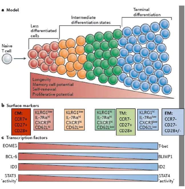
Figure 1: Different stages of CD8+ T cell differentiation:
Memory CD8+ T cells are heterogeneous and can be distinguished based on cell surface markers or the expression of different sets of transcriptional factors: central memory are less differentiated and express CD27, CCR7, and CD28 expression on their cell surface while showing greater self-renewal and proliferative capabilities. At the transcriptional level, Tcm express high levels of BCL-6, Id3, STAT3 and eomes. As cells differentiate in effector cells, they lose expression of CCR7, CD27, and CD28 on their cell surface and
express high levels of transcriptional factors T-bet, Id2, Blimp-1 and STAT4 that regulate their effector functions. Adapted from (Kaech and Cui 2012)
1.5.1 T-bet and eomesodermin
T-bet and eomesodermin are two T box transcription factors that play important roles in promoting effector and memory CD8+ T cell differentiation. Their role in mediating effector CD8+ T cell functions is confirmed by studies where they show that the two transcription factors are required for the expression of INF, granzyme B, perforine, CXCR3 and CXCR4 (Intlekofer, Takemoto et al. 2007; Joshi, Cui et al. 2007; Banerjee, Gordon et al. 2010; Pipkin, Sacks et al. 2010). Expression of T-bet is upregulated in the effector phase of the immune response by antigen presentation and by IL-12 and mammalian target of rapamycin (mTOR) activity (Takemoto, Intlekofer et al. 2006; Joshi, Cui et al. 2007; Rao, Li et al. 2010; Joshi, Cui et al. 2011).When memory CD8+ T cell are generated, T-bet expression decreases whereas eomes is induced by RUNX-3 and IL-2 (Banerjee, Gordon et al. 2010; Joshi, Cui et al. 2011).
1.5.2 Blimp-1 and Bcl-6
Blimp-1 and Bcl-6 are another pair of transcription factors involved in B and T cells differentiation. Blimp-1 expression is high on effector CD8+ T cells and its levels are increased by IL-2, IL-12 and IL-21 cytokines (Gong and Malek 2007; Kwon, Thierry-Mieg et al. 2009; Kalia, Sarkar et al. 2010; Pipkin, Sacks et al. 2010). In these cells Blimp-1 regulate effector functions such as T cell trafficking to sites of inflammation and INF and Granzyme B expression (Kallies, Xin et al. 2009; Rutishauser, Martins et al. 2009). As memory CD8+ T cells form Blimp-1 expression levels decrease and Bcl-6 levels increase (Cui, Liu et al. 2011). In mice, IL7Rhi KLRGlo memory precursors express high levels of Bcl-6 and its levels are maintained and increased in antigen-specific memory cells after LCMV infection, and this coincides with decreased levels of Blimp-1 (Gong and Malek 2007). Other studies have shown that Bcl-6 is important for the formation of Tcm and its
expression may be mediated by IL-10 and IL-21 (Ichii, Sakamoto et al. 2002; Gong and Malek 2007; Ichii, Sakamoto et al. 2007).
2. Id proteins
Inhibitor of DNA binding or differentiation (Id) proteins belong to a family of helix-loop-helix (bHLH) transcription factors. There are two main families of the HLH proteins: E-proteins and Id E-proteins. Id E-proteins were named due to their ability to inhibit DNA binding (Benezra, Davis et al. 1990). In mammals, four known members of Id proteins exist; Id,1 Id2, Id3 and Id4, all encoded by four different genes (Benezra, Davis et al. 1990; Deed, Jasiok et al. 1994).
2.1 The properties of Id proteins
Id proteins and E proteins both have a highly conserved helix-loop-helix (HLH) domain that consist of two amphipathic α-helices separated by a loop of variable sequence and length (Pesce and Benezra 1993). The HLH domain is required for homo- and hetero-dimerizzation of these proteins. Next to the HLH domain, E proteins contain a basic amino acid domain which is fundamental for their binding to DNA within the canonical “E-box” recognition sequence, CANNTG (Figure 2) (Barone, Pepperkok et al. 1994). However, Id proteins do not possess the basic domain, thus they are not able to bind DNA and act by inhibiting the transcriptional regulation of bHLH factors, in particular, the E-proteins (Figure 3) (Lasorella, Uo et al. 2001) (Zebedee and Hara 2001; O'Toole, Inoue et al. 2003) Id proteins, through the inhibition of E-protein transcriptional activity, control various critical cells function, including cell cycle, differentiation, apoptosis, cell senescence, tumorigenesis and neoplastic transformation (Barone, Pepperkok et al. 1994) (Andres-Barquin, Hernandez et al. 2000).
Figure 2: Schematic structure of E-proteins and Id- proteins
The basic DNA binding region (b) that bound the HLH region is shown. (Adapted from (Norton 2000)).
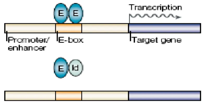
Figure 3: Id and E-protein interaction
E-proteins form heterodimers that bind DNA and induce transcription of genes involved in apoptosis and blockage of cell cycle progression. When Id proteins bind to E-proteins, they form a complex that cannot bind to DNA and thus inhibits E-proteins transcriptional activity (adapted from (Engel and Murre 2001)).
(
E-proteins)
(ID proteins)2.2 Id proteins in cell cycle control
The role of Id proteins in regulating cell cycle has been shown in mice and cell line models. These studies have shown that Id proteins are implicated in the G1/S transition of the cell cycle. Lasorella et al. have demonstrated that resting and undifferentiated cells have lower levels of Id proteins and its expression increases when cells enter the cell cycle and proliferate, suggesting that Id proteins may play a role in regulating cell growth (Lasorella, Uo et al. 2001). Mechanistic studies via the use of small interfering RNA have shown that the silencing of Id proteins results in cell cycle arrest (Barone, Pepperkok et al. 1994). Two main pathways have been shown to correlate Id proteins to cell cycle control. The first one is by inhibiting the binding of E-protein transcription factors: following mitogenic signals, increased levels of Id may sequester E-proteins from forming heterodimers and prevent them from binding the DNA. The Id-E protein interaction results in a consequent inhibition of E-protein target gene expression such as the cyclin-dependent kinase (CDK) inhibitors, (i.e. p21CIP1 and p27KIP1) (Trabosh, Divito et al. 2009). Moreover, Ids can inhibit the expression of p16INK4a promoter through an E-protein-dependent mechanism, while Id2 inhibits p15INK4b (Alani, Young et al. 2001). Id proteins have been shown to preferentially target the ubiquitously expressed E-proteins (E12, E47, E2-2, and HEB), (Benezra, Davis et al. 1990; Norton and Atherton 1998). In addition to E proteins, Ets transcription factors and the tumor suppressor proteins of the Retinoblastoma (pRb) family are also targets of Id proteins (Iavarone, Garg et al. 1994; Biggs, Zhang et al. 1995; Lasorella, Iavarone et al. 1996). The direct interaction between pRB and Id proteins (Id2 in particular) is thought to potentiate the S phase. This interaction in fact results in the dissociation of pRB from the E2F transcription factor. Once dissociated from pRB, (p107 and p130) E2F is free to bind to DNA and induce the expression of genes involved in cell cycle progression. Free E2F transcriptional complex then activates the expression of genes required for progression through S phase of the cell cycle (Figure 4).
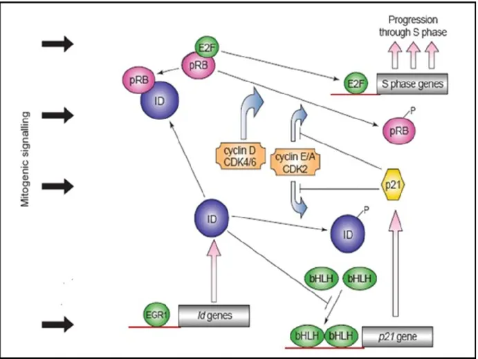
Figure 4: Id proteins in cell cycle control
Schematic representation shows how Id proteins regulate cell cycle progression. Mitogenic signaling pathways, during G1 phase of the cell cycle, promotes events that induce the phosphorylation of the cyclin-dependent kinase (CDKs). CDKs phosphorylate pRB (p107 and p130), leading to release of the transcription factor E2F. Free E2F is able to bind to DNA and thus to induce the expression of genes important for the S phase of the cell cycle. (Norton 2000).
2.3 Ids in Development and Differentiation
Because of the ability of Id proteins in regulating proliferation and differentiation, these proteins regulate many aspects of development. Studies in mice with Id protein knockout have shown that the absence of Id1 and Id3 together leads to altered differentiation and
proliferation of neural cells and are embryonic lethal, whereas the absence of Id2 resulted in decreased proliferation of many cell types and decreased body size was observed in these mice (Lasorella, Uo et al. 2001; Sikder, Devlin et al. 2003). Id1 knockout mice alone have no evident defects whereas Id3 knockout mice exhibit defects in B cell proliferation and humoral immunity. The importance of Id2 in regulating development was further confirmed by a study where the loss of Id2 lead to the absence of Peyer’s patches and lymph nodes as well as decreased numbers of epidermal Langerhans cells and natural killer cells (Sikder, Devlin et al. 2003).
2.4 Role Id2 and Id3 in memory CD8+ T cell differentiation
Few studies in mice have recently shown the new role of Id2 and Id3 in regulating the differentiation of effector and memory CD8+ T cell formation. The first study from Cannarile et al in 2006 had shown that after antigen stimulation, Id2 expression was increased in effector cells and its expression remained high in the memory compartment (Cannarile, Lind et al. 2006). CD8+ T cells from Id2 knockout mice infected with Listeria
monocytogenes were able to respond to the antigen, to proliferate, and differentiate into
effector cells but were more susceptible to spontaneous apoptosis compared to wild-type cells, resulting in a decreased ability to clear Listeria after infection (Cannarile, Lind et al. 2006). CD8+ T cells in absence of Id2 have decreased levels of the pro-survival Bcl-2 molecule and increased levels of the proapoptotic Bim and Ctla-4, suggesting that Id2 was mediating the survival of effector cells that undergo contraction during the immune response. Moreover, the few cells that survived in the absence of Id2 expression expressed a different phenotype, maintaining CD27 and CD127 expression and exhibiting a more central memory phenotype in comparison to wild-type cells that had instead differentiated into effector memory CD8+ T cells (Cannarile, Lind et al. 2006).
Another more recent study in mice showed that the ratio between Id2 and Id3 expression levels are important for determining which cells become long-lived memory cells and those that become short-lived effector cells after an immune response (Yang, Best et al. 2011). Indeed, high levels of Id3 preferentially lead to the differentiation of long-lived memory
cells while low levels of Id3 promote short-lived effector cell differentiation. Contrary to Id2 expression that is upregulated early after antigen stimulation, Id3 expression levels were decreased in effector cells and its expression became high when memory cells were generated. Id3 levels were found to be modulated right before the change in KLRG-1 and CD127 expression on the cell surface. Therefore, Id3 expression could be an indicator for the identification of memory precursors (CD127hiKLRG-1lo). Id3 deficient mice were not able to accumulate CD127hi memory cells. Moreover, Id2 knockout cells were lacking KLRG-1hi short lived effector cells. This study suggest that Id2 and Id3 regulate the differentiation of effector and memory cells differently and that their expression could be used to predict memory precursors.
3. CD8+ T cell exhaustion
During an acute infection, naïve T cells are stimulated by antigen to undergo clonal expansion, differentiate into effector T cells that secrete cytokines and exert cytolytic activity. Once the infection is resolved, a small subset of these functional effector T cells survive the contraction phase and further differentiate into highly polyfunctional memory T cells. Memory cells have the capacity to rapidly respond to previously encountered antigens, which include the ability to reacquire the homing receptors to secondary lymphoid tissues, high proliferative capabilities, and the ability to self-renew by undergoing homeostatic proliferation driven by IL-7 and IL-15 (Becker, Wherry et al. 2002; Goldrath, Sivakumar et al. 2002). These properties allow memory T cells to confer protective immunity. However, during a chronic viral infection, T cells lose these characteristics because of the continued exposure to the antigen, a functional impairment known as exhaustion. T cell exhaustion was described for the first time almost 10 years ago in murine LCMV models where researchers showed that during chronic infection, antigen-specific T cells were dysfunctional and were not able to kill virally infected cells (Barber, Wherry et al. 2006). During exhaustion, CD8+ T cells undergo a progressive loss of their effector functions. The events that regulate this loss are influenced by the level and persistence of antigen stimulation and by CD4+ T cell help. If the viremia is low and CD4+ T cell
numbers are not lost, CD8+ T cells are still functional, as during some latent infections. Typically, when viral infection persists, IL-2 production, proliferation, and ex vivo killing are the first effector functions to be lost during chronic infection (exhaustion I) (Wherry, Blattman et al. 2003). This stage is followed by partial exhaustion II when TNF-production begins to be impaired. Reduction in INF- production following antigen stimulation follows, a stage of exhaustion that is the result of high viral load and low CD4+ T cell numbers (Wherry, Blattman et al. 2003). Finally, the continuous presentation of the antigen to antigen-specific CD8+ T cells leads to death by apoptosis. After these first studies in mice, it became evident that humans had similar kinds of CD8+ T cell exhaustion. For example during human immunodeficiency virus (HIV), hepatitis C virus (HCV) or hepatitis B virus (HBV) infections, virus-specific CD8+ T cells appear to lack ex
vivo effector functions.
3.1 HIV infection
Human immunodeficiency virus (HIV) is a lentivirus, a member of the retrovirus family, and if untreated, leads to acquired immunodeficiency syndrome (AIDS). HIV is an enveloped viral particle that contains two copies of positive single-stranded RNA that code for nine genes: gag, pol, env, tat, rev, nef, vif, vpr and vpu. HIV infects CD4+ T cells, macrophages and dendritic cells. HIV infection leads to a gradual loss of CD4+ T cells through direct or indirect mechanisms: killing of CD4+ T cells directly mediated by the virus, indirect killing of infected cells by cytotoxic CD8+ T cells, killing of CD4+ T cells mediated by abortive HIV infection and/or increased rates of apoptosis of uninfected cells (Banda, Bernier et al. 1992; Groux, Torpier et al. 1992; Cottrez, Manca et al. 1997; Herbein, Van Lint et al. 1998; Badley, Pilon et al. 2000; Esser, Graham et al. 2001; McCune 2001; Alimonti, Ball et al. 2003; Doitsh, Cavrois et al. 2010). When the numbers of CD4+ T cells become very low, the immune system is compromised and the disease is characterized by susceptibility to infection with organisms that are not typically pathogenic (McCune 2001; Mattapallil, Douek et al. 2005).
3.1.1 Clinical Course of HIV infection
HIV infection is characterized by a peak in plasma viremia, usually reaching more than a million RNA copies/ml, and occurring 3-4 weeks after HIV entry. This period is characterized by non-specific flu-like symptoms and coincides with decreased numbers of circulating CD4+ T cells (McMichael, Borrow et al. 2010). The partial viral control correlates temporally with an HIV-specific CD8+ T cell response which peaks just prior to the decline in viremia and is important in maintaining the viral load at stable levels (Koup, Safrit et al. 1994). This essential role of CD8+ T cells in controlling HIV disease progression has been confirmed in non-human primate models (Matano, Shibata et al. 1998; Jin, Bauer et al. 1999; Schmitz, Kuroda et al. 1999). Indeed, it has been shown that depletion of CD8+ T cells leads to a rapid increase in viremia while upon reappearance of CD8+ T cells, viremia decreases during acute and chronic SIV (Jin, Bauer et al. 1999; Schmitz, Kuroda et al. 1999). Chronic HIV infection, also known as the latent phase, lasts an average of 10 years and begins when the viral load decreases and CD4 T cell counts are stabilized. Throughout this phase, CD4+ T cells are gradually depleted from the circulation (McMichael, Borrow et al. 2010). When CD4+ T cells numbers decrease below 200 cells/μl, the immune system is susceptible to opportunistic infections and tumors and this immune dysfunction leads to AIDS. The rate of this depletion varies from one individual to another, for reasons that remain to be fully elucidated (Sun, Williams et al. 2004). The depletion of CD4+ T cells is thought to contribute to the inability of CD8+ T cells to mount effective responses and control viremia during chronic infection.
3.1.2 Role of CD8+ T cells in HIV infection:
CD8+ T cells play an important role during HIV infection and are essential in controlling viral replication and for the killing of virally infected cells. Their ability in controlling viremia is mediated by the secretion of soluble factors (CAF, MIP-1α, MIP-1β, RANTES) and by the release of perforin and Granzyme A and B that mediate direct lysis of infected cells. HIV-specific CD8+ T cells from patients in the acute phase of HIV infection are still able to control viremia and exert strong cytotoxic activity. Indeed, the development of
tetramer stainings have provided the data showing that the frequency of antigen-specific CD8+ T cells can reach up to 18% during acute infection (Gea-Banacloche, Migueles et al. 2000; Addo, Yu et al. 2002). As the infection progresses, the CD4:CD8 T cell ratio is inverted as a consequence of CD4+ T cells loss (Chun, Justement et al. 2002). During the late stages of infection, the frequency of CD8+ T cells begins to decline (Margolick, Munoz et al. 1995). Furthermore, during chronic HIV infection, the capability of CD8+ T cells to exert a cytolytic response diminishes and usually, in the absence of treatment, the disease progresses to AIDS (Klein, van Baalen et al. 1995). How CD8+ T cells are lost during HIV infection is still under debate and different mechanisms have been proposed (Reviewed in: (Gougeon 2003)). HIV specific CD8+ T cells have been shown to be more sensitive to Fas/CD95-induced apoptosis when compared to cytomegalovirus (CMV)-specific CD8+ T cells (Mueller, De Rosa et al. 2001). One possible mechanism of CD8+ T cell apoptosis was shown to be the effect mediated by HIV infected macrophages on bystander cells (Cottrez, Manca et al. 1997; Herbein, Van Lint et al. 1998; Badley, Pilon et al. 2000; Vlahakis, Algeciras-Schimnich et al. 2001). In contrast, other groups have shown that increased apoptosis of antigen-specific CD8+ T cells was not dependent on Fas but these cells were more sensitive to TNF receptor 2 and caspase mediated apoptosis leading to decreased levels of the pro-survival Bcl-2 molecule (Derby, Snyder et al. 2001). Other groups have shown that apoptosis of antigen-specific and total CD8+ T cells can occur in
vitro by interaction between gp120 and CXCR4 that is expressed on the cell surface of
CD8+ T cells. However, other mechanisms of CD8+ T cell depletion as a consequence of T cell exhaustion have been more recently described during HIV infection.
3.2 T cell exhaustion during HIV infection
It has been shown that negative regulatory pathways play an important role in T cell exhaustion. Among them, three different pathways are the principal mediators of exhaustion: cell surface inhibitory receptors (such as PD-1), cytokines (such as IL-10) and immunoregulatory cells (such as Treg cells). Inhibitory receptors have the important function of limiting T cell activation by reducing downstream signaling of the T-cell
receptor (TCR). Thus, a robust immune response is mediated by a perfect balance between positive and negative signals. However, the persistant expression of negative regulatory receptors during chronic infection leads to an impaired ability to respond to infections, further leading to exhaustion.
3.2.1 Programmed death 1 (PD1)
3.2.1.1 Structure
Programmed death 1 (PD-1) and its ligands, PD-L1 and PD-L2, mediate inhibitory signals that control the balance between T cell activation, tolerance and autoimmunity (Nishimura, Nose et al. 1999; Nishimura, Okazaki et al. 2001). PD-1 is a 288 amino acid type I transmembrane protein consisting of one immunoglobulin (Ig) superfamily domain and an intracellular domain containing an immunorecepor tyrosine-based inhibitory motif (ITIM) as well as an immunoreceptor tyrosine-based switch motif (ITSM). PD-1 expression is very low or undetectable on resting cells and its expression is induced transiently on activated T cells, B cells, natural killer T cells, activated monocytes, and dendritic cells (DCs). PD-1 binds to two ligands, both members of the B7 family, called PD-L1 (programmed death ligand-1 or B7-H1) and PD-L2 (B7-DC) (Chen 2004; Greenwald, Freeman et al. 2005). PD-L1 is constitutively expressed on many cell types whereas PD-L2 is induced on antigen-presenting cells during inflammation. 1 binding to its ligands (L1 and PD-L2) induces a cascade of events that inhibit TCR signaling. This is a mechanism to control autoimmunity and to prevent constant T cell activation, proliferation, cytokine production, particularly IFN-, TNF- IL-2, and cytolytic function. The mechanisms that PD-1 uses to dampen TCR signaling are mediated, in part, by the inhibition of ZAP70 phosphorylation and the association of the TCR with CD3. Moreover, engagement of PD-1 inhibits glucose metabolism mediated by CD3-CD28 and also inhibits downstream signaling of Akt by inhibiting CD28 mediated activation of phosphatidylinositol 3-kinase. The role of PD-1 in regulating peripheral tolerance to self-antigen is mediated by its function in the development of regulatory T cells and in inhibiting self-reactive T cells. The role of PD-1
in regulating immune tolerance has been described with PD-1 knockout mice that develop autoimmune disease (Nishimura, Nose et al. 1999; Nishimura, Okazaki et al. 2001). Other studies have demonstrated the treatment with blocking PD-1 antibodies as a therapeutic intervention for immunotherapy to cancer.
3.2.1.2 Role of PD-1 in CD8+ T cell exhaustion
The signaling pathways that are mediated by PD-1 are still not well understood, however, extensive studies have been conducted in the role of PD-1 for mediating T cell exhaustion during immune response. The first studies that were done in order to understand PD-1 function during chronic viral infection have been conducted in a lymphocytic choriomeningitis virus (LCMV) mouse model (Barber, Wherry et al. 2006). Barber et al. were the first to describe that PD-1 expression regulates CD8+ T cell exhaustion in mice infected with LCMV clone 13 (a LCMV variant that generates chronic viral infection). 1 expression levels were high on virus-specific CD8+ T cells and blocking the 1: PD-L1 interaction during chronic viral infection rescues T cell function while decreasing viral load, suggesting that the inhibition of PD-1 crosslinking may be used as a potential application in the treatment of chronic infection. These first studies were conducted in mice, however, the role of PD-1 in regulating exhaustion of CD8+ T cells was then confirmed in humans during chronic viral infections, such as HIV (Petrovas, Casazza et al. 2006; Trautmann, Janbazian et al. 2006; Zhang, Zhang et al. 2007) and in SIV infected Rhesus macaques (Petrovas, Price et al. 2007), (Velu, Titanji et al. 2009). Different groups have shown that PD-1 expression levels from chronically HIV infected donors are upregulated in particular on HIV-specific CD8+ T cells and its levels were positively correlated with viremia and negatively with CD4+ T cell number. Moreover, PD-1hi cells show a decreased capacity to proliferate and to produce cytokines. Longitudinal studies have shown that the use of highly active antiretroviral therapy (HAART) was able to decrease PD-1 levels on HIV-specific CD8+ T cells, suggesting that the continuous antigen presentation to these cells and viremia are responsible for the increased PD-1 expression
(Day, Kaufmann et al. 2006; Trautmann, Janbazian et al. 2006). In support of this, other groups have shown that SIV-specific CD8+ T cells for a specific epitope that had undergone mutational escape had decreased PD-1 expression on their cell surface. (Petrovas, Price et al. 2007; Salisch, Kaufmann et al. 2010). Other groups have shown that dominant clonotypes within the HIV-specific CD8+ T cell pool with increased levels of PD-1 failed to survive in culture and produced lower levels of cytokine when compared to subdominant clonotypes (Conrad, Ramalingam et al. 2011). The role of PD-1 in regulating apoptosis has also been shown by Petrovas et al. in 2006 where they showed that PD-1 expression levels during HIV-infection correlated with increased susceptibility to spontaneous and CD95/Fas-induced apoptosis and is associated with decreased Bcl-2 expression levels (Petrovas, Casazza et al. 2006). HIV-specific CD8+ T cells have been shown to be more susceptible to apoptosis when compared to CMV-specific CD8+ T cells from the same HIV-infected patients, possibly due to the higher levels of PD-1 on their cells surface. In fact, PD-1hi populations were found to be more susceptible to apoptosis than PD-1 negative CD8+ T cells, independent of antigen specificity. Moreover, increased PD-1 expression on CD8+ T cells from HIV-infected donors correlates with a pro-apoptotic phenotype with lower levels of the pro-survival Bcl-2 molecule, decreased levels of CD127, and higher levels of CD95/Fas surface receptor when compared to the PD-1lo compartment. PD-1hi cells also showed increased binding of MitoTracker suggesting that modification within the mitochondria of these cells may be responsible for increased susceptibility to apoptosis (Petrovas, Mueller et al. 2007).
3.3 HIV Impairment of CD8+ T cell maturation
As discussed earlier, during an acute infection, naïve T cells after antigen stimulation give rise to effector and memory T cells. These populations are distinguished based on their specific phenotype. Naïve T cells express CD45RA, costimulatory molecules, and homing receptors. As cells differentiate into effector cells (CD45RA+ CD28-CD27-CCR7-), Tem (CD45RA-CCR7-CD28-CD27-) and Tcm (CD45RA-CD28+CCR7+CD27+), certain markers, such as CCR7 for Tem and CD27 for Tcm, are gained or lost. Appay at al. showed
that during the chronic phase of HIV infection, antigen-specific cells that were generated expressed a distinguishable differentiation phenotype (based on CD28 and CD27) when compared to Epstein-Barr Virus (EBV) and HCV-specific CD8+ T cells (Appay, Dunbar et al. 2002). Other groups have shown that HIV-specific CD8+ T cells expressed lower levels of perforin on their cell surface and these cells were not able to exert cytotoxic functions in relation to CMV and EBV-specific cells in HIV-infected patients that instead are granzyme A/perforin+ CD45RA+ CD27- (Strengell, Matikainen et al. 2003; Zhang, Shankar et al. 2003). Moreover, different groups demonstrated that HIV-specific CD8+ T cells have lower CD28 expression and are CD27+, suggesting a decreased effector phenotype (Ogg, Kostense et al. 1999; Roos, van Lier et al. 2000). CD28 and CD27 are costimulatory receptors. CD28 mediates TCR activation upon TCR triggering while CD27 facilitates the generation of antigen-specific CD8+ T cells. CD28+ cells have longer telomere lengths and therefore, have increased proliferative potential in contrast to CD28- cells. As a result, decreased levels of CD28 on HIV-specific CD8+ T cells are associated with diminished proliferation (Speiser, Migliaccio et al. 2001). CD27 negative cells that are associated with an effector phenotype express more Granzyme A and perforin when compared to cells that express CD27 on their cell surface (van Baarle, Kostense et al. 2002). Therefore, HIV-specific CD8+ T cells that express high levels of CD27 and have lower proliferative capabilities and decreased perforin and cytotoxic activity. Moreover, HIV-specific memory CD8+ T cells were also found to have a pre-terminally differentiated phenotype (CD45RA-CCR7-), when compared to CMV-specific cells that instead expressed a terminally differentiated (CD45RA+CCR7-) phenotype (Champagne, Ogg et al. 2001; Ellefsen, Harari et al. 2002). In summary, during the chronic phase of HIV infection, antigen-specific CD8+ T cells should have an effector/effector memory phenotype, however these cells express a pre-terminally differentiated phenotype. The skewed maturation of these cells is responsible for decreased effector functions and this leads to viral escape from immune containment.
3.4 Highly Active antiretroviral Therapy
Highly active antiretroviral therapy (HAART) is the treatment for HIV infected individuals. HAART is able to decrease the rates of viral replication thus delaying the progression to AIDS. HAART is a combination therapy of three different drugs: two nucleoside reverse transcriptase inhibitors (NRTIs) and protease inhibitors (PI), two NRTIs and non-nucleoside reverse transcriptase inhibitors (NNRTI) or other such combinations. HAART therapy has been shown to efficiently decrease viremia and increase CD4+ T cell numbers in the majority of cases. Upon initiation with HAART, the plasma viral load undergoes two exponential phases of decay (Perelson, Essunger et al. 1997). The first phase is characterized by a rapid decline of the viral load as result of the loss of productively infected CD4+ T cells, which are mainly activated CD4+ T cells. The second phase is less rapid and consists of the loss of infected macrophages and dendritic cells, which are more resistant to virus–induced cytopathic effects than CD4+ T cells. The majority of the patients treated with HAART have a reduction in viremia that drops below 25 copies of viral RNA per ml of plasma (the detection limit of sensitive assay) by week 20 and remains low to undetectable except for occasional increases in viral load called viral “blips” (Perelson, Essunger et al. 1997; Posevitz, Arndt et al. 2008). Following HAART, the recovery of CD4+ T cell numbers undergoes two phases. First, there is a rapid increase in the CD4+ T cells that mostly have a memory phenotype, which happens during the first weeks from the treatment (Autran, Carcelain et al. 1997). This phase is followed by an increase in the naïve CD4 + T cell numbers (Autran, Carcelain et al. 1997).
HYPOTHESIS AND AIMS:
We have previously shown that during chronic HIV infection, there is an impaired distribution of the memory CD8+ T cell subsets that is associated with high levels of PD-1. The majority of CD8+ T cells are arrested in a transitional memory (Ttm) phenotype, are not able to differentiate to effector/effector memory (Tem) cells and lack cytotoxic functions. Functional and phenotypical defects are not restored even under HAART treatment. PD-1 limits T cell activation by attenuating T cell receptor signaling, however, it is not known whether PD-1 also acts by downregulating genes important for differentiation and T cell functions.
In the first part of my thesis (Chapter 2) I used a system biology approach as an unbiased methodology in order to identify pathways downstream of PD-1 that are associated with CD8+ T cell dysfunction and T cell exhaustion. I analyzed global gene expression profile of highly purified PD-1 high HIV-specific CD8+ T cells when compared to PD-1 low CMV-specific CD8+ T cells from the same chronic HIV-infected individuals and found that HIV-specific CD8+ T cells express low levels of the transcriptional modulator Id2. Due to the fundamental role of Id2 in mediating effector CD8+ T cell differentiation we decided to further investigate whether defects in Id2 expression could be responsible for the impaired maturation of the PD-1 high cells during chronic HIV infection. We hypothesized that:
- PD-1 crosslinking inhibits T cell function by interfering with the expression of Id2, leading to decreased maturation of CD8+ T cells and exhaustion.
In the second part of my thesis (Chapter 5) I wanted to identify upstream signals that can upregulate Id2 expression and that could restore CD8+ differentiation during HIV-infection. Id2 and the common -chain cytokine IL-15 play similar roles in mediating survival and differentiation of effector CD8+ T cells; both Id2 or IL-15 knock-out mouse lack cells with an effector/Tem phenotype. Moreover, Id2 has two potential STAT5 binding sites at its promoter and IL-15 mediates survival of effector cells by inducing phosphorylation and transcriptional activity of STAT5. Finally, both Id2 and IL-15