O
pen
A
rchive
T
OULOUSE
A
rchive
O
uverte (
OATAO
)
OATAO is an open access repository that collects the work of Toulouse researchers and
makes it freely available over the web where possible.
This is an author-deposited version published in :
http://oatao.univ-toulouse.fr/
Eprints ID : 10815
To link to this article : DOI : 10.1099/ijs.0.039099-0
URL :
http://dx.doi.org/10.1099/ijs.0.039099-0
To cite this version :
Boubetra, Dalila and Zitouni, Abdelghani and
Bouras, Noureddine and Mathieu, Florence and Lebrihi, Ahmed and
Schumann, Peter and Spröer, Cathrin and Klenk, Hans-Peter and
Sabaou, Nasserdine Saccharothrix hoggarensis sp. nov., an
actinomycete isolated from Saharan soil. (2013) International
Journal of Systematic and Evolutionary Microbiology, vol. 63 (Pt
2). pp. 549-553. ISSN
1466-5026
Any correspondance concerning this service should be sent to the repository
administrator:
[email protected]
Saccharothrix hoggarensis
sp. nov., an
actinomycete isolated from Saharan soil
Dalila Boubetra,
1Abdelghani Zitouni,
1Noureddine Bouras,
1Florence Mathieu,
2Ahmed Lebrihi,
2Peter Schumann,
3Cathrin Spro¨er,
3Hans-Peter Klenk
3and Nasserdine Sabaou
1Correspondence Nasserdine Sabaou [email protected]
1Laboratoire de Biologie des Syste`mes Microbiens (LBSM), Ecole Normale Supe´rieure de Kouba,
BP 92, 16 050 Kouba, Algiers, Algeria 2
Universite´ de Toulouse, INPT-ENSAT, Laboratoire de Ge´nie Chimique, UMR 5503 (CNRS/INPT/ UPS), 1 Avenue de l’Agrobiopole BP 32607 Auzeville Tolosane 31326 Castanet-Tolosan, France
3DSMZ – German Collection of Microorganisms and Cell Cultures, Inhoffenstraße 7B, 38124
Braunschweig, Germany
An actinomycete, designated SA181T, was isolated from Saharan soil in the Hoggar region (south Algeria) and was characterized taxonomically by using a polyphasic approach. The morphological and chemotaxonomic characteristics of the isolate were consistent with the genus Saccharothrix,
and 16S rRNA gene sequence analysis confirmed that strain SA181Twas a novel member of the
genus Saccharothrix. DNA–DNA hybridization values between strain SA181Tand its closest
phylogenetic neighbours, the type strains of Saccharothrix longispora, Saccharothrix texasensis and Saccharothrix xinjiangensis, were clearly below the 70 % threshold. The genotypic and phenotypic data showed that the isolate represents a novel species of the genus Saccharothrix, for which the name Saccharothrix hoggarensis sp. nov. is proposed, with the type strain SA181T
(5DSM 45457T
5CCUG 60214T).
The genus Saccharothrix was described for the first time by Labeda et al. (1984). Several species of this genus have been transferred to other new taxa, including the genera Lentzea (Yassin et al., 1995), Crossiella (Labeda, 2001), Lechevalieria (Labeda et al., 2001), Goodfellowia (Labeda & Kroppenstedt, 2006) and Umezawaea (Labeda & Kroppenstedt, 2007). Currently, 12 species of the genus Saccharothrix have been described based on chemical analysis, physiological prop-erties, cellular fatty acid composition, phylogeny and DNA– DNA hybridization data. The genus is characterized by fragmentation of both substrate and aerial mycelia into rods and ovoid elements, type III cell-wall meso-diaminopimelic acid without glycine, galactose, rhamnose and small amounts of mannose as diagnostic whole-cell sugars, a phospholipid type PII (phosphatidylethanolamine) or PIV (phosphatidyl-ethanolamine and glucosamine-containing phospholipids) pattern (Labeda & Lechevalier, 1989), the presence of MK-9(H4) as the predominant menaquinone and the absence of mycolic acids (Labeda & Kroppenstedt, 2000).
During a study of Saharan actinomycetes, strain SA181T was isolated from a soil sample from Tamanrasset (22u479
00 N 5u 319 00 E), an arid area in the south of Algeria (Hoggar), by a dilution-plating method using humic acid-vitamin agar (Hayakawa & Nonomura, 1987) supplemen-ted with 50 mg actidione ml21.
Cultural characteristics were investigated on media from the International Streptomyces Project (ISP; Shirling & Gottlieb, 1966), nutrient agar and Bennett’s agar (Waksman, 1961). The degree of growth, aerial mycelia, pigmentation and other features were recorded after 7, 14 and 21 days of incubation at 30uC. Peptone-yeast extract-iron agar (ISP 6) and tyrosine agar (ISP 7) were used to determine melanoid pigment production. The colours of substrate and aerial mycelia and any soluble pigments produced were determined according to the ISCC-NBS centroid colour chart (Kelly & Judd, 1976). Strain SA181T exhibited good growth on ISP 2, ISP 3, ISP 4, nutrient agar and Bennett’s agar, with greyish blue, yellowish white and pinkish brown aerial mycelium on ISP 2, nutrient agar and Bennett’s agar, respectively. No aerial mycelium was observed on ISP 3 or ISP 4. The colour of substrate mycelium was pale to light yellow on nutrient agar, moderate to deep yellowish brown on ISP 2 and ISP 4, light brown on Bennett’s agar and dark brown on ISP 3. The substrate mycelium was fragmented into non-motile rods. The isolate did not produce diffusible or melanoid pigments. Spores and mycelia were examined by light
The GenBank/EMBL/DDBJ accession number for the 16S rRNA gene sequence of strain SA181Tis HQ399564.
Two supplementary figures are available with the online version of this paper.
microscopy (B1 series; Motic) and scanning electron microscopy after 2 weeks on ISP 2. The substrate mycelium exhibited an abundant fragmentation on both solid and liquid media. The aerial mycelium was fragmented into rod-shaped spores (0.8–1.061.0–1.5 mm). The spores had a smooth surface (Fig. 1) and were non-motile. No endospores, sporangia, sclerotia or synnemata were observed.
Chemotaxonomic characteristics were studied using bio-mass obtained after 4 days of incubation at 30uC in shake flasks containing ISP 2, harvested by centrifugation and washed several times with distilled water. The isomeric form of diaminopimelic acid and predominant whole-cell sugars were detected following standard procedures described by Becker et al. (1964) and Lechevalier & Lechevalier (1970). Phospholipids and mycolic acids were analysed using the procedure of Minnikin et al. (1977, 1980). The fatty acid composition was determined by the method of Sasser (1990), using the TSBA40 database in the Sherlock Microbial Identification System version (MIDI) version Sherlock 6.1. The menaquinones were extracted following the procedure of Minnikin et al. (1984) and analysed by HPLC (Kroppenstedt, 1982, 1985). Strain SA181T contained meso-diaminopimelic acid with alanine and glutamic acid, but not glycine. The whole-cell hydro-lysates contained galactose, rhamnose, traces of mannose and ribose, typical of cell-wall type IIIE (Stackebrandt et al., 1994). The diagnostic phospholipid detected was phospha-tidylethanolamine, which corresponds to phospholipid type PII (Lechevalier et al., 1977) (Fig. S1, available in IJSEM Online). Mycolic acids were not detected. The predominant fatty acid was iso-branched hexadecanoate (iso-C16 : 0), and
significant amounts of iso-C15 : 0, 9-methyl-C16 : 0,
anteiso-C17 : 0, iso-C16 : 1H, iso-C16 : 02-OH, anteiso-C15 : 0, iso-C17 : 0,
cis9-C17 : 1and anteiso-C17 : 02-OH were also detected (Table
1). Furthermore, a small fraction (3.04 %) of iso-C15 : 02-OH
could be detected in summed feature 4. Strain SA181T contained menaquinones MK-9(H4), MK-7(H4),
MK-10(H4), MK-10(H0) and MK-11(H4) (55, 12, 9, 3 and 2 %,
respectively, with traces of MK-9(H0), MK-9(H2),
MK-9(H6) and MK-10(H2). The morphological and chemical
characteristics described above clearly support the place-ment of strain SA181Twithin the genus Saccharothrix. Media and procedures for the determination of physio-logical features and carbon source utilization have been described by Gordon et al. (1974) and Williams et al. (1989). The results showed that strain SA181T is physio-logically different from its closest neighbours in the genus Saccharothrix, as can be seen from the differential characters given in Table 2. The physiological character-istics of strain SA181Tare given in the species description. Genomic DNA was extracted with a DNA extraction kit (MasterPure Gram Positive DNA Purification kit; Epicentre Biotechnologies). PCR-mediated amplification of the 16S rRNA gene was performed as described by Rainey et al. (1996). The sequence obtained was compared with sequences in public databases as well as in the EzTaxon-e server (http://eztaxon-e.ezbiocloud.net/; Kim et al., 2012), a web-based tool for the identification of
Fig. 1. Scanning electron micrograph of spore chains of strain SA181T grown on yeast extract-malt extract agar (ISP 2) for
10 days at 30 6C. Bar, 5 mm.
Table 1.Cellular fatty acid compositions of strain SA181Tand its most closely related neighbours in the genus Saccharothrix
Strains: 1, Saccharothrix hoggarensis sp. nov. SA181T; 2, S. longispora DSM 43749T; 3, S. texasensis DSM 44231T; 4, S. xinjiangensis DSM 44896T. All data were taken from this study. G and H indicate that the double bonds are in different locations. Fatty acid identities were determined using the TSBA40 database in the Sherlock Microbial Identification System version 6.1.
Fatty acid (%) 1 2 3 4 iso-C14 : 0 1.34 5.40 2.76 2.80 C14 : 0 2 3.17 2 0.15 iso-C15 : 0 14.87 14.60 23.97 5.70 anteiso-C15 : 0 4.03 5.06 5.63 7.93 iso-C16 : 1H 6.81 3.72 4.55 2 iso-C16 : 1G 2 2 2 5.67 iso-C16 : 0 27.00 24.63 24.73 24.89 cis9-C16 : 1 2 1.64 2 6.94 C16 : 0 0.27 6.40 0.26 1.19 9-Methyl-C16 : 0 9.45 2.13 4.59 2.66 iso-C17 : 0 3.99 3.14 3.67 1.83 anteiso-C17 : 0 9.38 4.37 4.35 9.04 cis9-C17 : 1 3.72 6.60 4.90 13.18 iso-C16 : 02-OH 5.80 2 4.35 4.45 C17 : 0 0.38 2.19 0.59 1.94 10-Methyl-C17 : 0 1.03 0.47 3.55 1.50 cis9-C18 : 1 0.46 7.71 0.22 3.41 anteiso-C17 : 02-OH 2.53 2 1.59 2.28 Summed feature 4 3.04 2 2 2
prokaryotes based on 16S rRNA gene sequences from type strains. Phylogenetic analysis was conducted using MEGA
version 5 (Tamura et al., 2011). The 16S rRNA gene sequence of strain SA181Twas aligned with neighbouring sequences using CLUSTAL W (with default parameters;
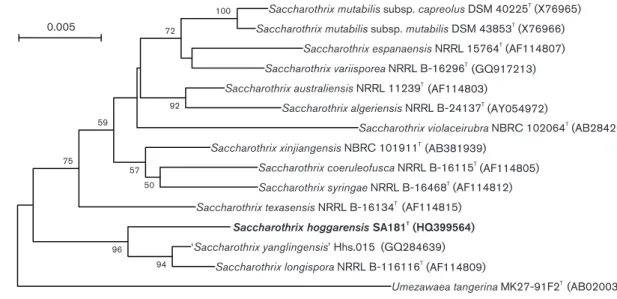
Thompson et al., 1994). Phylogenetic trees were con-structed by using neighbour-joining (Saitou & Nei, 1987) with the model of Jukes & Cantor (1969), maximum-likelihood (Felsenstein, 1981) with Kimura’s two-para-meter model (Kimura, 1980) and maximum-parsimony (Fitch, 1977). Bootstrap analysis (Felsenstein, 1985) was performed to evaluate the reliability of the tree topology. The phylogenetic relationships between strain SA181Tand members of the genus Saccharothrix are shown in the neighbour-joining dendrogram (Fig. 2). Maximum-par-simony and maximum-likelihood calculations resulted in similar tree topologies (Fig. S2). The similarity of the 16S rRNA gene sequence of strain SA181T to those of other members of the genus Saccharothrix ranged from 96.8 to 98.9 %. Strain SA181T displayed highest 16S rRNA gene sequence similarity with Saccharothrix longispora NRRL B-16113T (98.9 %), Saccharothrix xinjiangensis NBRC 101911T (98.4 %) and Saccharothrix texasensis NRRL B-61634T(98.2 %) and 16S rRNA gene sequence similarities with the other remaining members of the genus Saccharothrix were below 97.9 %.
For DNA–DNA hybridization, cells were disrupted by using a French pressure cell (Thermo Spectronic). DNA in the crude lysate was purified by chromatography on hydro-xyapatite as described by Cashion et al. (1977). DNA–DNA hybridization was carried out as described by De Ley et al. (1970) under consideration of the modifications described by Huss et al. (1983), using a model Cary 100 Bio UV/VIS spectrophotometer equipped with a Peltier-thermostatted 666 multicell changer and a temperature controller with in-situ temperature probe (Varian). DNA–DNA hybridization experiments were done in duplicate in 26 SSC in the presence of 10 % formamide at 71uC. DNA of strain SA181T was hybridized with that of its closest phylogenetic neighbours. Mean DNA–DNA relatedness values between strain SA181Tand S. longispora DSM 43749T, S. texasensis DSM 44231Tand S. xinjiangensis DSM 44896Twere 16.05 % (14.7 and 17.4 %), 50.05 % (46.5 and 53.6 %) and 22.0 % (24.2 and 19.8 %), respectively, which are clearly below the 70 % threshold proposed by Wayne et al. (1987) for the delineation of separate species.
A comparison of physiological characteristics of the isolate and S. longispora DSM 43749T showed differences in the utilization of adenine, hypoxanthine, sodium butyrate, sodium citrate, lactose, mannitol, melibiose, salicin and sorbitol, production of nitrate reductase, growth at 45uC and with 4 and 5 % (w/v) NaCl. Furthermore, S. texasensis DSM 44231T showed differences in the utilization of inositol, lactose, mannitol, methyl a-D-glucoside, raffinose
and sorbitol, production of nitrate reductase and growth at 45uC, and S. xinjiangensis DSM 44896Tshowed differences in the utilization of sodium butyrate, inositol, lactose, mannitol, methyl a-D-glucoside, raffinose and sorbitol, production of nitrate reductase and growth at 45uC and with 4 and 5 % (w/v) NaCl.
Recently, ‘Saccharothrix yanglingensis’ was described (Yan et al., 2012). Strain SA181T differs from that species in several morphological and physiological characteristics, including colour of substrate and aerial mycelia, produc-tion of diffusible pigment, degradaproduc-tion of gelatin, hypox-anthine and tyrosine, reduction of nitrate, use of arabinose, lactose, melibiose, rhamnose, xylose and sorbitol and growth at pH 5, at 45uC and in the presence of 4 % (w/ v) NaCl. Unlike strain SA181T, ‘S. yanglingensis’ contains no rhamnose in its whole cell.
All data support the conclusion that strain SA181Trepresents a novel species of the genus Saccharothrix, for which we propose the name Saccharothrix hoggarensis sp. nov. Description ofSaccharothrix hoggarensis sp. nov.
Saccharothrix hoggarensis (hog.gar.en9sis. N.L. fem. adj. hoggarensis pertaining to Hoggar, the source of the soil from which the type strain was isolated).
Aerobic, Gram-positive, filamentous actinomycete. Aerial mycelium is light greyish blue on ISP 2, yellowish white on
Table 2. Differential phenotypic characteristics of strain
SA181T and its most closely related neighbours in the genus
Saccharothrix
Strains: 1, Saccharothrix hoggarensis sp. nov. SA181T; 2, S. longispora DSM 43749T; 3, S. texasensis DSM 44231T; 4, S. xinjiangensis DSM 44896T. All data were taken from this study.
Characteristic 1 2 3 4 Decomposition of: Adenine 2 + 2 2 Hypoxanthine + 2 + + Production of: Nitrate reductase 2 + + + Assimilation of: Butyrate + 2 + 2 Citrate 2 + 2 2 Acid from: Inositol 2 2 + + Lactose 2 + + + Mannitol 2 + + + Melibiose + 2 + + Methyl a-D-glucoside 2 2 + + Raffinose 2 2 + + Salicin 2 + 2 2 Sorbitol + 2 2 2 Growth at/with: 45 uC + 2 2 2 4 % (w/v) NaCl + 2 + 2 5 % (w/v) NaCl + 2 + 2
nutrient agar and pinkish brown on Bennett’s agar and is well fragmented into rod-shaped spores (0.8–1.061.0– 1.5 mm). Substrate mycelium is pale to light yellow on nutrient agar, moderate to deep yellowish brown on ISP 2 and ISP 4, light brown on Bennett’s agar and dark brown on ISP 3. The substrate mycelium fragments into non-motile rods. Diffusible and melanoid pigments are not produced. Growth occurs at 20–45 uC (optimum 30uC), but not 10 uC, at pH 6–9 (optimum pH 7), but not at pH 5 or 11, and with 4–5 % (w/v) NaCl, but not with 7 %. Aesculin, casein, gelatin, hypoxanthine, Tween 80, starch, tyrosine and urea are degraded, but adenine, arbutin, testosterone and xanthine are not. UtilizesL-arabinose, cellobiose,D-fructose, D-galactose, D-glucose, glycerol, maltose, melibiose, D -ribose, L-rhamnose, D-sorbitol, sucrose, D-xylose, acetate,
butyrate, lactate, propionate, pyruvate and succinate as carbon sources, but adonitol, lactose, D-mannitol,
melezi-tose, myo-inositol,D-mannose, methyl a-D-glucoside, raffi-nose, salicin, trehalose, benzoate, citrate, oxalate and tartrate are not utilized.L-Proline is used as a source of nitrogen, but L-alanine andL-serine are not utilized. Nitrate reductase is not produced. Resistant to (mg ml21) chloramphenicol (30),
erythromycin (15) and novobiocin (5). The cell wall is type IIIE (meso-diaminopimelic acid, galactose, mannose, rham-nose and ribose in whole-cell hydrolysates). Exhibits phospholipid type PII (phosphatidylethanolamine, with phosphatidylmethylethanolamine, phosphatidylinositol, phosphatidylinositol mannosides and diphosphatidylgly-cerol). The predominant menaquinones are MK-9(H4),
MK-7(H4), MK-10(H4), MK-10(H0) and MK-11(H4), with
traces of MK-9(H0), MK-9(H2), MK-9(H6) and
MK-10(H2). The major fatty acids are iso-C16 : 0, iso-C15 : 0,
9-methyl-C16 : 0and anteiso-C17 : 0.
The type strain is SA181T (5DSM 45457T 5CCUG 60214T), isolated from a Saharan soil sample collected from Hoggar region (south Algeria).
Acknowledgements
We would like to gratefully acknowledge the help of Gabriele Po¨tter (DSMZ) for growing S. hoggarensis cultures and for assistance with chemotaxonomic analyses and Bettina Stra¨ubler for assistance with DNA–DNA hybridizations.
References
Becker, B., Lechevalier, M. P., Gordon, R. E. & Lechevalier, H. A. (1964).
Rapid differentiation between Nocardia and Streptomyces by paper chromatography of whole-cell hydrolysates. Appl Microbiol 12, 421–423.
Cashion, P., Holder-Franklin, M. A., McCully, J. & Franklin, M. (1977).
A rapid method for the base ratio determination of bacterial DNA.
Anal Biochem 81, 461–466.
De Ley, J., Cattoir, H. & Reynaerts, A. (1970). The quantitative measurement of DNA hybridization from renaturation rates. Eur J
Biochem 12, 133–142.
Felsenstein, J. (1981). Evolutionary trees from DNA sequences: a maximum likelihood approach. J Mol Evol 17, 368–376.
Felsenstein, J. (1985).Confidence limits on phylogenies: an approach using the bootstrap. Evolution 39, 783–791.
Fitch, W. M. (1977). On the problem of discovering the most parsimonious tree. Am Nat 111, 223–257.
Gordon, R. E., Barnett, D. A., Handerhan, J. E. & Pang, C. H. N. (1974). Nocardia coeliaca, Nocardia autotrophica, and the nocardin
strain. Int J Syst Bacteriol 24, 54–63.
Hayakawa, M. & Nonomura, H. (1987).Humic acid–vitamin agar, a new medium for the selective isolation of soil actinomycetes.
J Ferment Technol 65, 501–509.
Saccharothrix mutabilis subsp. capreolus DSM 40225T
(X76965)
Saccharothrix mutabilis subsp. mutabilis DSM 43853T
(X76966) Saccharothrix espanaensis NRRL 15764T (AF114807) Saccharothrix variisporea NRRL B-16296T (GQ917213) Saccharothrix australiensis NRRL 11239T (AF114803) Saccharothrix algeriensis NRRL B-24137T (AY054972) Saccharothrix violaceirubra NBRC 102064T (AB284261) Saccharothrix xinjiangensis NBRC 101911T (AB381939) Saccharothrix coeruleofusca NRRL B-16115T (AF114805) Saccharothrix syringae NRRL B-16468T (AF114812) Saccharothrix texasensis NRRL B-16134T (AF114815)
Saccharothrix hoggarensis SA181T
(HQ399564) ‘Saccharothrix yanglingensis’ Hhs.015 (GQ284639) Saccharothrix longispora NRRL B-116116T (AF114809) Umezawaea tangerina MK27-91F2T (AB020031) 100 72 92 50 57 59 75 94 96 0.005
Fig. 2.Neighbour-joining phylogenetic tree based on almost-complete 16S rRNA gene sequences showing the position of strain SA181Tin the genus Saccharothrix. This illustrates the taxonomic position of strain SA181Trelative to the other species
of the genus, including ‘Saccharothrix yanglingensis’ (Yan et al., 2012). Bootstrap values (.50 %) based on 1000 resamplings are shown at branch nodes. Bar, 0.005 substitutions per site.
Huss, V. A. R., Festl, H. & Schleifer, K. H. (1983). Studies on the spectrophotometric determination of DNA hybridization from renaturation rates. Syst Appl Microbiol 4, 184–192.
Jukes, T. H. & Cantor, C. R. (1969).Evolution of protein molecules. In
Mammalian Protein Metabolism, pp. 21–132. Edited by H. N. Munro.
New York: Academic Press.
Kelly, K. L. & Judd, D. B. (1976). Color: Universal Language and Dictionary of Names (National Bureau of Standards Special Publication 440). Washington, DC: US Department of Commerce.
Kim, O. S., Cho, Y. J., Lee, K., Yoon, S. H., Kim, M., Na, H., Park, S. C., Jeon, Y. S., Lee, J. H. & other authors (2012).Introducing EzTaxon-e: a prokaryotic 16S rRNA gene sequence database with phylotypes that represent uncultured species. Int J Syst Evol Microbiol 62, 716–721.
Kimura, M. (1980).A simple method for estimating evolutionary rates of base substitutions through comparative studies of nucleotide sequences. J Mol Evol 16, 111–120.
Kroppenstedt, R. M. (1982).Separation of bacterial menaquinones by HPLC using reverse phase (RP18) and a silver loaded ion exchanger as stationary phases. J Liq Chromatogr 5, 2359–2367.
Kroppenstedt, R. M. (1985).Fatty acid and menaquinone analysis of actinomycetes and related organisms. In Chemical Methods in
Bacterial Systematics, pp. 173–179. Edited by M. Goodfellow &
D. E. Minnikin. London: Academic Press.
Labeda, D. P. (2001).Crossiella gen. nov., a new genus related to Streptoalloteichus. Int J Syst Evol Microbiol 51, 1575–1579.
Labeda, D. P. & Kroppenstedt, R. M. (2000).Phylogenetic analysis of
Saccharothrix and related taxa: proposal for Actinosynnemataceae fam.
nov. Int J Syst Evol Microbiol 50, 331–336.
Labeda, D. P. & Kroppenstedt, R. M. (2006).Goodfellowia gen. nov., a
new genus of the Pseudonocardineae related to Actinoalloteichus, containing Goodfellowia coeruleoviolacea gen. nov., comb. nov. Int J
Syst Evol Microbiol 56, 1203–1207.
Labeda, D. P. & Kroppenstedt, R. M. (2007).Proposal of Umezawaea gen. nov., a new genus of the Actinosynnemataceae related to
Saccharothrix, and transfer of Saccharothrix tangerinus Kinoshita et al. 2000 as Umezawaea tangerina gen. nov., comb. nov. Int J Syst Evol Microbiol 57, 2758–2761.
Labeda, D. P. & Lechevalier, M. P. (1989).Amendment of the genus
Saccharothrix Labeda et al. 1984 and descriptions of Saccharothrix
espanaensis sp. nov., Saccharothrix cryophilis sp. nov., and
Saccharothrix mutabilis comb. nov. Int J Syst Bacteriol 39, 420–423.
Labeda, D. P., Testa, R. T., Lechevalier, M. P. & Lechevalier, H. A. (1984).Saccharothrix: a new genus of the Actinomycetales related to Nocardiopsis. Int J Syst Bacteriol 34, 426–431.
Labeda, D. P., Hatano, K., Kroppenstedt, R. M. & Tamura, T. (2001).
Revival of the genus Lentzea and proposal for Lechevalieria gen. nov.
Int J Syst Evol Microbiol 51, 1045–1050.
Lechevalier, M. P. & Lechevalier, H. A. (1970).Chemical composition as a criterion in the classification of aerobic actinomycetes. Int J Syst
Bacteriol 20, 435–443.
Lechevalier, M. P., de Bie`vre, C. & Lechevalier, H. A. (1977).
Chemotaxonomy of aerobic actinomycetes: phospholipid composi-tion. Biochem Syst Ecol 5, 249–260.
Minnikin, D. E., Patel, P. V., Alshamaony, L. & Goodfellow, M. (1977).
Polar lipid composition in the classification of Nocardia and related bacteria. Int J Syst Bacteriol 27, 104–117.
Minnikin, D. E., Hutchinson, I. G., Caldicott, A. B. & Goodfellow, M. (1980). Thin-layer chromatography of methanolysates of mycolic acid-containing bacteria. J Chromatogr A 188, 221–233.
Minnikin, D. E., O’Donnell, A. G., Goodfellow, M., Alderson, G., Athalye, M., Schaal, A. & Parlett, J. H. (1984). An integrated procedure for the extraction of bacterial isoprenoid quinones and polar lipids. J Microbiol Methods 2, 233–241.
Rainey, F. A., Ward-Rainey, N., Kroppenstedt, R. M. & Stackebrandt, E. (1996). The genus Nocardiopsis represents a phylogenetically coherent taxon and a distinct actinomycete lineage: proposal of
Nocardiopsaceae fam. nov. Int J Syst Bacteriol 46, 1088–1092.
Saitou, N. & Nei, M. (1987). The neighbor-joining method: a new method for reconstructing phylogenetic trees. Mol Biol Evol 4, 406– 425.
Sasser, M. (1990).Identification of bacteria by gas chromatography of cellular fatty acids, MIDI Technical Note 101. Newark, DE: MIDI Inc.
Shirling, E. B. & Gottlieb, D. (1966).Methods for characterization of
Streptomyces species. Int J Syst Bacteriol 16, 313–340.
Stackebrandt, E., Kroppenstedt, R. M., Jahnke, K. D., Kemmerling, C. & Qurther, H. (1994). Transfer of Streptosporangium viridogriseum (Okuda et al., 1966), Streptosporangium viridogriseum subsp. kofuensis (Nonomura, Ohara, 1969), Streptosporangium albidum (Furumal
et al., 1968) to Kutzneria gen. nov. as Kutzneria viridogrisea comb.
nov., Kutzneria kofuensis comb. nov., Kutzneria albida comb. nov., respectively, and Emendation of the genus Streptosporangium. Int J
Syst Bacteriol 44, 265–269.
Tamura, K., Peterson, D., Peterson, N., Stecher, G., Nei, M. & Kumar, S. (2011). MEGA5: molecular evolutionary genetics analysis using maximum likelihood, evolutionary distance, and maximum par-simony methods. Mol Biol Evol 28, 2731–2739.
Thompson, J. D., Higgins, D. G. & Gibson, T. J. (1994).CLUSTAL W:
improving the sensitivity of progressive multiple sequence alignment through sequence weighting, position-specific gap penalties and weight matrix choice. Nucleic Acids Res 22, 4673–4680.
Waksman, S. A. (1961). The Actinomycetes, Classification, Identifi-cation, Descriptions of Genera and Species, vol. 2. Baltimore: Williams
& Wilkins.
Wayne, L. G., Brenner, D. J., Colwell, R. R., Grimont, P. A. D., Kandler, O., Krichevsky, M. I., Moore, L. H., Moore, W. E. C., Murray, R. G. E. & other authors (1987). International Committee on Systematic Bacteriology. Report of the ad hoc committee on reconciliation of approaches to bacterial systematics. Int J Syst Bacteriol 37, 463– 464.
Williams, S. T., Goodfellow, M. & Alderson, G. (1989). Genus
Streptomyces Waksman and Henrici 1943, 339AL. In Bergey’s Manual
of Systematic Bacteriology, vol. 4, pp. 2452–2492. Edited by
S. T. Williams, M. E. Sharpe & J. G. Holt. Baltimore: Williams & Wilkins.
Yan, X., Huang, L. L., Tu, X., Gao, X. N. & Kang, Z. S. (2012).
Saccharothrix yanglingensis sp. nov., an antagonistic endophytic
actinomycete isolated from cucumber plant. Antonie van Leeuwenhoek 101, 141–146.
Yassin, A. F., Rainey, F. A., Brzezinka, H., Jahnke, K. D., Weissbrodt, H., Budzikiewicz, H., Stackebrandt, E. & Schaal, K. P. (1995).Lentzea
gen. nov., a new genus of the order Actinomycetales. Int J Syst Bacteriol 45, 357–363.