DEPARTMENT OF BIOLOGY AND ANIMAL PHYSIOLOGY
ThEsIS
Presented by
MERGHEM Mounira
For the fulfillment of the requirements for the degree of
DOCTORATE OF SCIENCES
BIOLOGY
Special filed:
ANIMAL PHYSIOLOGY
Topic
Evaluation of toxicity in mice and rats and antioxydant
activities of Ruta montana L. extracts
Presented publically in: …/…./2015
Jury:
President: BAGHIANI A. Pr. UFA Setif 1 Supervisor: DAHAMNA SERAICHE S. Pr. UFA Setif 1 Examiners: LALAOUI K. Pr. Univ Cons 1 NECIB Y. Pr. Univ Cons 1 DJENIDI R. Dr. Univ B.B.A
Laboratory of Phytotherapy Applied to Chronic Diseases
N°……….……/SNV/2015
ةيبعشلا ةيطارقميدلا ةيرئازجلا ةيروهمجلا
ملعلا ثحبلا و يلاعلا ميلعتلا ةرازو
ي
فيطس ،سابع تاحرف ةعماج
1ةيلك
مولع
ةايحلا و ةعيبطلا
Université Ferhat Abbas Sétif 1 Faculté des Sciences de la
ACKNOWLEDGMENTS
First and foremost, praises and thanks to the God, the Almighty, for providing me the blessings throughout my research work to complete the research successfully.
I wish to express my sincere gratitude to my supervisor Pr. Dahamna Saliha, for his valuable advices and guidance, timely suggestions and whole-hearted supports. His continuous interest and great generosity helped my work to be accomplished and also supported me during a course of this study.
I would like to thank my committee members, Professor Baghiani Abderrahmane University Ferhat Abbas Setif 1, Professor Laalaoui Nacer University of Constantine,
Professor Necib Youcef University of Constantine and Dr Djenidi Redha University of Borj Boariridj for critical reading of the thesis, and for their valuable comments, discussions and suggestions.
I would like to thank Pr. Bourriche Hammama, Pr. Harzallah Daoud, Pr. Khennouf Seddik, Pr. Baghiani Abderrahmane, Pr. Arrar Lakhmisi University Ferhat Abbas Sétif 1, Pr. Touabti and Dr. Krache Kaouala Samira, University Hospital of Sétif for their great help.
I would like to thank my family, especially my mother and father for always believing in me, for their continuous love and their supports in my decisions. Without whom I could not have made it here.
I also would like to thank my friend Abir Razagui for his great help, advices and consultation
Finally, my thanks go to all the people who have supported me to complete the research work directly or indirectly.
Abstract
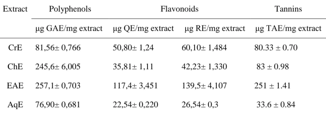
The aim of this study is to evaluate the acute and chronic toxicity of Ruta montana aerial part aqueous extract (Fidjel) used in folk medicine in Algeria in Albino Wistar male and female mice and rats as well as the antioxidant activity of the aqueous extract (AqE), methanolic extract (CrE), chloroformic extract (ChE) and ethyl acetate extract (EAE). The results showed that the ethyl acetate extract contains the highest amount of total polyphenols, tannins and flavonoids with values of 257.1±0.7 EAG/mg, 251 ± 1.41 EAT/mg, 117.4± 3.457EQ/mg, 136.2±9.876ER/ mg of extract, respectively. The study of the acute toxicity of aqueous extract at doses of 2, 4, 6, 8, 10 and 12 g/kg body weight by oral route in both sexes of mice, recorded no mortality, and showed no change in the general behavior of the treated animals. The study of chronic toxicity at the doses 100, 300 and 600mg/kg body weight in male and female rats for 90 days, recorded no mortality, and showed no change in the hematological and biochemical parameters. Treated animals showed a normal weight change compared with contral. The parametres of male fertility showed a significant decrease in the weight of testis, epididymis and seminal vesicle as well as a reduction in the number and the motility of spermatozoids at treated groups by the doses 300 and 600 mg/kg body weight compared to control group. DPPH scavenging assay showed that EAE has a higher anti-radical capacity (IC50 = 0.044 ± 0.001 mg/ml) followed by CrE, AqE and ChE with IC50 of
0.067 ± 0.002 ; 0.083 ± 0.003 et 0.146 ± 0.001mg/ml, respectively. Whereas, AqE showed the best inhibitory capacity of the coupled oxidation of linoleic acid/ β-carotene (90.34 ± 0.46%) and the best chelating capacity (IC50 = 0.051 ± 0.004mg/ml). The antioxidant activity of
aqueous extract (AqE) and methanolic extract (CrE) in vivo was estimated using the antioxidant plasma capacity (APC), DPPH and reducing power tests. All extracts did not show any significant change. The activity of catalase (CAT), the level of MDA and GSH were evaluated in rat liver and kidney homogenate. The results showed a not significant increase in CAT activity in the groups treated with AqE 300mg/kg and CrE 100mg/kg by 30.76%, 32.07% for liver tissue and 24.48%, 32.11% for kidney tissue, and also in the GSH levels by 18.57%, 22.97% for liver tissue and 12.3%, 37.36% for kidney tissue. However no significant decrease in the level of MDA in the treated groups with AqE 300mg/kg and CrE 100mg/kg.
Résumé
Lobjectif de cette étude est d’évaluer la toxicité aigu et chronique de l’extrait aqueux de la partie aérienne de Ruta montana (Fidjel) utilisée en médecine traditionnelle en Algérie sur des souris et rats Albino Wistar de sexe mâle et femelle. ainsi que l’activité antioxydante de l’extrait aqueux (AqE), l’extrait methanolique (CrE), l’extrait chloroformique (ChE) et l’extrait d’acétate éthyle (EAE). Les résultats ont montré que l’extrait d’acetate d’éthyle représente la quantité la plus elevée en polyphenols, tanins et flavonoides avec des valeurs de 257.1 ± 0.7 μg EAG/mg, 251 ± 1.41 μg EAT/mg, 117.4 ± 3.451 μg EQ/mg, 139.5 ± 4.107 μg ER/ mg d’extrait respectivement. L’étude de la toxicité aiguë de l’extrait aqueux avec les doses 2,4,6,8,10 et 12 g/kg du poids corporel par voie orale chez les souris mâles et femelles, n’a enregistré aucune mortalité, et n’a montré aucun changement dans le comportement générale des animaux traités, l’étude de la toxicité chronique avec les doses 100, 300 et 600 mg/kg du poids corporel chez les rats pendant 90 jours, n’a enregistré aucune mortalité et aucun changement dans les paramètres hématologiques et biochimiques. Les animaux traités ont connu une évolution pondérale normale en comparaison avec les témoins. Concernant les paramètres de la fertilité masculine, une diminution significative des poids des testicules, épididyme, vésicules séminales ainsi que le nombre et la mobilité des spermatozoïdes chez les rats traités par les doses 300 et 600 mg/kg du poids corporel. Le test du scavenger du DPPH a montré que l’extrait EAE a un pouvoir anti-radicalaire plus élevé (IC50 = 0.044 ± 0.001 mg/ml) suivi par CrE, AqE et
ChE avec IC50 de 0.067 ± 0.002 , 0.083 ± 0.003 et 0.146 ± 0.015 mg/ml, respectivement.
Alors que AqE a montré une meilleur capacité inhibitrice de l’oxydation couplée de l’acide linoléique/β-carotene (90.34 ± 0.46%) et une meilleure capacité chelatrice des ions (IC50 =
0.005 ± 0.004 mg/ml). L’activité antioxydante in vivo de l’extrait aqueux (AqE) et l’extrait méthanolique (CrE) est estimée par la capacité antioxydante plasmatique (CAP), le test de DPPH et du pouvoir réducteur n’a montré aucun changement significatif. L’activité de la catalase (CAT) et les taux du MDA et du GSH ont été évalués dans l’homogénat du foie et du rein des rats. Les résultats à montré une augmentation non significative dans l’activité de la CAT dans les groupes traités par AqE 300mg/kg et CrE 100mg/kg par 30.76%, 32.07% pour le tissu hépatique et 24.48%, 32.11% pour le tissu rénal, et aussi dans les taux du GSH par 18.57%, 22.97% pour le tissu hépatique et 12.3%, 37.36% pour le tissu rénal. Cependant, une diminution non significative du taux du MDA dans les groupes traités par AqE 300mg/kg et CrE 100mg/kg.
Mots clés : Ruta montana L., toxicité aigüe, toxicité chronique, activité antioxidante,
صخلم فدهت ةساردلا هذه ىلإ يئاملا صلختسملل نمزملا و داحلا يمسلا ريثأتلا ةفرعم ةتبنل يئاوهلا ءزجلل Ruta montana رئازجلا يف يديلقتلا بطلا يف ةلمعتسملا )لجيفلا( .ثانا ىرخأ و روكذ ءاضيب ناذرج و نارأف ىلع ىلإ ةفاضلااب ةيطاشنلا ريدقت صلختسملل ةدسكلأل ةداضملا ئاملا ي ( (AqE ( يلوناثيملا صلختسملا, CrE مروفورولكلا صلختسم ,) ( ChE ليثيا صلختسم و ) ( تاتيسلأا EAE ) . ليثيا صلختسم نأ اهيلع لصحتملا جئاتنلا تنيب ىلع يوتحي تاتيسلأا تلاونيفلا نم ةيمك ربكأ ، تاديونوفلافلا و غابدلا : 257.1 ± 0.7 مارغوركيم ئفاكم كيلاغلا ضمح / ,صلختسم غ 251 ± 1.41 مارغوركيم ئفاكم ضمح ناتلا كي / غ صلختسم , ±117.4 3.451 مارغوركيم ئفاكم نيتسركلا / غ صلختسم , 5 ±139. 107 4. مارغوركيم ئفاكم نيترلا / غ صلختسم ، .بيترتلا ىلع يمسلا ريثأتلا ةسارد يدبت مل تاعرجب ثانا و روكذ نارأف ىلع يئاملا صلختسملل داحلا 2 , 4 , 6 , 10 و 12 مفلا قيرط نع مسجلا نزو نم غك/غ ةافو ةلاح يأ لجسن مل امك ,ماعلا كولسلا يف ةظوحلم تاريغت يأ ناذرجلا ىلع ةنمزملا ةيمسلا ةسارد لجست مل . تاعرجب 100 , 300 و 600 ةدمل مسجلا نزو نم غك/غم 90 تارشؤملا يف رييغت يأ لجست مل امك ةافو ةلاح يأ موي لا و ةيومدلا يب و ةجلاعملا ناذرجلا تفرع .ةيئايميك نزولا يف ةيداع ةدايز صخي اميف امأ .ةدهاشلا تاناويحلاب ةنراقم و ةيونملا ةلصيوحلا و خبربلا ,ةيصخلا نم لك نزو يف يونعم ضافخنا لجس دقف روكذلا دنع ةبوصخلا تارشؤم ب ةجلاعملا ناذرجلا دنع ةيونملا تاناويحلا ةكرح و ددع يف ضافخنا كلذك نيتعرجلا 300 و 600 نزو نم غك/غم رذج رابتخا نيب .مسجلا DPPH نا EAE ( ةحازا ةردق ىلعا كلتمي 50 IC = 0.044 ± 0.001 نم لك هيلي )لم/غم ChE , AqE و CrE ب : 0.067 ± 0.002 , 0.083 ± 0.003 و 0.146 ± 0.015 نيح يف .بيترتلا ىلع لم/غم يب ن AqE ةميقب ديدحلا تانويا بلاختسا ىلع ةيلاع ةردق 50 IC = 0.005 ± 0.004 ىلع ةيلاع ةردق كلذك لم/غم يحلا نئاكلا يف ةدسكلال ةداضملا ةيطاشنلا ريدقت مت .كييلونيللا ضمح/نيتوراك اتيبل ةجودزملا ةدسكلاا طيبثت لل ةدسكلال ةداضملا ةيطاشنلا ةطساوب يلوناثيملا و يئاملا صلختسملل رابتخا رهظي مل ثيح .امزلاب DPPH ةردقلا و ةيطاشن ريدقت مت امك .يونعم رييغت يا ةيعاجرلاا CAT ةبسن و MDA و GSH ترهظا .ةيلكلا و دبكلا جيسن يف ةيطاشن يف يونعم ريغ عافترا جئاتنلا CAT ةجلاعملا تاعومجملا يف يئاملا صلختسملاب 300 صلختسملا و غك/غم يلوناثيملا 100 ك/غم ب غ 30.76 % , 32.07 % و دبكلا جيسنل ةبسنلاب 24.48 % , 32.11 % و ةيلكلا جيسنل ةبسنلاب يف يونعم ريغ عافترا كلذك GSH ب 18.57 % , 22.97 % ةبسنلاب جيسنل دبكلا و 12.3 % , 37.36 % ةبسنلاب جيسنل ةيلكلا ضافخنا و ريغ يونعم ةبسنل MDA يف تاعومجملا ةجلاعملا صلختسملاب يئاملا 300 غم / غك و صلختسملا يلوناثيملا 100 غم / غك . حاتفملا تاملكلا : Ruta montana , لونفلا تاديدع ,ةدسكلال ةداضملا ةيطاشنلا ,ةنمزملا ةيمسلا ,ةداحلا ةيمسلا .
ABREVIATIONS
ALT alanine aminotransferaseALB albumin
ALP alkaline phosphatase
AqE aqueous extract
AST aspartate aminotransferase
BHT butylated hydroxytoluene
ChE chloroform extract
CHOL cholesterol
CK creatine kinase
CREA creatinine
CrE crud extract
DPPH 2,2'-diphenyl-1-picrylhydrazyl
EAE ethyl acetate extract
EDTA ethylene diamine tetra acetic acid
GAE gallic acid equivalents
GLU glucose
GPx glutathion peroxidase
GSH gluthathion reduced
GSSG glutathion disulfure oxidized
Hct hematocrit
Hb hemoglobin
HxE hexane extract
H2O2 hydrogen peroxide HOCl hypochlorous acid
IC50 % inhibitory concentration for 50% of activity
LOO° lipid peroxide radical
MCH mean corpuscular hemoglobin
MCHC mean corpuscular hemoglobin concentration
VGM mean corpuscular volume
MeOH methanol
NO° radical nitroxid
NOS nitric oxidesynthase
1O
2 singlet oxygen O2°- superoxid Radical
TG triglycerides
OH° hydroxyl Radical
ONOO- peroxynitrite
MPV platelets count and mean platelet volume
QE quercetin equivalents
RBC red blood cells
RNS reactive nitrogene species
ROS reactive oxygen species
RE rutin equivalent
SD standard deviation
SOD superoxyde dismutase
AU uric acid
LIST OF FIGURES
Figure 1: Structural organization of the hepatic lobule………13
Figure 2: Structural organization of the nephron……….16
Figure 3:Generation of reactive oxigene species……….23
Figure 4: Basic structure of flavonoids………30
Figure 5:Ruta montana L. plant………..34
Figure 6: The steps of fractionation of the crude extract of Ruta montana L……….40
Figure 7:Malondialdehyde assay Principle………..49
Figure 8:Ellman reaction………..50
Figure 9: Standard curve of reduced glutathione (GSH)……….51
Figure 10: Standard curve of gallic acid for determination of total polyphenols in Ruta montana L………56
Figure 11: Standards curve of Rutin and Quercetine for determination of total flavonoids in Ruta montana L. extracts. ………57
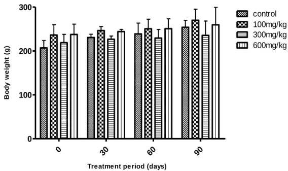
Figure 12: Changes in body weight of male rats after chronic oral treatment with aqueous extract of Ruta montana L………59
Figure 13: Changes in body weight of female rats after chronic oral treatment with aqueous extract of Ruta montana L………59
Figure 14: Effect of chronic administration of aqueous extract of Ruta montana L. on plasma red blood cells………..63
Figure 15: Effect of chronic administration of aqueous extract of Ruta montana L. on plasma white blood cells……….63
Figure 16: Effect of chronic administration of aqueous extract of Ruta montana L. on plasma hemoglobin……….64
Figure 17: Effect of chronic administration of aqueous extract of Ruta montana L. on
platelets……….64
Figure 18: Changes in relative weight of Liver after chronic oral treatment with aqueous extract of Ruta montana L………....69
Figure 19: Changes in relative weight of Kidneys after chronic oral treatment with aqueous extract of Ruta montana L………69
Figure 20: Changes in relative weight of Brain after chronic oral treatment with aqueous extract of Ruta montana L………70
Figure 21: Histological sections of liver………..73
Figure 22: Histological sections of kidneys………75
Figure 23: Histological sections of Brain……….77
Figure 24: Histological sections of Ovary………...79
Fifure 25:Histomorphology of testes………...81
Figure 26:Histomorphology of the epididymis………83
Figure 27: The percentage inhibition of free DPPH radical in the presence of different concentration of Ruta montana L. extracts………..86
Figure 28: The changes in the percentage of the inhibition ratios of linoleic acid oxidation under the influence of Ruta montana L. extracts (2mg/ml), compared to BHT as a positive control during 48h………87
Figure 29: Inhibition percentage of extracts of Ruta montana L. in β-carotene/linoleic acid assay after 48 h………..88
Figure 30: Metal chelating activity of different Ruta montana L. extracts………..89
Figure 31: Plasma antioxidant capacity toward DPPH radical for different groups…………90
LIST OF TABLES
Table 1: Classification of toxicity based on LD50 dose ranges………10
Table 2: Classification and main effects of free radicals……….20
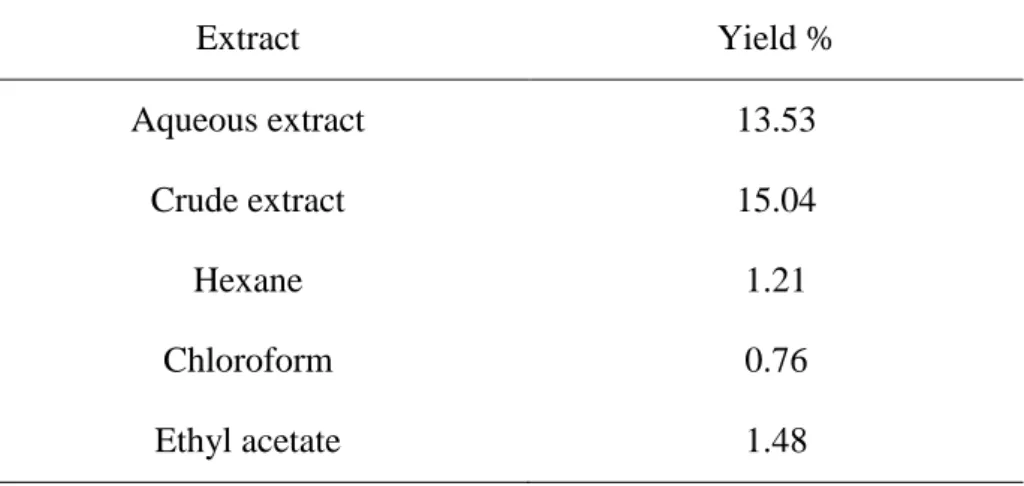
Table 3:Yield of different extracts from Ruta montana L………...54
Table 4: Total polyphenols, flavonoids and tannins content of Ruta montana L. extracts….55
Table 5: Effect of aqueous extract of Ruta montana L. (100, 300 and 600mg/kg) on
hematological parameters of male and female Wistar rats treated for 30 days………61
Table 6: Effect of aqueous extract of Ruta montana L. (100, 300 and 600mg/kg) on
hematological parameters in male and female Wistar rats treated for 90 days……….62
Table 7: Effect of aqueous extract of Ruta montana L. (100, 300 and 600mg/kg) on
bio-chemical parameters in male and female Wistar rats treated for 30 days……….66
Table 8: Effect of aqueous extract of Ruta montana L. (100, 300 and 600mg/kg) on
bio-chemical parameters in male and female Wistar rats treated for 90 days……….67
Table 9: Effect of chronic oral administration of aqueous extract of Ruta montana L. on rats
organs weight………68
Table 10: Effect of Ruta montana L. extract on Sperm count and Motility………71
Table 11:DPPH scavenging activity of Ruta montana L. extracts and phenolic standards….85
Table 12:Metal chelating activity of Ruta montana L. extracts and EDTA………89
Table 13: Effect of Ruta montana L. extracts on lipid peroxidation in liver and kidney……92
Table 14: Effect of Rutamontana L. extracts on reduced Glutathione (GSH)……….93
Summary
Acknowledgment………..……….I Summary in English………...………….……....II Summary in French……..……….………..…………...III Summary in Arabic………...…………..IV Abbreviations ………...V List of figures………..VI List of tables……….………..VII INTRODUCTION ………11. Toxicity of medicinal plants ………..4
1.1. Goals of toxicity testing of herbal drugs………..8
1.2. Difinition of Toxicity………...8 1.3. Acute toxicity………...9 1.4. Sub-acute toxicity………..…10 1.5. Chronic toxicity………..10 1.6. Routes of administration………11 1.6.1. Intra-peritoneal injection……….11 1.6.2. Oral administration……….11
1.7. Target tissue Toxicity ………12
1.7.1. Hepatotoxicity……….12
1.7.2. Nephrotoxicity………14
1.7.3. Neurotoxicity………..16
2. Oxidative stress………18
2.1. Free radicals………..18
2.1.1. Production of ROS……….20
2.1.2. Molecular damage induced by free radicals………23
2.1.2.1. Lipid peroxidation………24 2.1.2.2. Protein oxidation………..24 2.1.2.3. DNA oxidation……….25 2.2. Antioxidants………...25 2.2.1. Enzymatic antioxidants………...26 2.2.2. Non-enzymatic antioxidants………...27 3. Plant description………31 3.1. Rutaceae……….31 3.2. Ruta montana L………..32
3.2.1. Nomenclature (Communally names)………..32
3.2.2. Taxonomy………...32
3.2.3. Botanical description………...33
3.2.4. Traditional uses and side effects……….34
3.2.5. Chemical composition……….35
3.2.6. Pharmacological properties……….36
MATERIALS AND METHODS………..37
1. Materials………37 1.1. Biological materials………37 1.1.1. Plant………37 1.1.2. Animals………..37 1.2. Chemicals ………...………..37 2. Methods………38
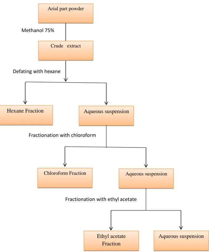
2.1. Preparation of plant extract………38
2.1.1. Aqueous extract………38
2.1.2. Methanolic extract………..38
2.1.3. Fractionation of the crude extract……….38
2.2. Determination of total polyphenols, flavonoids and tannins content in plant extracts...41
2.2.1. Determination of total polyphenols content in plant extracts ………..41
2.2.2. Determination of total flavonoids content in plants extracts ………...41
2.2.3. Determination of tannins content in plant extracts………...42
2.3. Evaluation of toxic effect of Ruta montana L. aqueous extract ………42
2.3.1. Acute toxicity of Ruta montana L. aqueous extract ……….42
2.3.2. Chronic toxicity of Ruta montana L. aqueous extract ……….43
2.3.2. 1. Effect of Ruta montana L. aqueous extract on heamatological and biochemical parameters ………43
2.3.2. 2. Effect of Ruta montana L. aqueous extract on organs weight………...44
2.3.2.3.histopathological examination……….44
2.3.2. 4. Effect of Ruta montana L. aqueous extract Fertility ……….44
2.4. Evaluation of antioxidant activity of plant extracts ……….45
2.4.1. In vitro antioxidant activity……….45
2.4.1.1. DPPH free radical-scavenging activity of plant extracts ……….45
2.4.1.2. Determination of antioxidant activity of plant extracts using β-carotene/linoleate model system……….46
2.4.1.3. Chelation of ferrous iron by plant extracts ……….46
2.4.2. In vivo antioxidant activity of plant extracts ………..47
2.4.2.1. Effect of extracts on plasma antioxidant capacity using DPPH radical ………..48
2.4.2.3. Estimation of lipid peroxidation……….49
2.4.2.4. Determination of reduced glutathione (GSH)……….50
2.4.2.5. Determination of catalase activity………...51
5.1.2.6. Estimation of total proteins……….52
Statistical analysis………52
RESULTATS AND DISCUSSION 1. Extraction yield………54
2. Total phenolics, flavonoids and tannins content in Ruta montana L. extracts……….55
3. Toxicity………57
3.1. Acute toxicity………57
3.2. Chronic toxicity……….57
3.2.1. Effect of Ruta montana L. aqueous extract on rats body weight……….58
3.2.2. Effect of Ruta montana L. aqueous extract on hematological parameters………..60
3.2.3. Effect of Ruta montana L. aqueous extract on biochemical parameters…………..65
3.2.4. Effect of Ruta montana L. aqueous extract on rat’s organs weight……….65
3.2.5. Effect of Ruta montana L. aqueous extract on Sperm count and Motility………..71
3.2.6. Effect of Ruta montana L. aqueous extract on histology of organs……….71
4. Antioxidant activity of Ruta montana L. extracts……….85
4.1. In Vitro Antioxidant Activity……….85
4.1.1. DPPH-scavenging assay……….85
4.1.2. β-carotene/linoleic acid bleaching assay………87
4.1.3. Chelation of ferrous iron by plant extracts ……….88
4.2. In vivo antioxidant activity……….90
4.2.1. Effect of extracts on plasma antioxidant capacity using DPPH radical ……….90
4.2.3. Effect of Ruta montana L. extracts on lipid peroxidation in liver and kidney ………92
4.2.4. Effect of Ruta montana L. extracts on reduced Glutathione (GSH)……….93
4.2.5. Effect of Ruta montana L. extracts on catalase activity……….94
DISCUSSION………..95
CONCLUSION………...114
1
Introduction
Medicinal plants have important contributions in the healthcare system. Use of herbal medicines represents a long history of human interactions with the environment. Plants used for traditional medicine contain a wide range of substances that can be used to treat chronic as well as infectious diseases. A number of modern drugs currently in use have been obtained through medicinal plants.
Despite the profound therapeutic advantages possessed by some of the medicinal plants, some constituents of medicinal plants have been found to be potentially toxic, mutagenic, carcinogenic and teratogenic. This raises concern about the potential toxic effects resulting from the short-term and long-term use of such medicinal plants. Therefore, evaluating the toxicity effects of any medicinal plants extracts intended to be used in humans and animals is of greatest significance.
In living systems, oxidation is a basic part of the normal metabolic process, in which reactive oxygen species (hydrogen peroxide and hypochlorous acid) and many free radicals (hydroxyl radical (OH) and superoxide anion) are generated. Rapid production of free radicals may cause alteration in the structure and function of cell constituents and membranes and can results in human neurologic and other disorders such as cancer, diabetes, inflammatory disease, asthma, cardiovascular, neurodegenerative diseases, and premature aging. Therefore, the prevention of the above conditions requires the presence of antioxidants or the free radical scavenging molecules in the body (Pinton et al., 2012).
2
Medicinal plants and products made from plants may contain a wide variety of free radical scavenging molecules, such as phenolic compounds (e.g. phenolic acids, flavonoids, quinones, coumarins, lignans, stilbenes, tannins), nitrogen compounds (alkaloids, amines, betalains), vitamins, terpenoids (including carotenoids), and some other endogenous metabolites, which are rich in antioxidant activity (Zheng and Wang, 2001; Cai et al, 2003). Epidemiological studies have shown that many of these antioxidant compounds possess anti-inflammatory, antiatherosclerotic, antitumor, antimutagenic, anticarcinogenic, antibacterial, or antiviral activities to a greater or lesser extent (Owen et
al, 2000; Sala et al, 2002).
Phenolic compounds from medicinal plants possess strong antioxidant activity and may help to protect the cells against the oxidative damage caused by free-radicals (Kahkonen et al, 1999). They are well known as radical scavengers, metal chelators, reducing agents, hydrogen donors, and singlet oxygen quenchers (Proestos et al, 2006).
The medicinal plant Ruta montana L. (Family Rutaceae), is used in folk medicine as hypoglycemic, antirhematic, antihelmintic, antiepileptic, antipyretic. It is also used in treating intestinal and hepatic diseases. This plant contains various active principles able to inhibit the growth of mycobacteria. The antioxidative effect of plant extracts is mainly due to phenolic components, such as flavonoids, phenolic acids, and phenolic diterpenes. In Algerian folk medicine, Ruta montana L. is also used against child fevers and as an abortive drug.
The main objectives of this study were:
- Determination of the LD50 of the Ruta montana L. aqueous extract.
- Evaluation of the acute and chronic toxicity of aqueous extract of aerial parts of
3
- Evaluation of the effect of Ruta Montana L. aqueous extract on fertility.
- Evaluation of the effect of Ruta montana L. aqueous extract on organs histology.
-
Evaluation of the in vitro and in vivo antioxidant activity.4
1. Toxicity of medicinal plants
In recent years, there has been growing interest in alternative therapies and the therapeutic use of natural products, especially, those derived from plants. This interest in drugs of plant origin is due to several reasons, namely, conventional medicine can be inefficient, abusive and/or incorrect use of synthetic drugs results in side effects and other problems (Kiranmai and Kiran, 2014).
Traditionally, herbs have been considered to be nontoxic and have been used for treating various problems by the general public and traditional medicine doctors worldwide (Oduola et al, 2007). Although, the literature has documented several toxicity resulting from the use of herbs on many occasions, still the potential toxicity of herbs has not been recognized by the general public or by professional groups of traditional medicine (O’Hara
et al, 1998). Poisonous plants are those which cause serious problems or even death
occur, if a small quantity of its stem, leaves, seeds, fruits and roots are ingested. Some other plants are normally harmless but they may become toxic if preparative from them are taken in excess in strong doses or for a long period of time (Khajja et al, 2011). Toxicity in herbal medicine may be due to: Accidents due to mistake in botanical identification, accidental ingestion of cardiotonic plants, intoxication by popular remedies and plants that interfere with conventional pharmacological therapy (Rates, 2001).
Poisonous plants can cause superficial irritation or discomfort through contact with the skin or serious poisoning when ingested (Ndhlala et al, 2013). Toxic substances from plants can affect the entire spectrum of vital human organs while some may affect key functional body systems like the central nervous system (CNS) ,thereby interfering with the coordination of nerve functions of the body. The most dominant toxins are neurotoxins that affect the brain and CNS, followed by cytotoxins and metabolic toxins that affect
5
organs such as kidneys, liver, heart and lungs. The severity of a toxic effect may depend on the route of administration, growth stage or part of the plant, the amount consumed, the species and susceptibility of the victim (Botha and Penrith, 2008). Other factors that may influence the severity of toxins include the solubility of the toxin in body fluids, frequency of intoxication as well as the age of the victim.
The toxic effect of plants is based on their chemical constituents which are classified into alkaloids, glycosides, proteins, oxalates, anti-vitamins, tannins etc. They act by altering specific mechanisms involving enzymes, receptors and even genetic material at particular cells and tissues (Chandra Sekhar et al, 2012).
- Alkaloids
Alkaloids are among the most potent toxic. It is a heterogeneous group of complex organic compounds to alkaline reaction with important physiological activity. Alkaloids in general have profound toxic effects on the nervous systems of mammals due to interaction with neuronal tissue (Donald and Billie, 1978).
The pyrrolizidine alkaloids, for example, exert potent hepatotoxicity and mutagenic activity (Donald and Billie, 1978). Also, the alkaloid ephedrine contained in ephedra herb can cause serious toxic reactions ranging from liver damage to severe high blood pressure and heart problems (George, 2011).
In addition, intoxications with tropane alkaloids are characterized by dryness of the mucosa in the upper digestive and respiratory tract, constipation, pupil dilation and disturbance of vision, photophobia and changes in heart rate, hypotension, nervousness, restlessness, irritability, disorientation, ataxia and respiratory depression (Alexander et al, 2008).
6
- Glycosides
The diterpene glycoside, atractyloside, is present in a number of plants used as ethnomedicines throughout Africa (Obatomi and Bach 1998; Dahamna et al., 2004). These plants are known to cause acute fatal renal and liver damage in humans and domestic animals foraging on atractyloside-containing plants (Stuart et al, 1981; Martin et al, 1986). Pure atractyloside also produces a similar lesion in animals (Carpenedo et al, 1974; Hatch
et al, 1982). Histologically, there was usually marked centrilobular degeneration of the
liver (Caravaca-Ma- garinos et al, 1985; Georgiou et al, 1988; Hedili et al, 1989) and proximal tubular necrosis of the kidney (Koechel and Krejci, 1993). Atractyloside was also found to alter catabolic and anabolic functions in vivo (Georgiou et al, 1988).
A number of studies have examined the acute, chronic and carcinogenic effects of coumarin in the rat and mouse. In studies involving the rat, hepatic biochemical and morphological changes have been examined for various periods of coumarin administration (1 week to 2 years). Depending on dose administered, coumarin treatment results in an increase in relative weight and changes in various hepatic biochemical parameters. Single oral doses of coumarin have been shown to produce liver necrosis and increase plasma transaminase activities in DBA/2 strain mice (Lake, 1999).
In addition to these, saponins inhibits cellular respiration (Wittstock and Gershenzon, 2002). And are potent surfactants that can disrupt lipid-rich cellular membranes of human erythrocytes and microorganisms which explain the potent antimicrobial properties of this group of phytochemicals (Francis, 2002).
7
- Proteins and peptides
Proteins from plants are an important source in food. Amino acids are absorbed from the intestine of man and animals and are built up into adapted proteins. Nevertheless, there are also plant proteins and peptides with bioactivity. They are often not hydrolyzed in the digestive tract, but may to a certain extent be absorbed and exert their specific action in the body. Euphorbiaceae (spurge family) include plants producing such proteins, for instance ricin (lectin) in seeds of Ricinus communis (castor bean) which inhibits protein synthesis and induce systemic effects in animals and humans, with gastrointestinal symptoms dominating. Far less potent lectins are also present in seeds of several species of Fabaceae (bean family). Colic and other gastrointestinal symptoms may occur if seeds are eaten without sufficient heat treatment, which inactivates many lectins (Bernhoft, 2010).
- Oxalates
According to Blood and Henderson (1974), the ingestion of an excessive amount of oxalate could cause gastrointestinal irritation, blockage of the renal tubules by calcium oxalate crystals, development of urinary calculi, hypocalcaemia, muscular weakness or paralysis.
- Minerals and heavy metals
Another implication in the toxicity of certain herbs is the presence of toxic minerals and heavy metals like mercury, arsenic, lead and cadmium (Dwivedi and Dey, 2002). Lead and mercury can cause serious neurological impairment when an herbal medicinal product contaminated with these metals is ingested (Amster et al, 2007).
8
1.1.
Goals of toxicity testing of herbal drugs
The primary goal of toxicological assessment of any herbal medicine is to identify adverse effects and to determine limits of exposure level at which such effects occur. Two important factors which are taken into consideration in evaluating the safety of any herbal drug are the nature and significance of the adverse effect and in addition, the exposure level where the effect is observed.
An equally important objective of toxicity testing is the detection of toxic plant extracts or compounds derived thereof in the early (pre-clinical) and late (clinical) stages of drug discovery and development from plant sources. This will facilitate the identification of toxicants which can be discarded or modified during the process and create an opportunity for extensive evaluation of safer, promising alternatives (Gamaniel, 2000).
1.2. Definition of Toxicity
Toxicity was defined as an aspect of pharmacology which deals with the adverse effects of bioactive substances on living organisms. Toxicological studies and experiments are very essential to establish the safety and efficiency of any new drug and to make a decision whether this drug should be adopted for clinical use or not (Anisuzzaman et al, 2001; Alam et al, 2006).
Depending on the duration of exposure of animals to drug, toxicological studies may be of three types: acute, sub-acute and chronic studies (Baki et al, 2007). Acute and chronic toxic effects differ principally from each other with respect to the amount of chemical compound involved and the time intervening before the effect is seen (Timbrell, 2002). Acute effects are normally observed soon after exposure and result from the uptake
9
of large amounts of poison, generally as a single dose. On the other hand, chronic effects are often detected over an extended period of time during which exposure may be continuous or intermittent, though obviously at levels which are too low to produce an acute effect (Loomis and Hayes, 1996; Pascoe, 1983).
1.3.
Acute toxicity
Acute toxicity represents the adverse effects occurring immediately after the administration of a single dose of the tested substance, or after multiple doses given within 24 hours (Duffus et al, 2009).
All acute toxicity tests are performed on either rats or mice because of the low cost, the availability of the animals and the fact that toxicological reference data for many compounds in these species are available (Loomis and Hayes, 1996). In addition, these animals may have a similar metabolism manner and metabolites pharmacodynamics as well as human.
Acute toxicity studies are commonly used to determine the Lethal Dose 50 (LD50)
of a drug or chemical (statistically, LD50 is the dose of a substance that can be expected to
cause the death of 50% of the tested animals). Since a great range of concentrations or doses of various chemicals may be involved in the production of harmful effects, the LD50
has been used by some authors to devise categories of toxicity on the basis of the amounts of the chemicals necessary to produce harm. An example of such a categorization, along with the respective lethal doses, is given in table 1.
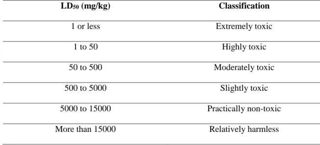
10
Table 1. Classification of toxicity based on LD50 dose ranges Hodge and Sterner (Frank,
1992).
LD50 (mg/kg) Classification
1 or less Extremely toxic
1 to 50 Highly toxic
50 to 500 Moderately toxic
500 to 5000 Slightly toxic
5000 to 15000 Practically non-toxic
More than 15000 Relatively harmless
1.4. Sub-acute toxicity
In sub-acute toxicity studies, repeated doses of drug are given in sub-lethal quantity for a period of 14 to 21 days. Sub-acute toxicity studies are used to determine effect of drug on biochemical and hematological parameters of blood as well as to determine histopathological changes (Baki et al, 2007; Dahamna, 1987; Dahamna et al., 2004; Belguet, 2010).
1.5. Chronic toxicity
Chronic toxicity is the adverse effects occurring as a result of the repeated daily exposure of experimental animals to a chemical. In chronic toxicity studies, drug is given in different doses for a period of 90 days to over a year to determine carcinogenic and mutagenic potential of drug (Baki et al, 2007; Belguet, 2010). The parameters of chronic toxicity studies are same as that of sub-acute study. Multiple dose studies are necessary to assure the safety of natural products. On the other hand, clinical observations of acute assays are valuable tools to define the doses to be tested in multiple dose experiments,
11
along with pharmacological studies in animals and in humans (Alvarez et al, 2004; Hasumura et al, 2004, Merghem et al.; 2013; Boussahel et al., 2013 ).
1.6. Routes of administration
This term refers to the way in which drugs or compounds are introduced to animals or humans. To evaluate toxicity of a compound in animals, various routes may be used but two most commonly used modes of administration for animals studies are via intra-peritoneal injection or the oral route (Poole and Leslie, 1989).
1.6.1. Intra-peritoneal injection
This is one of the methods of dosing, which may occasionally provide information about local as well as systemic toxicity. To give drugs by intra-peritoneal injection, the animal is laid on its back and the abdomen shaved. This area is thoroughly cleansed and, using an appropriate syringe and needle, the abdominal wall is punctured. To ensure minimal danger of perforation of abdominal viscera, the injection should be made rostral and lateral to the bladder at an angle of about 15º to the abdomen. The depth of penetration should not exceed 5mm (Poole and Leslie, 1989; Waynforth, 1980).
1.6.2. Oral administration
The oral route is probably one of the most common means by which a chemical enters the body. In short, the oral administration is the form of administration involving the gastrointestinal tract, which may be viewed as a tube going through the body, starting at the mouth and ending at the anus. Although it is within the body, its contents are essentially exterior to the body fluids. Most orally administered chemicals can otherwise have a systemic effect on the organism only after absorption has occurred from the mouth or the gastrointestinal tract. Oral administration of chemicals that are rapidly absorbed
12
from the gastrointestinal tract would theoretically expose the liver to concentrations of the agent that would not be obtained if other routes of administration were used (Loomis and Hayes, 1996). Furthermore, if a compound entered the entero-hepatic cycle, at least a portion of the compound would be localized in the organs involved in the cycle. Compounds that are known to be toxic to the liver would be expected to be more toxic following oral administration on repeated occasions, whereas their administration by other routes may be less hazardous (Loomis and Hayes, 1996; Waynforth, 1980).
1.7. Target tissue Toxicity
The extent to which an organ is susceptible to toxicity varies from tissue to tissue. For example the kidneys and liver are more highly vascularised making them more susceptible to toxicity than the bone tissues (Viala and Botta, 2007).
1.7.1. Hepatotoxicity
The liver is the largest internal organ in the body and is divided by the falciform ligament into two lobes: a large right lobe and a smaller left lobe. Each lobe is further divided into lobules which are the functioning units of the liver (Figure 1).There are approximately a million lobules in the liver filled with hepatocytes. The hepatocytes are responsible for bile secretion and also perform a variety of metabolic functions. Between each row of hepatocytes are small cavities called sinusoids (Berne and Levy, 1998). The main functions of the sinusoids are to destroy old or defective red blood cells, to remove bacteria and foreign particles from the blood, and to detoxify toxins and other harmful substances (Silverthorn, 2007).
13
Figure 1. Structural organization of the hepatic lobule (Jacquelyn and Maher, 1997).
The liver is the target organ for chemically induced injuries. Several important factors are known to contribute to the liver’s susceptibility. First, compounds absorbed in the gut tract are transported by the hepatic portal vein to the liver. Thus the liver is the first organ perfused by chemicals absorbed in the gut. A second factor is the high concentration in the liver of xenobiotic metabolizing enzymes (Deshpande, 2005; Wallace and Meyer, 2010).The liver receives 25% of the blood supply from the heart. Toxic substances absorbed from the gut are transported directly to the liver which is therefore the first target organ exposed after the gut itself. Hepatocytes are cells that make up most of the structure of the liver and are very metabolically active. Normally, they are involved in many essential biochemical processes, such as removal of nitrogen as urea, synthesis of glycogen as a glucose store, and lipid metabolism. Many toxic substances inhibit protein synthesis
14
because of their action in the liver. The liver also has a key role in removing external poisons (xenobiotic) (Baker, 2012).
Several plant extracts have been examined for use in a wide variety of liver disorders, Many studies have already led to the characterization of more than170 constituents isolated from 110 plants belonging to 55 families have been reported to treat liver diseases and boost liver functions (Mukazayire et al, 2010). While others have been reported to cause injury to the liver (Atawodi et al, 2011), includes elevated liver enzymes, acute or chronic hepatitis, cholestasis, hepatic necrosis or fibrosis, cirrhosis, liver failure, and hepatic veno-occlusive disease (Abdualmjid et Sergi, 2013).
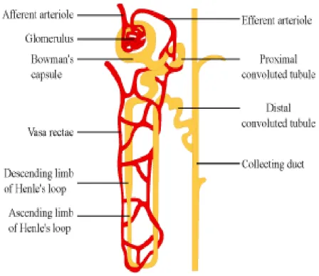
1.7.2. Nephrotoxicity
The kidneys are an essential part of the urinary system responsible for producing and excreting urine into a ureter and also serve homeostatic functions such as the regulation of electrolytes. They serve the body as a natural filter of the blood, and remove wastes which are diverted to the urinary bladder. In producing urine, the kidneys excrete wastes such as urea and ammonia; the kidneys also are responsible for the reabsorption of water, glucose, and amino acids. The renal capsule is the outer skin of the kidney. The outer portion of the kidney consists of the cortex and the medulla containing millions of tiny structures called nephrons (Wheater et al, 1982) (Figure 2). The nephrons are responsible for filtering water out of the bloodstream. The nephron is composed of two main parts: the renal corpuscle and the renal tubule. Inside the renal corpuscle is the glomerulus, a network of blood capillaries that is surrounded, first, by a double membrane (the glomerular capsular membrane) and then is surrounded by Bowman's capsule. The renal tubule consists of the proximal tubule, the loop of Henle, and the distal tubule (which eventually leads into collecting tubules) (Silverthorn, 2007).
15
Frequently, exposure to large amounts of a chemical can cause kidney effects that are not observed at lesser exposures. Effects of kidney damage are frequently assessed in non specific terms such as changes in kidney weight (both increases and decreases) or increases in protein content of the urine (proteinuria) or changes in volume of urine. Acute renal failure is one of the more common responses of the kidney to toxicants. Acute renal failure is characterized by a rapid decline in glomerular filtration rate and an increase in the concentration of nitrogenous compounds in the blood. Compounds that cause renal vasoconstriction reduce the amount of blood that reaches the glomerulus (Middendorf and Williams, 2000).
In addition to the organ-level response of the kidney, many toxicants affect specific regions of the nephron. They may damage the glomerulus, the proximal tubule, or the further tubule elements such as the loop of Henle, distal tubule, or collecting duct. The most common site of injury for toxicants is the proximal tubule (Middendorf and Williams, 2000).
16
Figure 2. Structural organization of the nephron (Tarloff and Wallace, 2010).
1.7.3. Neurotoxicity
The brain is a complex array of neurons grouped to control motor, sensory, posture and higher cognitive function. The brainstem controls much of the essential physiological activity (Baker, 2012). Neurotoxicology is the study of the adverse effects of chemical, biological and certain physical agents on the nervous system and/or behavior during development and in maturity (Harry et al, 1998).
Like the other organ toxicities, neurotoxicity can result from different types of exposure to a substance. The major routes of exposure are oral, dermal, or inhalation. Neurotoxicity may be observed after a single (acute) dose or after repeated (chronic) dosing. (Crofton et al, 2011). Neurotoxicants affect the nervous system in different ways: some neurotoxicants damage the distal portions of axons without much effect on the remainder of the cell, some produce outright cell death, while others affect signaling processes in the nervous system without causing structural damage. Neurons may also be secondarily affected by neurotoxicants that target other cells in the nervous system,
17
disrupting normal homeostatic function and causing structural or functional damage (Blake, 2010).
1.7.4. Hematotoxicity
Blood which forms the main medium of transport in the body is a very important tissue. It serves to transport many drugs and xenobiotic. Since all foreign compounds are distributed via the bloodstream, the various components, cellular and non-cellular, are initially exposed to significant concentrations of toxic compounds (Timbrel, 2009).
Toxic injury to the blood cells and blood-forming tissues is known as hematotoxicity. In humans, the bone marrow constitutes the principal blood-forming tissue. The bonne marrow produces stem cells which are precursors of the red blood cells, white blood cells, and platelets. White cells have an essential role in inflammatory, coagulation, and immune function. Red cells primarily deliver oxygen to all cells in the body and remove carbon dioxide from these cells (Baker, 2012; Deshpande, 2005).
The bone marrow is a major target for many toxic substances. As a result of failure of generation of new cells, there may be failure of the red cell system (anemia) and failure of the white cell system, causing both overwhelming infection due to absence of granulocytes and failure of the immune system from a total reduction on the white cell count (Baker, 2012).
Some plant materials when ingested either in raw state or as extract have been reported to cause anaemia which may result from sequestration of red blood cell in the spleen, impaired red cell production or primary bone marrow dysfunction (Cheeke, 1998).
Damage and destruction of the blood cells results in a variety of consequences such as a reduction in the oxygen-carrying capacity of the blood if the cells affected are the red
18
blood cells. The assessment of blood is relevant to the evaluation of risks since the haematological system carries a higher predictive value for toxicity in humans (Olson et al, 2000).
2. Oxidative stress
Oxidative stress has been defined as a disturbance in the antioxidants/oxidants balance in favor of oxidants (Kirschvink et al, 2008; Lykkesfeldt and Svendsen, 2007; Abdal Dayem et al, 2010; Mühl et al, 2011). This imbalance occurs by increasing the production of oxidants, decreasing the level of antioxidants or both (Mühl et al, 2011; Kirschvink et al, 2008). Oxidative stress may cause cellular damages in macromolecules like proteins, lipids and nucleic acids, leading to cellular pathology and ultimately to cell death (zadak et al, 2009 ; Abdal Dayem et al, 2010).
Oxidative stress plays an important role in the development and progression of many human pathologies including cardiovascular and neurodegenerative disorders (Alzheimer’s and Parkinson’s diseases) (Pham-Huy et al, 2008 ; Durackova, 2010 ; Pizarro
et al, 2009), atherosclerosis, cancer, diabetes, liver damages, rheumatoid arthritis,
cataracts, inflammatory bowel disease, ulcers, pneumonia and human aging (Makker et al, 2009).
2.1. Free radicals
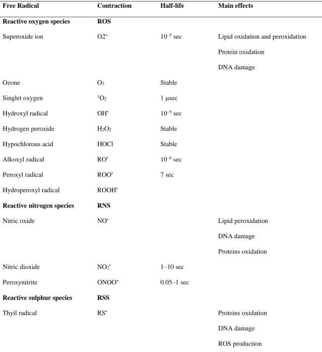
Free radicals are defined as atoms, molecules or parts of molecules containing one or more unpaired electrons in their outer orbits. They are characterized by a very short half- life and a considerable degree of reactivity (Valko et al, 2006).
Free radicals derivatives of oxygen like superoxide free radical anion (O2●-), hydroxyl free radical (OH●), nitric oxide radical (NO●), lipid alkoxyl (LOO●) and lipid
19
peroxide (LOOH) as well as non-radical derivatives such as hydrogen peroxide (H2O2) and
singlet oxygen (1O2) are collectively known as reactive oxygen species (ROS). Whereas,
reactive nitrogen species (RNS) include free radicals like nitric oxide (NO●) and nitrogen dioxide (NO2●), as well as non-radicals such as peroxynitrite (ONOO-) (Kalam et al, 2012)
(Table 2).
ROS are known to play an important role in biological systems, since they can be either harmful or beneficial to living systems. Beneficial effects of ROS involve physiological roles in defense against infections agents and the function of a number of cellular signaling systems. In contrast, at high concentrations, ROS can be important mediators of damage to cell structures, including lipids, proteins and nucleic acids. The harmful effects of ROS are balanced by the action of antioxidants (Palmieri and Sblendorio, 2007; Al-Rubae'i and Al-Musawi, 2011; Rahman, 2007).
20
Table 2. Classification and main effects of free radicals (Finaud et al, 2006).
Free Radical Contraction Half-life Main effects Reactive oxygen species ROS
Superoxide ion O2•– 10–5 sec Lipid oxidation and peroxidation
Protein oxidation DNA damage
Ozone O3 Stable
Singlet oxygen 1O
2 1 μsec
Hydroxyl radical OH• 10–9 sec Hydrogen peroxide H2O2 Stable
Hypochlorous acid HOCl Stable Alkoxyl radical RO• 10–6 sec Peroxyl radical ROO• 7 sec Hydroperoxyl radical ROOH•
Reactive nitrogen species RNS
Nitric oxide NO• Lipid peroxidation
DNA damage Proteins oxidation Nitric dioxide NO2• 1–10 sec
Peroxynitrite ONOO•– 0.05–1 sec
Reactive sulphur species RSS
Thyil radical RS• Proteins oxidation
DNA damage ROS production
2.1.1. Production of ROS
The major biological process leading to O2●- generation is the electron transport chain associated with the mitochondrial membrane (Bahorun et al, 2006). At this level, the ubiquinone – cytochrome b removes an electron from each one of the four cytochrome reduced molecules, oxidizing them and add the four electrons to the O2 (tetravalent oxygen
21
O2 + 4e- + 4H+ 2H2O + energy
However, univalent conversion of oxygen generates the superoxide radical (O2●-)
(Gardès-Albert et al, 2003).
O2 + e- O2●-
Superoxide radical (O2●-) is also formed by the NADPH oxidase of phagocytic cells
in order to facilitate the elimination of pathogens. Activation of this enzyme leads to a significant increase of oxygen consumption by neutrophils and macrophages called “respiratory burst” (Bartoz, 2003).
NADPH + 2O2 NADP+ H+ + O2●-
Superoxide radical (O2●-) undergoes a dismutation reaction catalyzed by the
Superoxide dismutase (SOD) to form H2O2 (Ahsan et al, 2003).
2O2●- + 2H +SOD H2O2
The hydroxyl radical (OH●) may be formed when H2O2 reacts with Iron or Copper
ions. This reaction is known as Fenton reaction (Schneider and Oliveira, 2004).
Fe2+ + H
2O2 OH●+ OH- + Fe+3
Cu+ + H2O2 Cu2+ + OH● + O
-On the other hand, transition metals ions may catalyze the reaction between H2O2
and superoxide leading to the production of hydroxyl radical, following Haber-Weiss reaction (Schneider and Oliveira, 2004).
22
H2O2 +O2●- Fe/Cu OH● + OH- + O2
H2O2 is converted by myeloperoxidase (MPO) in neutrophils to hypochlorous acid
(HOCl), a strong oxidant that acts as a bactericidal agent in phagocytic cells (Ahsan et al, 2003).
H2O2 + Cl- MPO HOCl + OH
-Besides, the superoxide radical may react directly with the nitric oxide (NO●) and generate the peroxinitrite. This compound may lead to the formation of an oxidant agent with hydroxyl radical features (Schneider and Oliveira, 2004).
O2●- + NO● ONOO● ONOO● + H+ OH●
The NO● is formed by the oxidation of L-arginine using nitric oxide synthase (NOS) (figure 3) (Bahorun et al, 2006).
Hydroxyl radical (OH●) is the most reactive free radical in vivo. It has a high reactivity which making it a very dangerous radical with a very short half-life. These potentially reactive molecules are able to attack healthy cells, causing structural and functional damages (Kalam et al, 2012). H2O2 since is long-lived and
membrane-permeable, may diffuse a considerable distance away from its site of generation. However, OH● is extremely reactive having a very short half-life but with a very limited diffusion capacity. It can attack and damage almost every molecule in its vicinity at a diffusion controlled rate (Bandyopadhyay et al, 1999).
Nitrogen dioxide NO2●is formed by oxidation of nitric oxide
23
Reaction of hypochlorous acid with hydrogen peroxide forms Singlet oxygen (1O 2)
(Bartoz, 2003).
Figure 3. Generation of reactive oxygen species (ROS) (Lucy Chen et al, 2012).
2.1.2. Molecular damage induced by free radicals
RONS may cause oxidative damage to proteins, lipids, and DNA, which inhibits the normal functions of proteins and lipids, and facilitates DNA mutagenesis; thus, these species play a pivotal role in various clinical conditions (Rice-Evans et al, 1996).
24
2.1.2.1. Lipid peroxidation
The polyunsaturated fatty acids (PUFAs) present in membrane phospholipids are particularly sensitive to attack by ROS (Sharma et al, 2012). Radicals, especially hydroxyl, alkoxyl, and peroxyl, can abstract a labile H atom from the methylene group of polyunsaturated fatty acids, generating carbon-centered free radicals. Thus, the initial reaction of hydroxyl radical with fatty acids produces a lipid radical which, with the reaction of oxygen, results in the lipid peroxyl radical, which further can react with fatty acids to produce lipid hydroperoxide. This chain reaction could significantly alter the structure of membranes and other lipids, resulting in altered fluidity, permeability, transport and metabolic processes (Radak et al, 2011).
2.1.2.2. Protein oxidation
Proteins are also targets for free radicals. Oxidative modification of proteins by reactive oxygen species (ROS) or reactive nitrogen species (RNS) is implicated in the pathogenesis of various diseases. Oxidative damage to a specific protein, especially at the active site, can induce a progressive loss of a particular biochemical function. Several types of ROS induced protein modifications have been demonstrated (Stadtman and Le vine, 2000), including the loss of sulfhyryl (SH) groups, formation of carbonyls, disulphide crosslink, methionine sulfoxide, dityrosine cross-links, nitro tyrosine, and glyoxidation and lipid peroxidation adducts, among others. Alterations of signal transduction mechanisms, transport systems, or enzyme activities have been shown (Stadtman, 1990). Protein oxidation may be at least in part responsible for atherosclerosis, many forms of cancer, ischemia-reperfusion injury and may also be associated with aging (Stadtman, 1992).
25
2.1.2.3.
DNA oxidation
ROS can interact with DNA and cause several types of damage such as modification of DNA bases, single and double strand DNA breaks, loss of purines, damage to the deoxyribose sugar, DNA-protein cross-linkage and damage to the DNA repair system (Kunwar and Priyadarsini, 2011).
Free radicals such as •OH and H• react with DNA by addition to bases or abstractions of hydrogen atoms from the sugar moiety. The C4-C5 double bond of pyrimidine is particularly sensitive to attack by •OH, generating a spectrum of oxidative pyrimidine damage products, including thymine glycol, uracil glycol, urea residue, 5-hydroxydeoxyuridine, 5-hydroxydeoxycytidine, hydantoin and others. Similarly, interaction of •OH with purines will generate 8-hydroxydeoxyguanosine (8-OHdG), 8- hydroxydeoxyadenosine, formamidopyrimidines and other less characterized purine oxidative products (Devasagayam et al, 2004). The consequence of DNA damage is the modification of genetic material resulting in to cell death, mutagenesis, carcinogenesis and ageing (Kunwar and Priyadarsini, 2011).
2.2. Antioxidants
Uncontrolled generation of ROS can lead to their accumulation in the cells, causing oxidative stress. Therefore, cells have evolved antioxidant mechanisms for the protection against ROS mediated oxidative damage. An antioxidant is defined as a molecule capable of slowing or preventing the oxidation of other molecules, or as any substance that when present at low concentrations compared to those of an oxidized substrate significantly delays or prevents oxidation of that substrate (Aher et al, 2011). Antioxidant systems are
26
classified into two major groups, enzymatic antioxidants and non-enzymatic antioxidants (Kunwar and Priyadarsini, 2011).
2.2.1. Enzymatic antioxidants
The enzymatic antioxidants include superoxide dismutase (SOD), catalase (CAT) and glutathione peroxidase (GPx) that act as body’s first line of defense against ROS by catalyzing their conversion to less reactive or stable species (Kunwar and Priyadarsini, 2011).
Superoxide dismutase (SOD) is the first line of defense against free radicals, and catalyzes the dismutation of superoxide anion radical (O2●-) into hydrogen peroxide (H2O2) (Pham-Huy, 2008). SOD exists in several isoforms, which differ in the nature of
the coenzyme (metal), the amino acid composition, the co-factors and other features. Three forms of SOD are present in humans: cytosolic (Cu/Zn-SOD), mitochondrial (Mn-SOD) and extracellular SOD (Rahman, 2007).
2O2●¯ + 2H +SOD H2O2 + O2
The hydrogen peroxide H2O2 is then transformed into water and oxygen (O2) by
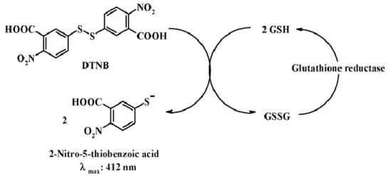
catalase (CAT) or glutathione peroxidase (GPx).
2H2O2 CAT 2H2O + O2
The selenoenzyme GPx removes H2O2 by using it to oxidize reduced glutathione
(GSH) into oxidized glutathione (GSSG). Glutathione reductase, a flavoenzyme, regenerates GSH from GSSG, with NADPH as a source of reducing power (Pham-Huy, 2008). Besides, hydrogen peroxide, GPx also reduces the lipid peroxides (ROOH) formed
27
by the oxidation of polyunsaturated fatty acids (PUFA) to a stable, non-toxic molecule- hydroxyl fatty acid (ROH) (Kumar, 2011).
2H2O2 + 2GSH GPX 2H2O + GSSG
2.2.2. Non-enzymatic antioxidants
The non-enzymatic antioxidants are divided into metabolic antioxidants and nutrient antioxidants. Metabolic antioxidants (endogenous antioxidants) are produced by the body metabolism such as glutathione, lipoid acid, L-arginine, etc. Whereas, nutrient antioxidants (exogenous antioxidants) cannot be produced in the body and must be provided through foods or supplements such as vitamin E, vitamin C, carotenoids, Polyphenols and other antioxidants(Pham-huy et al, 2008).
- Vitamin E
This is a fat-soluble vitamin existing in eight different forms. In humans, α-tocopherol is the most active form and is the major powerful membrane bond antioxidant
employed by the cell. The main function of vitamin E is to protect against lipid peroxidation, and there is also evidence to suggest that α-tocopherol and ascorbic acid function together in a cyclic-type of process. During the antioxidant reaction α-tocopherol is converted to α-tocopherol radical by the donation of labile hydrogen to a lipid or lipid peroxyl radical, and the tocopherol radical can therefore be reduced to the original α-tocopherol form by ascorbic acid (Rahman, 2007).
- Vitamin C
Also known as ascorbic acid and it is the major essential water-soluble antioxidant in human serum. It is able to neutralize ROS in the aqueous phase before that the lipid
28
peroxidation is initiated (Percival, 1998). Vitamin C is an electron donor; and this property accounts for all its known functions. It can scavenge the O2●-, 1O2, OH● and neutralize the
hypochlorous acid (HOCl) (Kelly and Tetley, 1997). Vitamin C works synergistically with vitamin E to quench free radicals and regenerate the reduced form of vitamin E (Pham-Huy, 2008).
- Glutathione
An important water-soluble antioxidant is synthesized from glycine, glutamate and cysteine (Fang et al, 2002). The main protective roles of glutathione against oxidative stress are:
1- Incorporation as cofactor in several antioxidant enzymes against oxidative stress. 2- Scavenger effect against hydroxyl radical and singlet oxygen directly, detoxifying
hydrogen peroxide and lipid peroxides by the catalytic action of glutathione peroxidase.
3- Regeneration of the most important antioxidant, vitamin C and E (Valko et al, 2007).
- Carotenoids
Carotenoids are the mainly colored pigments present in plants and microorganisms. These molecules contain conjugated double bonds and their antioxidant activity arises due to the ability of these bonds to delocalize unpaired electrons (Rahman, 2007). The β-carotene or pro-vitamin A is a fat soluble member of the carotenoids. It is considered as the strongest antioxidant and the best scavenger of singlet oxygen (Pham-Huy, 2008).