Université de Montréal
Prediction of hypertensive disorders in pregnancy by
combined uterine artery Doppler, serum biomarkers
and maternal characteristics
Par An Na
Département de Sciences Biomédicales Faculté de Médecine
Mémoire présenté à la Faculté des études supérieures en vue de l’obtention du grade de maîtrise
en Sciences Biomédicales option Recherche Clinique Biomédicale
Juin 2008
Université de Montréal Faculté des études supérieures
Ce mémoire intitulé:
Prediction of hypertensive disorders of pregnancy by combined uterine artery Doppler, serum markers and maternal characteristics
Présenté par : Na AN
a été évalué par un jury composé des personnes suivantes:
Emmanuel Bujold, président-rapporteur François Audibert, directeur de recherche
Zhong-Cheng Luo, co-directeur Nan Okun, membre du jury
Résumé
Objectif: Évaluer l'efficacité du dépistage de l’hypertension gestationnelle par les caractéristiques démographiques maternelles, les biomarqueurs sériques et le Doppler de l'artère utérine au premier et au deuxième trimestre de grossesse. Élaborer des modèles prédictifs de l’hypertension gestationnelle fondées sur ces paramètres.
Methods: Il s'agit d'une étude prospective de cohorte incluant 598 femmes nullipares. Le Doppler utérin a été étudié par échographie transabdominale entre 11 +0 à 13 +6
semaines (1er trimestre) et entre 17 +0 à 21 +6 semaines (2e trimestre). Tous les échantillons de sérum pour la mesure de plusieurs biomarqueurs placentaires ont été recueillis au 1er trimestre. Les caractéristiques démographiques maternelles ont été enregistrées en même temps. Des courbes ROC et les valeurs prédictives ont été utilisés pour analyser la puissance prédictive des paramètres ci-dessus. Différentes combinaisons et leurs modèles de régression logistique ont été également analysés.
Résultats: Parmi 598 femmes, on a observé 20 pré-éclampsies (3,3%), 7 pré-éclampsies précoces (1,2%), 52 cas d’hypertension gestationnelle (8,7%) , 10 cas d’hypertension gestationnelle avant 37 semaines (1,7%). L’index de pulsatilité des artères utérines au 2e trimestre est le meilleur prédicteur. En analyse de régression logistique multivariée, la meilleure valeur prédictive au 1er et au 2e trimestre a été obtenue pour la prévision de la pré-éclampsie précoce. Le dépistage combiné a montré des résultats nettement meilleurs comparés avec les paramètres maternels ou Doppler seuls.
Conclusion: Comme seul marqueur, le Doppler utérin du deuxième trimestre a la meilleure prédictive pour l'hypertension, la naissance prématurée et la restriction de croissance. La combinaison des caractéristiques démographiques maternelles, des biomarqueurs sériques maternels et du Doppler utérin améliore l'efficacité du dépistage, en particulier pour la pré-éclampsie nécessitant un accouchement prématuré.
Mot clés: Hypertension gestationnelle, Doppler utérins, Biomarqueurs sériques maternels, Caractéristiques démographiques maternelles, Dépistage, Modèle prédictif Multivarié.
Abstract
Objective: To evaluate the screening efficacy of maternal demographic characteristics, serum biomarkers and uterine artery Doppler (uaD) during the first and the second trimester for the hypertensive disorders of pregnancy. To elaborate prediction models of these diseases based on the combination of selected maternal demographic
characteristics, maternal serum biomarkers and uaD indexes.
Methods: This is a prospective pregnant cohort study of 598 singleton nulliparous consecutive women. UaD investigation was performed by transabdominal sonography between 11+0 and 13+6 weeks, and between 17+0 and 21+6 weeks. All the serum samples for measurement of several placental biomarkers were collected at the first trimester. Maternal demographic characteristics were recorded at the same time. Receiver operating characteristic curves and predictive values were used to analyze the predictive powers of the above parameters. Different combinations and their logistic regression predictive models were analyzed.
Results: Among 598 women, 20 developed preeclampsia (3.3%), 7 developed early-onset preeclampsia (1.2%), 52 developed gestational hypertension (8.7%), 10 developed gestational hypertension with delivery before 37 weeks (1.7%). Second trimester uterine artery pulsatility index was the best predictor with statistical significance for all the outcomes. In the multivariable logistic regression analysis, the best predictive value in the first and second trimester was obtained for the prediction of early onset preeclampsia. The combined screening showed significantly better results compared to either maternal parameters or Doppler alone.
Conclusion: As a single marker, second trimester Doppler has the highest predictive value for hypertensive disorders, preterm birth and SGA. Combination of the maternal demographic characteristics, maternal serum biomarker and uaD improves the screening efficacy, especially when this necessitates early delivery.
Key words: Hypertensive disorders of pregnancy, Doppler, Maternal serum biomarkers, Maternal demographic characteristics, Screening, Multivariable predictive model.
Table of contents
Résumé ... iii
Abstract...iv
List of tables ... viii
List of figures ...ix
List of abbreviations ...x
Acknowledgment...xi
Introduction ...1
1. Literature review ...2
1.1. Hypertensive disorders in pregnancy ...2
1.1.1. Classification of hypertensive disorders in pregnancy...2
1.1.1.1. Definition of chronic hypertension...2
1.1.1.2. Definition of pre-eclampsia/eclampsia...3
1.1.1.3. Definition of pre-eclampsia superimposed upon chronic hypertension ...4
1.1.1.4. Definition of gestational hypertension (GH)...5
1.2. Measurement of blood pressure...5
1.3. Epidemiology ...6
1.3.1. Prevalence...6
1.3.2. Impact on maternal and fetal mortality ...6
1.4. Risk factors of hypertensive disorders of pregnancy ...8
1.4.1. Pregnancy associated risk factors...8
1.4.1.1. Nulliparity...8
1.4.1.2. High-risk pregnancy, abnormal pregnancy ...8
1.4.1.3. Sperm exposure and time interval between deliveries ...9
1.4.2. Chronic risk factors ...9
1.4.2.1. Cardiovascular disorders ...10 1.4.2.2. Chronic hypertension...10 1.4.2.3. Renal diseases...10 1.4.2.4. Obesity...10 1.4.2.5. Diabetes ...10 1.4.2.6. Smoking...11
1.4.3. Hereditary risk factors ...11
1.4.4. Other risk factors ...11
1.5. Doppler analysis and hypertensive disorders of pregnancy ...12
1.5.1. Pathophysiology of hypertensive disorders of pregnancy...12
1.5.1.1. Normal adaptation of the uteroplacental vasculature in pregnancy ...12
1.5.1.2. Uterine vascular pathology in hypertensive disorders of pregnancy...12
1.5.2. Principles of Doppler analysis of ulteroplacental blood flow ...14
1.5.3. The Doppler indices ...15
1.5.4.1. Doppler ultrasound as a diagnostic and prognostic tool in complicated
pregnancies ...15
1.5.4.2. Doppler analysis of the uteroplacental circulation as a screening test for pregnancy complications ...16
1.6. Serum markers and hypertensive disorders of pregnancy...18
1.6.1. Human Chorionic Gonadotropin (hCG)...18
1.6.2. Inhibin A...19
1.6.3. Pregnancy-associated plasma protein A (PAPP-A) ...19
1.6.4. A disintegrin and metalloprotease (ADAM12) ...20
1.6.5. Maternal serum placental protein 13 (PP13) ...20
1.7. Screening test for hypertensive disorders of pregnancy...21
1.7.1. Serum Biomarkers screening test for hypertensive disorders of pregnancy ...21
1.7.1.1. Single maternal biomarker screening test...22
1.7.1.2. Combinations of maternal biochemical markers test ...22
1.7.2. Combinations of maternal factors, maternal biochemical markers and uterine artery Doppler test ...23
1.7.2.1. First trimester screening ...23
1.7.2.2. Second trimester screening...24
1.7.2.3. First trimester maternal serum markers in combination with second trimester uterine artery Doppler screening ...24
2. Thesis project...25
2.1. Viewpoints and objectives for the current study ...25
2.1.1. Study objectives...26 2.1.2. Hypothesis: ...26 2.1.3. Objectives:...26 2.2. Methods ...27 2.2.1 Data sources...27 2.2.2. Study design ...27 2.2.2.1. Population...27 2.2.2.2. Definition:...28 2.2.2.3 Measurement: ...28 2.2.3 Statistic analysis ...30 2.2.3.1 Variables...30 2.2.3.2 Statistic analysis ...31 2.3. Results ...34 2.3.1. General descriptions ...34
2.3.1.1. Maternal demographic characteristics...34
2.3.1.2. Maternal serum biochemical markers ...35
2.3.1.3. Maternal Doppler results ...36
2.3.2. Prediction of hypertensive disorders of pregnancy in the first trimester (11 to 13+6 gestational weeks)...36
2.3.2.1. Prediction of early onset preeclampsia...36
2.3.2.2. Prediction of gestational hypertension with delivery before 37 weeks....38
2.3.2.3. Prediction of other hypertensive disorders of pregnancies, preterm birth and SGA ...39
2.3.3. Prediction of hypertensive disorders of pregnancy in second trimester (17 to
21+6 gestational weeks)...39
2.3.3.1. Prediction of early onset preeclampsia...39
2.3.3.2. Prediction of gestational hypertension with delivery before 37 weeks....41
2.3.3.3. Prediction of preeclampsia ...42
2.3.3.4. Prediction of other hypertensive disorders of pregnancies, preterm birth and SGA ...43
Conclusion...45
List of references ...74
List of tables
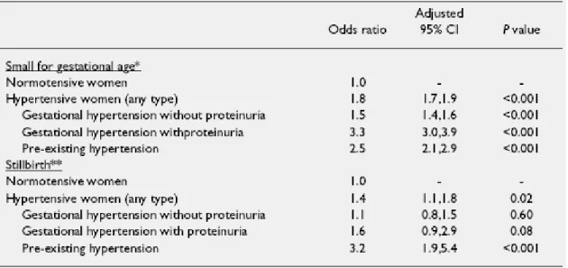
Table 1. Effect of hypertensive disorders in pregnancy on small for gestational age (< the
10th percentile) and stillbirth...51
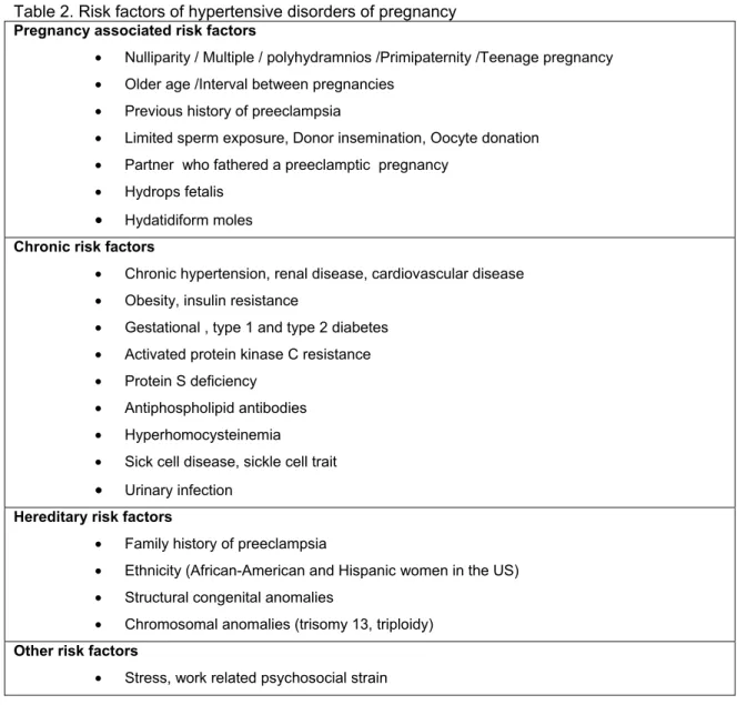
Table 2. Risk factors of hypertensive disorders of pregnancy ...52
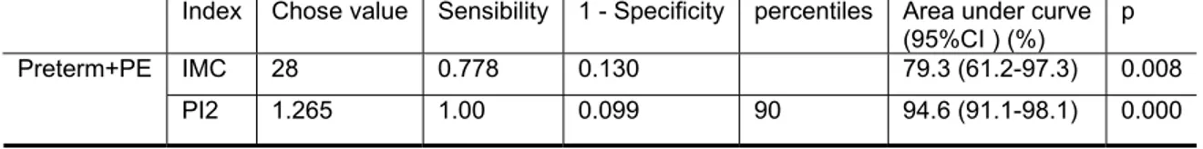
Table 3. Receiver-operating characteristics (ROC) curve analysis for appropriate cutoff point of IMC and Mean Pulsatility index at the second trimester...53
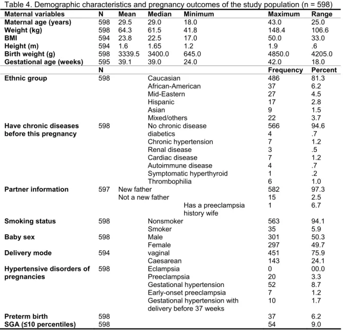
Table 4. Demographic characteristics and pregnancy outcomes of the study population (n = 598)...54
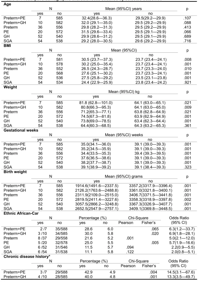
Table 5. Prescriptive demographic characteristics stratified by selected outcomes...55
Table 6. Maternal serum biochemical markers in *normal pregnancies from 11 to 13+6 gestational weeks...57
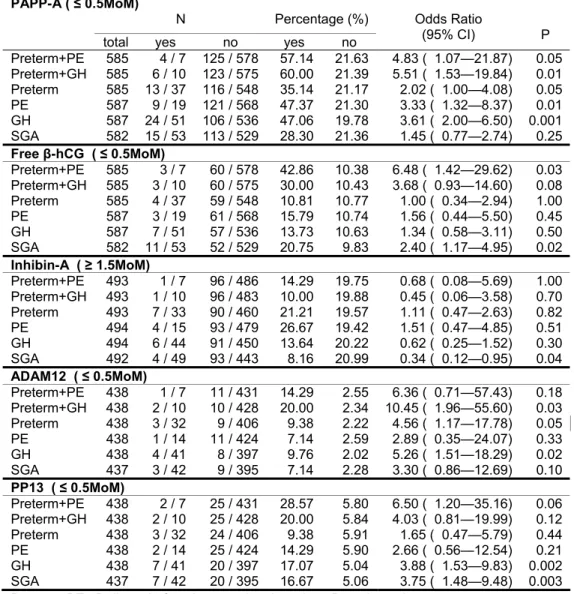
Table 7. The relationship between serum biochemical markers and diseases studied...58
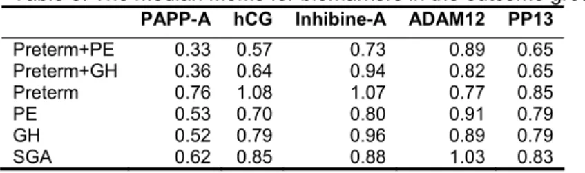
Table 8. The median MoMs for biomarkers in the outcome groups ...59
Table 9. Abnormal doppler pulsatility index in the 1st and 2nd trimester outcomes ...60
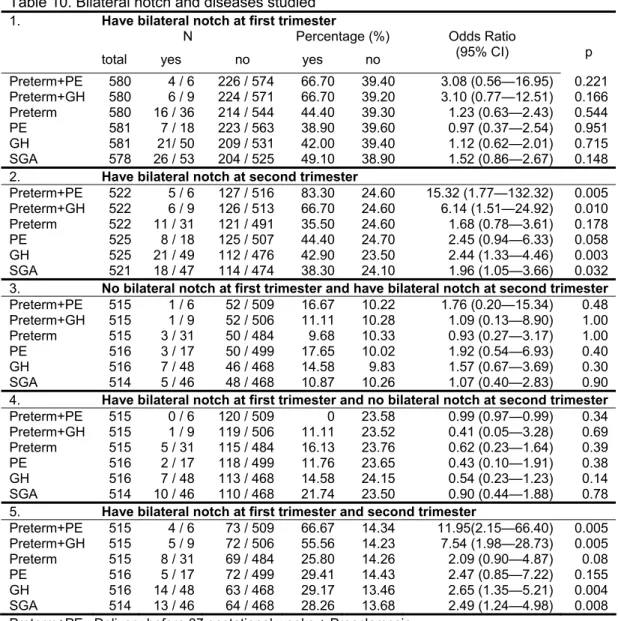
Table 10.Bilateral notch and diseases studied...61
Table 11. Mean *PI and the prevalence of the 95th percentile PI, the 90th percentile PI and Bilateral Notch in *normal pregnancies ...62
Table 12 .Area under ROC curve and the validity parameters of the predictive model ...63
Table 13. Predictive values of different combinations for early on-set preeclampsia (2nd trimester)...64
List of figures
Figure 1. Country distribution of hypertensive disorders of pregnancy as cause of
maternal deaths...65
Figure 2. Different hypertensions and their preterm birth rates. ...66
Figure 3. The uteroplacental blood supply ...67
Figure 4. Normal third trimester uteroplacental flow velocity waveform with high diastolic flow. ...68
Figure 5. Diagrammatic representation of the Doppler indices and diastolic notch ...69
Figure 6. Abnormal FVW with decreased diastolic flow and pronounced diastolic notch. ...70
Figure 7. Maternal biochemical markers median tendency in first trimester...71
Figure 8. Mean PI in different gestational weeks in normal pregnancy...72
List of abbreviations
ADAM12 : A disintegrin and metalloprotease 12 BMI : Body Mass Index (kg/m2)
CRL : Crown-rump length DIAST : Diastolic flot (D) FPV : False Positive value FVW : Flow Velocity Waveform GH : Gestational hypertension
β-HCG : Free human chorionic gonadotropin-β LR : Likelihood ratio
MoM : Multiples of medians
NOTCH : Protodiastolic notch (a velocity range reserved for non-displayed clutter) NT : Nuchal translucency thickness
OR : Odds ratio
PAPP-A : Pregnancy associated plasma protein A PE : Preeclampsia
PI : Pulsatility index (PI=(S-D/Vm)) PP13 : Placental protein 13
RI : Resistance index (RI=(S-D/S)) SGA : Small for gestational age SYST : Systolic flot (S)
Acknowledgment
This thesis owes its existence to the help, support, and inspiration of many people. I would like to express my sincere appreciation and gratitude to my supervisor, Dr. Francois Audibert, for his guidance, encouragement, support and friendship during the course of this study.His truly scientist intuition and passion inspire and enriche my growth as a student and a scientist wanted to be.
Deepest gratitude is also due to my co-supervisor, Dr. Zhong-Cheng Luo, for his instruction, support, and unconditional help whenever I ask for. Without his help and support, this study would not have been successful.
I gratefully acknowledge Dr. William D Fraser, director of our research group, who provided me with unflinching encouragement and support in various ways.
Special thanks to Valérie Tremblay, Susanne, Barbara Torres, Yuquan Wu, Hairong Xu, and all of our unit members, for their friendship, their always readiness to help, and for many valuable discussions we have shared.
Finally, I wish to express my love and gratitude to my parents, my husband and my little son, for their understanding and endless love, through the duration of my study.
Na An
Introduction
With the development of our medical technology and maternal/child care system,
complications of illegal abortions, infections and death from postpartum haemorrhage are seldom seen in recent years. The maternal and perinatal mortality numbers have declined dramatically in many countries. However, hypertensive disorder of pregnancy and their complications, a major cause of maternal mortality, still complicate approximately 0.34-11.5% of pregnancies1. These variations depend on age distribution, ethnic differences, socioeconomic status, and number of previous deliveries. In addition, hypertensive disorders of pregnancy are also found to be strongly associated with fetal growth restriction and prematurity, contributing largely to perinatal morbidity and mortality. 2,3
Hypertensive disorders of pregnancy is generally regarded as a multisystem disorder specific to pregnant women characterised by widespread endothelial damage which originates from the uteroplacental circulation but ultimately involves a variety of other organs such as the kidney, liver and brain. Its pathophysiology remains unclear, despite the progress made in the past decades4. Therefore, it is still a challenge to obtain an accurate prediction of women who are at risk of developing hypertensive disorders of pregnancy.
A theory regarding the etiology of hypertensive disorders of pregnancy has emerged in which maladaptation of the vasculature of the uteroplacental unit due to impaired trophoblast invasion has been implicated as the main causal factor5. Therefore, it should be possible to predict the risk by using Doppler ultrasonography to analyse the
uteroplacental circulation in the pregnancy. This view is now widely accepted and many of these efforts have focused on biochemical markers, primarily those suggesting
1. Literature review
1.1. Hypertensive disorders in pregnancy
1.1.1. Classification of hypertensive disorders in pregnancy
Many classifications focus on diastolic blood pressure, or changes in diastolic blood pressure. Because of the great variation in clinical expression of the syndrome and the inability to distinguish symptoms induced by pregnancy from underlying (but often latent) maternal disorders, we have difficulty defining the symptoms, the various clinical forms, and the pathophysiology that becomes more complex every time new evidence is found. In fact, the criteria used to identify the disorder remain a subject of confusion and controversy. Obviously, a comprehensive and easily obtainable classification is needed to improve prognostics and decision making and to enable comparison of research work.
American College of Obstetricians and Gynecologists (ACOG) has recommended a classification to define hypertensive disorders of pregnancy. This classification is widely accepted and consists of four terms as follow:8
• Chronic hypertension • Pre-eclampsia / Eclampsia
• Pre-eclampsia superimposed upon chronic hypertension
• Gestational hypertension: (1) transient hypertension of pregnancy if pre-eclampsia is not present at the time of delivery and blood pressure returns to normal by 12 weeks postpartum (a retrospective diagnosis) or (2) chronic hypertension if the elevation persists.
1.1.1.1. Definition of chronic hypertension
Chronic hypertension is defined as hypertension that is present and observable before pregnancy or that is diagnosed before the 20th week of gestation. Hypertension is defined as a blood pressure equal to or greater than 140 mm Hg systolic or 90 mm Hg diastolic. During pregnancy the hypertension remains, but proteinuria does not occur. Women who develop hypertension during pregnancy, without proteinuria or seizures, and whose blood
pressure remains elevated after pregnancy are also diagnosed with chronic hypertension. Hypertension that is diagnosed for the first time during pregnancy and that does not resolve postpartum is classified as chronic hypertension too.
1.1.1.2. Definition of pre-eclampsia/eclampsia
Pre-eclampsia is characterized by blood pressure greater than or equal to 140 mm Hg systolic or 90 mm Hg diastolic occurring after midpregnancy (20 weeks gestation), and accompanied by proteinuria. Preeclampsia may be further categorized as mild or severe. In the absence of proteinuria the disease is highly suspected when increased blood pressure appears accompanied by the symptoms of headache, blurred vision, and abdominal pain, or with abnormal laboratory tests, specifically, low platelet counts and abnormal liver enzymes. It is recommended that gestational blood pressure elevation be defined on the basis of at least two determinations. The repeat blood pressure should be performed in a manner that will reduce the likelihood of artifact and/or patient anxiety.
Proteinuria is defined as the urinary excretion of 0.3 g protein or greater in a 24-hour specimen. This will usually correlate with 30 mg/dL ("1+ dipstick") or greater in a random urine determination with no evidence of urinary tract infection. However, because of the discrepancy between random protein determinations and 24-hour urine protein in pre-eclampsia (which may be either higher or lower), it is recommended that the diagnosis be based on a 24-hour urine if at all possible or a timed collection corrected for creatinine excretion if this is not feasible. Pre-eclampsia always presents potential danger to mother and baby. Other conditions may increase blood pressure and even result in proteinuria; thus, as the certainty of the diagnosis increases, the requirements for careful assessment and consideration for delivery also increase.
According to the severe of syndrome, ACOG made three categories for pre-eclampsia: a. Mild preeclampsia
BP 140/90
300mg of proteinuria in 24hrs b. Severe preeclampsia (any of these)
5g of proteinuria in 24hrs Oliguria or <500 ml in 24hrs Cerebral of visual disturbances Pulmonary edema or cyanosis
Persistent pain of right-upper quadrant of abdomen Fetal growth restriction
Thrombocytopenia (Platelet count is less than 100,000 cells/mm3 and/or evidence of microangiopathic hemolytic anemia, with increased lactic acid dehydrogenase).
Impaired liver function (Elevated hepatic enzymes -alanine aminotransferase [ALT] or aspartate aminotransferase [AST]).
c. Eclampsia
Presence of new-onset seizures in a patient with preeclampsia
1.1.1.3. Definition of pre-eclampsia superimposed upon chronic hypertension
Lots of evidence indicates that pre-eclampsia may occur in women already hypertensive (i.e., who have chronic hypertension) and that the prognosis for mother and fetus is much worse than with either condition alone. Skills are needed to distinguish superimposed pre-eclampsia from worsening chronic hypertension. The principle of high sensitivity and unavoidable overdiagnosis is appropriate in order to facilitate clinical management. Close observation, with delivery indicated by the overall assessment of maternal-fetal well-being shall be provided at suspicion of superimposed pre-eclampsia. The diagnosis of superimposed pre-eclampsia is highly likely associated with the following findings:
• In women with hypertension and no proteinuria early in pregnancy (<20 weeks), new-onset proteinuria, defined as the urinary excretion of 0.3 g protein or greater in a 24-hour specimen.
• In women with hypertension and proteinuria before 20 weeks' gestation. • Sudden increase in proteinuria.
• A sudden increase in blood pressure in a woman whose hypertension has previously been well controlled.
• Thrombocytopenia (platelet count <100,000 cells/mm3). • An increase in ALT or AST to abnormal levels.
1.1.1.4. Definition of gestational hypertension (GH)
The woman who has blood pressure elevation detected for the first time after 20 weeks of gestation, without proteinuria, is classified as having gestational hypertension. This nonspecific term includes women with the pre-eclampsia syndrome who have not yet manifested proteinuria as well as women who do not have the syndrome. The
hypertension may be accompanied by other signs of the syndrome, which will influence management. The final differentiation that the woman does not have the pre-eclampsia syndrome is made only postpartum. If pre-eclampsia has not developed and blood pressure has returned to normal by 12 weeks postpartum, the diagnosis of transient hypertension of pregnancy can be assigned. If blood pressure elevation persists, the woman is diagnosed as having chronic hypertension. Note that the diagnosis of
gestational hypertension is used during pregnancy only until a more specific diagnosis can be assigned postpartum.
It is the elevation of blood pressure (>140/90mm Hg) during pregnancy or in the first 24 hours postpartum without other signs of preeclampsia or preexisting hypertension. The hypertension usually resolves in days to weeks after delivery. Rarely, women will become persistently hypertensive following a pregnancy complicated by transient hypertension. This condition is thought to foreshadow the development of essential hypertension later in life.
1.2. Measurement of blood pressure
For the purposes of accuracy and standardization, health professionals should take blood pressure measurements in pregnant women with the patient seated rather than lying on her side, because substantial differences exist between the blood pressures in the upper and lower arms when the patient is lying on her side. In addition, the National Institutes of Health (NIH) recommends that the diastolic pressure reading should be taken at
Korotkoff 5, with the disappearance of sound—not at Korotkoff 4, when sound becomes muffled. To meet strict criteria for hypertension, the patient's readings must be elevated on at least two separate occasions at least six hours apart.
a. Mercury manometer is still the gold standard.
b. Comfortable sitting position, patient seated with feet supported for 2-3 minutes, arm at the level of the heart.
c. Use appropriately sized cuff (large cuff if arm circumference > 33 cm). d. Record systolic and diastolic pressures, the latter as Korotkoff Vth
(disappearance). Only use Korotkoff IVth when Korotkoff Vth is absent.
e. If the Korotkoff Vth sound is not present, use the Korotkoff IVth but should note as such
f. In serial readings use the higher set of values
g. Relative BP change of 30mmHg/15mmHg is no longer used as hypertension
1.3. Epidemiology
1.3.1. Prevalence
The overall prevalence of hypertensive disorders of pregnancy was 7.5% in Brazilian women9, 6.4% in African American, 4.8% in other American women10, 3.7% in the United States11, 3.3 % in southern Iran12, 3% in northwest Saudi Arabia 13 and 2.6% in southwest Saudi Arabia14. These figures may not be accurate because the information is
usually based on hospital populations and therefore biased, especially in countries where access to health care is low. The differences in classification and definitions also
contribute to the variation of these figures, which may be different even among reports from the same region.
1.3.2. Impact on maternal and fetal mortality
Research shows that the occurrence of hypertensive disorders of pregnancy is inversely related to assess to health care of that region. In developed countries, hypertensive disorders of pregnancy were responsible for 16.1% (6.7-24.3%) of all maternal deaths.
The number was much higher (25.7%, 7.9-52.4%) in developing countries, especially in Latin America and the Caribbean. 15 (Fig. 1)
Fetal mortality from preeclampsia was usually caused by associated prematurity, IUGR, or hypermagnesemia. Hypertension and the poor placentation often seen with
preeclampsia may result in placental insufficiency and eventually IUGR which is associated with several problems, including fetal and neonatal hypoxemia, acidosis and newborn resuscitation.
Svein Rasmussen and Lorentz M Irgens’s study on 223541 single births in Norway from 1999 to 2002 showed that mothers with severe preeclampsia or preeclampsia
superimposed on chronic hypertension gave birth earlier than those who had mild preeclampsia or transient hypertension. Median gestational age at birth in transient hypertension, mild-, and severe preeclampsia were 279, 277, and 259 days, respectively, against 280 days in normotensive pregnancies, while those with chronic hypertension with and without preeclampsia had median gestational age of 268 and 279 days, respectively. The rates of preterm birth (less than 37 weeks of gestation) in transient hypertension, mild-, and severe preeclampsia were 8.1, 9.7, and 48.2%, respectively, against 5.0% in normotensive women. The rates were also significantly higher in chronic hypertension with or without preeclampsia(34.5% and 8.9%).16(Fig. 2)
Victoria M Allen and their colleagues performed a study on all pregnant women and births in the Canadian province of Nova Scotia between 1988 and 2000, including 135,466 pregnancies. In their study, after controlling for potential confounders, women with any hypertensive disorder were 1.4 (95% CI 1.1, 1.8, P = .02) times more likely to have a stillbirth and 1.8 (95% CI 1.7, 1.9, P < .001) times more likely to have SGA as compared with normotensive women. Women with pre-existing hypertension were 3.2 (95% CI 1.9, 5.4, P < .001) times more likely to have a stillbirth and 2.5 (95% CI 2.1, 2.9, P < .001)times more likely to have SGA as compared with normotensive women.17 (Table 1)
1.4. Risk factors of hypertensive disorders of pregnancy
Incidence of hypertensive disorders of pregnancy is associated with a number of risk factors (Table 2). Based on these factors, a comprehensive medical history and physical examination at the first antenatal visit can be used to estimate a mother’s risk of
developing the disorder.
Risk factors can be divided into four major groups as follows:
1.Pregnancy associated risk factors 2.Chronic risk factors
3.Hereditary risk factors 4.Other risk factors
Section 1.4.1.-1.4.4. provide detail descriptions of each of these risk factors.
1.4.1. Pregnancy associated risk factors
1.4.1.1. Nulliparity
Women in their first pregnancy are at an increased risk of hypertensive disorders of pregnancy. Previous pregnancies appear to have a protective effect, even if they ended early in abortion. In contrast, women with a history of hypertensive disorders in previous pregnancies are at an increased risk.18 It is likely that women who have recurrent pre-eclampsia have an underlying pathological phenotype that puts them at risk of hypertension.19
1.4.1.2. High-risk pregnancy, abnormal pregnancy
Factors associated with the pregnant state itself that increase the risk of hypertensive disease are twin pregnancy20, polyhydramnion21,hydatidiform molar pregnancies and
hydrops fetalis.4 Extremely young (teenage) or relatively old (over 35) women are at an elevated risk too.22
1.4.1.3. Sperm exposure and time interval between deliveries
Normal pregnancy requires adaptation of the maternal immune response, so that the foetus and placenta, being partly allogenic, are not rejected. In pre-eclampsia, this adaptation may be inadequate in a first pregnancy with a new partner (and limited sperm exposure, primipaternity theory); while in subsequent pregnancies with the same partner the risk is lower.23-25
Although numerous studies have shown an increased risk of pre-eclampsia associated with a new partner26, some investigators challenged this theory. In Trogstad’s study on 547 238 women, time interval between pregnancies have shown a significant impact on the risk of pre-eclampsia. They also found that a change of paternity for the second pregnancy was associated with a reduced risk of pre-eclampsiaafter controlling for the time since first delivery, but theinteraction between change in paternity and time between deliverieswas significant only for women with no previous pre-eclampsia(P = 0.04) and the interaction between history of pre-eclampsia andtime interval between the two deliveries was highly significant(P < 0.001). For women with no previous pre-eclampsia therisk of pre-eclampsia in second pregnancy increased with increasingtime interval, whereas for women with previous pre-eclampsiathe risk tended to decrease with increasing time interval betweendeliveries.27
The increased incidence of pre-eclampsiain donor insemination pregnancies indicates that prolonged exposureto paternal spermatozoa prior to conception, as in the partner insemination group, reduces the risk. Since recipients of donatedspermatozoa are exposed to foreign fetal antigens encoded bythe paternally derived genes, they are at higher risk. The findingof an increased incidence of gestational hypertension in ovum donation pregnancies provides further support to the hypothesisthat the development of pre-eclampsia may be due to alteredor inadequate immunoprotection of the fetoplacental unit inoocyte recipients due to short duration of exposure to non-maternalantigens.28
1.4.2. Chronic risk factors
of pregnancy, as detailed in the following paragraphs.
1.4.2.1. Cardiovascular disorders
Preeclampsia and cardiovascular disorders share similar risk factors like obesity, hyperlipidemia, increased insulin resistance, and analogous pathophysiology such as increased oxidative stress and vascular injury. 29 Cardiovascular disorders are risk factors for hypertensive disorders of pregnancy. Unfavourable cardiovascular and metabolic profiles may represent primary causes of pre-eclampsia, as well as subsequent cardiovascular disease.19
1.4.2.2. Chronic hypertension
Pregnancy has detrimental effects on chronic hypertension. The probability of
exacerbating of hypertension and development of preeclampsia and eclampsia in patients with chronic hypertension is much higher than previously normotensive population.30
1.4.2.3. Renal diseases
Women suffering from renal disease have a high risk too, because hyperuricaemia precedes significant proteinuria in pre-eclampsia and is thought to result from either enhanced tubular reabsorption of uric acid, or breakdown of nuclear (or therefore purine) rich syncytiotrophoblast.31
1.4.2.4. Obesity
Obese women and those with hypercholesterolemia have an increased risk of developing or having preexisting manifestations of the metabolic syndrome. So, there are lots of potential risk factors to develop this disease.32
1.4.2.5. Diabetes
Diabetes is a condition in which the body cannot use the sugars and starches (carbohydrates) it takes in as food to make energy. The body either makes too little insulin in the pancreas or cannot use the insulin it makes to change those sugars and starches into energy. As a result, the body collects extra sugar in the blood and gets rid of some sugar in the urine. The extra sugar in the blood can damage organs of the body, such as the heart, eyes, and kidneys, if it is allowed to collect in the body too long. And,
insulin resistance might affect the pathogenesis of hypertensive disorders in pregnancy since mid-trimester. Women with hypertensive disorders in pregnancy were more likely to have gestational diabetes and pre-existing diabetes compared with normotensive women (2.3% and 0.3%, respectively). 17
1.4.2.6. Smoking
An interesting observation is that smoking appears to have a protective effect. Women with a smoking history, even though they quit smoking in early pregnancy, still
demonstrate lower probability of developing hypertensive disorders in pregnancy33
1.4.3. Hereditary risk factors
Research indicates that women with positive family history have a higher risk of developing preeclampsia. They may have inherent abnormalities, which predispose to vascular disease.34 And, some ethnic groups, like African-American and Hispanic women in the US, have a higher incidence of hypertensive disorders of pregnancy compared to white women.35 Some investigators believe that the hypothesis of a single gene whose expression is pregnancy-specific and which is alone responsible for the progression from preeclampsia to eclampsia. Various candidate genes have been proposed. Arngrimsson and colleagues have reported the highest lod score for a region on 2p13 without any obvious candidate genes, which seems has been confirmed.36,37 But, in fact, no gene has gained common acceptance as the one that causes the hereditary component of
preeclampsia.
1.4.4. Other risk factors
Stress and work related psychosocial strain increase the risk of developing hypertensive disorders of pregnancy. One possibility suggested that stress contributes to the etiology of preeclampsia via priming the immune system to produce inflammatory cytokines. 38 On the other hand, Annelies Rep’s study mentioned that severe hypertensive disorders of pregnancy have a high psychological impact, especially when gestational age at onset of disease is below 30 weeks or if adverse infant outcome occurs.39
1.5. Doppler analysis and hypertensive disorders of pregnancy
1.5.1. Pathophysiology of hypertensive disorders of pregnancy
Blood supply to uterus is mainly provided by the left and right uterine arteries and to a less extent by the ovarian arteries. In the parametrium, the uterine arteries branches into the arcuate artery which in turn branch into the radial arteries. After crossing the
myometrium, these radial arteries issued spiral arteries at the junction of the myometrial-endometrial and terminate in the basal endometrium. Radial artery also issue branches named basal arteries which do not pass endometrium.
1.5.1.1. Normal adaptation of the uteroplacental vasculature in pregnancy
In the first few weeks of pregnancy, conceptus cytotrophoblast cells invade the basal decidua and the decidual portion of the spiral arteries. Cytotrophoblast is divided into two different types at this stage. One is called endovascular trophoblast. The other is called interstitial trophoblast which penetrates the basal deciduas.
The interstitial trophoblast migrates retrogradely to colonise the myometrium at 10 to 12 weeks of pregnancy. Between 12 to 16 gestational weeks, endovascular trophoblast invades the myometrial portions of the spiral arteries, replaces the endothelium and disrupts the muscular vessel wall.
Finally, distended fibrinoid tubes incapable of vasoconstriction replace the spiral arteries from the myometrium to the intervillous space. This results in a low-resistance, high-capacity vascular circulatory system which facilities adequate maternal blood supply to the growing fetus.
1.5.1.2. Uterine vascular pathology in hypertensive disorders of pregnancy
Since the 1950s it has been acknowledged that hypertensive disorders of pregnancy were associated with extensive placental infarction. Thanks to increasing knowledge of the normal physiological adaptation of the arteries of the placental bed and more
sophisticated microscopic techniques in recent days, people have better understanding of the pathogenesis of hypertensive disorders of pregnancy with two major distinct
abnormalities found in the uteroplacental circulation. First, normal physiological adaptation of pregnancy is impaired or absent in hypertensive disorders of pregnancy. Second, a distinctive vaso-occlusive lesion termed "acute atherosis" was found. These abnormalities impair maternal blood flow which results in placental insufficiency during pregnancy.
a. Impaired physiological adaptation:
Physiological changes of uterine artery only exist at the endometrial segments of the spiral arteries while the myometrial segments remain unchanged. Besides, trophoblast cells do not exist in the vessel wall of the spiral arteries that failed to undergo
physiological changes. The extent of endovascular trophoblast invasion and physiological changes varies between spiral arteries. Some arteries do not show any physiological changes along their entire length.40,41
The association between impaired adaptation of spiral arteries, hypertensive disorders of pregnancy, and fetal growth restriction has been confirmed in recent studies.42,43 The
studies also show that endovascular trophoblast invasion is not an all-or-none
phenomenon and that there is a gradient in the extent of trophoblast invasion of decidual and myometrial arteries in both normal pregnancy and pre-eclampsia. Uteroplacental vascular pathology, although being present in a percentage of normal pregnancies, has also been related to spontaneous prematurity and placental abruption. 44
b. Acute atherosis:
Research shows that acute atherosis is related to pre-eclampsia and fetal growth
restriction. An atheromatous infiltrate consisting of lipid-laden "foam cells" is a typical lesion characteristics, which originated from myocytes or macrophages, fibrin, and mononuclear cells. In addition, there is large amount of fibrin deposited in the vascular endothelium and the media. Cellulose necrosis and macrophage infiltration appeared at the earlier stages of the lesions.45
It was believed that acute atherosis only occurs in spiral arteries which fail to undergo physiological changes. Latest research showed that it can also occur in physiologically adapted spiral arteries.45,46
It is not clear what causes impaired trophoblast invasion and acute atherosis. The fact that trophoblast cells do not exist in the vessel wall of unchanged spiral arteries suggests a specific defect in the invasion of the endovascular type of trophoblast. The failure of endovascular trophoblast to express vascular adhesion phenotype molecules could play a role by impairing adhesion to the vessel wall. In recent studies, an increased number of activated macrophages were found in the placental bed of patients with pre-clampsia and widespread apoptosis of placental cytotrophoblast cells.47,48 This phenomenon could be caused by a maternal immunological response against endovascular trophoblast, perhaps triggered by the deficiency in the expression of vascular adhesion molecules. In addition, recent reports indicate that a deficiency of angiogenic growth factors such as vascular endothelial growth factor (VEGF) and placental growth factor (PIGF) is associated with pre-eclampsia.49,50 Further investigation is required to understand the precise meaning of these findings.
1.5.2. Principles of Doppler analysis of ulteroplacental blood flow
Doppler signals from the maternal side of the uteroplacental circulation can be derived from the uterine artery, the arcuate arteries, or the spiral arteries (Fig. 3).
These vessels undergo profound changes in early pregnancy resulting in increased volume flow.
Cytotrophoblastic invasion of the maternal spiral arteries and replacement of their endothelium is central to the successful outcome of pregnancy, and results in the establishment of a low-resistance high-flow uteroplacental circulation. This process, known as physiologic transformation, begins as early as the 10th day after conception and continues throughout the pregnancy.
The noninvasive assessment of uterine artery resistance by means of Doppler ultrasound has demonstrated that a high-resistance blood flow pattern is invariably present at the beginning of the pregnancy. As a result of the physiological transformation of the spiral
arteries, a low-resistance pattern develops at varying gestational ages in different individual pregnancies.42
The mature placenta is a low resistance, high capacity system in which arteries have lost their elastic wall and drain directly into large venous pools without an interposed
capillary bed. This results in low systolic maximum velocity and high forward diastolic flow. These changes are completed in normal pregnancy between 20 and 24 weeks (Fig.4)
1.5.3. The Doppler indices
Several indices have been described to quantify the information from the Doppler waveform. (Fig. 5)
The Resistance Index (RI) was introduced in 1974 by Planiol.51 The Pulsatility Index (PI), a modified version of the resistance index, was introduced in 1971 by Gosling RG.52 The systolic/diastolic ratio is also used frequently in clinical practice. Several authors have noticed the presence of a diastolic “notch”(Fig.6) in waveforms from the
uteroplacental circulation and used it in the prediction of pregnancy complications. This "notch" is present in most waveforms in nonpregnant women and early pregnancy but disappears in the second trimester in normal pregnancy. The presence of a diastolic notch beyond the 22nd week is generally considered to be abnormal. The physiological
background of the changes in uteroplacental flow velocity waveform (FVW)s has been investigated using computer models to simulate the uteroplacental circulation.
To obtain reliable information about the uteroplacental circulation, the uterine artery is the best sample site; it is easy to locate on real time imaging, provides good
reproducibility, and reflects the resistance in the entire distal placental bed. 53,54
1.5.4. Prediction of hypertensive disorders of pregnancy by Doppler
ultrasound
1.5.4.1. Doppler ultrasound as a diagnostic and prognostic tool in complicated pregnancies
Doppler ultrasound of the uteroplacental circulation could be used to predict poor pregnancy outcome in hypertensive pregnancies. In 1983, Campbell et al. used a pulsed
Doppler system to detect abnormal flow velocity waveforms (FVWs) in the arcuate arteries of 31 women with hypertension or suspected FGR, and found that abnormal FVWs were associated with a higher risk of proteinuric hypertension, premature delivery, and low birthweight. In addition, women with normal FVWs were likely to have a normal pregnancy outcome.55
1.5.4.2. Doppler analysis of the uteroplacental circulation as a screening test for pregnancy complications
The recognition of abnormal uteroplacental FVWs in patients with hypertensive disorders of pregnancy suggests the possibility of a screening test in early pregnancy. It is widely believed that in case of hypertensive disorders of pregnancy, uteroplacental resistance is increased already in the beginning of the second trimester, when normal adaptation of the spiral arteries has failed to occur. In 1986, Campbell and his colleagues performed the first screening study. In their study, a pulsed Doppler apparatus was used to assess the blood flow velocity profiles in uterine vessels (arcuate arteries) at 16 to 18 weeks' gestation. 56 Their purpose is to determine if complications associated with impaired trophoblastic invasion of the placental bed (ie, pregnancy-induced hypertension, intrauterine growth retardation, and fetal asphyxia) could be predicted by this
measurement. Thirty-one of 126 consecutive pregnancies developed one or more of the above complications. The sensitivity was 68% and the specificity 69%; the predictive value of a positive test was 42% and that of a negative test 87%.
Presently, more and more investigators use Doppler as a screening test, and found that there are several points worth of attention.
a. The gestational age at the time of measurement
• First trimester
In normal pregnancy, uteroplacental resistance falls gradually between the late first trimester and mid second trimester. Screening before 20 weeks might result in a false-positive test result in subjects in which spiral artery adaptation is in normal progress but not yet completed. 57,58
Good predictive values are reported in studies where an initial scan at 18-20 weeks is followed –if abnormal- by a repeat scan at 24 weeks.59 Two-stage screening (repeating an abnormal test after 4 weeks) have better predictive result in comparison with single sampling.60
b. Doppler screening is more accurate in predicting proteinuric pre-eclampsia compared to pregnancy-induced hypertension.
Some investigators found that abnormal uterine Dopplers have better predictive values for subgroups such as “severe” pre-eclampsia, in comparison to "uncomplicated" pregnancy-induced hypertension and pre-eclampsia.7,61, 62 In their studies, “severe” pre-eclampsia includes the following symptoms: diastolic blood pressure over 90 mmHg, severe proteinuria, pre-eclampsia requiring delivery before 34 weeks, and pre-eclampsia combined with SGA (≤ the 5th centile, ≤ the 10th centile).
c. The location of the placenta:
Lower PI and RI values are found in the uterine artery on the placental side at 16-20 weeks. In pregnancies with unilateral placental location, the uteroplacental Doppler flow measurements show significant side-to-side differences. In strictly unilateral placentas, there is a greater incidence of abnormal FVWs compared to central placentas and a greater incidence of pregnancy complications. Gonser’s study suggests that a laterally located placenta is associated with a significantly increased incidence of preeclampsia, with a risk ratio of 3.1 when compared to pregnancies with centrally located placentas.63
d. The persistence of PI and Notch:
Gomez found that the persistence of an abnormal mean PI from the first to the second trimester identified the group with the greatest risk for adverse perinatal outcome (OR, 10.7; 95% CI, 3.7-30.9). In addition, women in whom the uterine artery mean PI shifted from abnormal to normal between the two trimesters and women in whom the reverse shift occurred showed a similar intermediate risk (OR, 5; 95% CI, 2.1-10.6), comparable to that in women with persistence of a bilateral notch (OR, 5.6; 95% CI, 2.9-10.7).64
And, the high prevalence of notching indicates that the high mean uterine artery index is a more reproducible and objective basis for screening than using bilateral notches alone.62
1.6. Serum markers and hypertensive disorders of pregnancy
Early placental abnormalities were targeted by most current hypotheses regarding the pathophysiologic mechanisms of pregnancy-induced hypertention. Human placenta synthesizes steroid, protein, and glycoprotein hormones throughout gestation. A large number of serum markers that are measured in the maternal circulation have been
evaluated in the prediction of hypertensive disorders of pregnancy. These include tests of fetal and placental endocrinology dysfunction, maternal renal dysfunction, endothelial dysfunction, and markers of abnormal oxidative stress.65It is becoming more and more
evident that circulating factors in the maternal circulation may be associated with or cause defective trophoblastic invasion.
1.6.1. Human Chorionic Gonadotropin (hCG)
The hormone human chorionic gonadotropin (hCG) is a glycoprotein produced by syncytiotrophoblasts of the placenta. It is a peptide hormone produced in pregnancy, that is made by the embryo soon after conception and later by the syncytiotrophoblast. Its role is to prevent the disintegration of the corpus luteum of the ovary and thereby maintain progesterone production that is critical for a pregnancy in humans. The production of hCG by the placenta in early pregnancy is critical for implantation and maintanance of the blastocyst. hCG interacts with the LHCG receptor and promotes the maintenance of the corpus luteum during the beginning of pregnancy causing it to secrete the hormone progesterone.66 Progesterone enriches the uterus with a thick lining of blood vessels and capillaries so that it can sustain the growing fetus. Due to its highly negative charge hCG may repel the immune cells of the mother, protecting the fetus during the first trimester. It has also been hypothesized that hCG may be a placental link for the development of local maternal immunotolerance.67
As preeclampsia is characterized by disturbed trophoblastic physiology, early placental dysfunction could be reflected by altered hCG concentrations.
Smith et al. reported increasing hCG Levels in severe preeclampsia for the first time in 193468. Since then numerous studies have suggested that elevated maternal hCG serum levels may be associated with preeclampsia. 69-71
1.6.2. Inhibin A
Inhibin is a peptide that is an inhibitor of Follicle-stimulating hormone (FSH) synthesis and secretion. Inhibin contains an alpha and beta subunit linked by disulfide bonds. Two forms of inhibin differ in their beta subunits (A or B), while their alpha subunits are identical. In women, FSH stimulates the secretion of inhibin from the granulosa cells of the ovarian follicles in ovary. In turn, inhibin suppresses FSH.
Inhibin A is glycoprotein hormone and member of the transforming growth factor b family. In pregnancy, it is predominantly secreted by the placenta. In normal pregnancies, its serum concentration rise during the third trimester. In vitro studies suggested that inhibin A increases human chorionic gonadotrophin (HCG) secretion,72 whereas, in turn, HCG stimulates the production of inhibin A.73 This causal chain might explain the generally observed simultaneous increase of inhibin A.
On the other hand, there is partial or complete failure of trophoblastic invasion of the myometrial segments of the spiral arteries for preeclampsia. The failure of trophoblastic invasion is associated with ischaemic damage to the syncytiotrophoblast causing
functional alteration of the surface layer of the syncytiotrophoblast. This alteration in the surface layer of the syncytiotrophoblast has been postulated as a contributory factor for the increased ‘leakage’ of inhibin A into the maternal circulation, which might explain the increase in concentration of maternal serum inhibin A in preeclampsia.74
1.6.3. Pregnancy-associated plasma protein A (PAPP-A)
Pregnancy-associated plasma protein A (PAPP-A) is an important pregnancy protein. The main site of PAPP-A synthesis during pregnancy is the placenta. In the women the levels of PAPP-A are highest during pregnancy, when plasma levels increase by a factor of about 150 as compared to the nonpregnant state. PAPP-A is most abundant in the peripheral maternal circulation. It cleaves the insulin-like growth factor-dependent
binding protein-4 (IGFBP-4) and consequently modulates the amount of bioactive IGF-2, a factor known to promote trophoblast invasion.
Fournier’s study suggested that PAPP-A, which can be localized in the trophoblast and in the deciduas, might be considered as an early marker of physiological trophoblast
invasion.75 Since trophoblast is a source of PAPP-A in vivo, PAPP-A has been used in prenatal genetic screening and studies of atherosclerosis.76,77 Low blood levels of
PAPP-A at first trimester of gestation suggest an increased risk of intrauterine growth restriction,78 trisomy 2179, premature delivery,80 preeclampsia,81 and stillbirth.82
1.6.4. A disintegrin and metalloprotease (ADAM12)
ADAM12 is a recently discovered pregnancy associated member of the ADAM family. ADAM12 is an IGFBP-3 and IGFBP-5 protease and is present in human pregnancy serum. The ADAMs constitute a multi-domain glycoprotein family with proteolytic and cell-adhesion activities. 83,84 Human ADAM12 exists in two forms, ADAM12-L (long)
and ADAM12-S (short), the latter being the secreted form of ADAM12. ADAM12-S differs from ADAM12-L at the C-terminal end in that it does not contain the
transmembrane and cytoplasmatic domains. The mRNA for ADAM12-S is particularly abundant in placenta.85
Laigaard’s study indicates that serum ADAM12 levels in women who developed pre-eclampsia during pregnancy had a mean log MoM which was significantly lower than the mean log MoM for ADAM12 levels observed in serum samples from women with
normal pregnancy (P < .008). They suggested that the IGF axis may play a role in preeclampsia. ADAM12 may be a useful early marker for preeclampsia.86
1.6.5. Maternal serum placental protein 13 (PP13)
First isolated from human placenta and characterized in 1983,87 Placental protein 13 (PP13) is a 32-kd dimer protein that is produced only in the placenta and is thought to be involved in normal implantation and maternal artery remodeling. It is the member of the so-called ‘pregnancy –related protein’ family, which consists of 56 different proteins diverging either in their structure or function and grouped earlier on the basis of their
increased expression in placenta, in some maternal tissues, especially in the liver or in the fetus during pregnancy.88
As a potential marker for early detection of PE in low risk groups, PP13 has been designated galectin-13 because of its homology with members of the galectin family. There is a single conserved carbohydrate recognition domain and a strong binding affinity to sugar residues widely expressed in the placenta.89 It was suggested that the unique dimerization via disulphide bonds might affect the activity of PP13upon the oxygenization changes in the placenta. As other galectins are involved in
immunobiological functions, PP13 and its placental homologues might also have immune functions at feto-maternal interfaces. 89
Recent tests of PP13 in pregnancy serum during the 1st and 2nd trimesters showed a decreased PP13 concentration in patients later developing pre-eclampsia , IUGR and PTD, 90-92 as well as a stably increased PP13 serum value during normal pregnancy period.
1.7. Screening test for hypertensive disorders of pregnancy
1.7.1. Serum Biomarkers screening test for hypertensive disorders of
pregnancy
Risk prediction for pregnancy hypertensive disorders is of great value. Early tests focused on detecting early manifestations of disease, such as hypertension, proteinuria, edema, excessive weight gain, and increased vascular resistance. Based on the test result, clinicians identify women who require closer surveillance and permit early referral for their treatment when signs or symptoms occur. More recently, investigators are looking for changes closer to the period when the pathogenic events leading to preeclampsia occur, searching for measurable manifestations of the abnormal placentation and reduced placental perfusion that is said to initiate this disorder. Many of these efforts have focused on biochemical markers, primarily those suggesting endothelial dysfunction and
1.7.1.1. Single maternal biomarker screening test
There are differences in the metabolism of women who subsequently develop
hypertensive disorders of pregnancy compared with women who remain normotensive during their pregnancy, as indicated by the serum biochemical assays.
Large amount of research has been done trying to identify a unique biomarker screening test that would predict the risk of developing hypertensive disorders of pregnancy before the classic symptoms appear. These include tests of the dysfunction of fetal and placental endocrinology, of maternal renal dysfunction, endothelial dysfunction, and markers of abnormal oxidative stress. 65 Only modest correlations were observed in most of these
studies. The low predictive values could not justify any of them as a screening test alone for this condition.93,94
1.7.1.2. Combinations of maternal biochemical markers test
To be used in a screening program covering all pregnant women indiscriminately, the test should combine a high sensitivity to justify the cost. It should also have a high positive predictive value so as to avoid unnecessary interventions.65 No a single biomarkers screening test available today can meet these requirements.
The use of more maternal biomarkers in screening program has much better predictive power.95 It could be used to identify among high-risk pregnant women those with a very low probability of developing preeclampsia in their present pregnancy. In Floro’s study, activin A, inhibin A, and a combination of activin A and inhibin A were tested
respectively in a high risk population, and the test result showed that combining both hormones improved the predictive value of the test (positive predictive value of 86% as compared to 55% with activin A alone and 70% with inhibin A alone )96 In this case, a multi-biomarkers test that achieves optimal negative predictor value could be used to reduce anxiety in the patient and to prevent unnecessary intervention and hospitalization.
1.7.2. Combinations of maternal factors, maternal biochemical markers
and uterine artery Doppler test
It is widely accepted that the etiology of preeclampsia is multifactorial. They detect the early placental changes that are part of the evolving disease and only predict the
imminent development of the preeclampsia syndrome. This explains why screening in the first trimester is unlikely to work as well as in the second or third trimester.
The use of multiple markers in the screening reflects different aspects of the disease process and increases the specificity and sensitivity of the screening. Combinations of maternal factors, maternal biochemical markers and uterine artery Doppler improve the screening efficacy for prediction of hypertensive disorders of pregnancy, especially for preeclampsia.
1.7.2.1. First trimester screening
One effective way of screening women in the second trimester is to use Doppler ultrasonography to assess increased impedance of blood flow in the maternal uterine arteries. However, the performance of this method became much poorer when
investigated in the first trimester. In Martin’s study, their Doppler index only showed 27% sensitivity for identifying pre-eclampsia in an unselected population at 11-14 weeks.97
One suggestion was to combine uterine artery Doppler with serum markers. This combination improves the effectiveness of the screening and has a relatively high detection rate. It also helps identify patients amenable to effective prevention early enough.98
Plasencia proposed a screening method in which maternal characteristics (such as
maternal age, ethnic origin, BMI, smoking status and medical and obstetric history), free β-hCG and PAPP-A, as well as uterine artery Doppler were combined together. This screening method appeared to be particularly effective in identifying women who develop severe early onset PE (Pre-eclampsia/toxemia) rather than late-onset disease, GH or SGA. For a false-positive rate of 10%, the predicted detection rate of PE requiring delivery
before 34 weeks was 82%, compared to 31% for late PET, 12% for GH and 18% for SGA.61
1.7.2.2. Second trimester screening
Doppler is an effective method for screening early onset PE. Combining uterine artery Doppler velocimetry around 22 weeks’ gestation with maternal biochemical markers further improves the sensitivity of the PE screening method.99 As shown in Aquilina’s study, for pre-eclampsia requiring delivery before 37 weeks, the sensitivity improved from 27% to 60% and the positive likelihood ratio improved from 9.2 to 20.8 at a false-positive rate of 3% when uterine artery notch data was combinedwith inhibin-A. The improvement in sensitivity was statistically significant for both preeclampsia (P < 0.05) and preterm pre-eclampsia (P < 0.02) when compared to either inhibin-A or uterine artery Dopplers alone.100
1.7.2.3. First trimester maternal serum markers in combination with second trimester uterine artery Doppler screening
The underlying pathology of hypertensive disorders of pregnancy is present at the early stages of pregnancy. There is evidence that it is associated with impaired placentation, which may produce unknown substance(s) leading to endothelial dysfunction. A variety of placental proteins and hormones have been studied as potential early markers of pre-eclampsia.
The use of Doppler ultrasonography to assess increased impedance to blood flow in the maternal uterine arteries is an effective method of screening women in the second trimester.
Therefore, the use of abnormal levels of first trimester biomarkers for selecting women for further follow-up with uterine artery Doppler may further improve the clinical discrimination.101
Spencer’s study showed that both PP-13 and PAPP-A, when coupled with
second-trimester mean PI as measured by Doppler velocimetry, provide better prediction over the use of Doppler ultrasonography alone. With a specificity set to 0.80, they got sensitivity for early onset preeclampsia and late onset preeclampsia 0.70 and 0.73 respectively. 81
2. Thesis project
2.1. Viewpoints and objectives for the current study
Prevention of hypertensive disorders of pregnancy is a big challenge in clinic research. During the past 2 decades, numerous clinical studies and randomized trials were reported. These reports described the use of various methods to reduce the incidence and severity of preeclampsia. Prevention of any disease process requires knowledge of its etiology and pathogenesis, as well as the availability of methods for prediction of those at high risk for this disorder. Hypertensive disorders of pregnancy are disorders of unknown etiology. In addition, it is a clinical syndrome rather than a single disease. The possible causes of this disease include immunologic factors, genetic factors, dietary factors, preexisting medical conditions, or combination of these factors. Therefore it is very unlikely that any single intervention will be effective in preventing this syndrome.
The pathophysiological process of PE begins early in pregnancy; effective preventive strategies depend on early detection of risk, ideally in the first trimester. First trimester screening would clearly represent a major advantage over a second trimester approach because it opens prospects for early and more efficient interventions. In addition, first trimester screening followed by a second trimester screening may improve the prediction of PE by allowing concomitant (data from the same trimester) and/or sequential (data from 2 distinct trimesters) combinations between biological and biophysical data. The purpose of this study is to develop and validate a practical predictive model for PE, based on a combination of tests performed during the routine first and second trimester clinics, associating clinical (risk factors), biological (serum maternal biomarker levels) and velocimetric (UAD indices) data in high-risk pregnant women. An ideal model will offer a good sensitivity at a reasonably low false-positive rate which allows the estimation of a woman’s individual risk for PE later in pregnancy and represents a valuable tool in clinical practice.
2.1.1. Study objectives
In this study , considering that the hypertensive disorders of pregnancy has multifactorial origins which involve fetoplacental and angiogenic factors, the combination of different tests represents a promising avenue for the early detection of these disorders. Based on the current knowledge, we propose to investigate combinations of uterine artery Doppler (uaD) analysis, biochemical tests (in early pregnancy, i.e. circulating maternal plasma levels of selected fetoplacental and endothelial biomarkers), socio-demographic and obstetrical characteristics as predictive indexes in our screening method.
2.1.2. Hypothesis:
The combination of uterine artery Doppler, biochemical tests, socio-demographic and obstetrical characteristics will provide a sensitive and specific strategy for an early prediction of the hypertensive disorders of pregnancy, leading to optimized management and prevention.
2.1.3. Objectives:
Primary – To elaborate a multivariable prediction model of hypertensive disorders of
pregnancy based on the combination of selected maternal serum markers, uterine artery Doppler, socio-demographic and obstetrical characteristics during the 1st trimester.
Secondary –
1. To elaborate a multivariable prediction model of hypertensive disorders of pregnancy
based on the combination of uterine artery Doppler, socio-demographic and obstetrical characteristics during the early 2st trimester (16 to 22 gestational weeks).
2. To elaborate a multivariable prediction model of SGA (small for gestational age) based
on the combination of selected maternal serum markers, uterine artery Doppler, socio-demographic and obstetrical characteristics during the 1st trimester.
3. To determine if the evolution of velocimetric tests between the 1st and 2nd trimesters improve the prediction of hypertensive disorders of pregnancy.
2.2. Methods
2.2.1 Data sources
This was a prospective cohort screening study for hypertensive disorders of pregnancy in unselected low-risk women with singleton pregnancies. It was based on maternal and perinatal records of women who booked for maternity care and delivered at Saint-Justine hospital, Montreal, in the period between November 2005 and November 2007. Six hundreds and seventy healthy women were included in our study.
The study was approved by the Saint-Justine hospital Research Ethics Committee. Written informed consent was obtained from the women agreeing to participate in the study.
2.2.2. Study design
2.2.2.1. Population Inclusion criteria:
• Singleton nulliparous pregnancy women • More than 18 years old
• Followed their antenatal care at Saint-Justine hospital in their pregnancy period • Expected delivery at Saint-Justine hospital
Exclusion criteria:
• Multiparous pregnant women
• Multiple gestations, e.g. twins, triplets, quadruplets
• Women who had suffered miscarriage or intrauterine death before 24 gestational weeks
2.2.2.2. Definition:
Gestational hypertension was defined as the onset of hypertension (systolic BP ≥ 140
mmHg and/or diastolic BP ≥ 90 mmHg) after 20 weeks of gestation which returned to normal within 3 months of delivery, without or with proteinuria of no greater than trace levels. Hypertension in these women was confirmed after either overnight rest in hospital or subsequent repeated BP measurements during the next few days visit. Early onset gestational hypertension was defined as gestational hypertension necessitating delivery before 37 week’s gestation.
Preeclampsia was defined as two recordings of a systolic / diastolic blood pressure (at
least one of which is ≥ 140 / 90 mmHg) measured at least 4 hours apart in previously normotensive women, and proteinuria ≥ 300 mg in a 24-hour, or two reading of at least 2+ on dipstick urinalysis of midstream or catheter urine specimens if 24 hour urine collection was unavailable. Eclampsia is diagnosed when convulsions occur in a woman with preeclampsia. Early on-set preeclampsia was defined as preeclampsia necessitating delivery before 37 week’s gestation.
Small-for-gestational-age (SGA) was defined as a newborn’s birth weight below the 10th
percentile for gestational age.
Preterm birth was defined as birth before 37 weeks' gestational age.
Gestational age was calculated from the last menstrual period, confirmed by ultrasound
measurement of crown-rump length at first-trimester. If the date of the last menstrual period was not consistent, gestational age was estimated by ultrasound measurement of crown-rump length at first-trimester.
2.2.2.3 Measurement: Doppler