Université de Montréal
Validité de construit d’un test d’évaluation de la stabilité lombo-pelvienne,
le soulèvement actif de la jambe tendue (SAJT) version objective
par
Guillaume Gingras
Département de kinésiologie, Université de Montréal
Mémoire présenté au Département de kinésiologie
En vue de l’obtention du grade de maître
en sciences de l'activité physique
juin, 2009
Université de Montréal
Faculté des études supérieures et postdoctorales
Ce mémoire intitulé :
Validité de construit d’un test d’évaluation de la stabilité lombo-pelvienne, le soulèvement actif de la jambe tendue (SAJT) version objective
Pérenté par : Guillaume Gingras
a été évalué par un jury composé des personnes suivantes :
Daniel Curnier Président-rapporteur Laurent Bosquet Directeur de recherche Yan Burelle Membre du jury
LE RÉSUMÉ EN FRANÇAIS :
Introduction : L’évaluation clinique du contrôle neuromusculaire des muscles du tronc
est criante en réhabilitation et en entraînement. L’objectif principal de cette étude a été d’évaluer la reproductibilité intra et inter-évaluateur d’un test clinique le soulèvement actif de la jambe tendue (SAJT) chez une population active et saine. Méthode : Chaque sujet (18 joueurs de tennis) a été testé à deux reprises par 2 physiothérapeutes avec le SAJT. Résultats : La reproductibilité intra-évaluateur s’est avérée élevée à très élevée (ICC = 0.81 à 0.93) tandis la reproductibilité inter-évaluateur s’est avérée cliniquement non viable. Conclusion : À notre connaissance, le SAJT est le premier test clinique reproductible évaluant le contrôle neuromusculaire du tronc avec des compensations musculaires et cinétiques chez des sujets actifs et sains. Une formation plus importante des évaluateurs aurait certainement amélioré la reproductibilité inter-évaluateur.
MOTS CLÉS FRANÇAIS :
1. Stabilité lombo-pelvienne 2. Colonne vertébrale 3. Douleur au dos 4. Contrôle neuromusculaire 5. Transversus abdominis 6. Multifidus,7. Soulèvement actif de la jambe tendue 8. Reproductibilité
9. Athlète
LE RÉSUMÉ EN ANGLAIS :
Introduction: The clinical assessment of the trunk muscles neuromuscular control is
lacking in rehabilitation and in conditioning. The main goal of this study was to assess the intra and inter-rater reliability of a clinical test, the active straight leg raise (ASLR), on an active and healthy population. Method: Each subject (18 tennis players) was tested twice by 2 physiotherapists with the ASLR. Results: The intra-rater reliability turned out to be high to very high (ICC = 0.81 à 0.93) while the inter-rater reliability came out clinically unsustainable. Conclusion: To our knowledge, the ASLR is the first reliable clinical test to assess the core neuromuscular control based on muscular and kinetic compensation observations with active and healthy subjects. A more exhaustive training of the assessors would definitely improve the inter-rater reliability.
MOTS CLÉS EN ANGLAIS:
1. Core stability 2. Spine
3. Low back pain
4. Neuromuscular control 5. Transversus abdominis 6. Multifidus
7. Active straight leg raise 8. Reliability
9. Athlete
TABLE DES MATIÈRES
1. INTRODUCTION : ... 1
2. RECENSEMENT DES ÉCRITS :... 1
1. INTRODUCTION... 1
2. CORE STABILITY... 2
2.1. The dynamic stability concept ... 2
2.2. The spine robustness ... 4
3. TRUNK MUSCLES AND CORE STABILITY... 5
3.1. The local and global trunk muscles ... 5
3.2. The abdominal canister muscles ... 6
3.2.1. The transversus abdominis muscle...7
3.2.2. The multifidus muscle ...7
3.2.3. The diaphragm muscle ...8
3.2.4. The pelvic floor muscles ...8
4. PAIN AND CORE STABILITY... 9
4.1. Altered postural strategy with pain... 9
4.2. The global muscles altered activity with pain ... 10
4.3. The local muscles altered activity with pain ... 11
5. REHABILITATION AND CORE STABILITY... 12
5.1. Altered core control when pain subsides... 12
5.2. Core stability training methods... 12
5.3. Motor control stability exercises... 13
5.4. Progression of core stability exercises ... 15
6. INJURY PREVENTION AND CORE STABILITY... 16
6.1. Altered core control without pain ... 16
6.2. Altered core control and lower-extremity... 17
6.3. Core control prevention interventions ... 17
7. CONCLUSION... 18
3. OBJECTIFS DE L’ÉTUDE : ... 19
4. REPRODUCTIBILITÉ INTRA ET INTER-ÉVALUATEUR DU SAJT : ... 19
4.1. MATÉRIEL ET MÉTHODES : ... 21
4.1.1. Sujets :... 21
4.1.2. Le protocole ou « design » expérimental : ... 21
4.1.3. Déroulement des tests :... 22
4.1.3.1. Le soulèvement actif de la jambe tendue ou « active straight leg raise » ...22
4.1.3.2. Le soulèvement actif de la jambe tendue avec compression antérieure de la ceinture pelvienne...24
4.1.3.3. Le soulèvement actif de la jambe tendue avec compression postérieure de la ceinture pelvienne...24
4.2. ANALYSE STATISTIQUE :... 25
4.3. RÉSULTATS : ... 25
5. VALIDITÉ DU SAJT :... 27
5.1. MATÉRIEL ET MÉTHODE : ... 28
5.1.1 Sujets :... 28
5.1.2. Le protocole ou « design » expérimental : ... 28
5.1.3. Intervention : ... 29
5.1.4. Déroulement des tests :... 30
5.2. ANALYSE STATISTIQUE :... 31
5.3. RÉSULTATS : ... 31
6. DISCUSSION GÉNÉRALE :... 33
6.1. RAPPEL DES OBJECTIFS :... 33
6.2. ANALYSE DES RÉSULTATS DE L’ÉTUDE SUR LA REPRODUCTIBILITÉ INTER ET INTRA-ÉVALUATEUR : ... 33
6.3. ANALYSE DES RÉSULTATS DE L’ÉTUDE SUR LA VALIDITÉ DU SAJT : ... 39
7. APPLICATION PRATIQUE :... 41
8. PERSPECTIVES : ... 42
LISTE DES TABLEAUX :
Tableau I Caractéristiques des sujets 1ère étude ...21
Tableau II Reproductibilité intra-évaluateur………...25
Tableau III Reproductibilité inter-évaluateurs………26
Tableau IV Caractéristique des sujets 2e étude...28
Tableau V Volume d’entraînement par semaine ...30
Tableau VI Effet de l’entraînement (Sans compression) ...31
Tableau VII Effet de l’entraînement (Compression antérieure)...32
LISTE DES FIGURES
Figure 1 Global muscles (A), local musles (B). ...5
Figure 2 The abdominal canister. ...6
Figure 3 Example of a segmental stabilization exercise progression. ...14
Figure 4 Le SAJT...24
LISTE DES ABRÉVIATIONS:
• ACL: anterior cruciate ligament • ASLR: active straight leg raise • cm: centimètre
• CNS: central nervous system • Cont.: groupe contrôle • CSA: cross-sectional area
• CVM: contraction volontaire maximale • D: droite
• dMF: deep fibers of multifidus
• ÉIAS: épines iliaques antéro-supérieures • ÉIPS: épines iliaques postéro-supérieures • EMG: électromyogramme
• ES: effect size
• Exp.: groupe expérimental • G: gauche
• h: heure
• H/S: hamstrings
• IAP: intra-abdominal pressure
• ICC: coefficient de corrélation intraclasse • kg: kilogramme
• LBP: low back pain • Lbs: livres • M: moyenne • MD: différence minimale • ms-1: mètre/seconde • MT: multifidus • N: Newtons • NWB: non-weight bearing • PBU: pressure biofeedback unit • PFM: pelvic floor muscles • PIA : pression intra-abdominale
• PT : physiotherapist ou physiothérapeute • Reps.: répétitions
• RUSI: rehabilitative ultrasound imaging • sec: seconde
• SAJT: soulèvement de la jambe tendue • SCS: strength and conditioning specialist • SD: écart type
• SEM: erreur type de la mesure • SNC: système nerveux central • TA: transversus abdominis • TLF: thoraco-lumbar fascia
1. Introduction :
Le mémoire qui suit discute d’un sujet populaire en entraînement et en réhabilitation, l’entraînement et l’évaluation des muscles de la région lombo-pelvienne ou les muscles du « core ». La première partie du document est une recension des écrits qui a comme objectif d’établir un concept commun sur la stabilité du « core » pour les spécialistes en conditionnement physique et les physiothérapeutes. Cette revue de la littérature est présentée en anglais et sous forme d’article puisqu’elle sera soumise sous peu au journal scientifique Sports Medicine. La deuxième partie du texte présente l’étude qui a été faite pour évaluer la validité de contruit d’un test clinique d’évaluation des muscles du « core » nommé : le soulèvement actif de la jambe tendue ou le « active straight leg raise ».
2. Recensement des écrits :
Core stability from rehabilitation to injury prevention: A literature
review
1. Introduction
Core stability training has been used in conditioning and rehabilitation fields for many years. Core stability exercises are prescribed to decrease lumbo-pelvic pain, to prevent injury to the lower back and lower extremity and to enhance performance. However, the rationale behind the implementation of a core stability program seems to differ between professions. This could be explained by the fact that health professionals deal with different populations. Their clients are usually having distinctive problems and goals, leading to different interventions. Physiotherapists (PT) mostly deal with an injured population while strength and conditioning specialists (SCS) deal more with a healthy
athletic population. In situations involving the collaboration of both PT and SCS (e.g. athlete with low back pain, or injury prevention programs) on which core stability concept should they base their intervention? It is important that both professions bring their knowledge together and talk the same language to give the best treatment to their patients or clients especially if they are working with a common athlete. Therefore, the goal of this article is to establish a core stability concept that fits both rehabilitation and strength and conditioning reality based on a review of the most recent research data. The article will inform the reader on the dynamic stability of the spine, on the core muscle functions, on the impact of pain on core stability and on the type of core stability exercises that should be prescribed with injured and healthy athlete.
2. Core stability
2.1. The dynamic stability concept
Authors coming from different backgrounds (e.g. strength and conditioning, rehabilitation, biomechanical engineering) have tried to define the concept of core stability, which explain the great number of definitions available in the literature[1]. It could be interesting to establish a common concept including the findings from these different fields, to create a common ground of understanding between the rehabilitation sector and the strength and conditioning sector. Previous model of stability were considering the spine as being static. Stability was defined as the ability of the spine to regain its equilibrium after a perturbation.[1, 2]. Following that concept, stability and balance would be obtained by increasing the spine stiffness [1, 3]. Co-contraction or bracing of the core muscles would be the way to increase that stability[3-5]. But what if a certain task needs the spine to move? For example, a tennis player needs spinal rotation to generate pace when he hits a forehand shot. Obviously that player wants his spine to be stable throughout the motion. According to the original model of spine
stability it would be achieved through co-activation of agonist and antagonist trunk muscles. We can imagine that these muscles activities would limit or stop the trunk rotation needed to hit the shot. Moreover, recent data showed that stiffening of the spine alters negatively the stability and body equilibrium[6]. Indeed, if a person sits on an uneven surface and actively stiffens his spine, this person will have more difficulty to stay in equilibrium. In opposition, if the person uses movement of the spine, his balance will be recovered quickly.[6]. Then we should stop thinking about a static model and see the spine stability as being dynamic[1, 2]. The dynamic spine stability can be defined as the ability of the spine to maintain or quickly get back to the intended path of motion[1, 2]. The spine can move to anticipate the movement (inertia)[7, 8] or to recover from a perturbation (damping) using the core muscles with different timing and different degree of force recruitment to optimize the desired movement and minimize the energy cost[1, 2]. Even in quiet standing the spine is moving to preserve the body equilibrium disturbed by breathing[9]. Panjabi mentioned the importance of passive, active and neural subsystem to stabilize the spine[10]. With the static model, emphasis has been put on the active system which stiffen the spine since the passive system can only prevent buckling of the spine with load under 90 N (20 lbs)[11]. With that model, the neural system has less to do since when the stability is challenged bracing will always be chosen. In contrary,within a dynamic model of spine stability the central nervous system (CNS) is the most important component. It should be able to assess the external and internal perturbation and select the appropriate motor or neuromuscular strategy to keep intersegmental control and perform the intended motion with a stable spine[2]. According to Hodges and Cholewicki, the CNS will control dynamically the spine by choosing strategies varying between the two ends of the spectrum: static (stiff) or dynamic (supple) spine[2]. These strategies will be selected according to multiple factors. High load imposed on the spine, small movement of the spine needed, low
predictability of movement, low error tolerance and poor proprioception are all factors that will favor a stiffening strategy of the spine. Low load imposed on the spine, large movement of the spine, high predictability of movement, high error tolerance and ideal proprioception will lead to a dynamic spine strategy[2]. For example a football player who is about to receive a tackle will brace the spine since the neural system is assessing that a high load will be imposed on the spine[1]. In addition, the motion of the spine cannot be predicted, the task needs the spine to move minimally and the spinal structures have to be protected. On the other end, some situations, like walking, are more predictable and they apply minimal load on the spine. In these situations, the CNS uses a supple spine strategy with anticipatory activity of certain muscles and phasic activity of others but no bracing[2, 12, 13]. We can imagine that there are infinite possible situations to which the CNS will respond by finding the optimal strategy available. We need to understand that the principle of dynamic stability should be applied to all human being, from the sedentary person to the elite athlete, from the toddler to the elderly, no matter their sex.
2.2. The spine robustness
Moreover, Reeves brought forward that, from a biomechanical point of view, there is no level of stability[1]. For example, the spine cannot be more stable because of a certain exercise regimen. The spine is stable or not. Then, core stability exercises should be done to improve the robustness of the spine not the stability itself [1]. Robustness is defined as the ability of the spine to stay stable using different strategies according to the demand imposed[1]. Physiotherapists or strength and conditioning specialists should train the core muscles with different exercises exposing the CNS to different situations to improve the spine robustness. Now, since core stability has been defined let us identify, classify and define the functions of the core muscles.
3. Trunk muscles and core stability
3.1. The local and global trunk muscles“Core muscles” in this article is synonym of “trunk muscles”; the hip musculature is excluded. Bergmark proposed a useful model of classification, which identified two categories of core muscles, the local system and the global system[4]. The local muscles also called local system, inner unit, deep core muscles or segmental muscles have direct attachment on the spine. They control the intervertebral movement (spine curvature) and provide a sagittal and lateral stiffness of the lumbar spine[4]. The global muscles also called global system, outer unit, superficial core muscles or the multi-segmental muscles attach on the rib cage and pelvis; they influence the general spine orientation and react to the external load[4].
Figure 1 Global muscles (A), local musles (B).
One new important characteristic that can be added to the model of Bergmark is the timing of recruitment that varies between the deep and superficial muscles[14]. Indeed, in situation where the CNS can predict the task certain deep muscles are activated in an anticipatory fashion and independently of the direction of the perturbation while superficial muscles are turned on in reaction to a perturbation from a certain direction[15-17]. Muscles that correspond to the local system definition are: the multifidus (MT), the transverse abdominis (TA), the pelvic floor muscles (PFM), the diaphragm, the intertransverse, the interspinal, the medial fibers of psoas and quadratus
lumborum, the posterior fibers of the internal oblique and the lumbar parts of longissimus and illiocostalis[18-20]. Muscles that correspond to the global system definition are: the rectus abdominis, the external oblique, the anterior fibers of internal oblique, the thoracic parts of longissimus and illiocostalis, the lateral fibers of quadratus lumborum and psoas[18-20]. We should understand that stability is maintained by the coordination of deep and superficial core muscles[20]. If the local system muscles do not participate properly the stability will be affected[2]. In addition to have responsibilities in lumbo-pelvic stability some local muscles have been identified to share breathing and continence function[2, 21].
3.2. The abdominal canister muscles
These muscles form the abdominal canister. The TA and the deep fibers of the multifidus (dMF) are the anterior and posterior wall respectively, while the diaphragm is the roof and the PFM are the floor of the canister[21] (fig.2). A closer look on these muscles will underline their importance.
3.2.1. The transversus abdominis muscle
Transversus abdominis is the deepest layer of the abdominal muscles, it forms a corset that wraps around the abdomen and attaches on the middle and posterior layers of the thoraco-lumbar fascia (TLF)[22, 23]. TA participates in lumbo-pelvic stability mainly by tensioning the TLF which modulates the intra-abdominal pressure (IAP) that in turns stiffens the spine[22, 24, 25]. TA as been also recognized to effectively stabilize the sacroiliac joint(SIJ) and the pubis symphysis[26-28]. This muscle can improve segmental stiffness with minimal compressive load and secondary to its anatomy can minimally affect the mobility of the spine [22]. TA is involved in trunk axial rotation which is an aspect of stability[29]. It has been recognized to fire or increase its thickness bilaterally[30] in anticipation to any upper and lower limb movements (feed-forward activation), independently of the direction, when vertical loads are applied to the spine[15, 16, 31]. TA is activated the same way with expected trunk loading[32]. During walking and running at speeds less than 3ms-1 TA is the only trunk muscle to be turned on at any time throughout the cycle[13]. TA participates in breathing during the expiratory phase[33, 34]. It participates to continence as a synergist to the PFM[35-37]. An important feature of TA is that it can achieve multiple tasks at the same time[38]. It can contract tonically to participate in spine stability while contracting phasically to participate to breathing[13, 29]. This is possible since different anatomic regions of the TA muscle have different functions[39-42].
3.2.2. The multifidus muscle
Deep fibers of multifidus originate from a lumbar spinous process and cross two spinal levels to insert on the lamina and zygapophyseal joint of the caudal vertebra or dorsal sacrum[43]. MT is responsible for 2/3 of the segmental stiffness at L4-5 segment as well as being important controller of the lumbar lordosis[19, 44, 45]. Indirectly multifidus is participating to spine stability through the “hydraulic amplifier mechanism”. The muscle
during its contraction pushes on the TLF and increases the IAP[19]. It also nutates the sacrum helping the stability of the SI joint[46]. MT is also an important contributor to sense the position of the spine (proprioception)[47]. The dMF are activated before superficial fibers when an external load is predicted[48] and like TA, it is also activated in an anticipatory manner independently of the arm movement direction[17]. The dMF possibly co-contract with TA during functions[19, 43]. From these results, dMF is better suited than superficial fibers to control lumbar and pelvis segmental stability by inducing compressive forces with a minimal load [17].
3.2.3. The diaphragm muscle
The diaphragm is the main inspiratory muscle but is also important for spinal stability[49]. The diaphragm is inserted on the L2-L3 vertebrae through its crural fibers. This has a direct impact on upper lumbar stability[24, 50]. The diaphragm modifies the overall lumbo-pelvic stiffness by acting on IAP[24, 25]. Moreover, like TA and dMF, the diaphragm contracts prior to the onset of the arm movement[49] as well as being tonically active with upper extremity motions[33, 51] and during walking[2]. Like TA, the diaphragm activity is managing breathing and postural task concomitantly[51]. Besides, diaphragm will favor its respiratory functions over posture control if respiratory demand is increased[52].
3.2.4. The pelvic floor muscles
Pelvic floor muscles are important to support abdominal and pelvic content. They are also vital contributor to continence since they participate to urinary and anal closure[53]. They are also important for the lumbo-pelvic stability. They stabilize the SIJ by compressing the iliac bones together and counternutate the sacrum[54, 55]. PFM are also contributing indirectly to the spine stability through IAP variations and possibly by tensioning the TLF[53]. As the TA, dMF and the diaphragm PFM are active before any
voluntary arm movement[53]. New data suggest that an anticipatory activity of PFM also occurs before a leg motion [56]. PFM like TA and diaphragm manage many tasks at the same time and they are also implicated in breathing[53]. Indeed PFM are active during the expiratory phase with TA[53].
In summary, the muscles of the abdominal cavity are involved in breathing and continence tasks as well as being important stabilizers of the lumbo-pelvic region[2, 53]. These abdominal canister muscles have the ability to stiffen the lumbo-pelvic back directly and indirectly (through IAP) by placing minimal compressive load on the spine without reducing its mobility[2]. They have the best ability to control the intersegmental motion. Thus, when the spinal column is moved by the global muscles the local system can preserve the quality of motion between each vertebra[2]. Furthermore, the deep segmental muscles are recruited in anticipation to a planned movement of the extremities and are tonically active in many postural tasks[2]. Additionally, in submaximal tasks, TA contracts without the participation of global muscles[57]. However with unexpected situations the deep core muscles are less utilized and are acting like superficial ones[57, 58]. By taking into consideration all this information, it is possible to incorporate the local and global muscle concept into the dynamic spine stability model. Generally, when the CNS needs a movable spine the local system is favored while a greater participation of the superficial muscle is seen when the spine has to be stiffen[2]. Having defined the core musculature, it will be interesting to see what is happening to the core stability when pain is present.
4. Pain and core stability
4.1. Altered postural strategy with pain
Pain has a profound effect on the core stability of the lombo-pelvic region. Pain reduces the robustness or the posturomotor variability or the dynamic activity of the spine
[59-65]. Indeed, the CNS looses his capacity to select different neuromuscular strategies to meet optimally the activity or the task demand[59, 66, 67]. With pain the brain selects a stiffening strategy [60, 66-68] even in low load activities[69-73]. This rigid postural strategy is taken possibly to protect the spine[2, 63, 67, 68, 74] and is also caused by altered proprioception[59, 64, 75-78]. If the sensory inputs are altered, the motor response will not be optimal[64, 77, 79, 80] then, a simpler and safer strategy is chosen[2]. Fear of pain will trigger a guarding strategy as well[2, 81, 82]. New exciting data are showing that a reorganization of the motor cortex is associated with altered postural control with LBP patients[83]. On the long term, bracing can be detrimental since it puts excessive compression on spine structures[63, 67, 68, 82] and can be responsible for the chronicity of the condition[84]. With the presence of pain, the natural behavior of the core muscles is altered[66, 85]. In general, we can observe a hyperactivation of the superficial muscles and a hypoactivation of the deep ones[14, 77, 84-87].
4.2. The global muscles altered activity with pain
This increased tone of the superficial muscles allows the brain to stiffen the spine [63, 66-68, 74, 84, 88, 89]. It seems that the firing pattern of these multisegmental muscles is changed too. In response to an unpredictable balance challenge LBP subjects’ global muscles are turned on and turned off later compared to their control[75, 76]. These reflex delays produce more strain and stress on the spinal tissues and can possibly account to the worsening of the condition[90]. With a predictable task, LBP patients are turning on earlier[91] and are turning off later their trunk muscles exposing the spine to greater load for a longer period of time compared to their control[92].
4.3. The local muscles altered activity with pain
The local muscles recruitment pattern is also altered. With pain, TA and MT are losing their anticipatory activation and act as global muscles[2, 93-95]. Also, people with LBP have reduced TA tonic activity[96] and thickness [97-100] . In addition spinal pain subjects have an altered capacity to recruit TA leading to a poor ability to reduce the cross sectional area of the trunk[89, 101, 102]. Moreover, delayed onset of TA is associated to a reorganization of the motor cortex with recurrent LBP patients[83]. The cortical representation of TA is bigger and located in a more posterior/lateral position compared to healthy subjects[83]. MT muscle with LBP person is atrophied, reduced in thickness[97] and is more difficult to activate at the spinal level of the lesion [88, 103-106]. In addition to the MT, psoas is also atrophied[107]. Lumbo-pelvic pain patients have delayed activation of the MT, internal oblique and gluteus maximus muscles on the side of the supporting leg during a standing hip flexion task[46]. With an active straight leg raise maneuver (ASLR) women with lumbopelvic pain have altered activity of TA, internal oblique, diaphragm and PFM[72, 100, 108]. This diminished use of the deep muscle causes a lack of segmental control[77, 85] and can lead to spine buckling and impaired load transfer through the pelvis which can trigger pain even in submaximal tasks[72, 109]. The overall non-optimal motor control affects spine stability but also alters body equilibrium and breathing mechanics[2, 21, 110]. In people with lumbosacral pain balance ability is decreased and is translated by larger displacement of spine in attempt to recover a steady state [60, 64, 75]. This impaired trunk postural control is caused by the co-contraction strategy witch limits the spine dynamic capacities to anticipate or dissipate force to recover equilibrium[59, 64, 111] Decreased movements of the rib cage caused by overactivation of the superficial muscles,(i.e.chest gripping)[21, 110], and diaphragm splinting [72, 87, 110] alter respiration mechanics[86,
87], which has important impact on physiological [21, 87, 112, 113] as well as psychological issues [112]. In brief, with pain, the CNS is loosing its capacity to use supple spine strategies because of an altered neuromuscular control of the core muscles[67]. This lack of spine robustness affects the spine stability, the intersegmental control and the balance as well as being a factor for chronic pain and breathing problems. Knowing how the body reacts to pain, core stability rehabilitation program should be implemented to improve neuromuscular control or spine robustness[1, 60, 67].
5. Rehabilitation and core stability
5.1. Altered core control when pain subsidesPain or fear of pain triggers an altered motor control of the trunk muscles. We could think that with pain resolution the trunk muscle behavior will return to normal, but this is not the case. Indeed an athlete or non-athlete who suffered from an episode of LBP does not recover either a normal core muscle behavior nor deep muscle normal size, even if pain has subsided[85, 114, 115]. This acquired non-optimal core comportment following a spine injury seems to be a cause of back episode recurrence [85] and reinforces the fact that an history of back pain is the single best predictor of future spinal injuries[116]. In addition, athletes having a history of LBP are 3 times more susceptible to suffer a future spinal incident[117]. All this information is stressing the importance of implementing core stability exercises to restore an optimal recruitment pattern of the spine muscles.
5.2. Core stability training methods
Two core stability methods have been stated regularly in the literature: the bracing (co-contraction) exercises and the motor control exercises (segmental stability exercises). The goal of bracing exercises is to increase the recruitment of superficial muscles by
locking the pelvis and the rib cage[118]. This type of training aims at protecting the spinal structure with exercise positions that stiffen the spine with the smallest possible load on it. Exercises are focusing on maintaining a neutral spine for the longest time possible[118]. However, knowing the healthy and pain related faulty motor patterns, bracing techniques seem counterproductive in the early stages of rehabilitation since the brain already reacts by increasing the stiffness of the trunk. Moreover, pelvic pain patients using rehabilitative superficial trunk exercises did not improve[119] and LBP patients who did a co-contraction training session did not reestablish the anticipatory activation of their deep segmental muscles[120, 121]. On the other end, motor control exercises seem to be more suited in early rehabilitation phase since the goal of these exercises is to reestablish the normal firing patterns and force magnitude of the deep and superficial muscles.
5.3. Motor control stability exercises
The motor learning approach is focusing on low-level co-recruitment of the abdominal canister local muscles (TA, MT, diaphragm and PFM). Emphasis is put on lateral costal breathing, neutral spine position and limiting the substitution of global muscles [18, 19, 122-126]. The exercises usually start with non-functional position (eg. Crook lying) to master the local muscle recruitment and endurance and global muscle downtraining[18, 19, 122-124]. When this task is mastered the program will progress towards daily living and sport specific functional positions with an active pre-activation of the local musculature[18, 19, 122, 123, 125](fig.3).
Figure 3 Example of a segmental stabilization exercise progression.
These skilled motor control exercises have the capacity to give back to the local muscles their anticipatory postural adjustment abilities[121, 127]. Motor control stability exercises reestablish the CSA of MT of non-athletes[114] and athletes[128]. This motor learning intervention can normalize the diaphragm, PFM and respiration activation pattern[129]. This type of training increases the activity of TA and obtains better results with patient having a poor recruitment of TA at the initial assessment[126]. Isolated deep muscle training also decreases the recurrence of low back incidents[130], decrease pain and disability [126, 131-136] and improves functions in lumbopelvic pain patients[137]. As deep neck muscle motor control exercises improve proprioception at the cervical level [138], then it could possibly be the same at the lombopelvic level. This type of core activation training could possibly trigger plastic changes at the motor cortex level that should translate into better postural control[83]. Furthermore, this type of training seems to improve the esthetic of the lumbo-pelvic region[124, 125].From these results we could say that the first goal of any rehabilitation program is to reactivate the deep muscles and reduce the hypertonicity of the superficial ones, with low load segmental core stability exercises[114, 124, 139-141].
5.4. Progression of core stability exercises
When a better balance between local and global muscle is obtained the rehabilitation is not over yet since all trunk muscles are participating to the spine stability[142] and we should not focus only on a few muscles[143]. Daily and sport activities can put high loads on the spinal structures. Then to prepare the spine muscles to react properly to these situations, exercises targeting the global muscles activity should be used, since they are more effective than motor control exercises to stiffen the spine[144]. One study showed a decrease in pain and disability in lombo-pelvic pain patients[145], while an other showed an increase in multifidus CSA with LBP patients [146] using that proposed concept. Many authors seem to agree with this rehabilitation approach[2, 20, 147-150]. Let us not forget that any type of exercise and training method can be consider a core stability exercise[151] and can be useful to improve the robustness of the spine. Balance exercises[152], breathing exercises[87], proprioceptive exercises[153], proprioceptive neuromuscular facilitation exercises[154], sudden trunk loading exercises[155]), Pilates exercises[156, 157], Yoga exercises[158], slings training [119, 159], ball exercises[153, 160-162], bracing exercises[118], ballistic abdominal exercises[163], unilateral resistance exercises[164], training on unstable surfaces[164], dynamic weight-training exercises(squat, deadlift)[165], plyometric exercises and agility exercises are all different core training method examples along the mobility-stiffness continuum. More research is needed to identify the most effective exercises, the optimal dose, the duration, the frequency and the progression[166] as well as subgroup of patients that will be more responsive to stability exercises[122, 166-173]. It is possible that an athlete should need a more robust spine compared to a sedentary person, then this should translate into a more exhaustive rehabilitation program, with a selection of exercises that covers the stability-stiffness continuum. However, no matter which type of core exercise he is using, every PT or SCS should think primarily about rebuilding the normal
neuromuscular control with skilled motor training exercises before going further toward functional and/or sport-specific core exercise prescription. Motor control deficit seems to be the result of back and pelvis pain, but could it be possible that this altered strategy is a pre-existing factor or the cause of spine and lower-extremity injuries?
6. Injury prevention and core stability
6.1. Altered core control without painAn altered motor control of the trunk muscle is a common finding with subjects suffering or recovering from lumbo-pelvic pain. We still do not know if this spine control strategy could also be the cause of spinal injury. Richardson mentioned that with a sedentary lifestyle where non-weight bearing (NWB) positions are often used, like slouch sitting, people are loosing the tonic capacity of certain deep muscles[19]. Slouch sitting is associated with low activity level of deep and superficial trunk muscles[45, 174]. With an extreme NWB condition such as bed rest, studies showed a shift from tonic to phasic activity and atrophy of MT as well as hyperactivity of the superficial lumbo-pelvic muscles[175-177]. These changes were still present six months after the end of the experimentations. Then, we could think that this altered motor control, similar to the one seen in lumbo-pelvic pain patients, can possibly affect healthy people that are not moving enough against gravity during a day. This postural control could favor the development of LBP. Is it also possible to see athletes with a poor spine robustness and could that be the cause of future injuries? To answer this question, prospective studies have been conducted[116, 178]. These researches concluded that impaired motor control of the lumbar spine is related to an increased risk of developing back injuries in an athletic population[116, 178].
6.2. Altered core control and lower-extremity
In addition, deficits in neuromuscular spine control and core proprioception seem to be related to lower-extremity injuries as well[28, 178-180]. A faulty core control can have an important impact on the alignment of the lower extremity and can put the leg structures in injury prone positions[179]. This impaired trunk control seems to be more obvious in female athletes[179-182]. Moreover, Kulas et al. observed that females are preactivating less their local musculature right before landing than males[181]. These data could explain in part the sex difference of certain lower-extremity injury occurrences. For example, ACL injuries are much higher in women (2-10 times greater then man)[183]. Exciting preliminary data are showing that hamstring (H/S) stiffness or lack of flexibility could come from an altered core control[184]. Indeed, the stiffness of the H/S would compensate for a weak local core musculature. These data showed that after four weeks of a segmental stability training the flexibility of the H/S improved. Therefore, core training could possibly be an alternative to stretching which showed poor effectiveness to improve the flexibility of two joint muscles[184]. Knowing that a non-optimal recruitment of the trunk muscles can cause injuries to the spine and to the lower-body and possibly reduce the flexibility of certain muscles, preventive interventions should be implemented.
6.3. Core control prevention interventions
Injury prevention programs should aim to identify people with deficit in spine dynamic strategies and should be followed by a prophylactic core stability training [182]. Using the same rationale than with LBP patient, PT and SCS should first establish the proper activity of deep segmental muscles before moving towards functional and/or sport-specific core exercises[182]. This type of training showed improved control of the lumbo-pelvic region with healthy female athletes[185] as well as an increased level of activation of the local abdominal muscles with healthy subjects[186]. Finally,
neuromuscular training, like technique feedback, strength training, proprioception/balance exercises and plyometric exercises, can improve lower-extremity biomechanics[187, 188] and reduce the incidence of knee joint injuries with female athletes[187-189].
7. Conclusion
This article, based on the most recent literature, brought to PT and SCS a common ground of understanding on core stability. Therapists need to understand that the CSN is using dynamic strategies to preserve spine stability. Optimal recruitment of the core muscles gives the spine an optimal robustness or an optimal capacity to adapt to different situations and demands. This finer motor control derives from a well-coordinated deep and superficial musculature. With pain, the body is loosing its dynamic adaptations and is choosing a rigid strategy to control the spine stability. This inflexible tactic goes with an altered neuromuscular control of the core muscles. That is seen as a hypoactivity of the deep segmental muscles and a hyperactivity of the superficial ones. This non-optimal motor control seems to stay, even when pain has subsided. Moreover, it has been observed that this poor robustness is pre-existing and could be the cause of an injury to the lombo-pelvic region or the lower-extremity. In light of these data, therapists should implement core stability training to the one in need. This starts by rebalancing the coordination between local and global muscles with skilled motor control exercises, before progressing to higher intensity functional and sport-specific situations. Obviously, more researches are needed to establish the best core stability program and identify groups of patients or athletes that will best profit from this approach. To conclude, PT and SCS now have a common core stability method that will help them to collaborate and optimize their interventions with injured and healthy athletes.
3. Objectifs de l’étude :
Un déficit du contrôle neuromusculaire du « core » a été identifié comme pouvant être la cause et/ou le résultat de blessures aux bas du dos et aux membres inférieurs. Il est donc important que le clinicien ait dans sa boîte à outil des tests reproductibles, valides, peu coûteux, faciles et rapides à administrer qui sont en mesure d’évaluer le recrutement des muscles du tronc. Ainsi, l’objectif de cette étude est d’évaluer la validité de construit de la version objective du soulèvement actif de la jambe tendue (SAJT) ou « active straight leg raise » (ASLR) qui semble avoir toutes les qualités citées ci-haut. La première partie de l’étude évalue la reproductibilité inter et intra-évaluateur du test tandis que la seconde tente de tester sa validité en plus d’évaluer un protocole d’entraînement des muscles lombo-pelviens.
4. Reproductibilité intra et inter-évaluateur du SAJT :
La stabilité dynamique de la colonne lombo-pelvienne nécessite une interaction optimale entre les muscles profonds et les muscles superficiels du « core » [2]. Chez les sujets souffrant de douleurs lombaires et/ou aux membres inférieurs, la colonne perd sa capacité à bouger et se raidit. Cette diminution de mobilité est provoquée par un mauvais recrutement des muscles du « core » [2, 59, 64, 77, 91, 93, 111, 179, 180, 182]. Ce contrôle neuromusculaire non-optimal est observé chez des athlètes sains et est un facteur de risques de blessure au bas du dos et aux membres inférieurs [28, 178-180, 182] . La plupart des données ont été obtenues avec des outils d’évaluation complexes et coûteux auxquels les cliniciens n’ont pas accès. Le développement de tests cliniques, valides, fiables et rapides à administrer pour évaluer le contrôle neuromusculaire du « core » chez les athlètes est urgent [178, 182] . Le soulèvement actif de la jambe tendue (SAJT) ou le « active straight leg raise test (ASLR test) » est un test permettant
d’évaluer le transfert des forces entre le tronc et les membres inférieurs [190-192], le mouvement de l’articulation sacro-iliaque [191] ainsi que le contrôle neuromusculaire de la région lombo-pelvienne [72, 192]. Cet outil d’évaluation est positif lorsqu’il y a de la douleur ou de la difficulté lors du soulèvement actif de la jambe tendue [190]. Le test est négatif et l’interaction entre les muscles locaux et globaux est optimale si le sujet soulève la jambe sans effort et sans mouvement du bassin [18, 191, 192]. Le SAJT est valide pour mesurer le degré de sévérité de l’incapacité causé par des douleurs au bassin, il est sensible [193, 194] en plus d’être un outil pour évaluer le pronostic de récupération de ces mêmes douleurs [195]. Objectivement, on a mesuré que lors du SAJT les femmes enceintes souffrant de douleurs lombo-pelviennes ont une activité musculaire des fléchisseurs des hanches plus élevée mais qu’elles ne peuvent produire autant de force que leurs contrôles saines. Alors, on peut conclure que cette inhabilité à développer de la force est causée par un mauvais contrôle moteur diminuant la stabilisation de la ceinture lombo-pelvienne. Ainsi, on peut supposer que cette incapacité est aussi présente lors d’activités quotidiennes [196] et sportives. Un délai de contraction du transverse de l’abdomen (TA) chez des athlètes souffrants de douleurs chroniques à l’aine a été identifié lors du SAJT [28]. On a aussi remarqué une diminution de l’épaisseur du TA et des obliques internes chez les sujets souffrants de douleur lombo-pelvienne lors du SAJT[100]. La plupart de la recherche sur le SAJT a été faite chez des sujets souffrant de douleur au bassin mais ce test est aussi reproductible pour évaluer des sujets souffrant de douleurs lombaires non spécifiques [192]. On a remarqué qu’une compression des os iliaques de façon manuelle ou à l’aide d’une ceinture pelvienne diminue la difficulté du levé de la jambe [191, 194, 197] et optimise l’activité musculaire de certains muscles du tronc [72] et des jambes [198]. On a observé des compensations neuromusculaires lorsque le contrôle de la région lombo-pelvienne n’est pas optimal [18, 72, 192]. En effet, des mouvements exagérés et/ou un
recrutement proéminent des muscles superficiels du complexe lombaire et pelvien peuvent être identifiés [18, 72, 192]. Stuge et al. ont utilisé ces patrons de compensation pour évaluer des patientes avec et sans douleurs au bassin. Des stratégies compensatoires étaient présentes chez 83 % des femmes souffrantes et chez 50 % de celles sans douleur [140]. À la lumière de tous ces résultats et dans le but de développer un test clinique pour évaluer le contrôle neuromusculaire du « core », on pose l’hypothèse que le la reproductibilité intra et inter-évaluateur du SAJT modifié (avec et sans ceinture pelvienne et avec les observations de compensations) sera élevé chez des athlètes sains (joueurs de tennis).
4.1. Matériel et méthodes :
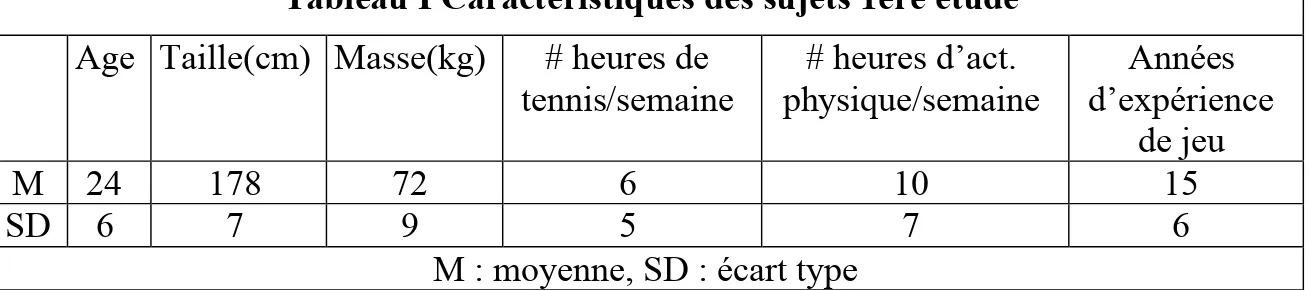
4.1.1. Sujets :Dix-huit joueurs de tennis (2 femmes, 16 hommes) en santé (10 ayant déjà souffert de douleur lombaire) ont été recrutés pour participer à cette étude (voir tableau I). Le protocole de l’étude a été révisé et approuvé par le comité d’éthique des sciences de la santé de l’université de Montréal.
Tableau I Caractéristiques des sujets 1ère étude
Age Taille(cm) Masse(kg) # heures de tennis/semaine # heures d’act. physique/semaine Années d’expérience de jeu M 24 178 72 6 10 15 SD 6 7 9 5 7 6
M : moyenne, SD : écart type
4.1.2. Le protocole ou « design » expérimental :
Les sujets intéressés ont disposé d’une période de réflexion de 48h pour donner leur accord et retourner les formulaires d’information et de consentement signés. Pour être inclus dans l’étude les sujets devaient : être un homme ou une femme âgé entre 18 et 45
ans et jouer au tennis au moins une heure par semaine. Pour être exclus de l’étude, les sujets devaient : (1) souffrir ou avoir souffert de douleur lombaire, sacro-iliaque ou à l’aine dans la dernière année, (2) souffrir d’un étirement musculaire des fléchisseurs de la hanche, des extenseurs du genou ou des muscles abdominaux, (3) être enceinte ou avoir accouché depuis moins d’un an, (4) souffrir d'une pathologie somatique ou psychiatrique évolutive connue. Suite à leur inclusion, les sujets ont été évalués avec le nouveau test du SAJT lors de deux rencontres, conjointement par deux physiothérapeutes, l’un avec 5 ans et l’autre avec 3 ans d’expérience clinique. Les rencontres ont duré approximativement vingt minutes. La deuxième évaluation a eu lieu au moins quarante-huit heures après la première. Les évaluateurs n’avaient pas accès à leurs évaluations précédentes. On a demandé aux participants d’éviter toute activité physique induisant de la fatigue lors des journées d’évaluation en plus d’éviter d’entraîner leurs muscles abdominaux entre les deux évaluations. Il faut ajouter qu’une rencontre d’une heure a été organisée avant le début de la recherche pour que les évaluateurs se familiarisent avec le test du soulèvement actif de la jambe tendue.
4.1.3. Déroulement des tests :
4.1.3.1. Le soulèvement actif de la jambe tendue ou « active straight leg raise »
On a demandé au sujet de s’étendre sur une table de traitement (Adapta 300, Chattanooga Medical Supply, Inc, Etats-Unis) en décubitus dorsal (sur le dos) avec les jambes tendues et les pieds écartés de 20 cm. Le sujet devait élever une jambe 20 cm au-dessus de la table sans plier le genou et tenir la position une à deux secondes avant de redescendre la jambe (voir figure 4). Le sujet devait relaxer complètement les muscles du tronc et du membre inférieur avant de soulever de nouveau la jambe. La jambe était re-soulevée à la demande des évaluateurs. La procédure a été la même pour le soulèvement actif de l’autre jambe. Les évaluateurs étaient placés du côté opposé à la jambe qui était levée, et placés entre le genou et la taille du sujet. Ce positionnement a
permis une vision optimale de la région abdominale. Les thérapeutes ont fait la lecture de leurs observations en même temps. Ils ont chacun inscrit leurs résultats sur une feuille différente sans voir l’évaluation de l’autre. Les évaluateurs avaient à observer les stratégies de stabilisation de la région lombo-pelvienne du sujet lors du soulèvement de chaque jambe. Ces stratégies ont été rapportées par Lee[18]. Ils devaient remarquer si certains patrons de compensations étaient présents. Trois mouvements de la région abdominale ont été évalués : la rotation lombo-pelvienne ipsilatérale, l’extension thoraco-lombaire et l’excursion latérale du thorax par rapport au bassin. Ces compensations ont été cotées pour chaque jambe sur une échelle de 3 points: pas de compensation observable = 0, compensation minimale = 1, compensation modéré = 2, compensation importante = 3. Le score des trois compensations a été additionné pour chaque jambe sur 9 et réadditionné sur 18 pour le total des deux jambes. Les évaluateurs avaient aussi à observer si deux types de compensations musculaires étaient visibles : le gonflement de l’abdomen ou « bulging » et la dépression de la cage thoracique ou « bracing ». Un point était ajouté pour chaque compensation remarquée. Ainsi, le score final pour chaque jambe était additionné sur 11 et réadditionné sur 22 pour le total des deux jambes. Par la suite, on a aussi demandé au sujet d’évaluer la difficulté du levé de chaque jambe selon une échelle de six points: pas de difficulté = 0, difficulté minime = 1, modérée = 2, importante = 3, sévère = 4, impossible = 5. Le score des deux jambes a été additionné sur 10. Cette échelle de cotation a été déjà utilisée à plusieurs reprises avec le SAJT[192, 194, 196]. Ainsi, le test comprend une partie objective basée sur les observations d’un thérapeute et une partie subjective basée sur les sensations du sujet. Le test a été aussi fait avec une ceinture pelvienne faisant compression au niveau du bassin.
4.1.3.2. Le soulèvement actif de la jambe tendue avec compression antérieure de la ceinture pelvienne
Selon Lee, faire une compression antérieure au niveau des épines iliaques antéro-supérieures (ÉIAS) simule davantage l’action du transversus abdominis (TA) tandis qu’une compression postérieure au niveau des épines iliaques postéro-supérieures (ÉIPS) simule davantage l’action du multifidus (MT)[18]. On a aussi rapporté une normalisation du recrutement moteur du diaphragme et des muscles du plancher pelvien[72] et un recrutement plus rapide des grands fessiers avec une compression des os iliaques[198]. Donc, cette simulation musculaire peut permettre d’améliorer le contrôle moteur lombo-pelvien[18]. On a donc installé une ceinture pelvienne (The Compressor, www.dianelee.ca ) sous les épines iliaques du sujet. Cette position semble optimale pour compresser les os iliaques[197]. Par la suite, on a installé les sangles de velcro de chaque côté en appliquant une pression antéro-médiale (voir figure 5). La pression exercée par les sangles de velcro n’a pas été mesurée. Pour l’installation de la ceinture, le sujet était debout. On a ensuite demandé au sujet de s’étendre de nouveau sur la table et le test a été évalué de la même manière que précédemment.
4.1.3.3. Le soulèvement actif de la jambe tendue avec compression postérieure de la ceinture pelvienne
La procédure a été la même qu’à la condition précédente. Cependant les sangles de velcro ont été appliquées avec une pression postéro-médiale.
Figure 5 La ceinture lombo-pelvienne
4.2. Analyse statistique :
La normalité de la distribution et l'homogénéité de la variance ont été vérifiés au moyen du test de Shapiro-Wilk et du test de Levenne, respectivement. Un test t pour données appariées a été réalisé pour tester l'hypothèse nulle. Le coefficient de corrélation intraclasse (ICC) et l'erreur type de la mesure (SEM) ont été calculés selon la méthode proposée par Hayen et al.[199] pour vérifier la reproductibilité absolue (SEM) et relative (ICC).
4.3. Résultats :
Tableau II Reproductibilité intra-évaluateur
Éval.1 Éval.2 Conditions M SD M SD ICC SEM MD Sans compression Jambe G /11 1.36 1.10 1.22 1.02 0,83 0,66 1,83 Jambe D /11 1.39 1.08 1.56 1.03 0,81 0,62 1,72 Total /22 2.75 1.95 2.78 1.81 0,88 1,05 2,91 Compression antérieure Jambe G /11 1.67 0.96 1,14 0,68 0,82 0,77 2,13 Jambe D /11 1.47 0.91 1,00 0,76 0,93 0,68 1,88 Total /22 3.14 1.62 2,14 1,15 0,92 1,19 3,30 Compression postérieure Jambe G /11 1.39 1.05 0,92 0,73 0,89 0,67 1,86 Jambe D /11 1.19 1.06 0,86 0,76 0,87 0,70 1,94 Total /22 2.58 1.89 1,78 1,15 0,92 1,10 3,05 ICC : coefficient de corrélation intraclasse , SEM : erreur standard de mesure,
Tableau III Reproductibilité inter-évaluateurs Éval.1 Éval.2 Conditions M SD M SD ICC SEM MD Sans compression Jambe G /11 1,64 1,07 0,94 0,92 0,79 2,88 7,98 Jambe D /11 1,78 1,12 1,17 0,88 0,73 2,88 7,98 Total /22 3,42 2,05 2,11 1,41 0,82 2,97 8,23 Compression antérieure Jambe G /11 1,94 0,95 1,14 0,68 0,84 2,89 8,01 Jambe D /11 2,14 0,83 1,00 0,76 0,92 2,88 7,98 Total /22 4,08 1,50 2,14 1,15 0,93 2,97 8,23 Compression postérieure Jambe G /11 1,81 1,04 0,92 0,73 0,86 2,88 7,98 Jambe D /11 1,72 0,97 0,86 0,76 0,81 2,88 7,98 Total /22 3,53 1,78 1,78 1,15 0,89 2,96 8,20 ICC : coefficient de corrélation intraclasse , SEM : erreur standard de mesure,
MD : différence minimale, , G : gauche, D : droite
Nous avons considéré qu’un ICC de plus de 0,90 comme étant très élevé, un ICC entre 0,70 et 0,89 comme étant élevé et un ICC entre 0,50 et 0,69 comme étant modéré[201]. Currier a suggéré qu’un ICC de plus de 0,80 comme cliniquement acceptable[202]. On a trouvé que le SAJT a une reproductibilité intra-examinateur élevé à très élevé démontrée par la reproductibilité relative variant de ICC = 0.81 à 0.93 (voir tableau II) . On a trouvé un MD , qui est la différence minimale pour considérer un changement réel d’environ 3 points sur 22 pour les trois conditions du test (voir tableau II). Les calculs pour la reproductibilité inter-évaluateurs montre une reproductibilité relative élevée à très élevée (ICC variant de 0.82 à 0.93) pour le total des deux jambes, pour chaque condition (voir tableau III). Cependant la MD est d’environ 8 points sur 22 (voir tableau III).
5. Validité du SAJT :
Le contrôle altéré des muscles de la région lombo-pelvienne est observé chez les sujets souffrant de douleur au bas du dos et aux membres inférieurs[14, 20, 28, 46, 66, 85, 86, 203]. Cependant, on s’aperçoit que cette activation neuromusculaire non-optimale ne se rétablit pas d’elle-même lorsque la douleur disparaît et qu’elle est fortement associée à des récidives de douleur lombaire[85]. De plus, des études prospectives ont rapporté qu’un déficit préexistant du recrutement moteur des muscles du tronc est un facteur de risque de blessures à la colonne vertébrale et aux structures du membre inférieur[116, 178, 179, 182]. De ces résultats, l’identification des sujets n’ayant pas une bonne coordination motrice est primordiale pour les cliniciens et entraîneurs physiques pour qu’ils puissent intervenir avec des exercices de stabilité efficaces. Le soulèvement actif de la jambe tendue (SAJT) semble un test fonctionnel tout à fait approprié pour observer le comportement des muscles du « core » et les mouvements compensatoires du bassin et de la cage thoracique. Dans cette deuxième partie d’étude, il sera intéressant d’évaluer la capacité à mesurer le changement du SAJT après un entraînement de six semaines d’exercices de stabilité mettant l’emphase sur le recrutement moteur des muscles profonds de la cavité abdominale soit le transverse de l’abdomen, le multifidus, le diaphragme et les muscles du plancher pelvien. Le choix de ce type d’entraînement est basé sur l’accumulation grandissante de données scientifiques qui vantent son efficacité à rétablir un équilibre musculaire entre les muscles locaux et globaux de la région lombo-pelvienne[2, 121, 127, 182]. En effet, un contrôle moteur altéré est associé à une hypoactivité des muscles profonds du tronc et à une hyperactivité de des muscles superficiels[66, 87]. Ainsi, en plus de récolter de l’information sur le SAJT on pourra ajouter des données à celles déjà existantes sur les exercices de stabilité segmentaire. On peut donc poser l’hypothèse que le SAJT est valide pour détecter un changement du contrôle moteur après un entraînement de six semaines de stabilité du « core ».
5.1. Matériel et méthode :
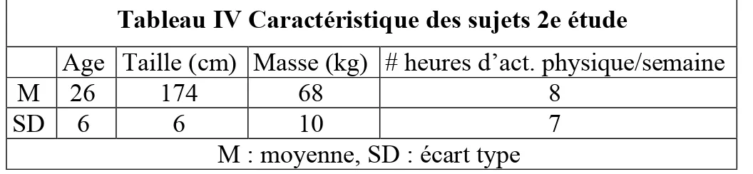
5.1.1 Sujets :Treize sujets actifs (4 femmes, 9 hommes) en santé (9 ayant déjà souffert de douleur lombaire) ont participé à la deuxième partie de l’étude (voir tableau IV). Deux autres sujets initialement inclus dans le groupe expérimental ont quitté le projet en cours de route. Le protocole de l’étude a été révisé et approuvé par le comité d’éthique des sciences de la santé de l’université de Montréal.
Tableau IV Caractéristique des sujets 2e étude
Age Taille (cm) Masse (kg) # heures d’act. physique/semaine
M 26 174 68 8
SD 6 6 10 7
M : moyenne, SD : écart type
5.1.2. Le protocole ou « design » expérimental :
Les sujets intéressés ont disposé d’une période de réflexion de 48h pour donner leur accord et retourner les formulaires d’information et de consentement signés. Pour être inclus dans l’étude les sujets devaient : (1) être un homme ou une femme âgé entre 18 et 45 ans, (2) faire au moins une heure d’activité physique par semaine, (3) avoir 3/22 avec le test du SAJT ou avoir un score au SAJT diminué par une compression des os iliaques (antérieure ou postérieure). On a supposé que les sujets ayant un pauvre recrutement des muscles locaux répondraient davantage à l’entraînement des muscles locaux ce qui est le cas avec des patients souffrant de douleur chronique au bas du dos[126]. Les sujets exclus devaient : (1) souffrir ou avoir souffert de douleur lombaire, sacro-iliaque ou à l’aine dans la dernière année, (2) souffrir d’un étirement musculaire soit des fléchisseurs de la hanche, des extenseurs du genou ou des muscles abdominaux, (3) être enceinte ou avoir accouché depuis moins d’un an, (4) souffrir d'une pathologie somatique ou psychiatrique évolutive connue, (5) avoir un score au SAJT inférieur à 3/22 ou avoir un
score qui augmentait ou qui ne variait pas avec la compression des os iliaques. Les sujets ont été aléatoirement séparés entre le groupe expérimental et le groupe contrôle au moment de leur inclusion. Les sujets ont été évalués à deux reprises avec le SAJT, par un physiothérapeute (responsable de l’étude), lors de leur inclusion et 6 semaines plus tard à la fin du programme d’exercices. Le groupe expérimental a eu à faire un entraînement de stabilité segmentaire de 6 semaines tandis que le groupe contrôle n’a eu qu’à continuer les activités préalables à son inclusion.
5.1.3. Intervention :
La période d’entraînement a été de six semaines. Elle a été divisée en deux phases, une de deux semaines et l’autre de quatre semaines. Le premier stade était constitué de quatre exercices n’impliquant aucun mouvement de la colonne ou des membres inférieurs et où l’emphase était mise sur le recrutement des muscles profonds du « core ». Le second stade était composé de quatre exercices plus dynamiques qui demandaient prioritairement la contraction des muscles profonds mais qui incluaient aussi le travail des muscles superficiels. Les exercices de la phase 1 avaient quelques points en commun : une co-contraction lente et douce (10-15 % CVM) du TA et du MT, le maintient d’une colonne neutre et d’un patron de respiration normale, ainsi que la relaxation des muscles superficiels. Puisque que les muscles locaux sont profonds, la façon de les recruter est plus subtile. Pour bien contracter ces muscles on a utilisé des images mentales. Il semble que chaque personne réponde différemment aux images mentales[18]. Ainsi, on a dû trouver pour chaque patient la pensée qui permettait la meilleure contraction du TA et du MT avec le moins possible de substitution des muscles superficiels. On a aussi montré au sujet comment palper le TA et le MT pour qu’il soit en mesure d’avoir plus de feedback lorsqu’il était seul à la maison. Lors des séances de groupe, le thérapeute (responsable de l’étude) a donné de la rétroaction visuelle, verbale et tactile aux sujets pour faciliter le nouvel apprentissage moteur. La
phase 2 demandait aux sujets de recruter les muscles profonds avant d’exécuter certains mouvements des jambes et du tronc. Le thérapeute s’assurant de la contraction de l’unité profonde a mis l’accent sur la qualité des patrons de mouvement. Ce type de programme d’entraînement a déjà été utilisé par le passé pour stimuler le recrutement des muscles de la cavité abdominale[18, 19, 122, 186]. Le volume d’entraînement a été augmenté après la première semaine et diminué au début de la troisième semaine avec l’introduction des exercices dynamiques. À partir de la troisième semaine, le volume d’entraînement a été maintenu jusqu’à la fin de l’étude (voir tableau V). Pendant toute la durée du projet, les sujets ont eu à faire les exercices six fois par semaine (5 fois à la maison + 1 session de groupe). La session de groupe a permis de s’assurer de la bonne exécution des exercices, de leur progression et de garder la motivation des participants. Pour s’assurer que les sujets contractaient bien le TA et le MT, le thérapeute s’est fié à ses qualités de palpation. Aucun ultrason, ni sphygmomanomètre (PBU « pressure biofeedback unit ») n’ont été utilisés. Lors de la première rencontre suite à la présentation des exercices un document d’éducation, rappelant la théorie et les exercices à faire, a été remis (voir annexe 1). De plus les sujets du groupe expérimental ont dû remplir un journal de bord qui était vérifié à chaque rencontre hebdomadaire.
Tableau V Volume d’entraînement par semaine
Programme d’entraînement de 6 semaines # de séries, # de répétitions et temps de contraction (secondes) Volume total d’entraînement : (# d’exercices x # de séries x # de répétitions x temps de contraction x 6 jours) Semaine 1 3 séries x 5 reps x 5 sec 4 x 3 x 5 x 5 x 6 = 1800 Semaine 2 3 séries x 10 reps x 10 sec 4 x 3 x 10 x 10 x 6 = 7200 Semaine 3,4,5,6 3 séries x 10 reps x 5sec 4 x 3 x 10 x 5 x 6 = 3600
5.1.4. Déroulement des tests :
5.2. Analyse statistique :
Une ANOVA à deux voies (groupe x temps) a été réalisée pour tester l'hypothèse nulle. L'amplitude de la différence entre les groupes a été calculée au moyen de l'effect size (ES). La différence était considérée comme triviale quand ES < 0.2, petite quand ES < 0.5, modérée quand ES < 0.8 et élevée quand ES > 0.8. Un risque alpha de 5% a été retenu pour tous les tests, qui ont été réalisés avec le logiciel Statistica (Statsoft, Tulsa, Etats Unis). D'après la méthode proposée par Bausel and Li[200] , il aurait fallu 10 participants dans chaque groupe pour que la probabilité qu'un effect size d'interaction comparable à celui obtenu dans notre étude (0.80 en moyenne) soit de 80% (alpha < 0.05), et 12 participants par groupe si nous avions voulu porter cette probabilité à 90% (alpha < 0.05).
5.3. Résultats :
Tableau VI Effet de l’entraînement (Sans compression)
Total 22 Exp. Cont.
Pré Moyenne 4,50 3,60
SD 1,93 1,52
Post Moyenne 3,25 4,60
SD 1,16 1,95
Effect Size -0,81 0,58
Pré : évaluation pré-entraînement, Post : évaluation post-entraînement, Exp. : groupe expérimental, Cont. : groupe
Tableau VII Effet de l’entraînement (Compression antérieure)
Total 22 Exp. Cont.
Pré Moyenne 4,63 4,20
SD 1,51 1,30
Post Moyenne 3,63 4,20
SD 0,74 1,79
Effect Size -0,89 0,00
Pré : évaluation pré-entraînement, Post : évaluation post-entraînement, Exp. : groupe expérimental, Cont. : groupe
contrôle, SD : écart type
Tableau VIII Effet de l’entraînement (Compression postérieure)
Total 22 Exp. Cont.
Pré Moyenne 4,13 2,60
SD 2,10 2,07
Post Moyenne 2,25 2,80
SD 1,28 1,30
Effect Size -1,11 0,12
Pré : évaluation pré-entraînement, Post : évaluation post-entraînement, Exp. : groupe expérimental, Cont. : groupe
contrôle, SD : écart type
Puisque le nombre de sujets inclus dans l’étude est faible et que le nombre de sujets dans chaque groupe est inégal, un calcul statistique, l’«effect size » (ES) a été utilisé pour comparer les groupes. Ce calcul statistique permet d’évaluer l’effet de l’entraînement indépendamment de la taille de l’échantillon. Un ES de plus de 0.8 considère qu’il y a une grande différence causée par le traitement. Un ES de 0.5 montre un changement modéré tandis qu’un ES de moins de 0.2 démontre un faible effet. Le groupe expérimental a un ES de -0.81, -0.89 et -1.11 pour les conditions sans compression, avec compression antérieure et avec compression postérieure respectivement. Le signe négatif indique une diminution du score final par rapport au score initial. De son côté, le groupe contrôle a un ES de 0.58, 0.00 et 0.12 pour les conditions sans compression, avec compression antérieure et avec compression postérieure respectivement.