Université de Montréal
Le Diabète Maternel Influence la Morphogenèse Rénale et la
Programmation Périnatale
par Yun-Wen Chen
Programme de Sciences biomédicales Faculté de Médecine
Thése présentée à la Faculté des études supérieures en vue de l’obtention du grade de docteurés sciences (Ph.D)
en Sciences biomédicales
Novembre, 2009 © Yun-Wen Chen, 2009
Université de Montréal Faculté des études supérieures
Cette thèse intitulée :
Le Diabète Maternel Influence la Morphogenèse Rénale et la
Programmation Périnatale
présentée par : Yun-Wen Chen
a été évaluée par un jury composé des personnes suivantes :
Dr Jean-Francois Cailhier Président rapporteur Dre Shao-Ling Zhang Directeur de recherche Dr John S.D.Chan Codirecteur de recherche Dre Anne-Monique Nuyt membre du jury Dre Indra Gupta Examinateur externe
Résumé
Le diabète maternel est un facteur de risque majeur pour le développement de malformations congénitales. Dans le syndrome de l’embryopathie diabétique, l’exposition prolongée du fœtus à de hautes concentrations ambientes de glucose induit des dommages qui peuvent affecter plusieurs organes, dont les reins. Les malformations rénales sont la cause de près de 40 pourcent des cas d’insuffisance rénale infantile. L’hyperglycémie constitue un environnement utérin adverse qui nuit à la néphrogenèse et peut causer l’agenèse, la dysplasie (aplasie) ou l’hypoplasie rénale. Les mécanismes moléculaires par lesquels les hautes concentrations ambientes de glucose mènent à la dysmorphogenèse et aux malformations demeurent toutefois mal définis.
Le diabète maternel prédispose aussi la progéniture au développement d’autres problèmes à l’âge adulte, tels l’hypertension, l’obésité et le diabète de type 2. Ce phénomène appelé ‘programmation périnatale’ a suscité l’intérêt au cours des dernières décennies, mais les mécanismes responsables demeurent mal compris.
Mes études doctorales visaient à élucider les mécanismes moléculaires par lesquels le diabète maternel ou un environnement in utero hyperglycémique affecte la néphrogenèse et programme par la suite la progéniture a développer de l’hypertension par des observations in vitro, ex vivo et in vivo. Nous avons utilisé les cellules MK4, des cellules embryonnaires du mésenchyme métanéphrique de souris, pour nos études in vitro et deux lignées de souris transgéniques (Tg) pour nos études
ex vivo et in vivo, soient les souris HoxB7-GFP-Tg et Nephrin-CFP-Tg. Les souris
HoxB7-GFP-Tg expriment la protéine fluorescente verte (GFP) dans le bourgeon urétérique (UB), sous le contrôle du promoteur HoxB7. Les souris Nephrin-CFP expriment la protéine fluorescente cyan (CFP) dans les glomérules, sous le contrôle du promoteur nephrin spécifique aux podocytes.
Nos études in vitro visaient à déterminer si les hautes concentrations de glucose modulent l’expression du gène Pax2 dans les cellules MK4. Les cellules MK4 ont été traitées pendant 24h avec du milieu contenant soit 5mM D-glucose et 20mM D-mannitol ou 25mM D-glucose et avec ou sans antioxydants ou inhibiteurs de p38 MAPK, p44/42 MAPK, PKC et NF-kB. Nos résultats ont démontré que le D-glucose élevé (25mM) augmente la génération des espèces réactives de l’oxygène (ROS) dans les cellules MK4 et induit spécifiquement l’expression du gène Pax2. Des analogues
du glucose tels le D-mannitol, L-glucose ou le 2-Deoxy-D-glucose n’induisent pas cette augmentation dans les cellules MK4. La stimulation de l’expression du gène Pax2 par le D-glucose dans les cellules MK4 peut être bloquée par des inhibiteurs des ROS et de NF-kB, mais pas par des inhibiteurs de p38 MAPK, p44/42 MAPK ou PKC. Ces résultats indiquent que la stimulation de l’expression du gène Pax2 par les concentrations élevées de glucose est due, au moins en partie, à la génération des ROS et l’activation de la voie de signalisation NF-kB, et non pas via les voies PKC, p38 MAPK et p44/42 MAPK.
Nos études ex vivo s’intéressaient aux effets d’un milieu hyperglycémique sur la morphogenèse de la ramification du bourgeon urétérique (UB). Des explants de reins embryonnaires (E12 à E18) ont été prélevés par micro-dissection de femelles HoxB7-GFP gestantes. Les explants ont ensuite été cultivés dans un milieu contenant soit 5mM D-glucose et 20mM D-mannitol ou 25mM D-glucose et avec ou sans antioxydants, catalase ou inhibiteur de PI3K/AKT pour diverses durées. Nos résultats ont démontré que le D-glucose stimule la ramification du UB de manière spécifique, et ce via l’expression du gène Pax2. Cette augmentation de la ramification et de l’expression du gène Pax2 peut être bloquée par des inhibiteurs des ROS et de PI3K/AKT. Ces études ont démontré que les hautes concentrations de glucose altèrent la morphogenèse de la ramification du UB via l’expression de Pax2. L’effet stimulant du glucose semble s’effectuer via la génération des ROS et l’activation de la voie de signalisation Akt.
Nos études in vivo visaient à déterminer le rôle fondamental du diabète maternel sur les défauts de morphogenèse rénale chez la progéniture. Dans notre modèle animal, le diabète maternel est induit par le streptozotocin (STZ) chez des femelles HoxB7-GFP gestantes (E13). Les souriceaux ont été étudiés à différents âges (naissants et âgés de une, deux ou trois semaines). Nous avons examiné leurs morphologie rénale, nombre de néphrons, expression génique et les événements apoptotiques lors de cette étude à court terme. La progéniture des mères diabétiques avait un plus faible poids, taille et poids des reins, et possédait des glomérules plus petits et moins de néphrons par rapport à la progéniture des mères contrôles. La dysmorphogenèse rénale observée est peut-être causée par l’augmentation de l’apoptose des cellules dans la région du glomérule. Nos résultats ont montré que les souriceaux nés de mères diabétiques possèdent plus de podocytes apoptotiques et plus de marquage contre la caspase-3 active dans leurs tubules rénaux que la
progéniture des mères contrôles. Les souriceaux des mères diabétiques montrent une augmentation de l’expression des composants du système rénine angiotensine (RAS) intrarénal comme l’angiotensinogène et la rénine, ainsi qu’une augmentation des isoformes p50 et p65 de NF-kB. Ces résultats indiquent que le diabète maternel active le RAS intrarénal et induit l’apoptose des glomérules, menant à une altération de la morphogenèse rénale de la progéniture.
En conclusion, nos études ont permis de démontrer que le glucose élevé ou l’environnement in utero diabétique altère la morphogenèse du UB, qui résulte en un retard dans la néphrogenèse et produit des reins plus petits. Cet effet est dû, au moins en partie, à la génération des ROS, à l’activation du RAS intrarénal et à la voie NF-kB.
Nos études futures se concentreront sur les mécanismes par lesquels le diabète maternel induit la programmation périnatale de l’hypertension chez la progéniture adulte. Cette étude à long terme porte sur trois types de progénitures : adultes nés de mères contrôles, diabétiques ou diabétiques traitées avec insuline pendant la gestation. Nous observerons la pression systolique, la morphologie rénale et l’expression de divers gènes et protéines. Nous voulons de plus déterminer si la présence d’un système antioxydant (catalase) peut protéger la progéniture des effets néfastes des ROS causés par l’environnement in utero hyperglycémique. Les souris Catalase-Tg expriment la catalase spécifiquement dans les tubules proximaux et nous permettrons d’explorer notre hypothèse sur le rôle des ROS dans notre modèle expérimental de diabète maternel.
Mots-clés: diabète maternel; hyperglycémie; néphrogenèse; système rénine
angiotensine; Pax2; apoptose; espèces réactives de l’oxygène; programmation périnatale
Abstract
Maternal diabetes is a major risk factor for congenital malformations. When the fetus is exposed to high, sustained, ambient glucose levels, widespread fetal damage may affect multiple organs, including the kidneys, evoking diabetic embryopathy syndrome. Renal malformations account for up to 40% of childhood renal failure cases. Hyperglycemia constitutes an adverse in utero environment that dynamically impairs nephrogenesis, resulting in renal agenesis, dysplasia, aplasia or hypoplasia. However, the molecular mechanisms by which high, ambient glucose levels lead to renal dysmorphogenesis and birth defects have not yet been delineated.
Maternal diabetes also programs the offspring to develop other problems later in life, such as hypertension, obesity and type 2 diabetes. This phenomenon, called ‘perinatal programming’, has attracted worldwide attention in recent decades, yet the mechanisms by which it occurs are incompletely understood.
My PhD studies are designed to elucidate the underlying molecular pathways by which maternal diabetes or hyperglycemic environments in utero impair nephrogenesis and subsequently make the offspring develop perinatal programming of hypertension in vitro, ex vivo and in vivo. We employed mouse embryonic metanephric mesenchyme cells, namely MK4 cells, for our in vitro experiments, and 2 transgenic (Tg) mouse lines, Hoxb7-GFP-Tg and Nephrin-CFP-Tg mice, for ex vivo and in vivo investigations. Hoxb7-GFP-Tg mice specifically express green fluorescent protein (GFP) in ureteric buds (UB), driven by the Hoxb7 promoter. Nephrin-CFP-Tg mice express cyan fluorescent protein (CFP) in glomeruli, driven by the podocyte-specific nephrin promoter.
In our in vitro studies, we examined whether high glucose alters Pax2 gene expresson in MK4 cells. The cells were treated with either 5 mM D-glucose plus 20 mM D-mannitol or 25 mM D-glucose media with or without reactive oxygen species (ROS) blockers (DPI, rotenone), and inhibitors of p38 mitogen-activated protein kinase (MAPK) (SB203580), p44/22 MAPK (PD98059), protein kinase C (PKC) (GF109203X), or nuclear factor kappa B (NK-kB) (PDTC) for 24-hr incubation. Our data showed that high D-glucose (25 mM) increased ROS generation and specifically induced Pax2 gene expression, but not other glucose analogs such as D-mannitol, L-glucose or 2-deoxy-D-glucose in MK4 cells. The stimulatory effect of high D-glucose on Pax2 gene expression could be blocked by ROS and NF-kB inhibitors in
MK4 cells but not by inhibitors of p38 MAPK (SB203580), p44/22 MAPK (PD98059), and PKC (GFX) in MK4 cells. These data indicated that the stimulatory effect of high glucose on Pax2 gene expression is mediated, at least in part, via ROS generation and activation of NF-κB, but not via the PKC, p38 MAPK and p44/42 MAPK signalling pathways.
In our ex vivo studies, we investigated the influence of a high-glucose milieu on UB branching morphogenesis. Kidney explants (E12 to E18) were microdissected from timed-pregnant Hoxb7-GFP mice and cultured with either 5 mM D-glucose plus 20 mM D-mannitol or 25 mM D-glucose media with or without ROS blockers (DPI, rotenone), catalase and phosphoinositide-3-kinase (PI3K)/AKT inhibitor at different time points, depending on the experiment. We found that high D-glucose specifically stimulated UB branching in a time-dependent manner. High D-glucose stimulation of UB branching morphogenesis was mediated via Pax2 gene expression. High D-glucose-induced UB branching and Pax2 gene expression could be blocked by ROS and PI3K/AKT inhibitors. These studies demonstrated that high glucose alters UB branching morphogenesis via Pax2 gene and protein expression. The stimulatory effect of high glucose seems to be mediated via ROS generation and activation of the AKT signalling pathway.
In our in vivo studies, we explored the fundamental role of maternal diabetes on renal morphogenesis impairment in offspring. In our experimental model, maternal diabetes was induced by streptozotocin in pregnant Hoxb7-GFP mice at embryonic day 13. The offspring were examined at several time points after birth (neonatal, 1 week, 2 weeks, and 3 weeks) with follow-up of kidney morphology, nephron number, gene expression, and apoptotic events in this short-term postnatal experiment. We observed that the offspring of diabetic mice had lower body weight, body size, kidney weight, small volume of glomeruli and a reduced number of nephrons in comparison to non-diabetic control offspring. Renal dysmorphogenesis may have been the result of increased cell apoptosis in glomeruli. Our findings showed that the offspring of diabetic mice displayed significantly more apoptotic podocytes as well as augmented active caspase-3 immunostaining in renal tubules compared to control mice offspring. Diabetic mice offspring presented heightened expression of intrarenal renin-angiotensin system (RAS) components, such as angiotensinogen and renin, with
upregulation of p50 and p65 NF-kB isoforms. These data indicated that maternal diabetes activates the intrarenal RAS and induces glomerular apoptosis, resulting in impairment of renal morphogenesis in diabetic offspring.
In conclusion, our findings indicated that a high-glucose milieu in utero or maternal diabetic alters UB morphogenesis, culminating in retardation of nephrogenesis with smaller kidney size. The underlying mechanism(s) is mediated, at least in part, via ROS generation and activation of the intrarenal RAS and NF-kB pathways.
In the future, we aim to investigate the underlying mechanism(s) of how maternal diabetes induces perinatal programming of adult hypertension in offspring in vivo. This long-term postnatal study will be undertaken in 3 groups: adult offspring (20 weeks) of control mice, adult offspring of diabetic pregnant mice, and adult offspring of insulin-treated, diabetic, pregnant mice. We will follow-up by tracking hypertension, kidney morphology, and gene expression. Furthermore, we also plan to determine whether an antioxidant system (catalase) can protect against an hyperglycemic environment in utero that affects embryonic organogenesis via an increase in ROS generation. Catalase-Tg mice that specifically overexpress catalase in proximal tubules will be tested. Such Tg mice with catalase overexpression represent a model for exploring our hypothesis on the role of ROS in gestational diabetes.
Keywords : maternal diabetes, hyperglycemia, nephrogenesis, renin-angiotensin
Table of Contents Résumé………...………iii Abstract………...vi Table of Contents.………...………..ix List of Tables..………..……….xvi List of Figures.………..………xvii List of Abbreviations..………...………..………..xxi Acknowledgments….……….xxvi Curriculum Vita...xxvii
CHAPTER 1 MATERNAL DIABETES 1 Maternal Diabetes or Gestational Diabetes Mellitus ...………….…...……….1
1.1. PGDM………..………...1
1.2. GDM………...………1
1.3. Prevalence and Epidemiological Studies in GDM…………...………1
1.4. Risk Factors Related to GDM……….……….………2
1.5. Pathophysiology of GDM……….………...3 1.6. GDM Complications………...5 1.6.1. Maternal Complications of GDM……….5 1.6.1.1. Diabetic Retinopathy………...…..5 1.6.1.2. DN……….………...….6 1.6.1.3. Hypertension………...…..7 1.6.1.4. Development of T2DM……….………7 1.6.1.5. Premature Labor…...………...9 1.6.1.6. Infection……….9 1.6.1.7. Cesarean Section………9
1.6.2. Impact of Maternal Diabetes Complications on the Progeny………..…..10
1.6.2.1. Short-term Impact of Maternal Diabetes Complications on ODMs………...11
1.6.2.1.1. Macrosomia………..………11
1.6.2.1.3. Stillbirth………..………..13
1.6.2.1.4. Birth Defects……….13
1.6.2.1.5. Perinatal Morbidity………...…15
1.6.2.1.5.1. Neonatal Hypoglycemia………..……….……15
1.6.2.1.5.2. Other Neonatal Metabolic Problem………...……..16
1.6.2.2. Long-term Impact of GDM Complications on ODMs ……….17
1.6.2.2.1. Obesity………..17
1.6.2.2.2. Diabetes Mellitus……….……….……...17
1.6.2.2.3. Perinatal Programming………...…………..18
CHAPETER 2 PERINATAL PROGRMMING 2.1. Perinatal Programming………...………...19
2.2. Perintal Programming related Hypothesis…...………..19
2.2.1. Barker’s Hypothesis……….……….19
2.2.2. Brenner’s Hypothesis………..…………...20
2.2.3. The “thrifty phenotype” Hypothesis………21
2.3. Clinical and Experimental Evidences on Perinatal Programming……….……..22
2.3.1. Clinical Studies of Perinatal Programming………...………22
2.3.2. Experimental Studies of Perinatal Progamming……….…..…….22
2.4. Possible Mechanisms of Perinatal Programming………..……....23
2.4.1. LBW and Low Nephron Numbers………...………….24
2.4.2. Low Nephron Number and Hypertension……..………..……….24
2.4.3. Possible Mechanisms Involved in Low Nephron Number Associated with Intrauterine Alteration……….…..……….……...………..25 2.4.3.1. Genetic Influence………...……….25 2.4.3.2. Nutrition……….….……….………...26 2.4.3.3. Vitamin A Deficiency………...………..27 2.4.3.4. ROS………...…………...27 2.4.3.5. Apoptosis………...……….28 2.4.3.6. RAS...………...………...29 2.4.3.7. Glucocorticolds………..………….…………30 2.4.3.8. Social/Behavioral……….30
CHAPTER 3 NEPHROGENESIS OR KIDNEY DEVLOPMENT 3. Nephrogenesis or Kidney Development……….…………..…...….………31
3.2. Mesonephros.………..…..33
3.3 Metanephros...……….………….….34
3.4. Gene Regulation..….……….35
3.4.1. Pax2/Pax8 and Lim1: Earliest Markers of Nephrogenesis…………....……..36
3.4.2. UB Outgrowth Requires Signaling: WT-1, Eyes-absent-1 (Eya 1) and the GDNF/c-ret pathway………...37 3.4.3. The Wnt Pathway……….…….……39 3.5. Renal Malformations……….…………40 3.5.1. Renal Agenesis………...…41 3.5.2. Multiple Ureters……….41 3.5.3. Renal Dysgenesis………..………...…42 3.5.4. Renal Hypoplasia………...…42 3.5.5. OMN………...……….….…43 3.5.6. Polycystic Kidney………...…43 3.6. Pax2 Pathways………...…43
3.6.1. Pax Proteins and Gene Structure……….…43
3.6.2. Pax Genes in Organogenesis………..…45
3.6.3. Pax2 Mutations Cause HumanRCS...………46
3.6.4. Animal Models of Pax2 Mutations………...…….…47
3.6.5. Pax2 in Kidney Development………...……..…48
3.7. The GenitoUrinary Development Molecular Anatomy project………50
CHAPTER 4 MATERNAL DIABETES MODULATES NEPROGENESIS 4. Maternal Diabetes Modulates Nephrogenesis...52
4.1. Clinical Phenotypes or Outcome……….…….……….…52
4.2. Possible Mechanisms in Maternal Diabetes Modulates Nephrogenesis...53
4.2.1. Hyperglycemia………..……….…54
4.2.1.1. Hyperglycemia and Renal Malformation………55
4.2.2. ROS……….………..56
4.2.2.1. Hyperglycemia and ROS………57
4.2.3. Apoptosis………58
4.2.3.1. Hyperglycemia and Apoptosis………59
4.2.4. The NF-kB Pathway……….…………59
4.2.4.2. Oxidative stress and the NF-kB Pathway………..…………61
4.2.4.3. Apoptosis and the NF-kB Pathway……….62
4.2.4.4. The NF-kB Pathway and Protein 53 or Tumor Protein 53 (p53) Pathways……….………..………...62
4.2.5. The p53 Pathway……..……….……….…63
4.2.5.1. Hyperglycemia and The p53 Pathway...……….…………..…64
4.2.5.2. The p53 Pathway and Kidney Development..………64
4.2.5.3. The p53 Pathway and Renal Malformation………...…65
4.2.6. The intrarenal RAS………..………..……66
4.2.6.1. The intrarenal RAS and Kidney Development………67
4.2.6.2. The intrarenal RAS and Renal Malformation...69
4.2.6.3. Hyperglycemia and the intrarenal RAS………….……….…70
CHAPTER 5 EXPERIMENTAL APPROACHES 5.1. In Vitro Studies……….………71
5.1.1. Cell Line………72
5.2. Ex Vivo Studies……….………73
5.2.1. Animals………..………73
5.2.2. Metanephric Organ Culture………74
5.3. In Vivo Studies………..75
5.3.1. Animals………75
5.3.2. Maternal Diabetes Animals Models………75
5.3.2.1. STZ injection………..………75
5.3.2.2. BioBreeding (BB) Diabetes-prone Rats………..76
5.3.2.3. Ins2 Akita Mice………76
5.3.2.4. Non-obese Diabetic (NOD) Mice………77
5.3.2.5. Heterozygous Leprdb/+ mice...77
5.3.2.6. Rich Diet-induced Rats………77
5.3.3. Our Maternal Diabetes Animal Models………78
CHAPTER 6 OBJECTIVES OF THE PRESENT STUDIES CHAPTER 7 ARTICLES 1
Reactive Oxygen Species and nuclear factor-kappa B pathway mediate high glucose-induced Pax-2 gene expression in mouse embryonic mesenchymal epithelial cells and kidney explants
7.1 Abstract……….……….………81
7.2 Introduction………81
7.3 Materials and Methods……….………..……84
7.4 Results……….………...………89
7.5 Discussion……….………….………92
7.6 Acknowledgments……….96
7.7 References……….96
7.8 Legends and Figures………..104
CHAPTER 8 ARTICLES 2 Reactive Oxygen Species in the Presence of High Glucose Alter Ureteric Bud Morphogenesis 8.1 Abstract………113 8.2 Introduction……….……113 8.3 Results……….……115 8.4 Discussion………118 8.5 Concise Methods……….………122 8.6 Acknowledgments………127 8.7 References……….128
8.8 Legends and Figures………136
CHAPTER 9 ARTICLES 3 Maternal Diabetes Modulates Renal Morphogenesis in Offspring 9.1 Abstract……….…148
9.2 Introduction………148
9.3 Results………...………150
9.4 Discussion……….………152
9.5 Materials and Methods………..…155
9.6 Acknowledgments……….………160
9.7 References………...160
9.8 Legends and Figures………172
CHAPTER 10 DISCUSSION 10.1. To Investigate Whether High Glucose Alters Pax2 Gene Expression in Mouse Embryonic MM (MK4) cells……….………….183
10.1.1. Hyperglycemia Induces Pax2 Gene Expression………183
10.1.2. High D-glucose Induces ROS Generation in MK4 cell…………..………184
10.1.3. High D-glucose Induces Pax2 Gene Expression via Activiation of NF-kB Pathway………...185
10.1.4. Conclusion………186
10.2. To Demonstrate the Influence of a High-Glucose Milieu on UB Branching Morphogenesis……….187
10.2.1. High D-Glucose Stimulates UB Branching in a Time-dependent Manner..188
10.2.2. High D-glucose Specifically Stimulates UB Branching………..188
10.2.3. Stimulatory Effect of High D-Glucose on UB Branching Morphogenesis is Mediated via Pax2 Gene Expression………189
10.2.4. ROS -induced UB Branching Morphogenesis………189
10.2.5. High Glucose Stimulated UB Branching via AKT Signaling………….…190
10.2.6. Conclusion………191
10.3. To Delineate the Fundamental Role of Maternal Diabetes in Modulating Renal Morphrogenesis in the Offspring………191
10.3.1. STZ-induced Gestational Diabetes Model and STZ Toxicity………192
10.3.2. LBW is Associated with Maternal Diabetes……….……194
10.3.3. Hyperglycemia Delays Renal Formation………195
10.3.4. Nascent Nephrons Hypoplasis is Due to Apoptosis………197
10.3.5. Effect of Hyperglycemia and Intrarenal RAS Activation on Renal Damage………..198
10.3.6. Effect of Hyperglyce mia and NF-Kb Activation on Re na l Damage………..…………..199
10.3.7 Conclusion………..………200
CHAPTER 11 PERSPECTIVE RESEARCH 11.1 Maternal Diabetes and Perinatal Programming 11.1.1. Maternal Diabetes and Perinatal Programming……….202
11.1.2. Maternal Diabetes related to the Development of Obesity………..…202
11.1.3. Maternal Diabetes Induces Impaired Glucose Tolerance, Insulin Resistance, and Type 2 diabetes in the offspring………...203
11.1.4 Maternal Diabetes related to the Development of Hypertension and Renal Disease……….……….204 11.1.5. Possible Mechanism(s) of Maternal Diabetes on Perinatal Programming of Hypertension………...………..205 11.1.6 Effects of Perinatal Programming by Hyperglycemia in Adult Offspring of Diabetic Mothers (Age 20 Weeks)………206 11.2.Underlying Mechanism by Which NF-kB Promotes Apoptosis via the p53 Pathway in UB and MM Lineages………209 11.3. To Determine Whether an Antioxidant System (Catalase) can Protect against the Effects of Hyperglycemia in utero on Embryonic Organgenesis Affected by an increased in ROS generation……….211
CHAPTER 12 UNPUBLISHED DATA CHAPTER 13 REFERENCES
List of Tables Chapter 1
Table 1-1 Risk Factors for Gestational Diabetes………..3 Table 1-2 Risk factors for the worsening of diabetic retinopathy during pregnancy…6
Chapter 3
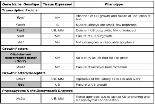
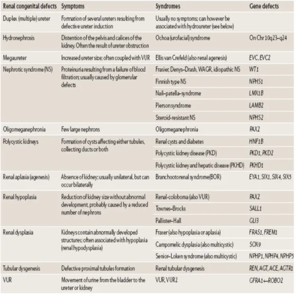
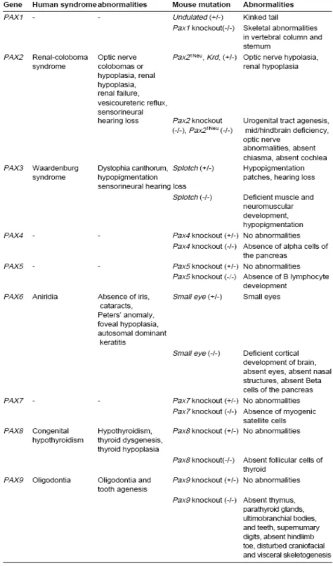
Table 3-1 A selection of genes in kidney development………..…36 Table 3-2 Congenital abnormalities and gene defects that have been identified in human patients………40 Table 3-3 Abnormalities in human and mice with heterozygous Pax gene
Mutations………46
Chapter 4
Table 4-1 Immunoreactivity to specific antibodies in the human mesonephros and metanephros during the 7th, 8th and 9th developmental weeks………..…65 Table 4-2 Expression of the renin-angiotensin system components during
metanephric kidney development………..67 Table 4-3 Effect of genetic inactivation of renin-angiotensin system genes in mice on the development of the renal collecting system………69
Chapter 6
Table 6-1 Average fetal birth weight and litter size born to wild-type (+/+) and
List of Figures Chapter 1
Figure 1-1 Cumulative incidence rate of diabetes mellitus………...8
Figure 1-2 Short- and Long-term complications in offspring of diabetic mother…10 Figure 1-3 Blood glucose levels correlate inversely to birth defect risk………15
Chapter 2 Figure 2-1 Hyperfiltration hypothesis………20
Figure 2-2 The original diagrammatic representation of the “thrifty phenotype” Hypothesis………...21
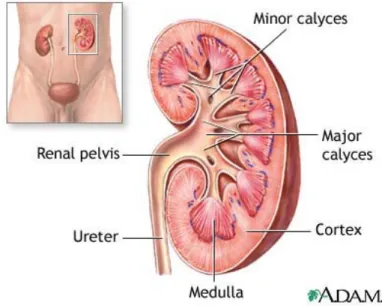
Chapter 3 Figure 3-1 Kidney structure ………31
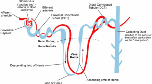
Figure 3-2 Simple diagram of kidney nephron ………32
Figure 3-3 Schematic representation of kidney development………32
Figure 3-4 Reciprocal induction in the development of the mammalian kidney..…35
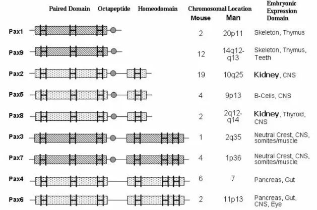
Figure 3-5 The Pax protein family……….…44
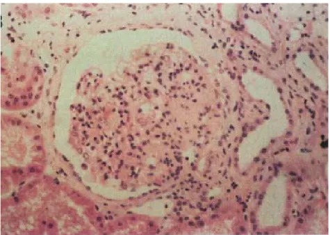
Figure 3-6 Renal cortical biopsy from a RCS patient ………..…47
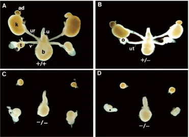
Figure 3-7 (A)wild type kidney (B) Pax21neu+/- mutant kidney………49
Figure 3-8 Analysis of the urogenital system defects in Pax2 mutant mice ………....49
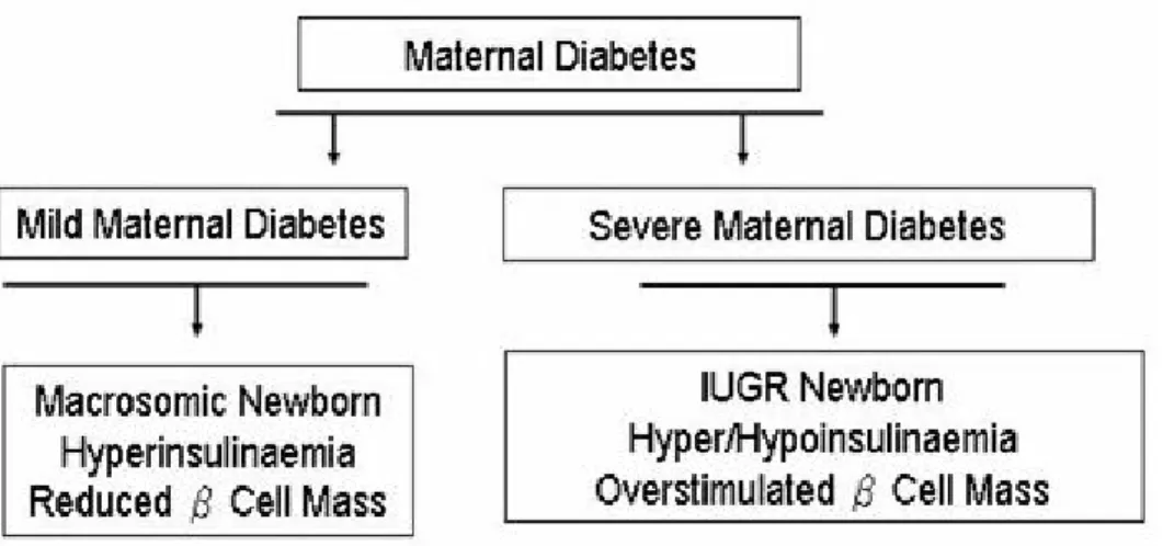
Chapter 4 Figure 4-1 Two opposite, abnormal fetal growth situation are associated with maternal diabetes……….53
Figure 4-2 Hypothetical model of diabetes-induced embryopathy………54
Figure 4-3 Potential mechanisms of hyperglycemic damage………55
Figure 4-4 Light micrographs depicting the effect of an high glucose enviroment on the morphology of murine embryonic metanephros……….…..56
Figure 4-5 The NF-kB pathway...60
Figure 4-6 A model of p53-mediated apoptosis………64
Figure 4-7 The RAS...66
Chapter 5 Figure 5-1 MK4 cell morphology………...…………71
Figure 5-2 The structure of D-gluocse, L-glucose, D-Mannitol and 2-Deoxy-D-glucose……….72
Figure 5-3 Expression of GFP in the ureteric bud derivatives of transgenic fetuses at
E12.5 through E16.5………74
Figure 5-4 Glomerular isolation under the dissecting microscope………75
Chapter 7 Figure 7-1 High glucose dose-dependent effect………104
Figure 7-2 High glucose specifical effect………..……105
Figure 7-3 Pax2 immunofluorescence staining……….106
Figure 7-4 p38 MAPK, p44/42 MAPK, and PKC inhibitor effect………107
Figure 7-5 High glucose induced ROS on Pax2 gene expression……….108
Figure 7-6 Inhibitor effect ROS and NF-kB inhibitors……….109
Figure 7-7 High glucose effect on Pax2 gene expression ex vivo………110
Figure 7-8 High glucose effect on the full length of 5’-franking region of pax2 promoter activity in MK4 cells……….111
Chapter 8 Figure 8-1 High D-glucose stimulates ureteric bud (UB) branching in a time-dependent manner………...136
Figure 8-2 High D-glucose on UB morphogenesis……….137
Figure 8-3 High D-glucose stimulates UB morphogenesis……….138
Figure 8-4 High glucose upregulates Pax2 expression in E18 kidney explants……139
Figure 8-5 Inhibitory effect of diphenylene iodinium (DPI) and rotenone on Pax2 expression in E16 kidney explants………...140
Figure 8-6 Inhibitory effect of DPI and rotenone on UB branching morphogenesis stimulated by high glucose………...……...141
Figure 8-7 H2O2 effect on UB branching morphogenesis in E13 kidney explants...142
Figure 8-8 Inhibitory effect of catalase and AKT inhibitor on UB branching morphogenesis in E12 kidney explants………...143
Figure 8-9 Inhibitory effect of catalase and AKT inhibitor on Pax2 gene expression in E17 kidney explants………...144
Figure 8-10 In vivo studies on the effect of maternal diabetes on the neonatal kidney of the offspring………...…..145
Figure 8-11 In vivo Pax2 expression in neonate kidney………...146
Chapter 9 Figure 9-1 Image of representative newborn Hoxb7-GFP mice from nondiabetic and diabetic mothers………...172
Figure 9-2 Physical parameters in offspring of Hoxb7-GFP-Tg mice…...173
Figure 9-3 Renal morphology and VG measurement………...174
Figure 9-4 STZ toxicity studies in Nephrin-CFP-Tg mice………...…….175
Figure 9-5 Apoptosis assay in kidneys of offspring from nondiabetic and diabetic mothers………...………..……...176
Figure 9-6 Active caspase-3 expression in kidneys of offspring from non-diabetic (control) and diabetic mothers………177
Figure 9-7 Mouse ANG and rennin mRNA expression assayed by RT-qPCR...178
Figure 9-8 ANG protein expression in kidneys of offspring from non-diabetic (control) and diabetic mothers………179
Figure 9-9 Renin protein expression in kidneys of offspring from non-diabetic (control) and diabetic mothers………180
Figure 9-10 NF-kB expression and localization as well as activation in neonate kidneys from control and diabetic mothers………181
Chapter 10 Figure 10-1 In vitro working diagram...187
Figure 10-2 ex vivo working diagram...191
Figure 10-3 Experimental design of short-term in vivo study………...193
Figure 10-4 Maternal diabetes working model...195
Figure 10-5 In vivo working diagram...200
Figure 10-6 our working model...201
Chapter 11 Figure 11-1 Experimental design of short-term in vivo study...206
Figure 11-2 Body weight of male adult offspring...207
Figure 11-3 Body weight of female adult offspring...207
Figure 11-4 Our working model for long-term in vivo study...208
Figure 11-5 High D-glucose stimulates NF-kB (p50/p65) gene expression in MK4 cells...………...209
Figure 11-6 High D-glucose effect on p53 gene in MK4 cells...…...210
Figure 11-7 Our working model...…...211
Figure 11-8 KAP2-rCAT construct...212
Figure 11-9 Our experiment design...………213
Chapter 12
Figure 12-1 Hyperglycemia in utero induced ROS generation on E19 kidney Explants………214 Figure 12-2 Catalase inhibits the stimulatory effect of high glucose on Pax2 gene expression in MK4 cells………215 Figure 12-3 Inhibitory effect of diphenylene iodinium (DPI) and rotenone on Pax-2 gene expression in E16 kidney explants………216 Figure 12-4 The effect of catalase and AKT inhibitor on UB branching morphogenesis in E13 kidney explants………217
List of Abbreviations
.O
2- Superoxide Radical
.OH- Hydroxyl Radical .R-OO- Peroxyl Radical
ACE Angiotensin Converting Enzyme ACE2 Angiotensin Converting Enzyme 2
AGE Advanced Glycation End-product Agt Angiotensinogen Ang I Angiotensin I
Ang II Angiotensin II
AP-1 Activated Protein-1
AT1R Ang II subtype I Receptor AT2R Ang II subtype II Receptor
ATM Ataxia Teleangiectasia Mutated ATR A-T-Related
Bax Bcl-2–associated X protein BB BioBreeding BMI Body Mass Index
Bmp Bone morphogenetic protein
BW Body Weight
CAKUT Congential Abnormalities of Kidney and Urinary Tract CFP Cyan Fluorescent Protein
CKD Chronic Kidney Disease CLG Capillary Loop Glomeruli CNS Central Nervous System CSR Caudal Regression Syndrome
DN Diabetic Nephropathy
DNA deoxyribonucleic acid
DNA-PK dsDNA-activated Protein Kinase DNIκBα Dominant negative IκBα
DPI diphenyene iodinium
E13 Embryonic day 13
ECM extracellular matrix
EDG Early Developing Glomeruli
ERK Extracellular signal-Regulated Kinase ESRD End-Stage Renal Disease
Eya 1 Eyes-absent-1
FAS Fetal Alcohol Syndrome
FGFR2 Fibroblast Growth Factor Receptor 2 FITC Fluorescien isothiocyanate Foxc1 Forkhead box c 1
GATA3 GATA binding protein 3
GDM Gestational Diabetes Mellitus GDNF Glial cell-line-Derived Neurotrophic Factor GFP Green Fluorescent Protein
GFR Glomerular Filtration Rate GFα1 GDNF family receptor alpha 1
GMSA Gel Motility Shift Assay
GPI glycosyl-phosphatidylinositol GSH glutathione
H2O2 Hydrogen Peroxide
HbA1c Hemoglobin A1c
HBW High Birth Weight
HE Hematoxylin/Eosin
HOCl Hydrochlorous acid
hPGH human placental growth hormone
HPK hypoplastic kidney
hPL human Placental Lactogen IDMs Infants of Diabetic Mothers
IkB Inhibitor of NF-kB
IKK IkB kinase
IKKγ IkB kinase gamma
IRPTCs Immortalized Renal Proximal Tubular Cells IUGR intrauterine growth restriction JG Juxta-Glomerular
KAP2 Kidney Androgen-regulated protein Promoter 2 KEEP Kidney Early Evaluation Program
Krd Kidney and retinal defects LBW Low Birth Weight
LFB3 Liver-enriched factor 3 Lim 1 LIM homeobox protein 1
MAPK Mitogen-Activated Protein Kinases
MEK MAPK kniase
MET Mesenchyme-to-Epithelial Transformation
MG Mature Glomeruli
MM Metanephric Mesenchyme
mRNA Messenger ribonucleic acid
NADPH nicotinamide-adenine dinucleotide phosphate NF-κB Nuclear Factor kappa B
NOD Non-Obese Diabetic
NOS Nitric Oxide Synthase
ODMs Offspring of Diabetic Mothers OMN oligomeganephronia
p53 protein 53
Pax2 paired box gene 2 Pax3 paired box gene 3 Pax4 paired box gene 4 Pax5 paired box gene 5 Pax6 paired box gene 6 Pax7 paired box gene 7 Pax8 paired box gene 8 Pax9 paired box gene 9
PDTC Pyrolidine dithiocarbamate PERP p53 apoptosis effector related to PMP-22
PGDM pregestational diabetes mellitus PI3K Phosphoinositide-3-kinase PKC Protein Kinase C
PKD1 Polycystic Kidney Disease 1 PKD2 Polycystic Kidney Disease 2
PKHD1 Polycystic Kidney and Hepatic Disease 1 PMP-22 peripheral myelin protein 22
RAR Retinoic Acid Receptor
RAS Renin-Angiotensin System
RCS Renal-Coloboma Syndrome
Ret Rearranged during Tranfection proto-oncogene Robo2 Roundabout homologue 2
ROS Reactive Oxygen Species RPF Renal Plasma Flow
RPTCs Renal Proximal Tubular Cells
Sall1 Sal-like 1
Six1 Sine oculis homeobox homolog 1 Six4 Sine oculis homeobox homolog 4 Slit2 Slit homologue 2
SM Stromal Mesenchyme
Spry1 sprouty 1
STZ streptozotocin T1DM Type 1 Diabetes Mellitus T2DM Type 2 Diabetes Mellitus Tg transgenic
TGF-β Transforming Growth Factor-beta
TK Tyrosine Kinase
TNF-α Tumor Necrosis Factor-alpha TP53 Tumor Protein 53
TUNEL Terminal deoxynucleotidyl transferase DUTP nick end labeling
UB Ureteric Bud
UV Ultraviolet
WD Wolffian Duct
WHO World Health Organization WT-1 Wilms' Tumor suppressor 1
Acknowledgement
~2000 days are just like a glance but have shaped my life entirely
What I experienced here in Montreal will be engraved in my memory eternally with countless gratitude
My first thank to my co-director, Dr. John S.D Chan, who gave me an opportunity to launch my research career in his laboratory by noticing me and patiently guiding me through my first step in Canada.
I would like to extend a very special thank to my supervisor, Dr. Shao-Ling Zhang, my mentor, who paid too much patience to guide me in the research, shoot the troubles, discuss with me, inspire me with great interpretations, broaden my understanding and directed this thesis as well as gave me numerous helps to adapt the life here.
I would also like to thank my committee members, Dr.Jean-Francois Cailhier, Dr.Anne-Monique Nuyt and Dr.Indra Gupta for reading and commenting my thesis manuscripts.
I gratefully thank you to all the members of Dr. John S.D Chan and Dr. Shao-Ling Zhang’s laboratory for indulging me in using English as most the only communicating tool, bearing my lousy French comprehension and assisting me when I need help. I truly thank Stella Tran with whom I shared three years in the laboratory for her kind, selfless need, suggestions and wonderful friendship. I especially thank Isabelle Chénier who is a wonderful friend and an incredibly reliable laboratory assistant for her technical supports and warm cares that makes me feel home. I would like to thank Nicole, SY Chang for giving me hands dealing with annoying but urgent stuffs, so are other colleagues, Dr. Marie-Luise Breziceanu, Dr. Chih-Chang Wei, Dr. Fang Liu, Dr. Cara Lau, Garent Lau, Michael Scotcher, Genevieve Bikond Nkoma and Nicolas Godin.
I also would like to thank all the friends -- past and present – of my life for sharing my pressure and joyful.
I dedicated this work to my parents for their endless encouragements and supports; without so, I would not dare to pursuit what I have earned and achieved now.
CURRICULUM VITAE
Yun-Wen Chen
Current Position
Ph.D. student………...………..2006/09 – present
Lab. of Molecular Nephrology and Endocrinology,
Department of Biomedical Science, University of Montreal, Montreal, QC, Canada
Education Background
Masters of Science………..…………..2000/09 – 2002/06
Department of Biochemistry, Kaohsiung Medical University, Kaohsiung, Taiwan – Mater Dissertation Title:
Effcets of ketone body on the expression and ubiquitination of transcription factor Smads and cyclin-dependent kinase inhibitor p21waf1and p27kip1 in LLC-PK1 cells
Bachelor of Science………..…………1995/09 – 1999/06
Department of Food Sciences, Tunghai University, Taichung, Taiwan
Working Experience
Invited Researcher………..……… 2005/07 – 2006/09
Lab. of Molecular Nephrology and Endocrinology,
Centre of Research, CHUM-Hotel-Dieu, Montreal, QC, Canada – Supervisor: Dr. Shao-Ling Zhang
– Research Title:
The possible mechanisms of high glucose – mediated regulation of Pax2 in Mouse Embryonic Mesenchymal Epithelial Cells
Invited Researcher………...………2004/09 – 2005/07
Lab. of Molecular Nephrology and Endocrinology,
Centre of Research, CHUM-Hotel-Dieu, Montreal, QC, Canada – Supervisor: Dr. John SD Chan
– Research Title:
Identification and cloning of novel genes in diabetic rat kidney proximal tubular cells
Research Assistant………..……… 2002/08 – 2003/04
Graduate Institute of Microbiology,
– Research Title:
Study on the possible mechanisms of LMP2A-mediated regulation of Syk in epithelial cells.
Award
2009/05 MRDC/DQ Travel Award,
2008/12 Bourse d'excellence, CHUM, Centre de Recherche
2008/12 Bourse d'excellence, Département de Sciences Biomédicales, UdeM, 2008/11 INMD Travel Award, CIHR, Canada
2008/04 – 2010/03 Bourse de formation en recherche en santé-Doctorat, FRSQ
2007/12 Bourse d'excellence, Département de Sciences Biomédicales, UdeM, 2007/03 INMD Travel Award, CIHR
2006/12 Bourse d'excellence, Département de Sciences Biomédicales, UdeM 2006/09 Merit-based scholarship covering the difference in tuition fees for
foreign students, Faculty of Graduate Studies, UdeM
2000 – 2002 Award for Great Achievement in Scientific Research at Postgraduate Study Level,Taiwan
Extracurricular Activities
1997 – 1998 Student Representative, Department of Food Science 1997 – 1998 Member of the Student Union Committee
2003/09 – 2004/06 English as second Language (ESL) courses in university of Regina,Canada
Publications
Chen YW, Tran S, Chenier I, Ingelfinger JR, Zhang SL: Deficiency of intrarenal AT2R
impairs Pax2/N-myc Expression during Nephrogenesis. Pediatric Nephrology.Oct;23(10):1769-77,2008
Tran S*, Chen YW*, Chenier I, Liu F, Brezniceanu, ML., Quaggin S, Chan J.S.D, Hébert MJ, Ingelfinger JR., Zhang SL: Maternal Diabetes Modulates Renal Morphogenesis in Offspring. J Am Soc Nephrol,19(5):945-52, 2008(*equal authorship)
Chen YW, Liu F, Tran S, Zhu Y.H., Hébert, MJ, Ingelfinger JR ,Zhang SL.: Reactive
Oxygen Species (ROS) and NF-kB Pathway Mediate High Glucose- Induced Pax-2 Gene Expression in Mouse Embryonic Mesenchymal Epithelial Cells and Kidney Explants. Kidney Int.70 (9):1607-1615, 2006
Zhang SL, Chen YW, Tran S, Chenier I , Hébert MJ, and Ingelfinger JR.:Reactive Oxygen Species in the presence of High Glucose Alter Ureteric Bud Morphogenesis. J Am Soc Nephrol.18(7):2105-2115, 2007
Zhang SL, Chen YW, Tran S, Liu F, Nestoridi E, Hébert MJ, and Ingelfinger J.R.: Pax-2 And N-myc, Regulate Epithelial Cell Proliferation and Apoptosis in a Positive Autocrine feedback loop. Pediatric Nephrology. 22(6):813-824, 2007.
Wei, CC, Zhang SL., Chen YW., Guo, DF., Ingelfinger, JR., Bomszytyk, K., and Chan,J.S.D.: Heterogenous nuclear ribonucleoprotein K modulates angiotensinogen gene expression in kidney cells. J Biol Chem, 281(35): 25344-25355, 2006
CHAPTER 1 : MATERNAL DIABETES
1. Maternal Diabetes or Gestational Diabetes Mellitus
Diabetes during pregnancy is associated with an increased risk of maternal and neonatal morbidities. Maternal diabetes includes pre-gestational diabetes mellitus (PGDM) and gestational diabetes mellitus (GDM). PGDM refers to pre-existing diabetes (Type 1 or Type 2) when women become pregnant. GDM occurs only during pregnancy.
1.1. PGDM
PGDM is defined as carbohydrate intolerance diagnosed prior to pregnancy. About 0.2-0.5% of all pregnancies are complicated by PGDM and are associated with adverse maternal and neonatal outcome (1;2).
1.2. GDM
GDM is formally defined as any degree of carbohydrate intolerance with onset or first recognition during pregnancy (3). It is classically diagnosed by oral glucose challenge during weeks 24-28 of gestation (4), because insulin resistance increases during the second trimester of pregnancy, and glucose levels will rise in women who do not have the ability to produce enough insulin to adapt to this resistance. GDM is a problem that affects a significant number of women during pregnancy. It could have lasting health impacts on both mother and fetus (as discussed below).
1.3. Prevalence and Epidemiological Studies of GDM
GDM affects 3-10% of pregnancies, depending on the population studied, and ranges from 1% to 14% among different racial/ethnic groups (5). Most commonly, it has been demonstrated that GDM complicates nearly 5-7% of all pregnancies,
accounting for more than 200,000 pregnancies annually in the U.S.A. (5;6). Several studies have provided evidence to support the assertion that the prevalence of GDM is strongly related to race, as consistently higher rates have been reported in African-Americans, Native Americans, Hispanics, Pacific islanders, and South and East Asians (7-10). In a Los Angeles survey, Baraban et al. (11) observed that the prevalence of GDM increased from 14.5 cases per 1,000 women in 1991 to 47.9 cases per 1,000 women in 2003. In another study, it was estimated that, between 1994 and 2002, among women of varied ethnic/racial backgrounds living in Colorado, the overall prevalence of GDM doubled from 2.1% in 1994 to 4.1% in 2002 (12). The prevalence of GDM was 12.8% among James Bay Cree women in northern Quebec in 1995-96 (13).
1.4. Risk Factors Related to GDM
The traditional and most common risk factors for GDM include high maternal age, obesity, parity, previous delivery of a child weighing more than 9 lbs at birth or with birth defects, stillbirth (fetus death in uterus), previous pregnancy with GDM, impaired glucose tolerance, and family history of diabetes. These and other reported risk factors are summarized in Table 1-1 (7). Being overweight or obese before pregnancy has also been associated with a high prevalence of GDM (14;15). Some studies have revealed a positive correlation between increasing pre-pregnancy body mass index (BMI) and the augmented rate of GDM (16;17). In a meta-analysis, the risk of developing GDM doubled in overweight women (BMI 25.0-30.0 kg⁄m2), quadrupled in obese women (BMI 30.0-35.0 kg⁄m2), and rose 8-fold in women with severe obesity (BMI >35.0 kg⁄m2) (18). Women with a history of GDM have an heightened risk of recurrent GDM in subsequent pregnancies. It has been found that GDM recurs in 30-69% of subsequent pregnancies after a pregnancy with GDM
(19). MacNeill et al. (20) demonstrated that the rate of GDM recurrence was 35.6% in a retrospective, longitudinal study performed in Nova Scotia, Canada. In addition, twin pregnancy has also been associated with a high prevalence of GDM (21).
Table 1-1. Risk Factors for Gestational Diabetes (7).
1.5. Pathophysiology of GDM
The precise mechanisms underlying GDM remain unknown. Chronic insulin resistance is a central element in the pathophysiology of GDM. Insulin resistance during pregnancy stems from various factors, including alteration of placental steroid and peptide hormone levels (e.g., estrogens, progesterone), human placental lactogen (hPL) and human placental growth hormone (hPGH) (22). During the 2nd and 3rd trimesters of pregnancy, rising estrogen and progesterone levels confer
increasing tissue insulin resistance, contributing to the disruption of glucose-insulin balance (23). hPL, the key hormone mediating insulin resistance in pregnancy, is produced by the placenta, affects fatty acid and glucose metabolism, promotes lipolysis, and decreases glucose uptake (24). However, hPL increases up to 30-fold during normal pregnancy and induces insulin release from the pancreas in pregnancy (25). Brelje et al. (25) reported that hPL stimulated the growth of pancreatic islets and insulin secretion during pregnancy, suggesting that it influences beta-cell function and peripheral tissue sensitivity to insulin.
hPGH is another major hormone that may be involved in the pathogenesis of
insulin resistance during pregnancy (26). It increases 6- to 8-fold during pregnancy and replaces normal pituitary growth hormone in the maternal circulation (24). hPGH overexpression in transgenic (Tg) mice causes severe peripheral insulin resistance (27). It has been demonstrated that the cytokine tumor necrosis factor-alpha (TNF-α), produced in the placenta, is one of the potential mediators of insulin resistance during pregnancy (28). Incubation of human tissue explants, such as the placenta and adipose tissue, in different glucose concentrations, showed that women with GDM release greater amounts of TNF-α in response to a glucose stimulus than those with normal glucose tolerance (29). Kirwan et al. (30) determined that TNF-α levels were higher in women with GDM than in those with normal glucose tolerance in late pregnancy. TNF-α levels were inversely correlated with insulin sensitivity. Studies have also disclosed increased circulating levels of leptin and decreased adiponectin in women with GDM (31;32). Autoimmune destruction of pancreatic beta cells is another potential cause of GDM. Type 1 diabetes mellitus (T1DM) resulting from pancreatic beta cell destruction is characterized by circulating immune markers against pancreatic islets, such as anti-islet cell antibodies or beta cell antigens, such as glutamic acid decarboxylase.
A small minority of women with GDM have been shown to present the same markers in their circulation, indicating that they most likely have inadequate insulin secretion due to autoimmune destruction of pancreatic beta cells (33;34).
1.6. Maternal diabetes Complications
Maternal diabetes complications affecting mother and fetus have been well demonstrated. This risk is largely related to high blood glucose and its consequences. Maternal complications include preterm labor and T2DM development, among others. Fetal complications comprise congenital anomalies, macrosomia, etc.
1.6.1. Maternal Complications of Maternal Diabetes
The maternal complications of maternal diabetes are diabetic retinopathy, diabetic nephropathy (DN), hypertension, preterm labor, infections, cesarean section, T2DM development, and so on.
1.6.1.1. Diabetic Retinopathy
Diabetic retinopathy is one of the diabetic microangiopathy complications generally occurring in T1DM and T2DM. It is the result of microvascular retinal changes. Hyperglycemia-induced pericyte death and basement membrane thickening lead to incompetence of the vascular walls. This damage changes the formation of the blood-retinal barrier and makes the retinal blood vessels more permeable. Studies have demonstrated worsening of retinopathy in diabetes during pregnancy (35;36). The risk factors for diabetic retinopathy during pregnancy include the duration of diabetes mellitus, metabolic control before and during pregnancy, and the presence of coexisting hypertension, as summarized in Table 1-2 (37).
Table 1-2. Risk factors for worsening of diabetic retinopathy during pregnancy
1.6.1.2. DN
DN is one of the diabetic complications generally occurring in T1DM and T2DM. DN, the leading cause of end-stage renal disease (ESRD), complicates approximately 5% of insulin-dependent diabetic pregnancies (38). Normal pregnancy is marked by increases of approximately 30-50% in the glomerular filtration rate (GFR) and proteinuria. Generally, women with underlying nephropathy can expect varying degrees of renal function deterioration during pregnancy. For example, an elevated GFR accompanying pregnancy may cause microvascular renal injury leading to glomerular changes; enhanced protein excretion can also cause renal damage. Kitzmiller et al. (39) reviewed 35 pregnancies complicated by nephropathy and found that proteinuria increased in 69% of all cases with a decline in 65% of them after delivery. Other complications, including preeclampsia, premature labor and hypertension, are all significantly more common in women with DN during pregnancy. The prevalence of preeclampsia in pregnant women with T1DM is higher than in pregnant women without diabetes (40). Pregnancy-induced hypertension is also twice more frequent in patients with T1DM than in non-diabetic women (41). Kidney disease can significantly raise the risk of high blood pressure problems during pregnancy.
1.6.1.3. Hypertension
Hypertensive disorders complicating 5-10% of all pregnancies (42) are classified into 4 categories, as recommended by the National High Blood Pressure Education Program Working Group on High Blood Pressure in Pregnancy: 1) chronic hypertension, 2) preeclampsia-eclampsia, 3) preeclampsia superimposed on chronic hypertension, and 4) gestational hypertension (43). Like pregnancy-induced hypertension, GDM is also relatively common and affects 3-5% of pregnancies (44). Chronic hypertension complicates nearly 1 in 10 diabetic pregnancies, and diabetic pregnant women with underlying renal or retinal vascular disease are at high risk (45). Women with diabetes and preexisting chronic hypertension are prone to developing preeclampsia. Preeclampsia and gestational hypertension are obviously more frequent in women with GDM. In a large population-based cohort in Washington State reported by Bryson et al. (46), GDM was more common in each of the pregnancy-induced hypertension case groups than in the controls. Another cohort study of 10,666 women in Sweden disclosed a significantly increased risk of preeclampsia among mothers with GDM compared to mothers without GDM (47). In Canada, a cohort study in Alberta showed that women with GDM are at 2-fold higher risk of presenting with preeclampsia than normal pregnant women (48). Recently, Lykke et al. (49) demonstrated that women who had severe preeclampsia in a first pregnancy ran a 7.58-fold increased risk of subsequent hypertension compared to women with no hypertensive disorder.
1.6.1.4. Development of T2DM
GDM affects nearly 5 to 7% of all pregnancies and increases the risk for women with a history of GDM of developing T2DM later in life. The factors that are associated with T2DM include increased BMI, history of impaired glucose tolerance,
gestational age at diagnosis of GDM, and insulin use during pregnancy. During pregnancy, women with GDM display metabolic abnormalities similar to those of people with T2DM, such as insulin resistance and reduced beta cell compensation (50). In a population-based study assessing all deliveries in the province of Ontario, Canada, Feig et al. (51) found that T2DM developed within 9 years after the index pregnancy in 18.9% of women with a history of GDM compared to 2.0% of women without GDM (Figure1-1) (51).
Figure 1-1 Cumulative incidence rate of diabetes mellitus (51)
Recently, in their meta-analysis of cohort studies, Bellamy et al. (52) reported an approximately 7.5-fold increased risk of T2DM in women who had gestational diabetes during pregnancy. In addition, Järvelä et al. (53) showed that about 10% of Finnish women with a history of GDM will incur diabetes over the next 6 years, and nearly half of them acquire T1DM while the other half develop T2DM. Their findings revealed that a small percentage of women with GDM may suffer from
T1DM postpartum. Taken together, the relationship between GDM and the risk of T2DM suggests that GDM can serve as a window to T2DM predisposition.
1.6.1.5. Premature Labor
Premature labor is defined as the beginning of labor before 37 weeks of gestation. Premature labor continues to be the leading cause of perinatal morbidity and mortality. It is estimated that approximately 6% of infants in the general population are born prematurely (54). Preterm labor occurs in approximately 12% of pregnancies and is the leading cause of neonatal mortality in the United States (55). Most women with GDM have normal labor and delivery but some with GDM may experience early or premature labor due to overstretching of the uterus by a macrosomic fetus or too much amniotic fluid (56). The rate of premature labor is 4 to 5 times higher among pregnant women with GDM compared to the general population (40). In addition, the preterm delivery rate is significantly higher in pregnant women with PGDM than in a control population (57;58).
1.6.1.6. Infection
Women with GDM have twice the number of urinary tract infections compared to women without GDM (22). This increased incidence of infection is thought to result from the high amount of glucose in urine during pregnancy. Nowakowaka et al. (59;60) found that the prevalence of fungi in the vagina is significantly higher in diabetic pregnant women with poor glycemic control compared to the controls.
1.6.1.7. Cesarean Section
An increasing number of newborns are being delivered by cesarean section, and GDM is among the high risk factors. Pregnancy weight gain, an important and modifiable risk factor for GDM, is also independently associated with elevated rates
of cesarean delivery (61). In the Tunisian population, Denguezli et al. (62) showed that the total cesarean rate was significantly higher in diabetic pregnancies (50.2%) than in the control group (28.2%). Fetal macrosomia was significantly correlated with the risk of cesarean delivery in diabetic women. In fact, women with diabetes often have macrosomic babies, and delivery can be difficult for both babies and mothers. In addition, the odds of requiring cesarean delivery can be much higher when babies are found to be macrosomic. Siggelkow et al. (63) noted a significantly higher number of secondary cesarean sections in the macrosomic group (27.4 vs. 16.7% in the normal-weight group; P<0.002). Furthermore, complications, such as preterm labor and infections, may also increase the risk of cesarean delivery.
1.6.2. Impact of Maternal Diabetes Complications on the Progeny
The maternal diabetic environment in utero has a serious impact on the fetus, neonate, child and adult, as shown in Figure 1-2 (64). I will briefly review the short-term (perinatal) and long-term complications that occur in the offspring of diabetic mothers (ODMs).
Figure 1-2. Short- and long-term complications in the offspring of diabetic mothers (modified from Weintrob et al.: J Diabetes Complications) (64)
1.6.2.1. Short-term Impact of maternal diabetes Complications on ODMs 1.6.2.1.1. Macrosomia
Fetal macrosomia is defined as birth weight of more than 4,000 g (8 lb, 13 oz) or birth at greater than the 90th percentile for gestational age. Based on these definitions, macrosomia affects 1-10% of all deliveries (65). Macrosomia arises in approximately 20% of GDM pregnancies with prevalence rates ranging from 10% to 32% in Caucasian populations (66). Among North American Pima Indians, a population with high diabetes prevalence in pregnancy, fetal macrosomia rates as high as 80% have been found in women with GDM, 94% in those with impaired gestational glucose tolerance, 43% in those with pre-gestational diabetes, and 23% in women who were nondiabetic (67). Fetal macrosomia is associated with high perinatal morbidity, including shoulder dystocia, birth trauma, and neonatal asphyxia (68).
Maternal diabetes is the most common cause of macrosomic babies. Augmented maternal plasma glucose levels produce fetal hyperglycemia, resulting in fetal hyperinsulinemia and elevated insulin-like growth factor values, which stimulate glycogen synthesis, fat deposition, and excessive fetal growth. Apart from glycemic control, various other factors, such as maternal overweight and obesity are likely to influence macrosomia. Olmos et al. (69) reported that the macrosomia in the infants of type-1 pregestational diabetics is mostly due to suboptimal glycemic control. On the other hand, the macrosomia in type-2 pregestational diabetic patients, who already have a near-optimal glycemic control, is related to maternal pre-pregnancy overweight/obesity.
1.6.2.1.2. Microsomia
Fetal microsomia is defined as birth weight of less than 2,500 g (5 lb, 8 oz) or birth at less than the 10th percentile for gestational age. Some babies are small due to genetics, but most microsomic babies are small because of fetal growth problems occurring during pregnancy. Although most fetuses of diabetic mothers exhibit fetal macrosomia, fetal microsomia is significantly more frequent in pregnancies complicated by preexisting T1DM. A high frequency of intrauterine growth restriction (IUGR) has been demonstrated in 44% of fetuses from T1DM mothers, compared to 3% in the controls (70;71). In Pima Indians, maternal diabetes during pregnancy usually produces low birth weight (LBW) infants (72). Fetal microsomia carries an increased risk of morbidity and mortalityboth in the perinatal period and in later life. IUGR fetuses tend to have more congenital malformations at birth and are at heightened risk of cardiovascular complications, T2DM, and ESRD in later life (73;74). In African-Americans, LBW rates are 2.3 times greater than in Caucasians, with severe LBW being 3.1 times higher and paralleling the increased risk of ESRD (75).
Many microsomic babies have a condition called IUGR. Severe maternal diabetes is associated with IUGR, resulting from nutrient limitation associated with chronic maternal hypertension, advanced diabetic retinal or renal vasculopathy (76-78). The elevated glucose concentration in the mother, accompanying hyperglycemia in the fetus, leads to degranulation of the fetal β-cells, resulting in fetal hypoinsulinemia. Severe hyperglycemia in maternal rats results in hyperglycemia and hypoinsulinemia of the fetuses and IUGR. IUGR is most commonly caused by reduced uteroplacental circulation or by maternal malnutrition. Increased IUGR rates in women with diabetic vasculopathy are likely due to impaired and decreased uterine blood flow (79).
1.6.2.1.3. Stillbirth
Stillbirth is the death of a baby still in the uterus. Every year, about 25,000 babies are stillborn in the U.S.A. (80). Dunne et al. (81) reported a high rate of stillbirth in PGDM. Similarly, Goldenberg et al. (82) found that the rate of stillbirths was no different in women with either T1DM or GDM, but it was 5-fold higher in women with T2DM.
The pathophysiology of stillbirth in diabetic pregnancies is unclear. It is generally accepted that unexplained stillbirths in diabetic pregnancies are associated with poor glycemic control. Rackham et al. (83) reported that women with pregestational diabetes have higher risk of stillbirth, which was related to mean daily blood glucose values. Stillbirth can also result from severe placental insufficiency and fetal growth restriction, especially in the presence of maternal vascular complications.
1.6.2.1.4. Birth Defects
Birth defects, also called “congenital anomalies” or “congenital abnormalities”, are defined as aberrations of structure, function, or body metabolism present in a baby at or before birth and affecting many organs. The prevalence of birth defects is 1-2% in the general population (45). In the U.S.A., 1 in 33 infants is born with a birth defect, the leading cause of infant deaths (84). The etiologies of most birth defects remain unknown. Environmental substances in utero that evoke birth defects are called teratogens, including alcohol, certain drugs/medications, cigarette smoking, and hyperglycemia. Hyperglycemia is a prominent teratogen for birth defects in diabetic pregnancies (85). When the fetus is exposed to high, sustained glucose concentrations, widespread damage to the fetus may affect multiple organs, including the cardiovascular, nervous, skeletal and urogenital systems, resulting in diabetic embryopathy syndrome (86). Experimental studies have shown a positive
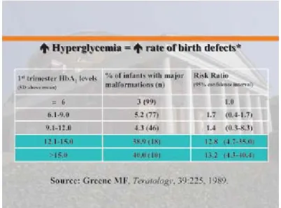
correlation between the level of hyperglycemia during embryogenesis and the risk of birth defects (87). Cardiac anomalies occur 4 to 5 times more frequently in infants of diabetic mothers than in those of control mothers. The risk of neural tube defects in diabetic pregnancies is nearly 3 times greater than in control pregnancies (88). There is approximately a 200-fold increase in the rate of caudal regression syndrome (CSR) in diabetic offspring compared to control offspring (89). Clinical studies have reported high rates of congenital malformation in T1DM and T2DM pregnancies (90;91). GDM has also been associated with birth defects (91;92). The risk of birth defects is 2-4 times higher in PGDM compared to 3% in the general population. Becerra et al. (93) noted that infants of mothers with GDM who required insulin during the 3rd trimester of pregnancy were 20.6 times more likely to have major cardiovascular system defects than infants of non-diabetic mothers. Hemoglobin A1c (HbA1c) levels are a direct measure of glucose control in diabetic mothers: high HbA1c values are predictive of risk for congenital complications (94). Recently, Reece et al. (95) observed high HbA1c levels in association with an increased rate of infants with major malformations, indicating that birth defects correlate very strongly with blood glucose levels (Figure 1-3). It has been shown that good glycemic control in diabetic mothers reduces the prevalence of birth defects to that in the general population (96).
Figure 1-3. Blood glucose levels correlate inversely to birth defect risk (adapted from Reece EA: J Matern Fetal Neonatal Med) (95)
In addition, some women diagnosed with GDM for the first time may actually have undiagnosed T2DM. Pre-pregnancy obesity, which is a risk for T2DM, has been found to be associated with birth defects (97). Maternal obesity and diabetes appear to increase the risk of birth defects through shared causal mechanisms. Studies have also shown that maternal smoking and alcohol intake during pregnancy are significant environmental causes of birth defects (98;99).
1.6.2.1.5. Perinatal Morbidity
Perinatal morbidity in diabetic pregnancies has decreased since the discovery of insulin in 1922. However, current perinatal mortality rates in ODMs remain high compared to the general population. I will briefly review several perinatal morbidities that occur in ODMs.
1.6.2.1.5.1. Neonatal Hypoglycemia
the baby in the first few days after birth, is a common neonatal problem in ODMs (100). The causes of neonatal hypoglycemia are many, such as premature birth, small or large for gestational age, maternal diabetes, and maternal hypertension, occurring more frequently in infants of diabetic mothers (IDMs). The most common risk factor for hypoglycemia in IDMs is hyperinsulinemia, a condition in which plasma insulin concentration exceeds 30 pmol/L. The pancreas of IDMs produces extra amounts of insulin in response to the high blood glucose levels of mothers with poorly-controlled diabetes mellitus. Since glucose supply from the mother is no longer present when the baby has been delivered, extra insulin remains and blood glucose can drop too low (hypoglycemia). About 10-25% of all IDMs have transient hypoglycemia during the first 4-6 hours after delivery. The frequency of neonatal hypoglycemia has been found to increase significantly if the maternal glucose level is greater than 90 mg/dl during delivery (101).
1.6.2.1.5.2. Other Neonatal Metabolic Problems
In addition to hypoglycemia, hypocalcemia and hypomagnesemia have been demonstrated to affect infants of women with T1DM (102). Neonatal hypocalcemia is a frequent event in the first hours of life of newborns from mothers with diabetes mellitus. Strict blood glucose control of diabetes during pregnancy seems to reduce the risk of neonatal hypocalcemia. Poor diabetic control leads to glucose excretion in urine with consequently increased urinary loss of magnesium, followed by low maternal blood magnesium concentration. Maternal hypomagnesemia causes fetal hypomagnesemia with consequent impairment of neonatal parathormone secretion, resulting in abnormally low calcium levels in blood of the fetus (103).
1.6.2.2. Long-term Impact of Maternal Diabetes Complications on ODMs
The long-term impact of maternal diabetes complications on ODMs has been well studied in animal models and clinical trials. There is increasing evidence that maternal diabetes augments the high risk for ODMs of developing obesity and diabetes mellitus later in life (104).
1.6.2.2.1. Obesity
According to the World Health Organization (WHO), obesity is defined as BMI >30 kg/m2(105). In 2005, the WHO reported that at least 400 million adults are
obese. The increasing prevalence of overweight and obese children is a major health problem. Gestational and perinatal factors have been shown to influence weight gained in childhood. Silverman et al. (106;107) demonstrated that the offspring of mothers with PGDM or GDM had a high BMI and developed childhood obesity in comparison to their controls. In another study, the offspring of Pima Indian mothers who had diabetes during pregnancy also had significantly higher body weight at 5 to 19 years of age compared to the controls (108;109). These findings strongly suggest that the intrauterine diabetic environment raises the risk of obesity in childhood and early adulthood.
1.6.2.2.2. Diabetes Mellitus
In utero exposure to high maternal glucose concentrations is a risk for the
long-term development of diabetes mellitus in ODMs (110), but the underlying mechanisms remain unclear. Both insufficient insulin production and insulin resistance are important factors for long-term diabetes mellitus in ODMs. The insulin secretion response is defective and glucose tolerance is impaired in the adult offspring of mothers with T1DM (111). The offspring of mothers with T2DM or
GDM tend to develop insulin resistance or T2DM. Hunter et al. (112) showed that the offspring of mothers with T2DM were overweight with a trend towards reduced insulin sensitivity compared to normal controls. Pettitt et al. (113) studied the prevalence of T2DM among Pima Indians aged 20-24 years and found that 1.4% of non-ODMs developed T2DM versus 45% of ODMs. Similarly, among Pima Indian youth aged 5-19 years, about 40% of ODMs developed T2DM (114).
1.6.2.2.3. Perinatal Programming
Evidence indicates that many adult diseases originate from events that happen in
utero (115). Exposure to alterations in the intrauterine and early postnatal nutritional,
metabolic, and hormonal environment may predispose to diseases in later life (116). However, exposure of the embryo to either genetic influences or environmental changes during a critical period of development can evoke adaptive responses that have a significant impact on its physiology and metabolism. In the next section, I will discuss perinatal programming, perinatal programming-related hypotheses, clinical and experimental investigations of perinatal programming, and possible mechanisms.