Open
Archive TOULOUSE Archive Ouverte (OATAO)
OATAO is an open access repository that collects the work of Toulouse researchers and makes it freely available over the web where possible.
This is an author-deposited version published in : http://oatao.univ-toulouse.fr/
Eprints ID : 17534
To cite this version :
Costet, Juliette. Inflammatory response to naturally occuring tiger
snake envenomation in dogs, with a special emphasis on il-6.
Thèse d'exercice, Médecine vétérinaire, Ecole Nationale Vétérinaire de Toulouse - ENVT, 2016, 110 p.
Any correspondence concerning this service should be sent to the repository administrator: [email protected].
1.
ANNEE 2016 THESE : 2016 – TOU 3 – 4081
INFLAMMATORY RESPONSE TO NATURALLY
OCCURRING TIGER SNAKE ENVENOMATION IN
DOGS, WITH A SPECIAL EMPHASIS ON IL-6
THESE
pour obtenir le grade de DOCTEUR VETERINAIRE
DIPLOME D’ETAT
présentée et soutenue publiquement devant l’Université Paul-Sabatier de Toulouse
COSTET Juliette
Née, le 21 Août 1990 à Nice (06)
Directeur de thèse : Mme Séverine BOULLIER
PRESIDENT :
Mme Bettina COUDERC
DIRECTEUR DE THESE:
Mme Séverine BOULLIER
ASSESSEUR :
M. Guillaume LE LOC’H
JURY
Professeur à l’Université Paul-Sabatier de TOULOUSE
Maître de Conférences à l’Ecole Nationale Vétérinaire de TOULOUSE
Répartition des Enseignants-Chercheurs par Département.
Mise à jour : 06/09/2016
DIRECTRICE :
I
SABELLE CHMITELINELEVAGEETPRODUITS/SANTE PUBLIQUEVETERINAIRE
SCIENCESBIOLOGIQUESET FONCTIONNELLES
SCIENCESCLINIQUESDESANIMAUX DECOMPAGNIE,DESPORTETDE
LOISIRS
Responsable : M. SANS
ALIMENTATION ANIMALE :
M. ENJALBERT Francis, PR Mme PRIYMENKO Nathalie, MC Mme MEYNADIER Annabelle, MC
EPIDEMIOLOGIE : Mathilde PAUL, MC
MALADIES REGLEMENTEES-ZOONOSES-MEDECINE PREVENTIVE DES
CARNIVORES DOMESTIQUES-DROIT VETERINAIRE : M. PICAVET Dominique, PR PARASITOLOGIE-ZOOLOGIE : M. FRANC Michel, PR M. JACQUIET Philippe, PR M. LIENARD Emmanuel, MC Mme BOUHSIRA Emilie, MC
HYGIENE ET INDUSTRIE DES ALIMENTS :
M. BRUGERE Hubert, PR M. BAILLY Jean-Denis, PR Mme BIBBAL Delphine, MC Mme COSTES Laura, AERC Mme DAVID Laure, MCC
PATHOLOGIE DE LA REPRODUCTION : M. BERTHELOT Xavier, PR
M. BERGONIER Dominique, MC Mme CHASTANT-MAILLARD Sylvie, PR Mme HAGEN-PICARD Nicole, PR M NOUVEL Laurent-Xavier, MC Mme MILA Hanna, MC
PATHOLOGIE DES RUMINANTS : M. SCHELCHER François, PR M. FOUCRAS Gilles, PR M CORBIERE Fabien, MC M. MAILLARD Renaud, MC M. M. MEYER Gilles, PR
PRODUCTION ET PATHOLOGIE AVIAIRE ET PORCINE :
Mme WARET-SZKUTA Agnès, MC M. JOUGLAR Jean-Yves, MC M. GUERIN Jean-Luc, PR M. LE LOC’H Guillaume, MC
Responsable : Mme GAYRARD
ANATOMIE :
M. MOGICATO Giovanni, MC M. LIGNEREUX Yves, PR Mme DEVIERS Alexandra, MC
ANATOMIE PATHOLOGIQUE - HISTOLOGIE : M. DELVERDIER Maxence, PR
Mme LETRON-RAYMOND Isabelle, MC Mme BOURGES-ABELLA Nathalie, PR Mme LACROUX Caroline, PR
BIOLOGIE MOLECULAIRE :
Mme BOUCLAINVILLE-CAMUS Christelle, MC
MICROBIOLOGIE – IMMUNOLOGIE - MALADIES INFECTIEUSES :
M. MILON Alain, PR
M. BERTAGNOLI Stéphane, PR M. VOLMER Romain, MC Mme BOULLIER Séverine, MC Mme DANIELS Hélène, MC
BIOSTATISTIQUES :
M. CONCORDET Didier, PR M. LYAZRHI Faouzi, MC
PHARMACIE-TOXICOLOGIE :
M. PETIT Claude, PR Mme CLAUW Martine, PR M. GUERRE Philippe, PR M. JAEG Philippe, MC
PHYSIOLOGIE –PHARMACOLOGIE THERAPEUTIQUE :
M. BOUSQUET-MELOU Alain, PR Mme GAYRARD-TROY Véronique, PR Mme FERRAN Aude, MC
M. LEFEBVRE Hervé, PR BIOCHIMIE. :
Mme BENNIS-BRET Lydie, MC
ANGLAIS :
M. SEVERAC Benoît, PLPA
Responsable : Mme CADIERGUES
ANESTHESIOLOGIE M. VERWAERDE Patrick, MC CHIRURGIE : M. AUTEFAGE André, PR M. ASIMUS Erik, MC M. MATHON Didier, MC
Mme MEYNAUD-COLLARD Patricia, MC Mme PALIERNE Sophie, MC
MEDECINE INTERNE :
Mme DIQUELOU Armelle, MC M. DOSSIN Olivier, MC Mme LAVOUE Rachel, MC
Mme GAILLARD-THOMAS Elodie, MCC
OPHTALMOLOGIE :
M. DOUET Jean-Yves, MC
DERMATOLOGIE :
Mme CADIERGUES Marie-Christine, PR
IMAGERIE MEDICALE M. CONCHOU Fabrice, MC BIOLOGIE MOLECULAIRE. : Mme TRUMEL Catherine, PR
PATHOLOGIE DES EQUIDES :
M. CUEVAS RAMOS Gabriel, MC Mme PRADIER Sophie, MC Mme LALLEMAND Elodie, AERC
Remerciements / Acknowledgements
A la président du jury, Madame le Professeur Bettina Couderc,
Professeur des Universités,
Pour m’avoir fait l’honneur d’accepter la présidence du jury de thèse, je vous prie de croire, Madame, en ma sincère gratitude.
To the inviting staff member, Dr. Manuel Boller
Dipl. ACVECC,
Thank you for letting me take part in this research project, for your time, your support and your help in my work. Thank you for your kindness and your generosity. You made me feel welcome from the very beginning and it was a great experience working with you on this project.
A la directrice de thèse, Madame le Docteur Séverine Boullier,
Maître de conférences à l’Ecole Nationale Vétérinaire de Toulouse,
Pour votre accompagnement au cours de cette thèse et pour m’avoir fait l’honneur de participer à ce jury de thèse, veuillez accepter mes sincères remerciements.
A l’assesseur, Monsieur le Docteur Guillaume Le Loch,
Maître de conférences à l’Ecole Nationale Vétérinaire de Toulouse, Nouveaux animaux de compagnie,
Pour m’avoir fait l’honneur de participer à ce jury de thèse, veuillez accepter mes sincères remerciements.
To the team involved in the study,
To Monique Stanley,
Thank you for your help with the research project. It was very nice to work with you.
A Julien Dandrieux,
Pour m'avoir accompagné dans la réalisation des manipulations, pour votre aide précieuse et votre sympathie, veuillez accepter toute ma gratitude.
To the Intensive Care Unit,
I am very grateful to all of you for your kindness and your help in collecting samples. .
TABLE OF CONTENTS
INDEX OF ILLUSTRATIONS...9
INDEX OF ABBREVIATIONS...13
INTRODUCTION...15
1. PART 1. BIBLIOGRAPHIC STUDY ON SNAKE ENVENOMATION ... 17
1.1 GENERALITIESONSNAKEENVENOMATION ... 17
1.1.1 DEFINITION OF SNAKE ENVENOMATION AND TIGER SNAKE GROUP OVERVIEW ...17
1.1.2 PRINCIPAL COMPONENTS OF SNAKE VENOM ... 23
1.1.2.1 Venom neurotoxins ... 23
1.1.2.2 Venom components acting on the hemostatic system ... 25
1.1.2.2.1 Venom proteins acting on blood coagulation factors ... 25
1.1.2.2.2 Venom proteins acting on vessel walls ... 27
1.1.2.2.3 Venom proteins acting on platelets ... 27
1.1.2.3 Lipases ... 28
1.1.2.4 Venom component specific to Elapid snakes ... 29
1.1.3 CLINICAL PRESENTATION OF SNAKE ENVENOMATION IN DOGS ... 30
1.1.4 CLINICOPATHOLOGICAL VARIABLES ASSOCIATED WITH OPHIDIAN ENVENOMATION ... 34 1.1.4.1 Biochemistry ... 34 1.1.4.2 Heamatology markers ... 36 1.1.4.2.1 RBC abnormalities ... 36 1.1.4.2.2 Leukocytes abnormalities ... 36 1.1.4.3 Hemostatic changes ... 37 1.1.4.3.1 Platelets abnormalities ... 37
1.1.4.3.2 Vascular abnormalities: coagulopathy ... 37
1.1.5 DIAGNOSIS OF SNAKE BITES ... 39
1.1.5.1 History ... 40
1.1.5.2 Clinical evidence ... 40
1.1.5.3 Laboratory tests ... 40
1.1.5.4 Snake venom detection kit (SVDK) ... 41
1.1.6 TREATMENT ... 43
1.1.6.1 First aid treatment ... 43
1.1.6.2 Antivenom treatment ... 43
1.1.6.3 Prophylactic treatment and symptomatic supportive treatment ... 46
1.1.7 PROGNOSIS ... 47
1.2 IMMUNERESPONSEAFTERENVENOMATION ... 49
1.2.1 IMMUNE SYSTEM: OVERVIEW ... 49
1.2.2.4.1 Cytokine overview ... 54
1.2.2.4.2 Multifunctional cytokines ... 54
1.2.2.4.3 Cytokines and acute Phase Protein synthesis ... 56
1.2.2.4.4 Cytokines and recruitment of leukocytes to inflammatory sites ... 56
1.2.2.4.5 Role of cytokines, neutrophil activity and neutrophil apoptosis: protective or deleterious inflammatory response ... 57
1.2.2.4.6 Fever induction ... 60
1.2.2.5 Interleukin-6: principal characteristics and interests ... 61
1.2.2.5.1 Pro and anti-inflammatory activities, hematopoiesis activity ... 61
1.2.2.5.2 Interleukin-6: beneficial and deleterious effects ... 62
1.2.2.5.3 Interleukin-6: cytokine measurement in body fluids ... 64
1.2.2.5.4 Interleukin-6: interests of investigation ... 65
1.2.3 IMMUNE SYSTEMIC RESPONSE TOWARDS SNAKE VENOM ... 66
1.2.3.1 Evidence and mechanisms of inflammation induced in snake envenomation ... 66
1.2.3.1.1 Oedema ... 66
1.2.3.1.2 Influx of leucocytes ... 66
1.2.3.1.3 Inflammatory mediators ... 67
1.2.3.2 Venom components acting in the inflammatory response ... 69
1.2.3.3 Venom components and inflammation in Elapids ... 73
1.2.3.4 Neutralization of the systemic inflammatory response by current treatment ... 73
2. PART 2. EXPERIMENTAL PROTOCOL, RESULTS AND DISCUSSION ... 75
2.1. OBJECTIVESANDHYPOTHESIS ... 75
2.2. MATERIALANDMETHODS ... 75
2.2.1. POPULATION: INCLUSION CRITERIA ... 75
2.2.2. GENERAL DATA ... 76
2.2.3. CLINICOPATHOLOGICAL DATA ... 77
2.2.4. INFLAMMATORY MARKERS MEASUREMENTS: IL-6 LEVELS ... 77
2.2.5. STATISTICS ... 78
2.3. RESULTS ... 78
2.3.1. DOGS AND SNAKEBITE INFORMATION ... 78
2.3.2. CLINICAL FINDINGS ... 79
2.3.3. BICHEMISTRY, HAEMATOLOGY AND CELL BLOOD COUNT, COAGULATION TIMES ...80
2.3.4. DIAGNOSIS OF SNAKE ENVENOMATION ... 82
2.3.5. KINETIC FOLLOW-UP OF IL-6 ... 83
2.3.6. TREATMENT AND OUTCOME ... 86
2.4. DISCUSSION ... 87
2.4.1. THE INFLAMMATORY RESPONSE ... 87
2.4.2. CLINICAL AND CLINICOPATHOLOGICAL FEATURES ... 92
2.4.3. LIMITATIONS ... 95
2.5. CONCLUSION ... 97
BIBLIOGRAPHY...99
INDEX OF ILLUSTRATIONS
Figures:
Figure I. Venom apparatus of Russell’s viper: dissected snake (A), annotated diagram (B) (Warrell 2010) ... 18 Figure II. Map of the distribution of the tiger snake, 2,366 records (Hardy, Cochrane, Allavena
2014). ... 20 Figure III. Site of action of presynaptic and postsynaptic snake venom neurotoxins (Van den
Enden MD, Erwin. 2015). ... 24 Figure IV. Site of action of venom proteins on the cascade of haemostasis (Lu, Clemetson,
Clemetson 2005, p1792). ... 26 Figure V. Snake venom detection kit: correspondence of wells and snakes immunotypes and
antivenom indicated (SVDK, manufacturer data) ... 42 Figure VI. The innate and adaptive system (Day Michael J, Veterinary Immunology book, p11,
2011) ... 50 Figure VII. The four complement pathways and their interaction (Day Michael J., Veterinary
Immunology book, p31, 2011) ... 52 Figure VIII. Roles of complement factors C3a and C5a (Day Michael J, Veterinary
Immunology book, p35, 2011) ... 53 Figure IX. Leukocytes adhesion and involvement of chemokines (Day Michael J, Veterinary
Immunology book, p63, 2011) ... 57 Figure X. Inflammatory response and role of neutrophils (Simon 2003). ... 59 Figure XI. Example of cytokine storm and deleterious inflammation in a case of pneumonia
(Bordon et al. 2013) ... 59 Figure XII. Summary of numerous activities of IL-1, IL-6 and TNF and interaction between
cytokines (modified from Paul William E., p656, 1989). ... 60 Figure XIII. Implication of IL-6 in the transition from neutrophil to monocyte recruitment
(Kaplanski et al. 2003) ... 62 Figure XIV. Main activities of IL-6. Simplified figure of beneficial versus deleterious effects of IL-6. ... 63 Figure XV. Factors affecting cytokine measurement in body fluids (Bienvenu et al. 2000). .... 64 Figure XVI. Effects of snake venom on the production and the activation of mediators
Figure XVIII. Schematic summary of the inflammatory action of a metalloproteinases and
phospholipases A2 of Bothrops asper venom (C. Teixeira et al. 2009) ... 72
Tables:
Table I. Geographical distribution of snakebites (Mirtschin et al. 1998) ... 21Table II. Distribution of snakebites in Australia in relation to species (Mirtschin et al. 1998). . 22
Table III. Summary of principal types of toxin effects on the haemostatic system (modified from Francis S. Markland 1998). ... 28
Table IV. Venom protein components of Elapid snake species and their main effects on the organism (modified from Birrell et al. 2007) ... 30
Table V. Clinical presentation of snake envenomation ... 33
Table VI. Laboratory evidences of snake envenomation ... 39
Table VII. List of monovalent antivenoms available from CSL and corresponding snake venoms neutralised (modified from SVDK, manufacturer data) ... 44
Table VIII. Properties of IL-1, IL-6 and TNF-alpha (from Akira et al. 1990) ... 55
Table IX. Signalment and history ... 79
Table X. Laboratory diagnosis: biochemistry ... 80
Table XI. Laboratory diagnosis: electrolytes ... 81
Table XII. Laboratory diagnosis: cell counts ... 81
Table XIII. Laboratory diagnosis: acid base status ... 81
Graphics:
Graphic 1. Mean, median and quartile of IL-6 levels for each time in affected and healthy dogs ... 84 Graphic 2. Evolution of mean IL-6 levels depending on time post admisison ... 85 Graphic 3. Evolution of IL-6 levels for each dog ... 85
Diagrams:
Diagram 1.Taxonomy of most venomous snake snakes. Focus on snakes observed in Australia (modified from Harris 1985; White, 2013). ... 17 Diagram 2. Key steps for snakebite diagnosis ... 40 Diagram 3. Clinical signs present at the admission (on 10 dogs) ... 79
Photographs:
Photograph 1. Common tiger snake (Notechis scutatus) (personal picture) ... 19 Photograph 2. Eastern Brown Snake (Pseudonaja textilis) (from Queensland museum website)
INDEX OF ABBREVIATIONS
APP = Acute Phase Proteins ACPs = Antigen Presenting Cells
aPTT = activated Partial Thromboplastin Time ALT = Alanine Amino Transferase
ALKP = Alkaline Phosphatase APPs = acute phase proteins BR = Breath Rate
CK = Creatine Kinase COX = Cyclooxygenases CRP = C-Reactive Protein
DIC = Disseminated Intravascular Coagulopathy F. V, VIII, IX, X, XII= Factor V, VIII, IX, X, XII FPD = Fibrinogen Product Degradation
GLDH = G- Lactate Deshydrogenase HR = Heart Rate
ICAM-1 = intercellular adhesion molecule 1 IL-1,6,8, etc. = Interleukin 1, 6, 8, etc . INF-γ = Interferon γ
iNOS = Inducible Nitric Oxide Synthetase INR = International Normalized Rates LTE = Leukotriene E
MCP-1 = Monocyte Chemoattractant Protein-1 NK = Natural killer
NO = Nitric Oxide
PECAM-1 = Platelet Endothelial Cell Adhesion Molecule 1 PGE = Prostaglandins E
PLA2 = Phosphatidylcholine 2- Acylhydrolase (phospholipase A2) PLT = Platelets
PMN = Polymorphonuclear Neutrophils PT = Prothrombine Time
RANTES = Regulated on Activation Normal T cell Expressed and Secreted RBC = Red Blood Cell
RT-PCR = Real-Time Reverse Transcriptase-Polymease Chain Reaction SAA = Serum Amyloid A
SVDK = Snake Venom Detection Kit SVMP = Snake Venom Metalloproteinases TNF-α = Tumor Necrosis Factor α
TGF-β = Transforming Growth Factors β T. snake = Tiger snake
INTRODUCTION
Venomous snakes are widespread in the world and Australia is home to many of them. The systematic classification of snakes recognises between 11 and 13 distinct families. Venomous snakes are identified in only 5 families: Elapidae, Hydrophiidae, Viperidae, Crotalidae and Colubridae (Order Squamata, class Reptilia) (Harris 1985). The genus Notechis belong to the family Elapidae, restricted to the wettest part of Southern and Eastern Australia. The species Notechus scutatus (also called tiger snake) presents variable morphs with different sizes and colourations that some authors recognise as several subspecies while others recognise
Notechis as a monotypic genus (Shea 2000).
Due to their high activity, dogs are particularly subject to be bitten and consequently snake envenomation is a common emergency in this species. It has been estimated that more than 6000 snake envenomed dogs are presented to veterinary clinics in Australia each year (Mirtschin et al. 1998). Tiger snake (Notechis scutatus) and eastern brown snake (Pseudonaja
textilis), both of them from the Elapidae family, were found to be the most commonly
implicated (White 1998, Mirtschin et al. 1998). If brown snakes are more often incriminated within Australia, tiger snakes are definitely the most commonly encountered species implicated in dog snake envenomation in the state of Victoria (Mirtschin et al. 1998) and thus represent a major threat to the health of humans and pets in this area.
Snake venoms are complex cocktails of toxins, containing numerous peptides, proteins and salts. These venom components act as neurotoxins, myotoxins, cardiotoxins and modulators of coagulation and inflammation (Fry 1999, Goddard et al. 2011). Tiger snakes are one of the world’s most venomous snakes based on their venom LD50 and because of their notexins – including the main neurotoxic phospholipase A2 (PLA2) and snake venom metalloproteinases (SVMP), which are important components of Elapids snake venom (Cher et al. 2003, Birrell et al. 2007).
Dogs envenomed by tiger snakes typically have mydriasis, decreased or absent pupillary light reflexes, depressed mentation, generalised muscle weakness, ataxia, tachypnoea or dyspnoea, vomiting and salivation (Hill 1979; Barr 1984). These clinical abnormalities, associated with the history, as well as laboratory test findings such as prolonged coagulation times, elevated serum creatine kinase and snake venom detection test, lead to the diagnosis of snake envenomation (Heller et al. 2007).
reported in several studies (Gutiérrez et al. 1995, Picolo et al. 2002, Moreira et al. 2012, Escocard et al. 2006) and represents the major part of the “post envenomation syndrome”. It was specifically described for vipers (Bothrops asper, Vipera russelli, Ammodytes,
Meridionalis, Aspis), rattlelsnakes (Crotalus) and Elapids (Maria Fernandes et al., 2006,
Zamuner et al., 2005, Langhorn et al., 2014, Santhosh et al., 2013, Szold et al., 2001, Tambourgi et al., 1994). Increased pro-inflammatory cytokines levels (e.g. interleukine IL-6, neutrophil attracting chemokine IL-8, the chemokine monocyte chemoattractant protein-1 MCP-1), induced by PLA2 and SVMP are implicated in these inflammatory processes (Rucavado et al. 2002, Moreira et al. 2012).
Out of the pro- inflammatory cytokines, interleukin-6 (IL-6) is one of the major mediators of acute phase proteins (APPs) synthesis in the liver (Murata, Shimada, Yoshioka 2004). IL-6 is induced by macrophages and other various cells, and is further amplified by IL-1 and TNFα. In dogs, its concentration markedly increases after inflammation stimulation (Yamashita et al. 1994) and therefore can be used for quantifying the induced systemic response to inflammation (Cerón, Eckersall, Martínez-Subiela 2005).
Antivenom is formulated from the plasma of horses immunised with the venom of snakes (hyperimmune IgG serum) (CSL, 2014). It is the current standard treatment that efficiently improves the main threatening systemic effects such as coagulopathy resulting from envenoming (MacDonald, White, Currie 2008). Nevertheless, its use seems to be partially inefficient against local manifestation induced by the inflammatory response to Viperidae snake envenomation (Araújo et al. 2007, Picolo et al. 2002, J. M. Gutiérrez 1981, Chaves, Barboza, Gutiérrez 1995). It was even found to further exacerbate the venom induced immune response and brings a second pro-inflammatory hit (León et al. 2013, Stone et al. 2013).
However, even though tiger snakebites hundreds of dogs every year in Australia, its impact on inflammation has not been examined. That is why there is a real need for understanding the mechanism and characteristics of the post-envenomation syndrome that follow after initial obvious crisis. While the relative contribution of inflammatory processes to the overall clinical picture remains insufficiently described at this time, their attenuation may be exploited as an exciting new therapeutic avenue and clinical trials using immune-modulatory drugs could be warranted.
The aim of this prospective study was to examine the systemic inflammatory response associated with snake envenomation and antivenom administration in dogs, with a special emphasis on IL-6.
2. PART 1. BIBLIOGRAPHIC STUDY ON SNAKE
ENVENOMATION
1.1 GENERALITIES ON SNAKE ENVENOMATION
1.1.1 DEFINITION OF SNAKE ENVENOMATION AND TIGER SNAKE
GROUP OVERVIEW
Definition:
Envenoming is the process where venom is introduced into a human being or an animal, and generally refers to the situation where enough venom is introduced to cause clinical effects. Snake venom can be defined as highly modified saliva able to immobilize and digest the prey by means of the actions of protein-degrading enzymes (Goddard et al. 2011; J. White, 2013).
Taxonomy of tiger snakes
Five venomous snake families are recognized: Elapidea, Hydrophiidae, Viperidae, Crotalidae and Colubridae (Diag. 1). The Hydrophiidae family includes sea snakes and is considered by some as a sub-family or family of the Elapidae. Crotalidae is considered by some as a sub-family of the Viperidae (Harris 1985).
Class Reptilia, Order Squamata
Elapidae
Observed in Australia: tiger snakes (genus Notechis), brown snakes (Pseudonaja), common copperhead (Austrelaps), taipan (Oxyuranus), death
adder (Acanthophis), mulga and red bellied black snake (Pseudochis)
No native or rarely observed king cobra (Ophiophagus), Mamba (Dendroaspis),
rough scales snakes (Tropedichis) Hydrophiidae Also considered as Elapidae, include snakes in marine environment
Viperidae No native in Australia (Genus Bothrops, Vipera)
Crotalidae No native: Rattlesnake (Genus Crotalus) Also considered as a Viperidae
Most of the research on the Elapidae family was focused on five major groups: brown snakes (genus Pseudonaja), tiger snakes (genus Notechis), black snakes (genus Pseudechis), death adders (genus Acanthophis) and taipans (genus Oxyuranus) (Hodgson and Wickramaratna 2006). Tiger snakes (Notechis scutatus) belong to the Elapidae family.
Description and identification of tiger snakes
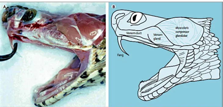
Venomous snakes use fangs to inject their venom (Fig.I).
Figure I. Venom apparatus of Russell’s viper: dissected snake (A), annotated diagram (B) (Warrell 2010)
The venom is stored in the venom gland; the activation of muscularis compressor glandulae allows venom ejection through the venom duct until the fangs.
Tiger snakes are, as all Elapid snakes, proteroglyphous carnivorous snakes, ie. snakes with permanently erect poisonous fangs at the front of the maxilla (Meier J. et al. 1995). Fangs of adult tiger snakes are about 3.5mm long and produce a moderate amount of very toxic venom.
Tiger snakes have an average length between 1.2m to 1.5m according to the authors. Colouration can vary from the classical banded dark grey/black and yellow skin to unbanded types with a range of colors from black to light grey (Geoffrey K. Isbister et al. 2012). Most of tiger snake species show some bands but unbanded species exist too (Shea 2000). As colouration and banding vary, brown snakes can be easily confused with tiger snakes (Photographies 1, 2).
Photograph 1. Common tiger snake (Notechis scutatus) (personal picture)
Photograph 2. Eastern Brown Snake (Pseudonaja textilis) (from Queensland museum website)
A study on the ability of Australians to recognize snake species has shown that identification is highly inaccurate (Morrison, Pearn, Covacevich, et al. 1983). In this study composed of 558 volunteers, only 19% of snakes were correctly identified, which includes the
factors, including inconsistent common nomenclature and similarities in appearance for different snake species.
The most reliable method of snake species’ identification is scale counting but it requires the snake to be dead at the time of the examination. Tiger snakes can be most readily differentiated from other snakes by the combination of a broad parietal scale - as wide as long, but shorter than the temporolabial scale and unpaired subcaudal scales (Shea 2000). Some authors have used a combination of detailed appearance and scalation for classification (Heller et al. 2007).
Geographic range, habitat and ecology:
The three most commonly encountered snakes causing envenomation of veterinary importance in Australia are the brown snake, the tiger snake, and the red-bellied black snake, from the Elapidae family (Hardy, Cochrane, Allavena 2014).
.
Figure II. Map of the distribution of the tiger snake, 2,366 records (Hardy, Cochrane, Allavena 2014).
It indicates the relative density of this species in Australia: tiger snakes are mainly concentrated along the South coast, especially along the South - East coast.
Tiger snakes are especially distributed near the coast, in Eastern, Southern and Western Australian waters. Fig.II indicates the relative density of this species in Australia. They are also found in Bass Strait Islands and Tasmania. This species is found near water areas such as riparian woodlands and watercourses, and eat amphibians and birds (IUCN 2009).
Epidemiology aspects of snake envenomation in dogs
The snakebite in domestic animals is frequent, with an average of four cases per year presented to each veterinary clinic and a total of about 6,240 cases reported annually. Moreover these numbers are probably underestimated because of the number of animals bitten but not treated, like those who survived alone and those who died before having been brought to a
clinic (Mirtschin et al. 1998). Tiger snakes, genus Notechis, are a common cause of snakebites and the second most common cause of snakebite deaths in humans in Australia (J. White, 2013).
A study describes some epidemiology aspects of 125 dogs envenomation enrolled in the Melbourne university hospital in Werribee. In this study, 77% of the dogs (n=125) were sporting breeds, around 4.1 years old with 40% being 2 years old or younger. 48% of the dogs were seen bitten by a snake, almost all by a tiger snake (only one case was a brown snake identified). The highest number of snakebite’ occurences in dogs occurred in December and January (50%), when the temperature rises to around 20°C or above (Barr 1984).
To determine the extent of the snakebite problem in domestic animals, a study was made based on a questionnaire sent to 10% of veterinary surgeons, selected randomly throughout Australia. Queensland was the region where most of snakebite cases were recorded (Tabl.I) over a 5-year period (413 dogs), followed by Victoria (88 dogs) and New South Wales (87dogs) (Mirtschin et al. 1998).
Table I. Geographical distribution of snakebites (Mirtschin et al. 1998)
Most of snakebites in dogs occurred in Queensland (413 dogs), Victoria (88 dogs) and New South Wales (87 dogs). Considering all animals, the total number of snakebites that occurred in Queensland is three times more important than in any other states.
QLD= Queensland, NSW= New South Whales, SA= South Australia, Vic= Victoria, WA= Western Australia, Tas= Tasmania, ACT Australian Capital territory, NT= Northern territory. Not known= address non-indicated by respondents, Other= Wallaby, Kangaroo and two unidentified animals.
In Victoria and Tasmania, more than half of dogs were bitten by tiger snakes (121 dogs), while in the other states, brown snakes were more often incriminated (Tabl. II).
Table II. Distribution of snakebites in Australia in relation to species (Mirtschin et al. 1998).
Different proportion of snake species are implied in snakebites depending on the state considered. Dogs have been bitten in majority by tiger snakes in Victoria whereas brown snakes are more frequently involved in other states.
QLD= Queensland, NSW= New South Whales, SA= South Australia, Vic= Victoria, WA= Western Australia, Tas= Tasmania, ACT Australian Capital territory, NT= Northern territory. Not known= address non-indicated by respondents
Bites have also been shown to be more prominent in rural areas (78%) compared to urban areas (22%) with brown, tiger and black snakes accounting for 76%, 13% and 6% of cases, respectively (Mirtschin et al. 1998).
Venom toxicity
The tiger snake is one of the world’s most venomous snakes based on its venom Lethal
Dose 50% (LD50= 0,118mg/Kg) (4th venomous species with the lower LD50) (Broad,
Sutherland, and Coulter 1979). Nevertheless, to assess the relative danger of each snake species, many other factors such as the relative toxicity should be taken into consideration. The amount of venom injected into the prey and the ‘efficiency’ of the bite (i.e. the masse of venom actually injected into tissues) are of great importance. They have been shown to depend on the number of consecutive bites, fang length and oral morphology, posture and ferocity in attack or defense. The age of the snake may influence the relative toxicity: for example, decreased coagulant activity has been reported with increasing age (Chippaux, Williams, and White 1991; Heller et al. 2007; Morrison et al. 1983). Dogs seem to be particularly sensitive, with lethal doses reported of 0,03mg/Kg, making them three times more sensitive than cats (Barr 1984).
1.1.2 PRINCIPAL COMPONENTS OF SNAKE VENOM
Snake venoms are the most complex of all natural venoms and poisons. They contain a large number of toxic and non-toxic proteins and peptides, non-protein toxins, carbohydrates, lipids, amines, and other small molecules. These components allow to digest the prey, to paralyse it or to kill it. Under evolutionary pressures, venoms have evolved to take full advantage of ecological niches and prey species. Venom toxins have been selected to be specific for many targets in animal tissue (Warrell 2010; Goddard et al. 2011).
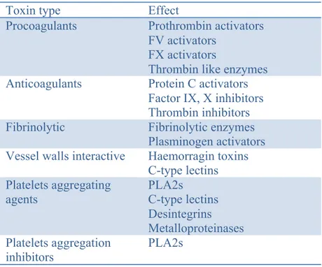
The tiger snake venom contains potent neurotoxins (nerve damaging, pre- and post-synaptic toxins), myotoxins and modulators of coagulation and inflammation. Neurotoxic components affect the central nervous system by blocking neurotransmission. Myotoxins lead to severe muscle damage. Venom constituents are also able to disrupt the haemostatic system by affecting blood coagulation (procoagulant and anticoagulant components, fibrino(geno)lytic components), by acting on vessel walls and by acting on platelets (aggregating agents and aggregating inhibitors agents) (Tabl.III).
Three main types of venom components have been characterised from Australian Elapids, family in which tiger snake belongs: powerful peptidic neurotoxins, proteins acting on the hemostatic system and lipases (Fry 1999; Goddard et al. 2011).
1.1.2.1 Venom neurotoxins
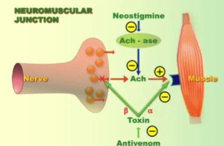
The neurotoxic components of Elapid snake venom may act either pre or post synaptic at the neuromuscular junction to disrupt nervous function.
Pre-synaptic action (β-neurotoxins)
Presynaptic neurotoxins have been identified in the venoms of four major families of venomous snakes (i.e. Crotalidae, Elapidae, Hydrophiidae and Viperidae). Some of the most well characterised β-neurotoxins have been identified in Australian snake venoms, including notexin (isolated from Notechis scutatus) the principle neurotoxic and myotoxic phospholipase A2 (PLA2) in Elapids (Cull-Candy et al. 1976; Hodgson and Wickramaratna 2006). These
(Fig.III). When the stores of neurotransmitter vesicles are depleted, a neuromuscular block results, leading to paralysis (Cull-Candy et al. 1976). Other less potent pre-synaptic neurotoxic PLA2 enzymes such as scutatoxin and notechis II-5 are also found in tiger snake venom (Fry 1999).
Post-synaptic action (α-neurotoxins)
The alpha neurotoxins found in Australian Elapids are reversible postsynaptic blocking neurotoxins. These toxins are present in small amounts and act in a similar way: they bind with high affinity to skeletal nicotinic acetylcholine receptors (Fig.III) and thus prevent the binding between ACh and its receptors (Fry 1999). Tiger snake antivenom counteracted the postsynaptic, but not the presynaptic effects of tiger snake venom when they had developed (Fig. III) (Datyner and Gage 1973).
Figure III. Site of action of presynaptic and postsynaptic snake venom neurotoxins (Van den Enden MD, Erwin. 2015).
Presynaptic (β-neurotoxin) and post synaptic (α-neurotoxin) toxins are represented with
a green cross. Antivenoms are able to counteract the post synaptic action. These mecanisms are the main one involved in Elapids snake.
Metabolic changes
Alvarez et al. investigated the effects of several snake venoms on the Ca2+-dependent K+ channels of human red cells. Venom of the snake Notechis scutatus is able to irreversibly inhibit Ca2+ dependant K+ transport. Thus, the neuromuscular block is further potentiated by venom-induced metabolic changes that interfere with ions (Alvarez and García-Sancho 1989).
1.1.2.2 Venom components acting on the hemostatic system
1.1.2.2.1 Venom proteins acting on blood coagulation factors
Haemorrhaging and incoagulable blood are common findings in Australian Elapids snakebites. Many species have the prothrombin activating enzymes in their venoms and the vast majority contains phospholipaseA2s (PLA2s). Elapids can be divided into two main classes: species with procoagagulant venom including Notechis scutatus (tiger snake) and species with no procoagulant venom (Fry 1999).
Venom proteins acting as coagulant factors
Venom proteins include metalloproteinases such as factors FV activators, FX activators, thrombin-like enzymes and prothrombin activators. Metalloproteinases affect coagulation by activating coagulation factors in the cascade of hemostasis (Fig.IV).
Prothrombin activators are snake proteins known in Notechis scutatus and many other species, in the form of factor Xa-like which function is to catalyse the formation of thrombin, resulting in a disturbance of the coagulation cascade (Heller et al. 2007). Prothrombin activators are able to convert prothrombin into meizothrombin or thrombin (Birrell et al. 2007). According to (Rosing and Tans 1992), based on their structure, venoms can be divided into four groups depending on what prothrombin can be converted and depending on the cofactors needed. Venoms from Australian Elapids fall into either group II or group III (serine proteinases), able to convert prothrombin to thrombin. More precisely, tiger snake belonged to
group II, in which activators are Ca2+ dependant and their activity increased by the presence of
Ca2+, factor Va, and phospholipid (Fry 1999). Then, thrombin act as a positive feedback in the
coagulation cascade, leading to the activation of other coagulation factors, subsequent fibrin formation and almost complete consumption of Factor V, Factor VIII, Factor XII and fibrinogen (G. K. Isbister et al. 2010). The latter consumption of coagulation factors results in the situation of venom-induced consumptive coagulopathy (VICC) that will be discussed further.
FV and FX activators have been isolated from Viperidae, Colubridae and a few snakes from Elapid family, and act as procoagulant in the coagulation cascade (Lu, Clemetson, and Clemetson 2005). Thrombin like enzymes, mainly found in Colubridae and Viperidae, do not
Figure IV. Site of action of venom proteins on the cascade of haemostasis (Lu, Clemetson, Clemetson 2005, p1792).
Venom proteins are surrouned in red, straight arrows represent the activation of factor. Straight arrow = activation, curved arrow = conversion.
: Inhibiton : Venom component
PLA2= Phospholipase A2; IX, X BP= IX, X binding protein, tPA= tissue type Plasminogene
activators; uPA= Urokinase plasminogene activators
Venom proteins acting as anticoagulant factors
While procoagulant effects are widely described, some venom proteins inhibit FIX and FX and thus act as anticoagulant (Morita 2004). Furthermore, Protein C activators are anticoagulant purified from some venoms, which degrade FVa and FVIIIa (Fig.IV) (Lu, Clemetson, and Clemetson 2005). Thrombin inhibitors have been found in Bothrops venom (Viperidae family). It has several roles including prolonged fibrinogen-clotting time by inhibiting competitively the binding of thrombin to fibrinogen (Zingali et al. 1993). PLA2s can
also be classified as strong anticoagulant due to inhibiton of FX and inhibition of the prothrombinase complexe (FVa, FXa, phospholipids and calcium ions) but it is not reported in tiger snake venom (Francis S. Markland 1998).
Venom proteins acting on fibrinolysis
Fibrinolytic and fibrinogenolytic activity has been described in the venoms of members of the Crotalidae, Viperidae and Elapidae families. They act by lysing fibrinogen in FPD (fibrinogen product degradation). Plasminogen activators (Fig.IV) act by activating plasminogen to plasmin, which can transform fibrin clot into FPD (F. S. Markland 1998; Lu, Clemetson, and Clemetson 2005).
1.1.2.2.2 Venom proteins acting on vessel walls
Many metalloproteinases described in Crotalidae, Viperidae and Elapidae venoms are haemorragic toxins that act in synergy to degrade the blood vessel extracellular matrix, and to play key roles in the development of such symptoms as haemorrhage, edema, hypotension, hypovolemia, inflammation, and necrosis (Hite et al. 1994; Birrell et al. 2007).
C-type lectins belong to the composition of some Elapidae venoms and can cause oedema, increased vascular permeability and agglutinate erythrocytes although mechanisms remain unclear (Lu, Clemetson, and Clemetson 2005; Birrell et al. 2007).
1.1.2.2.3 Venom proteins acting on platelets
Snake venoms contain proteins acting on platelets such as PLA2s, C-type lectins, disintegrins and metalloproteinases binding to collagen or collagen receptors on platelets. PLA2s affect platelets function by several mechanisms (for example: cleaving platelet membrane phospholipids). Tough in many cases PLA2s induce platelet aggregations initially, in high concentration they will often inhibit it (Hutton and Warrell 1993).
Table III. Summary of principal types of toxin effects on the haemostatic system (modified from Francis S. Markland 1998).
Toxin type Effect
Procoagulants Prothrombin activators
FV activators FX activators
Thrombin like enzymes
Anticoagulants Protein C activators
Factor IX, X inhibitors Thrombin inhibitors
Fibrinolytic Fibrinolytic enzymes
Plasminogen activators
Vessel walls interactive Haemorragin toxins
C-type lectins Platelets aggregating agents PLA2s C-type lectins Desintegrins Metalloproteinases Platelets aggregation inhibitors PLA2s
Examples of different venom toxins acting as procoagulant, anticoagulant, fibrinolytic agent, acting on vessel walls and on platelets. For example, FV activators have been described in Naja naja oxiana and Naja nigricollis nigricollis venoms, prothrombin activators in Pseudonaja textilis.
1.1.2.3 Lipases
The vast majority of lipases found in Australian snake venoms are phospholipases. There are four main species of phospholipases (A1, A2, C, D). Most venom phospholipases are Phosphatidylcholine 2 - Acylhydrolase (PLA2): they are the most frequently encountered proteins identified across the species (Fry 1999; Birrell et al. 2007). The PLA2s found in Elapid venom may be classified into five types: neurotoxic, haemotoxic, myotoxic, non-toxic enzymatically active and non-toxic non-enzymatically active (Heller et al. 2007).
Neurotoxic PLA2s produce neurological effects not through the enzymatic esterase activity but rather through direct blockage (presynaptic acethylcholine release blocker) (see part 1.1.2.1). Notexin is the principle neurotoxic and myonecrotic phospholipase A2 in
Haemotoxic PLA2s produce haemorrhage through blockage of factors in the coagulation cascade. It doesn’t produce net anticoagulation through fibrinolysis but rather bind to specific factors essential in the coagulation cascade (see previous part 1.1.2.2). Hemotoxic PLA2s isolated from the tiger snake are responsible for hypotension and haemorrhage (Fry 1999).
Myotoxins, defined as venom components acting on skeletal muscle, act by causing damage to the integrity of the sarcolemma, resulting in local haemorrhage and necrosis to systemic skeletal rhabdomyolysis producing severe myoglobinuria (Heller et al. 2007; Mebs, Ehrenfeld, and Samejima 1983). The degeneration of muscle induced by the inoculation of either venom or pure myotoxins is rapid and followed by regeneration of the affected tissue (Harris et al. 2003; Heller et al. 2007). This myotoxin PLA2s has been well described in a particular species of Elapidae (Pseudechis australis), which differs from the other species with the primary toxins being myotoxic rather than neurotoxic (Fry 1999).
Non-toxic PLA2s have also been isolated from the tiger snake and do not present any toxic activity (Fry 1999).
Moreover, PLA2s are known to play a role in the hyperalgesia induced by Bothrops asper venom (Chacur et al. 2001) and may play a role in the inflammatory process ( Fry 1999; Cher et al. 2003), which will be further detailed (see part 1.2.3.2).
Other lipases such as lyophospholipases and phospholipases also exist and are responsible for haemolysis and myolysis (Heller et al. 2007).
1.1.2.4 Venom component specific to Elapid snakes
The venom protein components of 18 Elapid snake species were separated and identified using mass spectrometry and peptide sequencing. Three groups of proteins account for a large proportion of the total venom proteins, confirming their high abundance: neurotoxins, prothrombin activators, and PLA2s (Tabl. IV).
SVMPs, known in Viperidae venoms has been identified in 7 out of the 18 Elapid species (species non precised). A limited number of C-type lectins have also been identified in this study in some Elapids.
Table IV. Venom protein components of Elapid snake species and their main effects on the organism (modified from Birrell et al. 2007)
Toxin type Effect
Neurotoxins Neurological signs (mydriasis, paralysis, etc.) Prothrombin activators Consumptive coagulopathy Phospholipases A2
Neurological signs, muscle degeneration, necrosis, inflammation
SVMP
Hemorrhage, oedema, hypotension, hypovolemia, inflammation, and necrosis
C-type lectins Oedema, increased vascular permeability
Neurotoxins, prothrombin activators and phospholipases A2 (highlighted) represent the three major groups of components found in tiger snake venom.
1.1.3 CLINICAL PRESENTATION OF SNAKE ENVENOMATION IN DOGS
Clinical effects of snake envenomation include local, general, neurological, haematological and myotoxicity signs (Tabl.V).
Clinical effects at the bite site
At the bite site, the envenomation can cause locally painful and redness, swelling, and bruising. It is usually local minor effects although necrosis may occur. Fang puncture or scratch marks can be visible and helpful for the diagnosis (Geoffrey K. Isbister et al. 2013). A study on 53 dogs envenomed by Vipera berus showed a varying degree of swelling in the area of the bite. The bite site was located in the head/nose in 77% of dogs, in the hind limb in 13%, the front limb in 6%, the neck and prepuce in 2% each (Lervik, Lilliehöök, and Frendin 2010). Nevertheless, in Elapids envenomation, snakebite is not always visualized, as in other studies of tiger snake bites in which wounds were seen in only 9 on 125 dogs (Barr 1984) and 1 on 104 dogs (Indrawirawan et al. 2014) and were generally found on the face. It can be due to the
difficulty of identification of the wound (and the fact that the face has less hair than other parts of the body) as well as a lower sever local response.
General signs
Non-specific signs such as ptyalism (10%), vomiting (38% to 42% according to the authors) and generalised muscle weakness/collapse (around 30%) or reduced level of consciousness (14%) are common signs in tiger snake envenomation on dogs (Barr 1984, Indrawirawan et al. 2014). Collapse is usually described but often followed by rapid recovery (29%, Indrawirawan et al. 2014). Tachycardia, tachypnea or dyspnea are usually found in tiger snake envenomation, controversially elevated rectal temperature is not usual. Draining lymph nodes may also be tender (Barr 1984). These findings are also observed in humans envenomed by tiger snake (Geoffrey K. Isbister, Brown, and ASP Investigators 2012).
Neurological signs
Neurologic signs are also a common finding on dogs following a snake envenomation, including by tiger snake. Experiments on mice have shown the relationship between the dose and the severity of the disease, suggesting that these signs depend on the amount of venom injected at the bite site (Lewis 1994).
The earliest signs are usually disorientation and ptosis, which develops from one to several hours after the bite. Mydriasis and loss of pupillary light reflex, which is consistent with a paralysis of oculomotor nerve, are common findings and more particularly the most commonly observed signs in a study on tiger snake envenomation (76% and 66% of dogs respectively) (Barr 1984). In a more recent study on 21 dogs envenomed by tiger snake, mydriasis was observed on 29% of the dogs and reduced pupillary reflex on 24%. These numbers can be underestimate since they only include tiger snake envenomation confirmed by SVDK, professional snake handler or scale counting and do not include “suspected tiger snake envenomation” (Indrawirawan et al. 2014). In a study on 53 dogs, 73% of them had an affected mental status on arrival at the hospital, but this study was concerning only Viper envenomation
(Lervik, Lilliehöök, and Frendin 2010). Then paralysis of facial, peripheral and
glossopharyngeal muscles may occur and thus conduct to respiratory failure. Generalized paralysis is an important feature of tiger snakebites, but clinical evidences of flaccid paralysis take several hours to become apparent.
These neurological effects are usually reversed with antivenom if given prior to development of major paralysis. If many hours after the bite, the patient presents a major paralysis requiring intubation and ventilation, it may be necessary to continue this treatment for days, weeks, and very occasionally, even over a month (Fry 1999; Hodgson et al. 2003; Geoffrey K. Isbister et al. 2012).
Haematological signs
Coagulopathies are well known in snake envenomation and may result in clinical signs such as increased bleeding (linked to decreased blood coagulability or to endothelial damages), hypovolemic shock and secondary organ damage, macro- and microthrombosis, such as pulmonary thromboembolism (Goddard et al. 2011). Australian Elapids venom toxins act as procoagulants, causing in-vivo activation of the coagulation system, but in most cases, this does not result in massive thrombosis and consequent embolic disease, but rather causes consumption of coagulation factors, resulting in clinical anticoagulation. It may cause important abnormalities of clinical laboratory tests, but generally does not result in clinically significant bleeding (White 2005).
Bleeding from bite site or venupuncture site (hematoma, ecchymosis), oral and nasal cavity (epistaxis, petechial haemorrhages), gastrointestinal (melena), urinary and intracranial sites, may occur (study non specific to tiger snake) (Geoffrey K. Isbister et al. 2013). Discolouration of the urine is a common finding, reported in 25% of the dogs envenomed by tiger snake (Barr 1984) but can be the result of haemoglobin, myoglobin or erythrocyte’s presence.
Myotoxicity signs
Severe myolysis can occur with tiger snakebites, particularly if treatment is delayed or inadequate. Myotoxicity was seen in 59% of the dogs envenomed by tiger snake (Indrawirawan et al. 2014) and is generally characterized by increased Creatine Kinase (CK). The usual signs may occur, e.g. muscle tenderness, muscle weakness, pain on contracting muscles, myoglobinuria and later development of muscle wasting (Geoffrey K. Isbister et al. 2012).
Renal failure
No renal toxins have so far been isolated from tiger snake venoms, but renal failure is a common effect of tiger snakebites in humans, for cases with significant envenoming. Nevertheless it is not usual in dogs, in which only a few isolated cases are reported (Warrell 2010).
Histopathology findings
A study has described the examination of post mortem heart, lungs, kidneys and gastrocnemius muscle tissue from one dog envenomed by a tiger snake. Pathological evidence of procoagulant venom activity with formed thrombi has been observed in the lungs, heart, muscles and kidneys. This supports the proposal that an initial thrombotic state occurs in envenomed dogs, leading to the depletion of vital clotting factors and thus to coagulopathy. Nevertheles, it is unclear whether renal failure is a direct result of the venom’s nephrotoxicity or a secondary effect of myoglobinuria (Jacoby-Alner et al. 2011).
Table V. Clinical presentation of snake envenomation
Clinical signs Presentation
Local signs Not usualy visualized. Pain, red, swelling and bruising Scratch marks
General signs
Ptyalism, vomiting
Collapse / Reduced level of consciousness Weakness
Tachycardia, tachypnea, dyspnea Tender lymph nodes
Neurological signs
Disorientation
Ptosis, mydriasis, loss of pupillary light reflex
Ataxia, generalized weakness, paralysis
Haematological signs
Mostly laboratory findings (bleeding, hypovolomic shock not frequent)
Discoloration of urines
Myotoxicity signs Muscle tenderness, weakness, pain
Clinical signs associated with snake envenomation include local, general, neurological, haematological and myotoxicity signs. The most frequently observed clinical signs in tiger snake envenomation on dogs are highlighted.
.
1.1.4 CLINICOPATHOLOGICAL VARIABLES ASSOCIATED WITH OPHIDIAN ENVENOMATION
1.1.4.1 Biochemistry
Many biochemical abnormalities have been described in snake envenomation (Tabl. VI) and may help the veterinarian to detect pathologic effects. Myotoxicity is the most affected biochemistry parameter described on tiger snake envenomation in the literature. Other biochemical abnormalities are described in various snake species.
Muscle activity: myotoxicity
In humans, CK (Creatine Kinase) activity is an indicator of many factors (like the amount of venom injected, the delay between envenomation to presentation and the quantification of myonecrosis) (Nakada et al. 1984) but its use is limited in dogs since it is often difficult to know the exact time of snakebite (Goddard et al. 2011). In an in-vivo study on mice, from six Australian Elapid venoms, only those from Pseudechis colletti guttatus and Pseudechis
australis produced a rise of CK-activity. They also showed that a fraction of the Elapid snake Pseudechis colletti guttatus venom, the Phospholipase A2, caused a dose-dependent increase of
CK-level and myoglobinuria (Mebs, Ehrenfeld, and Samejima 1983). Elevated CK were found in most cases of several studies: 15 out of the 28 sampled dogs (54%) 24h after admission on Viperidae envenomation (Lervik, Lilliehöök, and Frendin 2010), 75 out of 109 dogs (69%) in another study on Viperidae envenomation (Aroch and Harrus 1999) and 43 out of the 45 dogs (96%) on tiger snake envenomation (Barr 1984). In a more recent study on tiger snake envenomation, myotoxicity was seen in 59% of dogs with tiger envenomation, with a median CK activity of 1,878 U/L; the highest CK value was 62,586 U/L (Indrawirawan et al. 2014). In some of these studies, it was suggested that CK values were within the reference values, mainly for samples that were collected too early after snakebite (before the release of plasma CK from degenerating muscle) or too late in the envenomation process (after excretion by the kidneys). The increased CK activity may be related to rhabdomyolysis induced by snake venom myotoxicity and also to intravascular hemolysis.
Hepatic variables: hepatotoxicity
Hepatic variables changes associated with snake envenomation have mostly been described in Viperidae family. In a study on 34 dogs envenomed by Vipera berus, 65% had a
serum concentration above reference values for at least one liver enzyme (Alanine Amino Transferase ALT, Alkaline Phosphatase ALP, Lactate Deshydrogenase GLDH and bile acids) during one or more examinations (on arrival, at 24h, at follow-up on the period of days 4-10 and days 9-23). But in most cases, the increase was mild, temporary and only one or two parameters were affected (Lervik, Lilliehöök, and Frendin 2010). High increased of these variables have also been described in dogs envenomed by Vipera palaestinae. These findings have suggested hepatocellular damages and cholestasis, as a consequence of vascular ischemia as well as direct damages by cytotoxin effects of snake venom. An increase to some degree in ALAT and ASAT may also be explained by muscle damages and by glucocorticoid treatments (Aroch and Harrus 1999, Goddard et al. 2011).
Renal variables: nephrotoxicity
According to Lervik, Lilliehöök, and Frendin (2010) and Segev et al. (2008), the serum creatinine was within the reference values for all dogs at all examinations. Nevertheless some cases of acute renal failure have been described in dogs’ envenomation. For example, a dog bitten by en Elapid snake, the Red-bellied Black snake (Pseudechis porphyriacus), with signs of rhabdomyolisis and hemolysis (increased CK, bilirubine and ASAT) presented moderate increased creatinine, azotaemia and marked hyperphosphataemia consistent with renal failure. This dog presented evidence of shock, dehydration, with discoloured and reduced urine production and became totally anuric before euthanasia (Heller et al. 2006). Renal failure may be due to renal ischemia caused by vasoconstriction and procoagulant microthrombotic effect, nephrotoxic effects of myoglobinuria, hemoglobinuria, DIC (disseminated intravascular coagulopathy), toxic nephropathy, and hypovolemic shock (Goddard et al. 2011, Heller et al. 2007).
Other biochemical abnormalities:
Other abnormalities have been especially shown in dogs envenomed by Vipera
palaestinae e.g.: hyperalbuminemia, hyperglycemia, hypocholesterolaemia, hypoalbuminemia
and hyperproteinemia (Aroch and Harrus 1999; Segev et al. 2004; Segev et al. 2008). Moderate hyperalbuminemia could be the consequence of hemolysis and/or ischemic hepatopathy (Goddard et al. 2011). Hyperglycemia can be linked to stress and pain (Segev et al. 2004). Hypocholesterolaemia and hypoalbuminemia may be explained by capillary damages and changes in lipoprotein transport and metabolism due to the phospholipase A2 (Segev et al.
envenomation in human cases (Winkler et al. 1993). These abnormalities are poorly described on tiger snake envenomation.
1.1.4.2 Heamatology markers
1.1.4.2.1 RBC abnormalities
Hemoconcentration, characterized by an increased in hematocrit and hemoglobin concentration is a common finding on envenomation by puff-adders from Viperidae family and cobras from Elapidae family (Segev et al. 2004; Segev et al. 2008; Lobetti and Joubert 2004; Aroch et al. 2010). This abnormality can be explained by fluid losses, associated with third spaces due to heamorrhagins inducing capillary damages and the shift of proteins and fluids from circulation to the inflamed tissue at the bite site. Splenic contraction may also be involved associated with excitement, pain, and stress and subsequent release of stored erythrocytes into circulation. Dehydration was eliminated as a prominent cause of increased hematocrit based on lack of clinical evidence (Lobetti and Joubert 2004; Goddard et al. 2011).
Haemolysis is well described in snake envenomation, especially in the Elapid red-bellied Black Snake. First evidence is a discolouration of serum (Heller et al. 2006). Another evidence is red urine, although it could be the result of bleeding, hemoglobinuria or myoglobinuria and thus imply hemolysis, rhabdomyolysis, or renal damages (Heller et al. 2006). Nevertheless, in an in-vivo study on experimental administration of tiger snake venoms in dogs, although haemolysis appears quickly with plasma haemolized in 30 mintes and even at low doses of venom injected, it didn’t seem to progress significantly. Haemolysis is thought to be the consequence of lytic factor or phospholipase activity which damages the erythrocyte cell membrane and results in an influx of water, leading to erythrocyte swelling, stretching and, ultimately, haemolysis (Goddard et al. 2011; Heller et al. 2006). The haemolysis is sometimes accompanied by echinocytosis and spherocytosis (Aroch and Harrus 1999).
Anemia has been observed less frequently in dogs with snake envenomation (Goddard et al. 2011).
1.1.4.2.2 Leukocytes abnormalities
Leukocytosis also characterizes envenomation in dogs, suggesting an acute inflammatory reaction. It has been described on envenomation by Viperidae such as the puff adders (Bitis
(Lobetti and Joubert 2004; Aroch and Harrus 1999; Goddard et al. 2011; Segev et al. 2004). Leukocytosis was identified as a risk factor for mortality in dogs with Vipera palaestinae envenomations (Aroch and Harrus 1999; Goddard et al. 2011).
1.1.4.3 Hemostatic changes
1.1.4.3.1 Platelets abnormalities
Various degrees of thrombocytopenia are commonly found: for example, on dogs envenomed by Vipera palestinae, 51.9% have shown thrombocytopenia at the venom admission and 52% 24h post admission (Aroch and Harrus 1999). A fall in the platelet count is usually evident within 24 hours of the bite (Geoffrey K. Isbister 2010). In a mouse model, platelet numbers were significantly decreased 30 minutes after Bothrops asper snake venom injection and returned to normal after 12 hours (Lomonte, Tarkowski, and Hanson 1993). Several mechanisms could explain thrombocytopenia in envenomation such as vasculitis, sequestration of platelets in inflamed tissues, and consumption of platelets with potential development of disseminated intravascular (Goddard et al. 2011). It is poorly described in tiger snake envenomation.
Interaction of snake venom with platelets aggregation are reported. Some PLA2s seem to induce platelet aggregation by cleaving platelet membrane phospholipids, releasing arachidonic acid and forming arachidonic acid metabolites such as thromboxane A2. Another group of PLA2s inhibit platelet aggregation via the cleavage products (Lu, Clemetson, and Clemetson 2005).
1.1.4.3.2 Vascular abnormalities: coagulopathy
As seen earlier (part 1.1.2.2.), a large number of venom proteins act as procoagulants, sometimes as anticoagulants, that then create thrombosis, consumption of coagulant factors and
thus may result in incoagulable blood and bleeding (Fry 1999; White 2005). Coagulopathy is a
common finding in snake envenomation and Australian Elapids are amongst the most potent in causing procoagulant coagulopathy (White 2005).
(aPTT), decreased fibrinogen, increased Fibrin/ fibrinogen degradation products (FDP) or D-Dimer levels, hypoantithrombinemia activity. An observation on blood coagulation in cats and dogs naturally envenomed by tiger snake showed that all dogs (n=6) had an increased PT and an increased aPTT, and half of them (n=3) had increased FDP levels (Holloway and Parry 1989). Later, other studies showed similar results in Viper envenomation with prolonged PT and aPTT at admission (68.4% and 21.1% of the dogs respectively (Segev et al. 2004), 29% and 39% of the dogs in (Aroch et al. 2010)) and further prolonged aPTT a day later (75.8% of the dogs). Prolonged PT and aPTT were observed in 54% and 44% of the dogs respectively, at least once during the experiment. Hypoantithrombinemia was observed at least once in 56% of the cases and D-dimer concentrations were positive (>250 ng/mL) in 90% (Aroch et al. 2010). In these studies, the authors recommended that dynamic multiple coagulation tests should be realized as the incidence of abnormalities was associated with higher mortality. Thromboelastography may also be an helpful diagnosis of coagulopathy (Goddard et al. 2011).
Presence of thrombocytopenia, increased PT and aPTT may lead to the diagnosis of DIC (Disseminated Intravascular Coagulopathy). Nevertheless, in Elapid envenomation, and especially with tiger snake species, this consumptive coagulopathy associated with snakebites has been referred to as a VICC, Venom Induced Consumption Coagulopathy (Goddard et al. 2011). VICC is characterized by prolonged clotting times, depletion of fibrinogen and cofactors V and VIII, and high concentrations of FDPs. However, it does not include evidence of thrombus formation and organ failure. In a study on tiger snake envenomation on dogs, laboratory evidence of a coagulopathy was detected in 12 out of 21 dogs (57%) envenomed by confirmed tiger snake and in 34 out of 41 dogs (83%) with suspected tiger snake envenomation (Indrawirawan et al. 2014). These numbers are lower than those observed in human literature where VICC was observed in 95% of tiger snake victims (Geoffrey K. Isbister et al. 2012). It was suggested that dogs are less sensitive to the procoagulant effects of tiger snake venom than humans (Indrawirawan et al. 2014).
Table VI. Laboratory evidences of snake envenomation
Laboratory
evidence Affected parameters
Myotoxicity High CK levels
Hepatotoxicity High ALP, ALT, GLDH, bile acids levels Nephrotoxicity High Creatinine levels
RBC
abnormalities Haemoconcentration (increased Ht) Haemolysis (haemolized plasma, red urines)
Anemia Leukocytes abnormalities Leukocytosis Platelet abnormalities Thrombocytopenia
Coagulopathy Increased PT, aPTT
Raised D-Dimer concentration
Low to undetectable fibrinogen (VICC)
Laboratory evidences highlighted represent those mostly altered in tiger snake envenomation.
CK = Creatine Kinase, ALT = Alanine Amino Transferase, ALPalkaline phosphatase ALP, lactate deshydrogenase GLDH, PT = Prothrombin Time, aPTT = Activated Partial Thromboplastin Time, VICC= Venom Induced Consumption Coagulopathy.
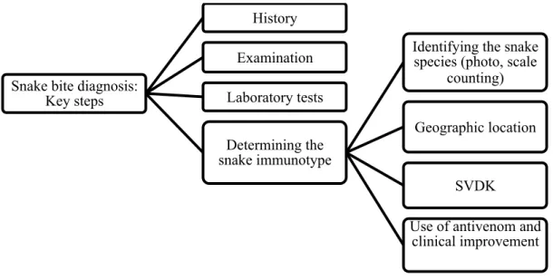
1.1.5 DIAGNOSIS OF SNAKE BITES
The diagnosis of snake envenomation is lead by the association of several factors (Diag.2). First of all, the presence of envenomation should be done on arrival with the history, clinical evidence and laboratory test. If none of the clinical or laboratory evidence can be observed, further exams should be run some hours later with a neurological examination in order to exclude snakebite. If envenomation has been identified, then the vet should determine which snake or group of snakes is most likely to be involved - with use of pictures, scale counting, geographic prevalence and SVDK (Snake Venom Detection Kit).
If the diagnosis is established, appropriate antivenom that covers the likely snake(s) is given. Nevertheless, in veterinary medicine, due to financial reason or to a long elapse time between the bite and the animal presentation, antivenom is sometimes used directly after the history and examination. Then, the diagnosis would be based on clinical improvements
Diagram 2. Key steps for snakebite diagnosis
Two main steps are important to consider: diagnosis of snake envenomation and determination of the snake immunotype.
1.1.5.1 History
History is essential to make a diagnosis of snakebite. A detailed history of the circumstances of the bite (time, temperature, geographic location), of the witnessed snake (length, colouring, use of scale counting), of the first symptoms (vomiting, period of unconsciousness, collapse, etc.) and medical history as well should be taken into account, according to human’s guide but applicable in domestic animals (White, 2013).
Nevertheless, as seen previously (see part 1.1.1), the identification of the snake by non-professional people, without any pictures taken or scale counting, is not reliable in the majority of cases.
1.1.5.2 Clinical evidence
Clinical evidence includes the clinical presentation (neurological, haematological and myotoxicity signs) presented previously (see part 1.1.3, Tabl. V). Fang marks at the bite site can be reliable clue of snakebite but is not always observed.
1.1.5.3 Laboratory tests
It is necessary to use clinical signs in conjunction with the history, the physical Snake bite diagnosis:
Key steps History Examination Laboratory tests Determining the snake immunotype
Identifying the snake species (photo, scale
counting) Geographic location
SVDK Use of antivenom and