OATAO is an open access repository that collects the work of Toulouse
researchers and makes it freely available over the web where possible
Any correspondence concerning this service should be sent
to the repository administrator:
[email protected]
This is an author’s version published in:
http://oatao.univ-toulouse.fr/24489
To cite this version:
Rodriguez, Gabriela Melo and Bowen, James and Grossin, David
and
Ben-Nissan, Besim and Stamboulis, Artemis Functionalisation of Ti6Al4V and
hydroxyapatite surfaces with combined peptides based on KKLPDA and
EEEEEEEE peptides. (2017) Colloids and Surfaces B Biointerfaces, 160. 154-160.
ISSN 0927-7765
Functionalisation of Ti6Al4V and hydroxyapatite surfaces with
combined peptides based on KKLPDA and EEEEEEEE peptides
Gabriela Melo Rodriguez a,James Bowenb,c , David Grossin<l, Besim Ben-Nissan e , Artemis Stamboulis a,+• School of Metallurgy and Materials, University of Birmingham, Edgbaston, Birmingham, BI 5 ZTT, UK b School of Chemical Engineering, University of Birmingham, Edgbaston, Birmingham, BI 5 ZTT, UK
c Faculty of Mathematics, Computing and Technology, School of Engineering and Innovation, The Open University, Walton Hall, Mi/ton Keynes, MK7 6AA, UK d C/R/MAT Université de Toulouse, CNRS, INPT, UPS, ENSIACET, 4 allée Emile Monso, BP44362, 31030 Toulouse Cedex 4, France
• Advanced Tissue Regeneration & Drug Delivery Croup, School of Life Sciences, University ofTechnology Sydney, P.O. Box 123, Broadway, NSW 2007,
Australia ARTIC L E INFO Keywords: Coatings Hydroxyapatite Peptides Surface Titanium alloys
1. Introduction
ABSTR ACTSurface modifications are usually performed on titanium alloys to improve osteo-integration and sur
face bioactivity. Modifications such as alkaline and acid etching, or coating with bioactive materials such as hydroxyapatite, have previously been demonstrated. The aim of this work is to develop a peptide
with combined titanium oxide and hydroxyapatite binders in order to achieve a biomimetic hydroxya
patite coating on titanium surfaces. The technology would also be applicable for the functionalisation
of titanium and hydroxyapatite surfaces for selective protein adsorption, conjugation of antimicrobial peptides, and adsorption of specialised drugs for drug delivery. ln this work. functionalisation of Ti6Al4V
and hydroxyapatite surfaces was achieved using combined titanium-hydroxyapatite (Ti-Hap) peptides based on titanium peptide binder (KKLPDA) and hydroxyapatite peptide binder (EEEEEEEE). Homoge
neous peptide coatings on Ti6Al4V surfaces were obtained after surface chemical treatments with a
30wt% aqueous solution of H2O2 for 24 and 48 h. The treated titanium surfaces presented an average
roughness of s. = 197 nm (24 h) and s. = 128 nm ( 48 h); an untreated mirror polished sample exhibited
an s. of 13 nm. The advancing water contact angle of the titanium oxide layer after 1 h of exposure to
30wt% aqueous solution ofH2O2 was around 65°, decreasing gradually with time until it reached 35° after a 48 h exposure, suggesting that the surface hydrophilicity increased over etching time. The presence of a
lysine (L) ami no acid in the sequence of the titanium binder resulted in fluorescence intensity roughly 16%
higher compared with the arginine (R) amino acid analogue and therefore the lysine containing titanium
peptide binder was used in this work. The Ti-Hap peptide KKLPDAEEEEEEEE (Ti-Hap 1) was not adsorbed by the treated Ti6Al4V surfaces and therefore was modified. The modifications involved the inclusion of
a glycine spacer between the binding terminais (Ti-Hap2) and the addition of a second titanium binder
(KKLPDA) (Ti-Hap3 and Ti-Hap4). The combined Ti-Hap peptide which exhibited the strongest inten
sity after the titanium surface dip coating was KKLPDAKKLPDAEEEEEEEE (Ti-HAp4). On the other hand,
hydroxyapatite surfaces, exhibiting an average roughness ors.= 1.42 µm, showed a higher fluorescence
for peptides with a higher negative net charge.
Surface modifications on titanium alloys are usually performed
to improve osteo-integration and surface bioactivity [ 1 ]. Bioactive
surfaces interact with their biological environment by water and
hydrated ions adsorption. inducing bone growth and biochemi cal bonding with proteins (2,3). Titanium and its alloys are widely used in orthopaedics due to properties such as excellent corro sion resistance, as well as biological-related properties such as the stimulation of bone cell growth [1,3.4). Modifications such as sur face etching with basic (5-7) and acidic solutions [8,9). or coatings with bioactive materials such as hydroxyapatite, have been shown to increase the bioactivity of titanium and titanium alloy surfaces (10-13).
* Corresponding author.
E-mail address: [email protected] (A Stamboulis).
A number of techniques used to obtain bioactive coatings
include plasma spray[14], electrophoretic deposition[15], micro
arc oxidation[16], sol gel[17]and biomimetic methods[18]. The
technique most commonly used in the orthopaedic and dental
industry is plasma spray[14]. During this method high
temper-atures are normally used in order to coat metallic surfaces with
hydroxyapatite followed by a rapid cooling of the coating[1,19].
As a result, in some cases, heterogeneous hydroxyapatite
coat-ings can be obtained [20,21]. Therefore, research efforts have
been focused to develop alternative surface modifications that are cheaper, use less energy and are more biomimetic. One example
is surface modification using NaOH or H2O2 to form surface gel
layers, sodium titanate gel and titania, respectively, which induce deposition of hydroxyapatite from simulated body fluid (SBF)
envi-ronments [22,23]. Apatite deposition onto sodium titanate gel
depends strongly on the sodium ion exchange from the gel with
H3O+ions in SBF[6], whereas apatite deposition onto titania gel
is associated with the abundance of OH−groups on the surface,
presenting a negative charge in SBF[24].
The titanium oxide layer formed on the titanium surface after treatment with H2O2presents both acidic (-Ti-O−) and basic
(-Ti-OH2+) moieties in aqueous solution at pH 7.5[25]. The amphoteric
character of the titanium oxide layer promotes electrostatic inter-actions with organic molecules such as peptides and proteins.
Roddick-Lanzilotta et al.[26–28]have reported electrostatic
inter-actions between modified titanium surfaces and amino acids such as glutamic acid, aspartic acid or lysine; these interactions were highly dependent on solution pH. For example, glutamic acid
adsorbs to TiO2at higher pH, whereas aspartic acid adsorption is
weaker at high pH, but becomes stronger at those pH where electro-static interactions are favourable[26]. It has also been reported that aspartic acid could bridge two titanium ions via a bidentate
coordi-nation at pH = 7[26]. Understanding the amphoteric behaviour of
the titanium oxide layer when interacting with organic materials led to the synthesis of a titanium peptide binder (RKLPDA) by Sano and Shiba, capable of binding to titanium oxide following treatment with H2O2[25].
Hydroxyapatite binding proteins such as dentin phospho-phoryn, osteopontin, osteonectin and bone sialoprotein (BSP)
contain domains rich in aspartic and/or glutamic acid[29]. These
two amino acids interact via their carboxyl group with the Ca2+
ions of the hydroxyapatite crystal surface[30]. Glutamic acid and
aspartic acid oligopeptides have been used in bone drug delivery
[31]due to their ionic interaction between the anionic
oligopep-tide and the cationic calcium ion of hydroxyapatite at pH = 7.4[32].
The aspartic acid oligopeptides with 6 and 20 amino acids had a high affinity with hydroxyapatite but Hunter et al. reported that the interaction had a negative effect on the nucleation of
hydroxya-patite during in vitro mineralization[29,33]. On the other hand, the
glutamic acid oligopeptides with 6 and 20 amino acids had a spon-taneous interaction with hydroxyapatite and allowed nucleation of
hydroxyapatite within the steady-state gel system[29,33,34]. The
main difference between the oligopeptide types was that at neutral pH, the glutamic acid had a left-handed extended helical conforma-tion, whereas the oligopeptide of aspartic acid had a non-helical
conformation[34].
Creating cost effective multifunctional orthopaedic surfaces that combine bioactivity as well as drug delivery capabilities is highly desirable. The aim of this project is to develop a peptide with the capability to bind to both titanium alloys and hydroxyapatite, and can be used to achieve a biomimetic hydroxyapatite coating on titanium surfaces. The peptide can also be used to functionalise titanium and hydroxyapatite surfaces for selective protein adsorp-tion as well as conjugaadsorp-tion of antimicrobial peptides and specialised drugs for drug delivery.
2. Materials and methods
2.1. Peptide synthesis, purification and analysis
All the peptide sequences were synthesised according to the principles of solid-phase peptide synthesis (SPPS). HPLC grade fluorenyl-methyloxy-carbonyl chloride (Fmoc-Cl) protected amino
acids and Wang resins were supplied by Novabiochem®.
Analyt-ical grade dimethyl-formamide (DMF), dichloromethane (DCM), and acetonitrile were supplied by Fisher Scientific. Analytical grade piperidine, tri-isopropyl-silane, tri-fluoro-acetic acid (TFA), nin-hydrin, 5(6)-carboxy-fluorescein dye (5-FAM) and other reagents used in the synthesis of peptides were supplied by Sigma Aldrich. The different sequences that were synthesized and labelled with
5-FAM are presented inTable 1together with their isoelectric points
that were calculated using the PepCalc online calculator[35].
The purification of the sequences was performed using a prepar-ative high performance liquid chromatographer (Dionex, USA) with a stationary phase of C18-bonded silica and a mobile phase gradi-ent of 0.1 vol% TFA in water and 0.1 vol% TFA in acetonitrile. The peptide aptamer sequences were analysed under the same mobile and stationary phase of the purification technique in an analyti-cal Shimadzu HPLC. The sequence compositions were verified by electrospray mass spectrometry on a Micromass LCT (Waters, UK). 2.2. Preparation of surfaces and coatings
2.2.1. Polished to a mirror finish Ti6Al4V plates
Preparation of single-sided polished Ti6Al4V plates of
dimen-sions 10 mm× 10 mm, cut with a guillotine, was carried out by the
Struers method of grinding using an MD-Piano disk with water, at 300 rpm and a force of 40 N, until the samples presented with plane surfaces. This was followed by polishing using the MD-Largo with
9m diamond solution in oil for 5 min, at 150 rpm with a force of
30 N. Final polishing was performed using an MD-Chem disk with
0.04m colloidal silica solution in 10% v/v aqueous solution of 30%
H2O2, at 150 rpm and a load of 30 N, until mirror finish. Polishing to
a mirror finish of Ti6Al4V plates was necessary in order to ensure homogeneity of surface roughness prior to surface modifications by etching and final growth of the titanium oxide layer. The plates were then washed following 15 min consequent ultrasonic baths
with deionised water (dH2O), acetone, and dH2O, before finally
being dried at 22◦C. After drying, the plates were etched using a
30 wt% aqueous solution of H2O2 in order to develop a
homoge-neous TiO2layer. After etching, the plates were rinsed three times
with deionised water (dH2O).
2.2.2. Hydroxyapatite tablets
Hydroxyapatite (HA) powder (Sigma Aldrich, UK, code 21223)
was used to make hydroxyapatite tablets; 700± 2 mg of HA
pow-der was added to 100L dH2O and the mixture was placed into a
home-made stainless steel mould with a 15 mm internal diameter. The tablets were compressed using a screw-driven Instron mechan-ical testing machine (Model 1195, Instron Corporation, UK), using a maximum compression load of 5 kN and a compression rate of
2 mm/min. The HA tablets were then sintered at 1100◦C for 2 h,
followed by controlled cooling at a rate of 20◦C/min, in a furnace
(Elite Thermal Systems, UK). After the sintering process three sam-ples were measured with a digital calliper; the mean diameter was
12.30± 0.01 mm, and the mean thickness was 2.80 ± 0.04 mm. The
tablets were polished with 400 grit carbide paper until the tablets were visibly flat and smooth; residual materials was removed by rinsing with acetone. After each tablet preparation, the mould was cleaned with acetone until the visible residue of hydroxyapatite
Table 1
Peptide aptamer sequences, nomenclature, target material for binding, and isoelectric point of each sequence calculated using PepCalc, available athttp://pepcalc.com/.
Peptide aptamer sequencea Name of peptide Target material Isoelectric point
RKLPDA Ti1 Ti6Al4V 8.86
KKLPDA Ti2 Ti6Al4V 8.72
EEEEEEEE Hap1 HA 3.11
KKLPDA EEEEEEEE Ti-Hap1 Ti6Al4V and HA 3.65
EEEEEEEE GGGG KKLPDA Ti-Hap2 Ti6Al4V and HA 3.65
EEEEEEEE KKLPDA KKLPDA Ti-Hap3 Ti6Al4V and HA 3.94
KKLPDA KKLPDA EEEEEEEE Ti-Hap4 Ti6Al4V and HA 3.94
aAll peptide sequences were labelled with 5-FAM.
2.2.3. Peptide coating
The titanium plates and hydroxyapatite tablets were dip coated
for 17 h in 0, 1, 5 and 10M of 5-FAM labelled peptide solution
in phosphate buffer saline (PBS) at pH 7.4 and at a temperature
of 20◦C. After the coating procedure, the solution was removed
using a micro-pipette (Gilson, USA). The coated titanium plates and hydroxyapatite tablets were soaked in water for 1 min, three
consecutive times, and the samples were left at 20◦C in a plastic
covered container with silica gel until dried. They were then kept in the fridge and covered with aluminium foil to protect them from exposure to ambient light.
2.3. Surface characterisation
The titanium surfaces were imaged with a JEOL 7000 scanning electron microscope (SEM) using an electron voltage of 20 kV and a magnification of X3000. For the fluorescence imaging, a DM6000 Leica microscope was used to track the 5-FAM labelled peptides on the Ti6Al4V and HA surfaces, using a filter according to the requirements for the fluorescent dye used and with an excitation wave length of 495 nm and an emission of 519 nm. The fluores-cence intensity is presented in arbitrary units and was calculated using ImageJ (NIH, USA) for three non-overlapping areas, for each sample that exhibited a homogeneous green colour on the surface;
the dimensions of each area was 1280m × 957 m. The
fluores-cence intensity was not measured for surfaces that did not exhibit a homogeneous colour in the fluorescence microscopy study. The surface roughness was measured using a MicroXAM interferome-ter (Omniscan, UK) and white light in the visible wavelength range
was used. Regions of dimensions 432m × 321 m were analysed
using a 20X lens. A minimum of three regions were recorded per sample in non-overlapping areas. The water contact angle was mea-sured three times per sample in a home-built apparatus using a
static drop of deionised water (dH2O); the captured images were
analysed using ImageJ.
Table 2
Average roughness, Sa, of Ti6Al4V after etching with 30 wt% H2O2aqueous solution for 0, 1, 3, 6, 13, 24 and 48 h, measured by white light interferometry. The errors correspond to standard deviation, with number of samples n = 3.
Etching time (h) Sa(nm) 0 13± 9 1 37± 3 3 29± 1 6 78± 4 13 33± 3 24 197± 7 48 128± 2 3. Results
The titanium surfaces obtained after etching using an aqueous solution of 30 wt% H2O2for 0, 1, 3, 6, 13, 24 and 48 h were analysed by SEM. Micrographs of (a) the untreated surface and (b) surfaces etched for 24 h are presented inFig. 1, showing the topography change caused by the etching treatment. The polished titanium sur-face was not completely free of sursur-face features, indicating some titanium oxide layer may be present already on the surface. The surface of the etched sample was clearly different with evidence of crystal growth on the surface. Values of surface roughness obtained
using white light interferometry are presented inTable 2. The
aver-age roughness (Sa) increased with increasing etching time, and was
significantly higher for the treated samples, compared with the polished to mirror finish Ti6Al4V sample.
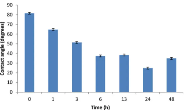
The wettability study using water contact angle on mirror
pol-ished and etched Ti6Al4V samples is presented inFig. 2. The study
demonstrated that the hydrophilicity of Ti6Al4V surfaces increased with increasing etching time of up to 6 h. The hydrophilicity of the samples etched for 6–48 h was similar for all samples, with a small decrease for the sample etched for 24 h.
Fig. 3 shows the fluorescence microscopy images of etched
titanium surfaces (etching times of 1 h and 24 h) coated with
Fig. 2. Water contact angle of etched Ti6Al4V using 30 wt% H2O2aqueous solution for 0, 1, 3, 6, 13, 24 and 48 h; error bars correspond to standard deviation, with number of samples n = 3.
Ti1 peptide binder. The titanium surface that was not etched prior to coating with the peptide, did not present any fluores-cence emission, whereas all etched surfaces showed some peptide
agglomerations/clusters. The coating shown in Fig. 3a was not
homogeneous (according to Materials and Methods, the fluores-cence intensity was measured only for the surfaces presenting a homogeneous green colour), but the fluorescence emission increased as peptide agglomerations were present when the time of etching was longer, suggesting a greater interaction with the peptides. On the other hand, Ti6Al4V etched for 24 h presented a homogeneous green colour, suggesting a homogeneous peptide coating (Fig. 3b) The fluorescence intensity for Ti1 (RKLPDA) was
157± 1 a.u. and for Ti2 (KKLPDA) it was 188 ± 8 a.u.. Ti2
exhib-ited 16% higher fluorescence intensity when adsorbed onto Ti6Al4V surface, compared with Ti1. Therefore, the titanium peptide binder used in this work was Ti2 (KKLPDA).
The first combined Ti-Hap peptide binder used in this study was the (5-FAM) labelled KKLPDA EEEEEEEE (Ti-Hap1). Fluores-cence microscopy of the Ti6Al4V surface etched for 24 h and dip
coated in a solution of 1M Ti-Hap1 in PBS at pH 7.4 exhibited
no emission, indicating that the peptide did not adsorb onto the surface. For this reason, three other peptides were designed; Ti-Hap2, Ti-Hap3 and Ti-Hap4. The coatings were prepared following exactly the same dip coating conditions, using Ti6Al4V etched for 24 h. Homogeneous coatings were obtained for all three combined Ti-Hap peptides and the fluorescence intensities were measured. The Ti-Hap4 coated sample exhibited higher fluorescence inten-sity compared with Ti-Hap2 and Ti-Hap3 (Fig. 4). For this reason,
Ti-Hap4 was used in solutions of concentration 1, 5 and 10M to
coat titanium surfaces etched for 24 h and 48 h, in order to study
Fig. 4. Fluorescence Intensity on Ti6Al4V surfaces etched for 24 h and HA surfaces.
Samples were immersed in a 1M concentration of peptide in PBS for 17 h; The error bars correspond to standard deviation, with number of samples n = 3. Control surfaces were uncoated; the Ti-Hap1 coating on the Ti6Al4V surface was visibly heterogeneous, and therefore the fluorescence intensity was not calculated. The set-up of the fluorescence microscope varied between the Ti6Al4V and HA surface measurements, and therefore intensity comparisons are not possible between the two different materials.
Table 3
Fluorescence intensity of Ti6Al4V after etching with 30 wt% H2O2aqueous solution for 24 and 48 h, followed by immersion in 1, 5 and 10M concentration of Ti-Hap4 in PBS solution. The errors correspond to standard deviation, with number of samples n = 3. Ti-Hap4 peptide concentration (M) Fluorescence intensity (a.u.) on Ti6Al4V surface after 24 h etching Fluorescence intensity (a.u.) on Ti6Al4V surface after 48 h etching 1 101± 3 121± 10 5 104± 2 164± 18 10 148± 29 246± 15
the effect of peptide concentration, as well as the effect of etching time, on the quality of the obtained coatings.
From the fluorescence intensities measured for the 24 h and 48 h etched Ti6Al4V samples (Table 3), it was noticed that the 48 h
sam-ples coated with 10M Ti-Hap4 exhibited 40% higher fluorescence
intensity compared with the rest of the samples, suggesting an enhanced interaction of the peptide with the Ti6Al4V surface. Also
fromTable 3, the fluorescence intensity increased with increasing
the peptide concentration in the coating solution, as well as with the etching time of the Ti6Al4V surface.
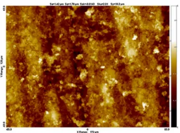
The average roughness (Sa) of the hydroxyapatite surface after
polishing, obtained by white light interferometry and shown in
Fig. 5, was Sa= 1.42m, much higher compared with all
tita-nium surfaces. The hydroxyapatite tablets were dip coated with
Fig. 3. Fluorescence microscopy images with 10X magnification (100m scale bar) of Ti6Al4V samples coated with Ti1 peptide, using a 1 M concentration in PBS solution. Prior to coating, samples were polished to mirror finish and then etched using 30 wt% H2O2aqueous solution for (a) 1 h and (b) 24 h.
Fig. 5. White light interferometry of the surface of a hydroxyapatite tablet prior to
coating with peptides. The average roughness was Sa= 1.42m.
the hydroxyapatite binder (Hap1) as well as the combined pep-tides Ti-Hap1-4. The hydroxyapatite samples coated with the hydroxyapatite binder Hap1 had the highest fluorescence intensity compared with the combined peptides, showing strong interac-tion with HA surfaces as expected (Fig. 4). The samples coated with the combined Ti-Hap peptides showed also some adsorption. The Ti-Hap1 peptide showed higher fluorescence intensity compared with the other combined Ti-Hap peptides. Among the modified combined Ti-Hap peptides, the sequence which presented higher fluorescence intensity was the peptide with the glycine spacer (Ti-Hap2). Ti-Hap3 and 4 exhibited similar fluorescence intensities, both lower than Ti-Hap2 (Fig. 4). It should be noted, that the set-up of the fluorescence microscope was different between Ti6Al4V and HA surface measurements, and therefore comparisons of the measured intensity should not be made between the two different materials inFig. 4.
4. Discussion
The roughness of Ti6Al4V surfaces increased with etching time
(24 h and 48 h in 30 wt% H2O2) compared with the untreated
sam-ple. This is in agreement with a similar study, where a commercial
pure titanium grade I was etched with 30% v/v H2O2; the surface
exhibited nanoscale topographical changes when etched for 1–6 h, but exhibited microscale topographical changes when etched for
24 h or more[36].
The Ti6Al4V samples etched with H2O2were more hydrophilic
than the polished to mirror finish control Ti6Al4V sample, whereas samples etched between 6 h and 48 h were more hydrophilic compared with those etched for up to 3 h (Fig. 2). Other stud-ies also showed that the hydrophilicity of titanium oxide and Ti6Al4V increased in samples treated with H2O2. For example,
tita-nium (IV) dioxide particles after treatment with peroxide became
more hydrophilic[37]. Similarly, Ti6Al4V surfaces became more
hydrophilic after treatment with aqueous 30 wt% H2O2[38]. It was
concluded that the use of H2O2resulted in topographical changes,
an increase in the oxide layer thickness, and the formation of per-oxide complexes such as Ti(IV)O21−, Ti(IV)O22−and Ti(III)O2[38].
Generally, it has been reported that hydrophilic surfaces exhibited improved adsorption of peptides and proteins, as well as enhancing
cell attachment[24,38–40].
Surfaces etched for 24 h and 48 h exhibited homogeneous coat-ings with Ti-Hap2-4 peptides, indicating improved interaction. Samples treated with peroxide for up to 13 h displayed visibly het-erogeneous coatings (Fig. 3). A recent fibronectin adsorption study
showed that samples of Ti6Al4V pre-treated with peroxide for short periods, e.g., 5 min, did not present increased fibronectin coverage
compared with the untreated control sample[38]. A separate study,
where the etching time ranged from 1 h to 4 weeks, showed that a significant increase in the adsorption of fluorescein isothiocyanate (FITC) conjugated albumin serum was observed, compared with the
untreated control sample[36]. The study suggested that the OH−
ions or peroxide complexes on the etched surfaces interacted with the charged protein. Hence, the ionic composition of surfaces plays an important role for the adsorption of proteins onto the etched surfaces.
Sano and Shiba[25]suggested that the Ti1 (RKLPDA) peptide
binder can bind to the titanium surface by the cis-folding of P causing an orientation of R and D amino acids to the surface. An
electrostatic interaction between the Ti-O−and R, a Lewis base,
and an electrostatic interaction between−Ti-OH2+and D, a Lewis
acid, summarises the attractive interactions between Ti1 and the titanium surface. This suggests that any other cationic amino acid in the first position in the peptide sequence, and/or anionic amino acid in the fifth position in the peptide sequence, can be used to bind to titanium surfaces.
In this study, Ti1 and Ti2 differ only in their first amino acid, which is either R or K. The coatings formed at pH 7.4 on Ti6Al4V with RKLPDA and KKLPDA were both homogeneous, and the fluo-rescence intensity inFig. 4was slightly higher for KKLPDA. It should be noted that the isoelectric points of both sequences are relatively close to 8.8 (Table 1). It is likely that the amphoteric behaviour of the etched titanium surfaces at pH 7.4 made the interaction with both peptides possible. The homogeneity of the coating for both cases suggests that R or K can be used in the first position of the sequence. In this study, Ti2, with K in the first position of the pep-tide sequence, was preferred because it resulted in greater surface adsorption compared with Ti1.
The peptide sequences Ti-Hap2-4 were modified from Ti-Hap1 by adding amino acids between the titanium and the hydrox-yapatite peptide binders. TiHap2-4 were found to adsorb on the Ti6Al4V surfaces, whereas the Ti-Hap1 did not. This may be a con-sequence of the additional negative charge from the glutamic acid coil that resulted in a lower isoelectric point of the peptide (3.65) compared with Ti2 (8.72); furthermore at pH = 7.4, the Ti-Hap1 pep-tide presented a negative charge that would interact weakly with the amphoteric titanium surface. Although the isoelectric point of Ti-Hap2, modified to include a 4-glycine spacer, was the same as Ti-Hap1, only Ti-Hap2 coating was found to adsorb onto the Ti6Al4V surface. This can be explained by the increase in confor-mational freedom conferred by the glycine spacer in the sequence, which improves the exposure of the titanium binder to the sur-face, resulting in improved peptide/surface interactions. The role of glycine spacers between a self-assembly sequence RADA 16-I and the functional biological motif PFSSTKT was reported by
Taraballi et al.[41], who concluded that a glycine spacer of four
residues or more can lead to a more stable self-assembling 3D nanostructure and improved exposure of the functional motif to neural stem cells, resulting in improved cell adhesion, viability and differentiation ability. The peptides Ti-Hap3-4 presented an extra sequence of titanium peptide binder KKLPDA, and hence the iso-electric point increased to 3.94 with the addition of two lysines. These sequences had increased net charge and greater distance between each peptide terminal, but the charge of both sequences at pH 7.4 remained negative. The difference between these two sequences was the position of hydroxyapatite and titanium pep-tide binders (Table 1). The Ti-Hap4 coating at pH 7.4 showed almost double the fluorescence intensity compared with the other sequences. This observation suggests that the order of the charged amino acids in the sequence affects the interaction with the surface.
The coatings on the hydroxyapatite surface were performed with all peptides containing the glutamic acid motif. It was noted that for peptides with higher negative net charge, the adsorption was higher (Fig. 4). Previous studies reported that the adsorption of proteins on hydroxyapatite was generally better than on titanium.
For example Zeng et al.[42]reported that bovine serum albumin
was adsorbed twice as much on hydroxyapatite than on pure
tita-nium, whereas Kilpadi et al.[43] reported that both fibronectin
and vitronectin were adsorbed on both materials, hydroxyapatite exhibiting higher adsorption than pure titanium once again. Shen
et al.[44]reported that the electrostatic energy plays a key role
in the interactions between proteins and hydroxyapatite surface,
whereas COO−and NH3+are the main chemical groups present in
model proteins interacting with calcium phosphate surfaces. Based on the above findings in the literature, one would expect that the adsorption of peptides onto hydroxyapatite and titanium surfaces in our case should follow a similar trend. However, direct com-parison of the fluorescence intensity between the titanium and hydroxyapatite surfaces was not possible as mentioned earlier and therefore such an argument would not be supported by the data presented here.
5. Conclusions
The ability of a number of peptide sequences to be adsorbed by Ti6Al4V and hydroxyapatite surfaces was studied. It was shown that longer periods of Ti6Al4V surface treatment with peroxide resulted in increased titanium peptide binder (RKLPDA or KKLPDA) adsorption which may be associated with changes in the topog-raphy and hydrophilicity of the surface. Further work needs to be conducted to establish the relationship between roughness, hydrophilicity and adsorption of peptides. A small modification of RKLPDA by replacing one arginine by one lysine, both amino acids exhibiting a cationic charge at physiological pH, resulted in a small improvement of the adsorption intensity for the KKLPDA peptide. Four combined Ti-Hap peptides were synthesised by combining the hydroxyapatite peptide binder EEEEEEEE with the titanium pep-tide binder KKLPDA in different ways using in one case a 4-glycine spacer. The combined Ti-Hap peptide with the highest adsorption to Ti6Al4V was the Ti-Hap4 at pH 7.4, whereas the highest adsorp-tion ability for hydroxyapatite at the same pH was exhibited by the more negatively charged Ti-Hap1 peptide. Both Ti-Hap2, contain-ing the 4-glycine spacer, and Ti-Hap3 were successfully adsorbed onto Ti6Al4V and hydroxyapatite surfaces. The peptide Ti-Hap4 also bound to both surfaces successfully, suggesting that it could be used for further experiments in an arrangement that contains both materials.
Acknowledgements
GMR would like to thank the School of Metallurgy and Materials/University of Birmingham for providing a PhD EPSRC studentship. This project has received funding from the European Union’s Horizon 2020 research and innovation programme under the Marie Sklodowska-Curie grant agreement No. 645749.
References
[1]X.Y. Liu, P.K. Chu, C.X. Ding, Surface modification of titanium, titanium alloys, and related materials for biomedical applications, Mater. Sci. Eng. R-Rep. 47 (2004) 49–121.
[2]Y.-T. Sul, C. Johansson, E. Byon, T. Albrektsson, The bone response of oxidized bioactive and non-bioactive titanium implants, Biomaterials 26 (2005) 6720–6730.
[3]M. Geetha, A.K. Singh, R. Asokamani, A.K. Gogia, Ti based biomaterials, the ultimate choice for orthopaedic implants – a review, Prog. Mater. Sci. 54 (2009) 397–425.
[4]L.T. de Jonge, S.C.G. Leeuwenburgh, J.G.C. Wolke, J.A. Jansen, Organic-inorganic surface modifications for titanium implant surfaces, Pharm. Res. 25 (2008) 2357–2369.
[5]H.B. Wen, J.R. de Wijn, F.Z. Cui, K. de Groot, Preparation of bioactive Ti6Al4V surfaces by a simple method, Biomaterials 19 (1998) 215–221.
[6]H.-M. Kim, F. Miyaji, T. Kokubo, T. Nakamura, Preparation of bioactive Ti and its alloys via simple chemical surface treatment, J. Biomed. Mater. Res. 32 (1996) 409–417.
[7]L. Jonáˇsová, F.A. Müller, A. Helebrant, J. Strnad, P. Greil, Biomimetic apatite formation on chemically treated titanium, Biomaterials 25 (2004) 1187–1194. [8]C. Ohtsuki, H. Iida, S. Hayakawa, A. Osaka, Bioactivity of titanium treated with
hydrogen peroxide solutions containing metal chlorides, J. Biomed. Mater. Res. 35 (1997) 39–47.
[9]F.M. He, G.L. Yang, Y.N. Li, X.X. Wang, S.F. Zhao, Early bone response to sandblasted, dual acid-etched and H2O2/HCl treated titanium implants: an experimental study in the rabbit, Int. J. Oral Maxillofac. Surg. 38 (2009) 677–681.
[10]K. de Groot, J.G. Wolke, J.A. Jansen, Calcium phosphate coatings for medical implants, J. Eng. Med. 212 (1998) 137–147.
[11]A. Bigi, E. Boanini, B. Bracci, A. Facchini, S. Panzavolta, F. Segatti, et al., Nanocrystalline hydroxyapatite coatings on titanium: a new fast biomimetic method, Biomaterials 26 (2005) 4085–4089.
[12]A.M. Smith, J.Z. Paxton, Y.-P. Hung, M.J. Hadley, J. Bowen, R.L. Williams, et al., Nanoscale crystallinity modulates cell proliferation on plasma sprayed surfaces, Mater. Sci. Eng. C 48 (2015) 5–10.
[13]H. Wang, N. Eliaz, Z. Xiang, H.-P. Hsu, M. Spector, L.W. Hobbs, Early bone apposition in vivo on plasma-sprayed and electrochemically deposited hydroxyapatite coatings on titanium alloy, Biomaterials 27 (2006) 4192–4203. [14]L. Sun, C.C. Berndt, K. Gross, a & kucuk, A. material fundamentals and clinical
performance of plasma- sprayed hydroxyapatite coatings: a review, J. Biomed. Mater. Res. 58 (2001) 570–592.
[15]O. Albayrak, O. El-Atwani, S. Altintas, Hydroxyapatite coating on titanium substrate by electrophoretic deposition method: effects of titanium dioxide inner layer on adhesion strength and hydroxyapatite decomposition, Surf. Coat. Technol. 202 (2008) 2482–2487.
[16]M.-S. Kim, J.-J. Ryu, Y.-M. Sung, One-step approach for nano-crystalline hydroxyapatite coating on titanium via micro-arc oxidation, Electrochem. Commun. 9 (2007).
[17]H.-W. Kim, Y.-H. Koh, L.-H. Li, S. Lee, H.-E. Kim, Hydroxyapatite coating on titanium substrate with titania buffer layer processed by sol–gel method, Biomaterials 25 (2004) 2533–2538.
[18]P.B. Habibovic, et al., Biomimetic hydroxyapatite coating on metal implants, J. Am. Ceram. Soc. 85 (2002) 517–522.
[19]E. Pfender, Fundamental studies associated with the plasma spray process, Surf. Coat. Technol. 34 (1988) 1–14.
[20]L. Yan, Y. Leng, L.-T. Weng, Characterization of chemical inhomogeneity in plasma-sprayed hydroxyapatite coatings, Biomaterials 24 (2003) 2585–2592. [21]F.M. Fazan, P.M. Marquis, Dissolution behavior of plasma-sprayed
hydroxyapatite coatings, J. Mater. Sci. Mater. Med. 11 (2000) 787–792. [22]X.-X. Wang, S. Hayakawa, K. Tsuru, A. Osaka, A comparative study of in vitro
apatite deposition on heat-, H2O2-, and NaOH-treated titanium surfaces, J. Biomed. Mater. Res. 54 (2001) 172–178.
[23]M. Navarro, A. Michiardi, O. Casta ˜no, J.A. Planell, Biomaterials in orthopaedics, J. R. Soc. Interface 5 (2008) 1137–1158.
[24]Li Pea, The role of hydrated silica, titania, and alumina in inducing apatite on implants, J. Biomed. Mater. Res. 28 (1994) 7–15.
[25]K.I.S. Sano, A hexapeptide motif that electrostatically binds to the surface of titanium, J. Am. Chem. Soc. 125 (2003) 14234–14235.
[26]A.D. Roddick-Lanzilotta, A.J. McQuillan, An in situ infrared spectroscopic study of glutamic acid and of aspartic acid adsorbed on TiO 2: implications for the biocompatibility of titanium, J. Colloid Interface Sci. 227 (2000) 48–54. [27]A.D. Roddick-Lanzilotta, P.A. Connor, A.J. McQuillan, An in situ infrared
spectroscopic study of the adsorption of lysine to TiO2 from an aqueous solution, Langmuir 14 (1998) 6479–6484.
[28]A.D. Roddick-Lanzilotta, A.J. McQuillan, An in situ infrared spectroscopic investigation of lysine peptide and polylysine adsorption to TiO2 from aqueous solutions, J. Colloid Interface Sci. 217 (1999) 194–202. [29]R. Fujisawa, Y. Wada, Y. Nodasaka, Y. Kuboki, Acidic amino acid-rich
sequences as binding sites of osteonectin to hydroxyapatite crystals, Biochim. Biophys. Acta (BBA)-Protein Struct. Mol. Enzymol. 1292 (1996) 53–60. [30]S. Koutsopoulos, E. Dalas, The effect of acidic amino acids on hydroxyapatite
crystallization, J. Cryst. Growth 217 (2000) 410–415.
[31]D. Wang, S. Miller, M. Sima, P. Kopeˇcková, Kopeˇcek J. Synthesis, Evaluation of water-soluble polymeric bone-targeted drug delivery systems, Bioconjugate Chem. 14 (2003) 853–859.
[32]M.B. Murphy, J.D. Hartgerink, A. Goepferich, A.G. Mikos, Synthesis and in vitro hydroxyapatite binding of peptides conjugated to calcium-binding moieties, Biomacromolecules 8 (2007) 2237–2243.
[33]G.K. Hunter, C.L. Kyle, H.A. Goldberg, Modulation of crystal formation by bone phosphoproteins: structural specificity of the osteopontin-mediated inhibition of hydroxyapatite formation, Biochem. J. 300 (1994) 723–728. [34]G.K. Hunter, H.A. Goldberg, Modulation of crystal formation by bone
phosphoproteins: role of glutamic acid-rich sequences in the nucleation of hydroxyapatite by bone sialoprotein, Biochem. J 302 (1994) 175–179. [35]http://www.pepcalc.com.
[36]M. Nagassa, A. Daw, W.G. Rowe, A. Carley, D.W. Thomas, R. Moseley, Optimisation of the hydrogen peroxide pre-treatment of titanium: surface characterisation and protein adsorption, Clin. Oral Implants Res. 19 (2008) 1317–1326.
[37]D.E. MacDonald, N. Deo, B. Markovic, M. Stranick, P. Somasundaran, Adsorption and dissolution behaviour of human plasma fibronectin on thermally and chemically modified titanium dioxide particles, Biomaterials 23 (2002) 1269–1279.
[38]D.E. MacDonald, B.E. Rapuano, N. Deo, M. Stranick, P. Somasundaran, A.L. Boskey, Thermal and chemical modification of titanium-aluminum-vanadium implant materials: effects on surface properties, glycoprotein adsorption, and MG63 cell attachment, Biomaterials 25 (2004) 3135–3146.
[39]B. Feng, J. Weng, B.C. Yang, S.X. Qu, X.D. Zhang, Characterization of surface oxide films on titanium and adhesion of osteoblast, Biomaterials 24 (2003) 4663–4670.
[40]L. Ponsonnet, et al., Relationship between surface properties (roughness, wettability) of titanium and titanium alloys and cell behaviour, Mater. Sci. Eng. C 23 (2003) 551–560.
[41]F. Taraballi, A. Natalello, M. Campione, O. Villa, S.M. Doglia, A. Paleari, et al., Glycine-spacers influence functional motifs exposure and self-assembling propensity of functionalized substrates tailored for neural stem cell cultures, Front. Neuroeng. 3 (2010) 1.
[42]H. Zeng, K.K. Chittur, W.R. Lacefield, Analysis of bovine serum albumin adsorption on calcium phosphate and titanium surfaces, Biomaterials 20 (1999) 377–384.
[43]K.L. Kilpadi, P.L. Chang, S.L. Bellis, Hydroxylapatite binds more serum proteins, purified integrins, and osteoblast precursor cells than titanium or steel, J. Biomed. Mater. Res. 57 (2001) 258–267.
[44]J.-W. Shen, T. Wu, Q. Wang, H.-H. Pan, Molecular simulation of protein adsorption and desorption on hydroxyapatite surfaces, Biomaterials 29 (2008) 513–532.