Genome-Wide Small Interfering RNA Screens Reveal VAMP3 as a
Novel Host Factor Required for Uukuniemi Virus Late Penetration
Roger Meier,aAndrea Franceschini,cPeter Horvath,a,bMarilou Tetard,a,dRoberta Mancini,aChristian von Mering,cAri Helenius,a Pierre-Yves Lozacha,dInstitute of Biochemistrya
and Light Microscopy and Screening Center,b
ETH Zurich, Zurich, Switzerland; Institute of Molecular Life Sciences and Swiss Institute of Bioinformatics, University of Zurich, Zurich, Switzerlandc
; INRS—Institut Armand-Frappier, Université du Québec, Laval, Québec, Canadad
ABSTRACT
The Bunyaviridae constitute a large family of enveloped animal viruses, many of which are important emerging pathogens. How
bunyaviruses enter and infect mammalian cells remains largely uncharacterized. We used two genome-wide silencing screens
with distinct small interfering RNA (siRNA) libraries to investigate host proteins required during infection of human cells by the
bunyavirus Uukuniemi virus (UUKV), a late-penetrating virus. Sequence analysis of the libraries revealed that many siRNAs in
the screens inhibited infection by silencing not only the intended targets but additional genes in a microRNA (miRNA)-like
manner. That the 7-nucleotide seed regions in the siRNAs can cause a perturbation in infection was confirmed by using synthetic
miRNAs (miRs). One of the miRs tested, miR-142-3p, was shown to interfere with the intracellular trafficking of incoming
vi-ruses by regulating the v-SNARE VAMP3, a strong hit shared by both siRNA screens. Inactivation of VAMP3 by the tetanus toxin
led to a block in infection. Using fluorescence-based techniques in fixed and live cells, we found that the viruses enter VAMP3
ⴙendosomal vesicles 5 min after internalization and that colocalization was maximal 15 min thereafter. At this time, LAMP1 was
associated with the VAMP3
ⴙvirus-containing endosomes. In cells depleted of VAMP3, viruses were mainly trapped in
LAMP1-negative compartments. Together, our results indicated that UUKV relies on VAMP3 for penetration, providing an indication of
added complexity in the trafficking of viruses through the endocytic network.
IMPORTANCE
Bunyaviruses represent a growing threat to humans and livestock globally. Unfortunately, relatively little is known about these
emerging pathogens. We report here the first human genome-wide siRNA screens for a bunyavirus. The screens resulted in the
identification of 562 host cell factors with a potential role in cell entry and virus replication. To demonstrate the robustness of
our approach, we confirmed and analyzed the role of the v-SNARE VAMP3 in Uukuniemi virus entry and infection. The
infor-mation gained lays the basis for future research into the cell biology of bunyavirus infection and new antiviral strategies. In
addi-tion, by shedding light on serious caveats in large-scale siRNA screening, our experimental and bioinformatics procedures will
be valuable in the comprehensive analysis of past and future high-content screening data.
W
ith over 350 identified isolates worldwide, bunyaviruses
represent a global threat to public health, livestock, and
ag-ricultural productivity (
1
). Many cause health problems in
hu-mans, such as fatal pulmonary syndromes, encephalitis, and
hem-orrhagic fever. Severe fever with thrombocytopenia syndrome
virus in China (2009), Rift Valley fever virus (RVFV) in South
Africa (2010), Schmallenberg virus in Europe (2011), and
hanta-virus in California (2012) are recent examples of outbreaks
dem-onstrating that bunyaviruses must be taken seriously as emerging
agents of disease. Currently, there are no approved vaccines or
treatments to protect humans from infection.
Bunyaviruses are enveloped, negative-strand RNA viruses that are
roughly spherical (
⬃100 nm), with a trisegmented genome that is
replicated in the cytosol. The envelope glycoproteins G
Nand G
Cform
spike-like projections responsible for virus attachment to host cells
and penetration by membrane fusion. Receptors, cellular factors, and
entry pathways remain poorly characterized. However, many
bunya-viruses use the C-type lectin CD209 as a receptor to enter dendritic
cells (DCs), and some members of the hantavirus subfamily have
been shown to use
3-integrin to infect endothelial cells (
2
,
3
).
Hepa-ran sulfate has been implicated in RVFV attachment (
4
). Sensitivity to
clathrin and dynamin-2 perturbation implies a role for
clathrin-me-diated endocytosis in uptake and internalization (
5–9
).
Several lines of evidence suggest that many bunyaviruses are
late-penetrating viruses (L-PVs), a large group of viruses that
de-pends on trafficking to late endosomes (LE) for productive
infec-tion (
10
,
11
). Inhibitor studies have shown that infection relies on
late endosomal maturation and vacuolar acidification (
11
,
12
).
Weak bases that neutralize the endosomal pH, such as ammonium
Received 8 February 2014 Accepted 12 May 2014 Published ahead of print 21 May 2014 Editor: T. S. Dermody
Address correspondence to Pierre-Yves Lozach, [email protected].
Present addresses: Roger Meier, Scientific Center for Optical and Electron Microscopy, ETH Zurich, Zurich, Switzerland; Peter Horvath, Synthetic and Systems Biology Unit, Biological Research Center, Szeged, Hungary, and Institute of Molecular Medicine Finland (FIMM), University of Helsinki, Helsinki, Finland; Pierre-Yves Lozach, CellNetworks—Cluster of Excellence and Department of Infectious Diseases, Virology, University Hospital Heidelberg, Heidelberg, Germany. Supplemental material for this article may be found athttp://dx.doi.org/10.1128 /JVI.00388-14.
Copyright © 2014, American Society for Microbiology. All Rights Reserved.
chloride (NH
4Cl), inhibit infection by most bunyaviruses (
1
).
Ac-id-activated viral membrane fusion, which results in the release of
the virus genome in the cytosol, occurs typically at pH levels below
5.8, which are characteristic of late endosomal vacuoles (
5
,
11
,
13
,
14
).
To further investigate the life cycle of bunyaviruses, we used
two separate libraries of small interfering RNAs (siRNAs) against
the whole human genome to screen for host factors involved in
infection by Uukuniemi virus (UUKV), a bunyavirus of the
Phle-bovirus genus. Since UUKV is not pathogenic to humans, it
en-ables state-of-the-art experimental approaches that are nearly
im-possible for pathogenic bunyaviruses, most of which must be
contained within high-level biosafety laboratories. Together, the
screens revealed 562 cellular factors with a potential role in UUKV
endocytosis and replication. Further analysis, however, showed
that many of the identified candidates were caused by off-target
effects due to microRNA (miRNA)-like functions of the siRNAs.
When the effects of a synthetic miRNA (miR) that mimicked the
endogenous miRNA-142-3p were analyzed in detail, we observed
that miR-142-3p inhibited UUKV entry and penetration. The
v-SNARE VAMP3, a hit shared between both of the siRNA screens,
was identified as an important target of miR-142-3p and a critical
player in the late penetration of the virus.
MATERIALS AND METHODS
Cells and viruses. All products used for cell culture were from Life
Tech-nologies. Human epithelial cells (HeLa) that stably express the human C-type lectin CD209 were cultured according to ATCC recommenda-tions and used as the cell model (3). The prototype strains of Uukuni-emi virus S23, influenza A virus X-31 (IAV; Virapur), and green fluo-rescent protein (GFP)-recombinant Semliki Forest virus (SFV; a kind gift from the University of Helsinki) have been described previously (15–17). Production, purification, and titration of UUKV were per-formed in BHK-21 cells as described elsewhere (11). Labeling of UUKV with AF-succinimidyl esters and Bodipy TR-thiosulfates (In-vitrogen) was performed in HEPES (20 mM) as recently described (11). Alternatively, UUKV (109focus-forming units [FFU/ml]) was labeled with the lipid dye R18 (20M; Life Technologies) according to a similar protocol. The multiplicity of infection (MOI) is given accord-ing to the titer determined on BHK-21 cells.
siRNAs, miRs, and plasmids. siRNAs (siR-VAMP3#1, CAGCAUGU
UUCUGAUAAUUAU [SI00759227]; siR-VAMP3#2, CCCAAAUAUGA AGAUAAACUA [SI00759248]; siR-N, GGAGAUCUUGGAUGCCAAU GA), AllStars negative-control siRNA (siR-Ctrl), and miRs were all from Qiagen. siR-N has been described previously (3). The plasmid carrying the gene for enhanced GFP (EGFP)-VAMP3 was generated using the human open reading frame library V3.1 (hORFeome v3.1) and Gateway cloning technology according to the manufacturer’s instructions (18). Plasmids carrying genes for red fluorescent protein (RFP)-LAMP1 and EGFP-LAMP1 were kindly provided by J. Gruenberg (University of Geneva, Geneva, Switzerland). The catalytic chain of tetanus toxin (TeTx) was a kind gift from G. Coppolino (University of Guelph, Guelph, Ontario, Canada).
Antibodies and reagents. The mouse monoclonal antibody (MAb)
8B11A3 (kind gift from Ludwig Institute for Cancer Research, Stockholm, Sweden) and HB-65 (ATCC) are directed against the nucleoproteins of UUKV and IAV, respectively (19). The phycoerythrin (PE)-conjugated fragment antigen binding (Fab) 1621P (R&D Systems) was used to detect CD209 at the cell surface. The mouse MAb against␣-tubulin and the polyclonal rabbit antibody (Ab) against VAMP3 were purchased from Abcam and Thermo Scientific, respectively. An AF647-conjugated anti-mouse antibody (Life Technologies) was used as a secondary Ab in a
fluorescence-activated cell sorting (FACS)-based infection assay. NH4Cl (Sigma) stock was dissolved in water.
High-throughput siRNA screening procedures. (i) Human genome-wide siRNA libraries. The human genome-genome-wide siRNA libraries
em-ployed in the screens were from Qiagen and Dharmacon. The library of Qiagen consists of four independent siRNAs against each gene, while that of Dharmacon involves pools of four different siRNAs per gene. The Qia-gen library (OnGuard) was comprised of the druggable Qia-genome version 3, the genome supplement version 1, and the predicted gene set version 1. The Dharmacon library (On-TargetPlus) included the G-104655-02 OTP drug targets, the G-104675-02 OTP druggable subsets, and the G-105005-02 OTP genome. The gene information released by the Na-tional Center for Biotechnology Information (NCBI) in November 2012 was used for the annotation of libraries.
Several siRNAs in each plate were used to control the successful con-duct of the general screening procedures. The AllStarsNegative and scrambled siRNAs from Qiagen and Dharmacon, respectively, as well as mock-transfected cells were used as negative controls. siRNA silencing of proteins known to be essential for UUKV infection, such as the vacuolar-type H⫹-ATPase V1 subunit A1 and the virus receptor CD209, but also against the viral genome, such as siR-N, were used as positive controls (3, 11). To assess the efficiency of transfection, cell death was induced with siRNAs that silence proteins critical for cell viability, such as the kinesin family member 11.
(ii) Liquid handling procedure. All liquid handling steps, including
preparation of cell plates, reverse transfection, virus infection, cell fixa-tion, and immunofluorescence staining, were performed with an EL406 microplate washer and dispenser as well as with a microplate Biostak stacker (BioTek).
(iii) siRNA screen reverse transfection. RNA interference screenings
were achieved in single runs (Qiagen) or in triplicate (Dhamarcon). Ex-periments were conducted in a 384-well plate format, amounting to 296 and 171 plates for the Qiagen and Dharmacon screens, respectively. Re-verse transfection was performed by seeding 400 cells in 50l of culture medium into a well containing 30l of transfection mix (24.9 l of Dul-becco’s modified Eagle’s medium [DMEM], 0.1 l of RNAiMax [LifeTechnologies], and 1.6 pmol siRNA in 5l [final siRNA concentra-tion of⬃20 nM]). Cells were then left at 37°C for 72 h prior to infection.
(iv) Infection, immunofluorescence staining, and imaging.
Trans-fected cells were washed with complete medium before infection with UUKV (15,000 FFU [MOI,⬃5], diluted in DMEM containing 20 mM HEPES and 0.2% bovine serum albumin [BSA; Life Technologies]) at 37°C for 1 h. After exposition, the virus inoculum was replaced by culture medium. Infected cells were fixed with 4% formaldehyde 19 h later and immunostained with the mouse MAb 8B11A3 against N (diluted 1:500 in phosphate-buffered saline [PBS], 0.1% Triton X-100, 0.2% BSA) at room temperature (RT) for 1.5 h. The primary Ab and nuclei were then detected with a mix containing an anti-mouse Alexa Fluor 488-conjugated second-ary Ab (1:1,000; Life Technologies) and Hoechst 33258 (1g ml⫺1; Life Technologies) in PBS with 0.2% BSA at RT in the dark for 1.5 h. After washing with 0.04% NaN3, plates were sealed and imaged by using auto-mated inverted epifluorescence microscopes (ImageXpress; Molecular Devices) with a 10⫻ objective, 0.3 numerical aperture (NA) Plan Fluor lens.
Image analysis. (i) Imaging error detection (out of focus or illumi-nation problems). As the first step, image quality was checked.
Out-of-focus and illumination problems were automatically detected. Eight global image properties were extracted based on image intensities and their gradients and total variation. There was not a single property that clearly indicated whether an image was in focus or had to be reimaged. Therefore, we used all global parameters and a supervised machine learn-ing algorithm based on the Random Forest classifier (20). Manually se-lected in-focus, out-of-focus, and illumination problem-affected images were used to train the model. The recognition accuracy was higher than 99%. Plates with out-of-focus wells were reacquired.
(ii) Image segmentation and feature extraction. The next step of the
image analysis was the identification of the cells, followed by the segmen-tation of their nuclear and cytoplasmic regions. We used a custom mod-ified version of the CellProfiler program (21). The extraction started with an adaptive threshold determination on the Hoechst channel and fol-lowed by a morphological filter to eliminate objects that were too small or too large. Cells were kept in areas between 176 and 1,963 pixels (73.2m2 and 816.7m2). To perform measurements of the cytoplasm, we ex-tended the nuclear mask by a 12-pixel-wide (7.7-m-wide) ring. The extension value was deduced from the homogeneous fluorescence throughout the cytoplasm of infected cells. Using the detected objects, 37 cellular properties were measured for every single cell. The properties were based on intensity, texture, and morphology. They were used to further characterize the cellular phenotypes. The CellProfiler pipeline, the refactoring function, and a detailed list of features can be found atwww .highcontentanalysis.org.
(iii) Phenotypic classification. Infected cells were visualized using
im-munofluorescence staining against the viral protein N, which is expressed in the cytoplasm. Using simple fluorescence intensity-based threshold determinations resulted in a 20% infection recognition error. This error could be reduced to less than 4% by using a multiparametric supervised machine learning algorithm (22). We tested several models successfully applied earlier for high-content screening for phenotypic discovery (the analyzed classifiers are described in reference23). By combining a com-mittee of such classifiers using a majority voting method (20), the accu-racy of the recognition rate was 96%.
Screen analysis and hit definition. (i) Transformation and normal-ization. While the number of cells seemed to be homogenous within
plates over all the screens, from raw data infections in outer wells were higher than inner wells for most of the plates (data not shown). Conse-quently, the systematic infection edge effect was normalized by using an adapted plate-wise local polynomial regression fitting Loess function (24). To this end, the sigmoidal distribution of the infection indices (i.e., values in the range 0 to 1) was first linearized with empirical logit transformation. From these processed values, the predicted values obtained with the Loess fit function were subtracted, and the mean of the predicted values was finally added (data not shown). In addition, plate-to-plate variations were corrected with a robust Z* score normalization function, and data were back-transformed by using the equation eZ* score/(1⫹ eZ* score) and
nor-malized to the median of the overall negative siRNA controls (25).
(ii) Quality control. After transformation and normalization, the data
quality of individual plates was controlled with the strictly standardized means difference (SSMD) method applied to each positive control within a plate (26). The criteria for quality control were fixed according to the SSMD definition for a “very strong control” (data not shown). Only plates with at least 75% of positive controls considered good or excellent were further processed for the hit analysis. Other parameters that may reflect experimental problems were also considered, such as the infection index and siRNA cytotoxicity. Plates with median infection indices below 10% in the negative siRNA controls were removed to avoid increased errors on potential inhibitory effects. Data for wells of the Qiagen screen were re-moved when the number of cells was less than 75% of the median. As the Dharmacon screen was performed in triplicate, data were removed when the mean cell number for a gene was 3 standard deviations away from the mean observed for the siRNA negative controls.
(iii) Hit definition. Plates and wells that successfully passed the quality
control step were further processed to quantitatively score and rank genes according to the impact of their depletion on UUKV infection. The re-dundant siRNA activity (RSA) method, which is based on a hypergeomet-ric distribution formula, was employed to rank siRNA efficiency and de-fine hits in the Qiagen screen (27). This method allowed generation of two ranked gene lists according to the overall rank of each siRNA against the same gene, for either inhibition or enhancement siRNA strength effects. Subsequently, siRNAs were ranked following the Z* score method, and inhibitor and enhancer hits were defined by P values of 0.01 and 0.001,
respectively. Different thresholds were used because the siRNA effects on infection were asymmetrically distributed. The SSMD method combined with fold change approaches was utilized to rank genes in the Dharmacon screen, as that screen was performed in triplicate. SSMD cutoffs of less than⫺3 and greater than 2 with changes of 0.5-fold and 1.5-fold were used for definitions of inhibitors and enhancers, respectively.
FACS-based infection and plasma membrane-virus fusion assays.
Cells, transfected or not, were infected at the indicated MOIs as previously described (11). After fixation and permeabilization with 0.1% saponin, UUKV and IAV infections were detected by standard immunofluores-cence staining against the nucleoprotein N with the mouse MAbs 8B11A3 and HB-65, respectively, and quantified by flow cytometry. In the case of SFV, GFP expression was used to monitor infection. For siRNA- and miR-based infection assays, transfected cells were washed 1 h before in-fection. For the VAMP3 inactivation assay, cells were transfected with the plasmid carrying the TeTx gene, washed 6 h later, and exposed to viruses 18 h posttransfection. Virus fusion at the surface of siRNA- and miR-transfected cells was performed as recently reported (11).
siRNA- and miR-mediated knockdown confirmation. siRNA and
miR reverse transfections were performed using Lipofectamine RNAiMAX transfection reagent according to the manufacturer’s reverse protocol (Life Technologies). Briefly, 40,000 cells were transfected with a 20 nM final concentration of siRNAs or miRs in fetal calf serum-free medium and seeded in a 24-well dish 3 days before infection or virus fusion.
Surface immunofluorescence staining. The expression of CD209 was
assessed at the surface of cells (not permeabilized) by FACS, using the PE-conjugated Fab 1621P (1:50) according to a standard procedure (3).
DNA transfection. Cells were transfected with plasmids carrying
genes for TeTx, EGFP-VAMP3, RFP-LAMP1, or EGFP-LAMP1 by using Lipofectamine 2000 (Life Technologies) according to the manufacturer’s instructions.
Binding and internalization assays. Viruses were bound to the cells
for up to 2 h on ice at the indicated MOIs in binding buffer (DMEM [pH ⬃7.4] containing 0.2% BSA, 1 mM CaCl2, and 2 mM MgCl2) as described before (3). In internalization assays, fluorescent virus-bound cells were rapidly warmed to 37°C for up to 40 min to trigger endocytosis. Binding and internalization were quantified by flow cytometry or analyzed by confocal microscopy. For FACS analysis, to distinguish between internal-ized and external particles, samples were treated with trypan blue (0.01%; Sigma) for 15 s at RT (3). To assess the role of VAMP3 in UUKV intracel-lular trafficking by using confocal microscopy, cells were transfected with the plasmids carrying genes for EGFP-VAMP3 and RFP-LAMP1, washed 6 h later, and incubated at 37°C for 12 h prior to the exposure to the virus. To further investigate the function of VAMP3 in UUKV late penetration, cells were first transfected with the control siRNA, siR-VAMP3#1, and miR-142-3p, and 48 h later with the plasmid harboring the gene for EGFP-LAMP1, before exposure to UUKV. Cells were fixed and analyzed by using a Zeiss LSM510 Meta microscope and a 63⫻, 1.4 NA Plan-Apochromat immersion oil objective lens.
R18-based virus fusion. Transfected cells were collected from the
cul-ture surface with PBS-EDTA (0.5 mM), washed, and exposed to R18-labeled UUKV (UUKV-R18; MOI,⬃5) in binding buffer (DMEM [pH ⬃7.4] containing 0.2% BSA and 20 mM HEPES) on ice for 1 h. Virus internalization was triggered by rapid warming at 37°C, in the presence or absence of NH4Cl (50 mM), directly inside a Varian Cary Eclipse fluorim-eter (Agilent Technologies), and the fluorescence was measured for 1 h.
Microarray gene expression analysis. Microarray experiments were
conducted using Agilent Human Gene Expression 8⫻60k chips according to the manufacturer’s recommendations. Briefly, total mRNA was iso-lated from cells transfected with siR-Ctrl by using TRIzol reagent, digested with DNase I (Qiagen), and purified. When RNA samples passed quality controls, i.e., when the A260/A280ratio was above 2 and no RNA degrada-tion was detected, microarray analysis was performed. Data were pro-cessed with the quantile normalization method and further analyzed with a two-group-based statistical method in combination with a fold change
approach using the Limma package under the open source software R (28). Down- and upregulated genes were defined by a false-discovery rate better than 0.01 and a change below 0.5-fold or above 1.5-fold, respec-tively.
Protein analysis. Proteins were analyzed by SDS-PAGE and
trans-ferred to polyvinylidene difluoride membranes (iBlot transfer stacks; Life Technologies). Incubation with primary Abs against␣-tubulin (1:5,000) and VAMP3 (1:500) was followed by incubation with rabbit or anti-mouse horseradish peroxidase-conjugated secondary Abs (1:5,000). RESULTS
Genome-wide siRNA screens to elucidate bunyavirus-host cell
interactions in human cells. We developed an automated,
high-content siRNA screening procedure with fluorescence
micros-copy as a readout to elucidate host cell factors and processes
im-portant during early UUKV-host cell interactions. As a cellular
system, we used HeLa cells expressing the surface lectin CD209.
Given that CD209 serves as a receptor for UUKV in DCs, these
HeLa cells provided a model system to investigate the early stages
of infection, including cell attachment, endocytosis, and
transla-tion of the viral proteins (
3
).
Each gene in the human genome was silenced by using two
independent siRNA libraries, as illustrated in
Fig. 1A
. While one of
the libraries (Qiagen) contained four unpooled, single siRNAs
targeting distinct regions of each gene, the second (Dharmacon)
relied on pools of four different siRNAs against each gene. The
screens were carried out as detailed in
Fig. 1A
and in Materials and
Methods. Automated transfection, infection, and
immunostain-ing against newly synthesized nucleoprotein N as well as
auto-mated fluorescence microscopy and image analysis were
em-ployed to analyze cell factors required during the replication cycle,
up to and including translation (
Fig. 1A
).
Before potential hits were defined, systematic errors, such as
batch-to-batch, plate-to-plate, or well-to-well differences and
plate-wise spatial effects, generally caused by the experimental
procedure itself, were normalized by computer-based
mathemat-ical procedures (data not shown). Parameters reflecting
experi-mental problems were also considered, such as siRNA cytotoxicity
and a variable infection index (i.e., the fraction of infected cells).
Plates with median infection indices lower than 10% in the
nega-tive siRNA controls (compared to the median values of 20% and
34% for all plates in the Qiagen and Dharmacon screens,
respec-tively) were removed to avoid any misinterpretation. Of the plates
and wells, 10 to 20% were not taken into consideration for further
analysis. This represented 28 plates out of 296 for Qiagen and 0 for
Dharmacon, or 21,514 and 1,453 wells out of 94,720 and 18,003
for Qiagen and Dharmacon, respectively.
Because the intrinsic features of the siRNA libraries were
dif-ferent (single unpooled versus pooled siRNAs), we used distinct
statistical analyses to define potential hits. Next, we determined
the transcriptome of our HeLa cell line by using an Agilent-based
microarray approach. Consistent with previous transcriptome
studies conducted in HeLa strains, we found that the cells
ex-pressed some 13,000 different mRNA transcripts (see Table
S1 at
http://www.highcontentanalysis.org/download/Table_S1
.xlsx
). As expected from previous HeLa transcriptome studies,
G-protein-coupled receptor signaling and immune defense
factors were poorly represented (
29
,
30
). The set of repressed
genes was therefore excluded from the analysis, to minimize
false-positive hits. For complete details on the screen
proce-dures and analysis as well as the hit definition, refer to Materials
and Methods.
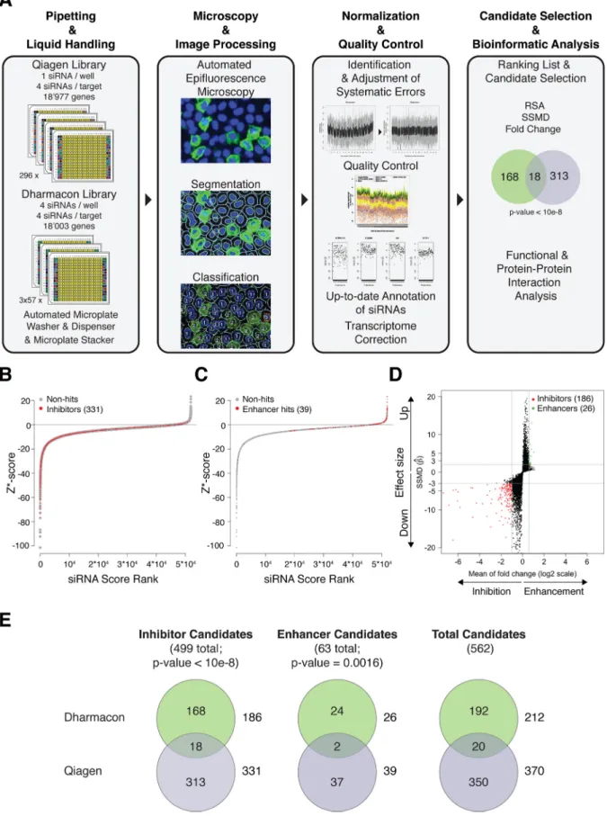
siRNA screens identified 562 candidate genes with a role in
UUKV infection. The Qiagen screen led to the identification of
370 genes with a potential role in UUKV entry and replication, 331
inhibitors and 39 enhancers (
Fig. 1B
,
C
, and
E
; see also Tables S2
and S3 at
http://www.highcontentanalysis.org/download/Table
_S2.xlsx
and
http://www.highcontentanalysis.org/download
/Table_S3.xlsx
, respectively). The Dharmacon screen revealed
212 candidate genes, 186 inhibitors and 26 enhancers (
Fig. 1D
and
E
; see also Table S4 at
http://www.highcontentanalysis.org
/download/Table_S4.xlsx
). Twenty candidates were present in
both the Qiagen and Dharmacon screens (
Fig. 1E
). Although the
direct overlap was modest, Fisher’s exact test clearly established
that the results of both screens were not statistically independent.
Bioinformatics analysis via open source software like STRING,
DAVID, and Reactome showed, moreover, that the potential hits
complemented each other with regard to cell functions (data not
shown). Consequently, we combined the results from both
siRNA libraries. The new list of candidates (shown in Table S5
at
http://www.highcontentanalysis.org/download/Table_S5
.xlsx
) contained 562 host factors representing a broad range of cell
functions. The majority of genes in the list had no prior
connec-tion to bunyavirus infecconnec-tion.
Some of the identified cell functions would be expected for any
RNA virus, such as those related to transcription. Others were
specific to viruses that depend on endosomal acidification and
membrane trafficking for infection, as illustrated by the presence
of many subunits of the vacuolar-type H
⫹-ATPases and several
proteins involved in intracellular protein transport (e.g., AP2M1
and CLTCL1). The largest and most significant groups of
poten-tial hits represented vacuolar acidification and other
membrane-related functions. Cell functions also included signaling, protein
localization, cellular homeostasis, and others that are likely to lay
the basis for future research into bunyaviruses.
siRNAs can silence genes through their seed regions in a
miRNA-like manner. When siRNA screens have been performed
in different laboratories to identify host factors against a virus, the
genes in published hit lists have shown poor overlap, suggesting
serious inherent problems in the approach. This is in part
ex-plained by different screening procedures and analysis, as well as
by siRNA off-target effects (
31–33
). When our analysis was
lim-ited to the 13,000 transcribed genes, the number of candidates was
reduced to about 70% of the original lists independently of the
Qiagen or Dharmacon screens. This implied that at least 30% of
the potential hits in both the Qiagen and Dharmacon original lists
were likely to represent false positives, since the corresponding
genes were not expressed in our cell model. More specifically, it
has been shown that siRNA antisense seed regions of 7 nucleotides
(nt) regulate the expression of many genes through mechanisms
identical to those involved in the silencing of mRNAs by miRNAs
(
Fig. 2A
) (
34–36
). This may result in off-target effects.
To investigate the possibility that siRNAs acted like miRNAs in
our screens, we first classified all the siRNAs in each library
ac-cording to their 7-nt seed regions, i.e., we sorted them into groups
according to the nucleotide sequences from position 2 to 8 in the
5= end of the antisense siRNA strand (data not shown). We could
identify 9,541 and 11,890 different seed sequences among the
72,773 and 71,557 siRNAs in the Qiagen and Dharmacon libraries,
respectively (data not shown). By applying the SSMD and fold
FIG 1 UUKV siRNA screens. (A) Outlines of screening and bioinformatics procedures. (B) Z* score distribution of infection indices obtained for each siRNA
in the Qiagen screen regarding inhibitor candidate genes. A redundant siRNA activity (RSA) P value below 0.01 defined potential hits. (C) Z* score distribution, as described for panel B but for enhancer candidate genes. An RSA P value below 0.001 defined potential hits. (D) Dual flash plot for Dharmacon siRNAs, showing selected candidates based on SSMD criteria and the fold change. Inhibitor genes were defined by SSMD and change values below⫺3-fold and 0.5-fold, respectively, and enhancers were defined as those with values above 2-fold and 1.5-fold (indicated by the gray horizontal lines). (E) Overlaps of candidate genes between the two siRNA screens. The numbers of potential hits that were specific to the Dharmacon and Qiagen screens are indicated in the top and bottom circles, respectively. The total numbers of potential hits for the Dharmacon and Qiagen screens are indicated next to the top and bottom circles, respectively. The P values, based on Fisher’s exact statistical tests, indicate that potential hits obtained for both libraries were not independent.
change approach to the seed sequences that were present in a
minimum of 3 different siRNAs (totals of 5,658 and 7,622 seeds in
the Qiagen and Dharmacon libraries, respectively), we found that
610 seeds in the Qiagen screen conferred the ability to block
UUKV infection. Only 6 such sequences were identified in the
Dharmacon screen. In other words, 5,257 siRNAs out of the
64,793 in the Qiagen screen and 54 siRNAs out of the 65,936 in
the Dharmacon screen had the potential to inhibit the UUKV
life cycle through miRNA-like mechanisms in addition to the
intended siRNA effect. Although this phenomenon may appear
relatively limited, the fraction of siRNAs that potentially acted
like miRNAs was dramatically higher within lists of potential
hits (see Tables S2 to S4 at
http://www.highcontentanalysis
.org
). This implied that miRNA-like mechanisms caused by
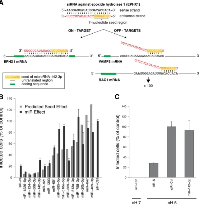
FIG 2 miRs, with sequences identical to those of siRNA seeds that inhibit the UUKV cycle, block infection. (A) Principles of gene silencing by siRNAs through
on- and off-target mechanisms. The siRNA against epoxide hydrolase 1 (EPHX1), the seed region of which is identical to that of miR-142-3p, is shown as an example. VAMP3, vesicular-associated membrane protein 3; RAC1, ras-related C3 botulinum toxin substrate 1. (B) Cells treated with miRs (20 nM), which have identical seeds to those of siRNAs that blocked UUKV infection in the two screens, were infected with UUKV (MOI,⬃5) and harvested 7 h later. Infection was quantified via FACS after immunostaining against the viral nucleoprotein N, and results were normalized to the infection of cells treated with a negative-control siRNA (siR-Ctrl). An siRNA directed against N was used as a positive-control siRNA (siR-N). The light gray graphs indicate the impact predicted for the corresponding siRNA seeds. The value for the correlation between predicted and measured effects was 0.7, according to the Pearson statistical test. Error bars indicate the standard errors (SE) of the means of three independent experiments. (C) To bypass the need of virus endocytosis for infection, cells treated with miRs (20 nM) were exposed to UUKV (MOI,⬃0.5) in the cold, washed, and treated at the indicated pHs at 37°C for 1.5 min. Infected cells were then incubated for 8 h in the presence of NH4Cl (50 mM) to block virus penetration from endosomes. Consequently, only the release of viral genomes from the plasma membrane
was monitored. Infection was analyzed via FACS, following immunostaining against the protein N. siR-Ctrl and siR-N were used as negative- and positive-control siRNAs, respectively. Error bars indicate SE of the means of three independent experiments.
seed regions are likely to play a major role in the generation of
off-target effects.
miRs bearing seed sequences that were predicted to impact
infection had adverse effects on the virus life cycle. Next, we
selected 10 seed sequences predicted to affect UUKV and analyzed
their potential miR functions in more detail. In addition, we
se-lected 5 seed sequences predicted to have no effect on UUKV
infection as controls. To predict whether a seed region has the
capacity to impair the UUKV life cycle, SSMD and fold change
values inferior to
⫺3 and superior to 0.5 were used as cutoffs,
respectively. When miRs with these seed sequences were tested for
their impact on the UUKV life cycle in the HeLa CD209 cell line,
we observed that the effect on infection strongly correlated with
the predicted seed effects (correlation coefficient of 0.7) (
Fig. 2B
).
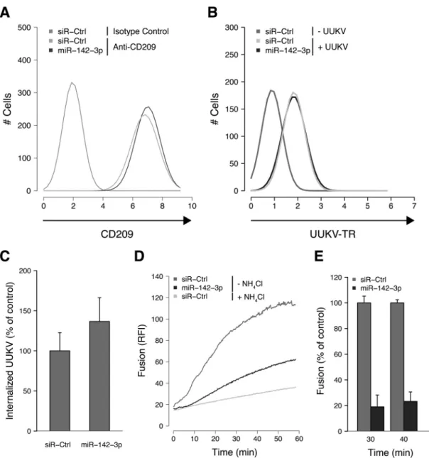
FIG 3 miR-142-3p regulates UUKV intracellular trafficking. (A) Cells transfected with a negative-control siRNA (siR-Ctrl) or miR-142-3p (20 nM) were
immunostained for CD209 expression before FACS analysis. The data shown are representative of three independent experiments. (B) AF488-labeled UUKV (UUKV-AF488) was bound to cells treated with siR-Ctrl or miR-142-3p for 1 h on ice before fixation and FACS analysis. The results are representative of three independent experiments. (C) UUKV-AF488 (MOI,⬃2) was bound to siR-Ctrl- or miR-142-3p-transfected cells on ice before warming to 37°C for 25 min. Internalization was analyzed by FACS after cell fixation and trypan blue treatment to quench fluorescence due to cell surface-bound viruses. Internalization was normalized to internalization in cells treated with siR-Ctrl. The mean value for the siR-Ctrl samples was 20%. Error bars indicate standard errors (SE) of three independent experiments. (D) R18-labeled UUKV (MOI,⬃5) was bound to siR-Ctrl- or miR-142-3p-transfected cells on ice before warming to 37°C for 1 h. The fluorescence increase corresponded to the dequenching of the lipid dye R18, following virus fusion within endosomes in living cells, and was measured with a fluorimeter. NH4Cl was used to block the virus fusion by raising the endosomal pH and thus to determine the fluorescence background due to spontaneous
flip-flop of the dye R18 between the virus envelope and the adjacent cell membrane. RFI, relative fluorescence intensity. The data are representative of three independent experiments. (E) Results of an experiment similar to that shown in panel D, but for two time points and with data normalized to the fusion in siR-Ctrl-transfected cells, as follows: 100⫻ [(fluorescence in miR-142-3p-transfected cells) ⫺ (fluorescence in siR-Ctrl-transfected cells in the presence of NH4Cl)]/[(fluorescence in siR-Ctrl-transfected cells) – (fluorescence in siR-Ctrl-transfected cells in the presence of NH4Cl)]. Error bars indicate SE of the means
of three independent experiments.
Interestingly, in the lung epithelium cell line A549, which lacks
endogenous CD209 expression, similar results were obtained for
many of the miRs, including miR-142-3p, miR-921, miR-1303,
and miR-369-3p (data not shown). This suggested that UUKV
uses mechanisms for penetration and infection that are conserved
in cell lines, beyond receptor-mediated virus uptake.
To determine whether the miRs that blocked UUKV infection
affected the viral entry process (endocytosis and penetration into
the cytosol), we used a bypass approach (
11
,
37
). Bypass allows for
the delivery of viral genomes into the cytosol of cells without
en-docytosis and endosomal trafficking. After allowing the virus to
bind to the cell surface, the pH in the medium was briefly lowered
to induce fusion of the viral envelope with the plasma membrane.
Of the six miRs tested, one, miR-142-3p, failed to reduce infection
after bypass, independently of the MOI (
Fig. 2C
). This was
con-sistent with an effect on prepenetration steps of UUKV entry.
To define which entry steps were affected by miR-142-3p, the
major stages in the entry program, from virus attachment to
fu-sion, were analyzed. The surface expression of the virus receptor
CD209 was unaffected by miR-142-3p-transfection (
Fig. 3A
).
Us-ing Alexa Fluor (AF) 488-labeled viral particles (UUKV-AF488),
we found that virus binding to the plasma membrane was normal
(
Fig. 3B
). To monitor virus endocytosis, a flow cytometry-based
assay was used to discriminate between virus particles on the cell
surface and those which had internalized (
3
). This assay relies on a
membrane-impermeable dye, trypan blue, that quenches the
flu-orescence emitted by surface-exposed UUKV-AF488 while
leav-ing intracellular viruses unquenched. When the amount of trypan
blue-resistant fluorescence of cell-associated AF488-conjugated
UUKV was analyzed 25 min after warming, no significant
differ-ence was observed between cells transfected with miR-142-3p and
control RNA (
Fig. 3C
). This indicated that virus internalization by
endocytosis was functional.
To analyze acid-activated membrane fusion in late endosomal
vacuoles, we relied on the autoquenching of the fluorescence of a
lipid dye, R18 (
38
). At high concentrations, the dye molecules in
the virus envelope of labeled UUKV resulted in the autoquenching
of the fluorescence signal. Fusion with cellular membranes
al-lowed the release of R18 in the target membrane, resulting in
dilution and dequenching. When a fluorimeter was used to
mon-itor living cells, we observed that virus fusion was strongly
im-paired in cells transfected with miR-142-3p (
Fig. 3D
). The extent
of fusion was 25% of control values (
Fig. 3E
).
These results indicated that in cells transfected with
miR-142-3p, virus binding and endocytosis were normal but the viruses
failed to fuse. The steps affected by miR-142-3p could therefore
include intracellular trafficking of the virus to a fusion-competent
compartment, acidification, or the fusion event itself. Of these, the
last one, fusion, was less likely, because acid-activated fusion at the
plasma membrane took place normally in bypass experiments.
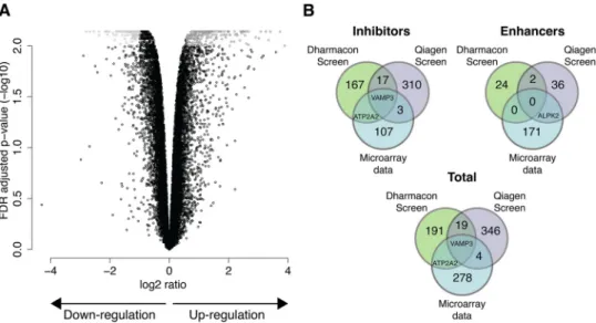
miR-142-3p regulates the expression of the v-SNARE VAMP3
and 283 other genes. With a sequence of only 7 nt, the seed
se-quences are so short that they are likely to match sese-quences in
many mRNAs. Thus, a single miRNA can have many mRNA
tar-gets and can influence a broad spectrum of cellular functions (
39
).
To identify which of the 13,000 genes expressed in our HeLa
CD209 cell line were silenced by miR-142-3p, we used Agilent
microarrays to compare the level of each mRNA in
miR-142-3p-transfected cells with control cells. We found that 172 genes were
upregulated and 112 were downregulated (
Fig. 4A
and
B
; see also
Table S6 at
http://www.highcontentanalysis.org/download/Table
_S6.xlsx
). The identity of the genes indicated that miR-142-3p had
effects on many cellular functions, ranging from cell death to
leu-kocyte activation. Many genes related to endocytosis, the
cytoskel-eton, and membrane trafficking were also included.
It was of interest to compare this list of genes with those obtained
for UUKV in the siRNA screens and determine whether any of the
genes were shared. If such genes existed, they would probably
repre-sent strong candidates. As shown in
Fig. 4B
, there was in fact one such
gene, the vesicle-associated membrane protein 3 (VAMP3). VAMP3
is an integral membrane protein in the v-SNARE family of membrane
fusion proteins with a role in late vesicular trafficking (
40
). Since
UUKV uses late endosomal vesicles for penetration into host cells, it
seemed worthwhile to follow up on VAMP3.
FIG 4 miR-142-3p regulates VAMP3. (A) Microarray chips were used to define the genes regulated by miR-142-3p. The x axis of the volcano plot indicates the
magnitude of gene expression changes, on a log2scale, and the y axis shows the corrected P value for multiple comparisons. The correction was performed using
the false-discovery rate (FDR) method, and values were log transformed and multiplied by⫺1. Data represent the combination of four independent experiments. (B) Overlaps of potential hits between microarray data obtained for miR-142-3p and both siRNA screens.
The v-SNARE VAMP3 is a key player in UUKV late
penetra-tion. We first depleted HeLa CD209 cells of VAMP3 by using
miR-142-3p and two nonoverlapping siRNAs. The level of
VAMP3 protein was reduced by 60% with the miR-142-3p and by
up to 75% with the siRNAs, as assayed by Western blotting (
Fig.
5A
). In the miR-142-3p-transfected cells, infection was inhibited
by 70%. In the siRNA-treated cells, the inhibition was 70 to 80% of
that in cells transfected with a control siRNA (
Fig. 5B
).
This indicated that VAMP3 was, indeed, needed for efficient
UUKV infection. To confirm its specific role in the UUKV
infec-tion cycle, we assessed infecinfec-tion in cells expressing the catalytic
light chain of TeTx, a protease that cleaves and inactivates VAMP
molecules, including VAMP3 (
41
). The expression of the toxin
resulted in a significant decrease in UUKV infection (about 40%)
(
Fig. 5C
).
We next determined whether UUKV enters VAMP3-positive
vesicles during its journey through the endocytic network. Bodipy
Texas Red-conjugated viral particles (UUKV-TR) were prepared
as previously described (
3
). They were allowed to attach to cells
expressing EGFP-VAMP3 (EGFP-tagged VAMP3) on ice. The
temperature was then rapidly shifted to 37°C for periods of up to
40 min. Forty minutes is the time needed for UUKV to penetrate
by membrane fusion from endosomes (
3
,
11
).
While a fraction of the particles remained at the plasma
mem-brane, as expected and previously reported (
3
,
11
), confocal
mi-croscopy 5 min postwarming showed some colocalizations
be-tween internalized UUKV and EGFP-VAMP3 in vesicles located
in the peripheral cytosol (
Fig. 6A
). Colocalization reached a
max-imum 20 min postwarming, when UUKV-positive vacuoles were
mainly in the nuclear periphery, a typical location for late
endo-somal vesicles (
Fig. 6A
and
B
). In addition, confocal microscopy
in live cells revealed that UUKV-TR actually moved in the
cyto-plasm together with the EGFP-VAMP3 in the same vesicles (see
Movie S1 in the supplemental material).
Thirty minutes after warming, several endocytosed viral
parti-cles were seen associated with large EGFP-VAMP3 and
RFP-LAMP1 containing vacuoles in the nuclear periphery (
Fig. 6C
).
LAMP1 is a marker for LEs and lysosomes. This indicated that
UUKV and VAMP3 were present in the same vacuoles and that
these most likely corresponded to LEs or endolysosomes. UUKV
intracellular trafficking was then assessed by confocal microscopy
in EGFP-LAMP1-expressing cells transfected with an siRNA
against VAMP3 or miR-142-3p. Both RNA interferences resulted
in a significant decrease in the colocalizations between LAMP1
and UUKV (
Fig. 7A
and
B
). This suggests that the viral particles
could not reach LAMP1-positive late endosomal vesicles. These
results highlight the functional importance of VAMP3 in UUKV
late penetration.
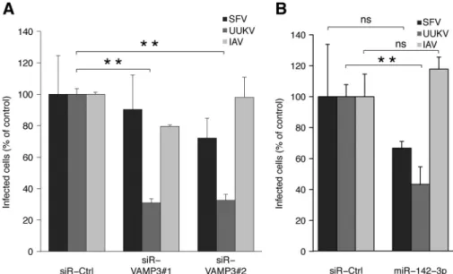
Silencing of VAMP3 by using siRNA or miR-142-3p had no
sig-nificant effect on infection by SFV. SFV is a virus that penetrates from
early endosomes (
37
,
42
). Interestingly, we also observed no effect on
IAV, another L-PV (
Fig. 8A
and
B
). This implied that UUKV and IAV
differently exploit late endocytic functions for entry.
FIG 5 VAMP3 depletion inhibits UUKV infection. (A) Efficiency of VAMP3
knockdown, assayed by Western blotting. VAMP3 protein levels are expressed as the percentage of VAMP3 levels in cells treated with interfering RNAs against VAMP3 (miR-142-3p and siR-VAMP3#1 and -#2) normalized to levels of␣-tubulin and VAMP3 in cells treated with negative-control siRNAs (siR-Ctrl). (B) Cells were treated with siRNAs against VAMP3 (20 nM) and de-tached with PBS-EDTA 72 h posttransfection, prior to the exposure to UUKV (MOI,⬃2). Infection was analyzed via FACS, after immunostaining against N, 8 h later. The values were normalized to the infection level in samples treated
with siR-Ctrl. Error bars indicate the standard error (SE) of the means of three independent experiments. (C) TeTx was transiently expressed in cells. The cells were then infected with UUKV (MOI,⬃2), and infection was quantified via FACS; data were normalized to results with cells transfected with the empty control plasmid. Error bars indicate SE of the means of three independent experiments. *, Pⱕ 0.05.
DISCUSSION
In the last few years, large-scale siRNA screens have become a
common tool for the analysis of virus-host cell interactions. For
bunyaviruses, a Drosophila melanogaster genome-wide siRNA
screen was used to show that mRNA uncapping restricts
bunyavi-rus replication (
43
). We describe here the results from the first
human genome-wide siRNA screen for a bunyavirus and report
the identity of 562 cellular factors in human cells with a potential
role in UUKV entry and replication. We were able to demonstrate
that of the candidate genes, a large fraction was included because
of off-target effects. Some of the siRNAs used could be shown to
interfere with infection through an miRNA-like mechanism. In
follow-up studies, we establish that one of the miRs, miR-142-3p,
interfered with intracellular trafficking of the incoming UUKV
and prevented its penetration into the cytosol. The identification
and validation of the v-SNARE VAMP3 as one of the critical genes
affected by miR-142-3p, as well as a true hit in the two siRNA
screens, allowed us to define the role of this cell protein as a late
endosomal factor required for UUKV infection.
Large-scale siRNA screens have in the last few years been
ap-FIG 6 UUKV associates with VAMP3 and LAMP1. (A) Bodipy Texas Red-conjugated UUKV (UUKV-TR; MOI,⬃10) was bound to EGFP-VAMP3-expressing
cells on ice. Cells were washed, rapidly warmed for up to 40 min, and fixed. UUKV (red) and EGFP-VAMP3 (green) were imaged in one focal plane by confocal microscopy. Magnifications of the association between UUKV and green vesicles (yellow squares) or noncolocalizing, internalized particles (white squares) are shown on the bottom. Bars, 10m. (B) The percentage of viruses colocalizing with the marker EGFP-VAMP3 at different times postwarming was calculated from series of z stacks, as described for those shown in panel A, and merged to one plane. Cells (n⫽ 10) were counted in 10 independent fields for each time point. Error bars indicate the standard error (SE) of the means of the counted cells. The data are representative of three independent experiments. (C) Entry of prebound AF647-labeled UUKV (UUKV-AF647; MOI,⬃10) into cells expressing EGFP-VAMP3 and RFP-LAMP1, based on confocal microscopy. Viruses were internal-ized at 37°C for 30 min. One focal plane is show; UUKV is seen in blue, cell vesicles containing EGFP-VAMP3 are green, and RFP-LAMP1 is red. Magnifications of association between VAMP3⫹LAMP1⫹vesicles and internalized UUKV are shown on the bottom (yellow squares). Bar, 5m.
plied to numerous viruses (
31
,
32
). The information gained has
led to the potential identification of hundreds of host cell factors
with a role in the replication cycle of these viruses. The
discrep-ancy in the hit lists reported for the same virus in different
labo-ratories has, however, been alarmingly high (
31
,
32
). Not more
than 10% overlap was observed between the hits reported between
IAV screens, with similar numbers observed for human
immuno-deficiency virus (HIV) screens (
31
,
32
). The poor reproducibility
has undermined confidence in the screening approach.
The comparison of two genome-wide siRNA libraries
under-taken by us provides further clarification. Here, the screens were
performed with the same virus stocks, cells, assays, and
instru-ments, in an attempt to eliminate any trivial reasons for
discrep-ancies. Still, the overlap between the lists of candidate genes
ob-FIG 7 Depletion of VAMP3 blocks UUKV intracellular trafficking. (A) Cells transfected with miR-142-3p or siR-VAMP3#1 were exposed to AF594-labeled
UUKV (UUKV-AF594; MOI,⬃5) on ice for 2 h, then washed and rapidly warmed to 37°C to allow virus endocytosis for 30 min. UUKV (red) and EGFP-LAMP1 (green) were imaged in one focal plane by confocal microscopy. Magnifications of association between internalized UUKV and LAMP1⫹vesicles (yellow squares) or noncolocalizing, internalized particles (white squares) are shown on the bottom. Bars, 5m. (B) The percentage of viruses colocalizing with EGFP-LAMP1 was calculated from series of z stacks, as described for those shown in panel A. Cells (nⱖ 10) were counted in 10 independent fields, at least. Error bars indicate standard errors of the means of the counted cells. ***, Pⱕ 0.001. The data are representative of two independent experiments.
tained with Qiagen and Dharmacon libraries was not significantly
higher than that seen in the IAV and HIV studies. It was apparent
that the roots of false-negative and -positive hits were inherent in
the design of the siRNA libraries and in the individual siRNA
sequences that they contained.
The magnitude of the problem of false positives became
evi-dent when our potential hits were compared with the
transcrip-tome. Nearly one-third of the candidates in our initial,
uncor-rected lists corresponded to genes in the human genome that were
not transcribed in detectable amounts in the HeLa cells used. This
allowed us to purge the initial 827 identified cellular factors of 265
false-positive candidates. It is clear that this represents a major
problem inherent in the siRNA screening approach. It may
ex-plain many of the divergent results observed in previous screens.
The extent of the problem probably also depends on factors such
as the readout, the cellular processes involved, and the threshold
settings.
One reason for the false positives was no doubt the large
num-ber of siRNAs with miRNA activity (
34–36
). We estimated that
more than 5,200 of the 64,793 siRNAs in the Qiagen screen and 54
siRNAs of the 65,936 in the Dharmacon library were likely to
affect UUKV infection through a miRNA-like mechanism. For a
proof of principle, we experimentally tested 10 miRNAs that
shared 7-nt seed sequences with those present among siRNAs that
blocked UUKV infection, and we observed similar inhibition in
infection. This indicated that many siRNAs in the libraries
af-fected UUKV infection through a miRNA mechanism, or through
combined siRNA and miRNA mechanisms.
Our results indicated that miRNA effects were less pronounced
in the Dharmacon screen with pooled libraries than in the Qiagen
screen, where cells were transfected with single siRNAs. However,
the use of the siRNA pools in the Dharmacon library makes the
analysis and correction of false-positive effects more difficult.
Anyhow, it is unlikely that the nature of libraries, pooled versus
unpooled, is by itself responsible for the off-target effect (
44
).
To increase the reliability of siRNA-based screens, there are
several options. A tighter control of the specificity of siRNAs is a
first obvious step. In this sense, the recent method developed by
Buehler and colleagues (called control C911) may represent an
important achievement (
45
). New approaches with built-in
ad-vantages for large loss-of-function screens also include the use of
large libraries of short hairpin RNAs (shRNAs) in combination
with the improvements in the plate design and the analysis of
population context-regulated cell-to-cell variability (
46–48
).
Despite the problems, it is evident that the siRNA screens can
provide valuable new information about the role of cellular factors
in virus infection when carefully performed and validated. By
fo-cusing on gene clusters and performing stringent validation, it has
been possible to identify legitimate hits of value (
49
,
50
). With
regard to UUKV infection, the screens did help us to identify
candidate genes that made sense and brought our understanding
of UUKV entry forward. Some of the potential hits confirmed
cellular processes already described in the literature, and others
were obvious and expected for RNA viruses. Among the
unex-pected findings was the v-SNARE VAMP3, a hit derived by
com-bining miRNA (miR-142-3p) and siRNA screening data.
VAMP3 was the only cellular factor shared as an inhibitor hit
between the miRNA screen and both siRNA screens. It is unlikely
that the block in infection was caused by an adverse effect of
VAMP3 depletion on the luminal pH of endosomes. Infection by
SFV and IAV, which both rely on endosomal acidification for
penetration, was not impaired in cells depleted of VAMP3.
Fol-low-up studies showed that VAMP3 was, rather, needed for
intra-cellular trafficking of UUKV to late endosomal compartments
competent to support acid-activated membrane fusion between
the virus and cellular membranes. About 20 min after
internaliza-tion, the virus was maximally present in vacuoles containing
LAMP1 and VAMP3. In cells depleted of VAMP3, viruses did not
reach LAMP1
⫹late endosomal vesicles, suggesting that UUKV
remained trapped in upstream endosomal compartments.
FIG 8 IAV does not rely on VAMP3 for penetration. (A) Cells were treated with siRNAs and then exposed to SFV, UUKV, or IAV as described in the legend for
Fig. 5B. Infection was quantified by FACS 8 h later, and data were normalized to infection of cells treated with siR-Ctrl. **, Pⱕ 0.01. Error bars indicate standard errors (SE) of the means of three independent experiments. (B) Cells transfected with miR-142-3p were exposed to SFV, UUKV, or IAV as described forFig. 2B. Infection was quantified by FACS 8 h later, and data were normalized to infection of cells treated with siR-Ctrl. Error bars indicate SE of the means of three independent experiments. **, Pⱕ 0.01; ns, nonsignificant.
Our data support the view that the v-SNARE VAMP3 plays an
active role in the maturation of the endosomal vacuoles used by
UUKV during its journey within the endocytic network. That the
intracellular trafficking of UUKV was abrogated in cells
trans-fected with miR-142-3p supported this model, although we
can-not completely exclude that genes other than VAMP3 that are
targeted by miR-142-3p participate in UUKV infection. The role
of VAMP3 was not restricted to cells expressing CD209.
Interest-ingly, the synaptosome-associated protein 23 (SNAP23) was also a
strong candidate. Consistent with the late timing for UUKV
pen-etration, SNAP23 is part of the t-SNARE complexes that mediate
fusion of LEs with each other and with other vacuoles (
51
).
To-gether, these observations suggested that VAMP3 may have a role
in late endosomal maturation and penetration of UUKV.
The observation that IAV was not dependent on VAMP3
indi-cated that the pathways of entry of the two L-PVs might diverge.
Rather than mediating endosome-plasma membrane fusion,
VAMP3 is actually thought to be involved in
autophagosome-endosome fusion (
40
,
41
,
52
,
53
). It is present on
autophago-somes. This suggests that UUKV fusion may depend on the
inter-section between the endocytic and autophagocytic pathways, with
VAMP3 acting like a bridge between both routes. Among the
other potential hits, FIP200 (also known as RB1CC) and Rab1B
also have functions related to autophagy and autophagosome
maturation (
54
,
55
). It has been recently proposed that high levels
of autophagy increase adenovirus type 2 penetration and that
echovirus 7 entry involves some cell factors of the autophagy
ma-chinery (
56
,
57
). Though it is likely that some viruses induce and
subvert autophagy for their own purposes, further investigations
will be needed to clarify whether UUKV and other bunyaviruses
depend on autophagy for productive entry.
ACKNOWLEDGMENTS
This work was supported by grants from InfectX to C.V.M., from the European Research Council, the Swiss National Foundation for Research, ETH Zurich, and LipidX to A.H., and from the Natural Sciences and Engineering Research Council of Canada and the Banting Research Foun-dation to P.Y.L.
We thank M. Krzyzaniak, J. Mercer, L. Burleigh, S. Noerrelykke, and N. Tischler for proofreading. We also acknowledge S. Wirth and H. Reh-rauer from the Functional Genomics Center Zurich, as well as G. Csúcs, A. Kaufman, T. Schwarz, and M. Stebler from the ScopeM ETH Zurich and A. Vonderheit from the IMB, Mainz, Germany.
REFERENCES
1. Walter CT, Barr JN. 2011. Recent advances in the molecular and cellular biology of bunyaviruses. J. Gen. Virol. 92:2467–2484.http://dx.doi.org/10 .1099/vir.0.035105-0.
2. Gavrilovskaya IN, Shepley M, Shaw R, Ginsberg MH, Mackow ER. 1998.3 integrins mediate the cellular entry of hantaviruses that cause respiratory failure. Proc. Natl. Acad. Sci. U. S. A. 95:7074 –7079.http://dx .doi.org/10.1073/pnas.95.12.7074.
3. Lozach PY, Kuhbacher A, Meier R, Mancini R, Bitto D, Bouloy M,
Helenius A. 2011. DC-SIGN as a receptor for phleboviruses. Cell Host
Microbe 10:75– 88.http://dx.doi.org/10.1016/j.chom.2011.06.007. 4. de Boer SM, Kortekaas J, de Haan CA, Rottier PJ, Moormann RJ, Bosch
BJ. 2012. Heparan sulfate facilitates Rift Valley fever virus entry into the
cell. J. Virol. 86:13767–13771.http://dx.doi.org/10.1128/JVI.01364-12. 5. de Boer SM, Kortekaas J, Spel L, Rottier PJ, Moormann RJ, Bosch BJ.
2012. Acid-activated structural reorganization of the Rift Valley fever vi-rus Gc fusion protein. J. Virol. 86:13642–13652.http://dx.doi.org/10.1128 /JVI.01973-12.
6. Hollidge BS, Nedelsky NB, Salzano MV, Fraser JW, Gonzalez-Scarano
F, Soldan SS. 2012. Orthobunyavirus entry into neurons and other
mam-malian cells occurs via clathrin-mediated endocytosis and requires traf-ficking into early endosomes. J. Virol. 86:7988 – 8001.http://dx.doi.org/10 .1128/JVI.00140-12.
7. Santos RI, Rodrigues AH, Silva ML, Mortara RA, Rossi MA, Jamur MC,
Oliver C, Arruda E. 2008. Oropouche virus entry into HeLa cells involves
clathrin and requires endosomal acidification. Virus Res. 138:139 –143. http://dx.doi.org/10.1016/j.virusres.2008.08.016.
8. Simon M, Johansson C, Mirazimi A. 2009. Crimean-Congo hemor-rhagic fever virus entry and replication is clathrin-, pH- and cholesterol-dependent. J. Gen. Virol. 90:210 –215. http://dx.doi.org/10.1099/vir.0 .006387-0.
9. Jin M, Park J, Lee S, Park B, Shin J, Song KJ, Ahn TI, Hwang SY, Ahn
BY, Ahn K. 2002. Hantaan virus enters cells by clathrin-dependent
recep-tor-mediated endocytosis. Virology 294:60 – 69.http://dx.doi.org/10.1006 /viro.2001.1303.
10. Lozach PY, Huotari J, Helenius A. 2011. Late-penetrating viruses. Curr. Opin. Virol. 1:35– 43.http://dx.doi.org/10.1016/j.coviro.2011.05.004. 11. Lozach PY, Mancini R, Bitto D, Meier R, Oestereich L, Overby AK,
Pettersson RF, Helenius A. 2010. Entry of bunyaviruses into mammalian
cells. Cell Host Microbe 7:488 – 499. http://dx.doi.org/10.1016/j.chom .2010.05.007.
12. Simon M, Johansson C, Lundkvist A, Mirazimi A. 2009. Microtubule-dependent and microtubule-inMicrotubule-dependent steps in Crimean-Congo hem-orrhagic fever virus replication cycle. Virology 385:313–322.http://dx.doi .org/10.1016/j.virol.2008.11.020.
13. Cifuentes-Munoz N, Barriga GP, Valenzuela PD, Tischler ND. 2011. Aromatic and polar residues spanning the candidate fusion peptide of the Andes virus Gc protein are essential for membrane fusion and infection. J. Gen. Virol. 92:552–563.http://dx.doi.org/10.1099/vir.0.027235-0. 14. Filone CM, Heise M, Doms RW, Bertolotti-Ciarlet A. 2006.
Develop-ment and characterization of a Rift Valley fever virus cell-cell fusion assay using alphavirus replicon vectors. Virology 356:155–164.http://dx.doi .org/10.1016/j.virol.2006.07.035.
15. Pettersson R, Kaariainen L. 1973. The ribonucleic acids of Uukuniemi virus, a noncubical tick-borne arbovirus. Virology 56:608 – 619.http://dx .doi.org/10.1016/0042-6822(73)90062-7.
16. Kilbourne ED. 1969. Future influenza vaccines and the use of genetic recombinants. Bull. World Health Organ. 41:643– 645.
17. Spuul P, Balistreri G, Kaariainen L, Ahola T. 2010. Phosphatidylinositol 3-kinase-, actin-, and microtubule-dependent transport of Semliki Forest virus replication complexes from the plasma membrane to modified lyso-somes. J. Virol. 84:7543–7557.http://dx.doi.org/10.1128/JVI.00477-10. 18. Lamesch P, Li N, Milstein S, Fan C, Hao T, Szabo G, Hu Z, Venkatesan
K, Bethel G, Martin P, Rogers J, Lawlor S, McLaren S, Dricot A, Borick H, Cusick ME, Vandenhaute J, Dunham I, Hill DE, Vidal M. 2007.
hORFeome v3.1: a resource of human open reading frames representing over 10,000 human genes. Genomics 89:307–315.http://dx.doi.org/10 .1016/j.ygeno.2006.11.012.
19. Persson R, Pettersson RF. 1991. Formation and intracellular transport of a heterodimeric viral spike protein complex. J. Cell Biol. 112:257–266. http://dx.doi.org/10.1083/jcb.112.2.257.
20. Witten IH, Frank E. 2005. Data mining: practical machine learning tools and techniques, 2nd ed. Elsevier, San Francisco, CA.
21. Carpenter AE, Jones TR, Lamprecht MR, Clarke C, Kang IH, Friman O,
Guertin DA, Chang JH, Lindquist RA, Moffat J, Golland P, Sabatini DM. 2006. CellProfiler: image analysis software for identifying and
quan-tifying cell phenotypes. Genome Biol. 7:R100.http://dx.doi.org/10.1186 /gb-2006-7-10-r100.
22. Horvath P, Wild T, Kutay U, Csucs G. 2011. Machine learning improves the precision and robustness of high-content screens: using nonlinear multiparametric methods to analyze screening results. J. Biomol. Screen.
16:1059 –1067.http://dx.doi.org/10.1177/1087057111414878.
23. Banerjee I, Yamauchi Y, Helenius A, Horvath P. 2013. High-content analysis of sequential events during the early phase of influenza A virus infection. PLoS One 8:e68450. http://dx.doi.org/10.1371/journal.pone .0068450.
24. Jacoby WG. 2000. Loess: a nonparametric, graphical tool for depicting relationships between variables. Electoral Stud. 19:577– 613.
25. Birmingham A, Selfors LM, Forster T, Wrobel D, Kennedy CJ, Shanks
E, Santoyo-Lopez J, Dunican DJ, Long A, Kelleher D, Smith Q, Beijer-sbergen RL, Ghazal P, Shamu CE. 2009. Statistical methods for analysis
of high-throughput RNA interference screens. Nat. Methods 6:569 –575. http://dx.doi.org/10.1038/nmeth.1351.