Complex autoregulation of the post-transcriptional
regulator RsmA in Pseudomonas aeruginosa
Fabrice Jean-Pierre, Jonathan Perreault and Eric De´ziel
CorrespondenceEric De´ziel
INRS-Institut Armand-Frappier, 531 Boul. des Prairies, Laval, QC, Canada
RsmA is a post-transcriptional RNA-binding protein that acts as a pleiotropic global regulator of mRNAs in the opportunistic pathogen Pseudomonas aeruginosa. Upon binding to its target, RsmA impedes the translation of the mRNA by the ribosome. The RsmA regulon affects over 500 genes, many of which have been identified as important in the pathogenicity of
P. aeruginosa. Whilst the regulatory function of RsmA is relatively well characterized, the genetic regulation of rsmA itself at the transcriptional and translational levels remains poorly understood. Here, we show that RsmA is capable of self-regulation through an unorthodox mechanism. This regulation occurs via direct interaction of the protein with an RsmA-binding site located in the early portion of its coding sequence. To the best of our knowledge this is the first report of such an unusual regulation in pseudomonads.
Received 21 May 2015 Accepted 9 July 2015
INTRODUCTION
Rapid adaptation of bacteria requires the detection of and response to diverse environmental cues. Post-transcrip-tional regulation is used by bacteria to quickly adapt to changing conditions. The well-characterized CsrA/RsmA family of post-transcriptional regulators is widespread among Gram-negative bacteria and can globally affect gene expression (Romeo et al., 2013). This family consists of small dimeric RNA-binding proteins that have the capacity to recognize a GGA trinucleotide present in the loop portion of a stem–loop located in the 59 untranslated region (UTR) of a mRNA (Lapouge et al., 2008). The GGA motif can be present in multiple copies in the 59 UTR of a target mRNA. However, one GGA trinucleotide is almost always positioned close to the Shine–Dalgarno region, thus hindering the attachment to the ribosome and block-ing the translation of the target mRNA (Baker et al., 2002, 2007; Dubey et al., 2005). Accordingly, CsrA/RsmA pro-teins typically act as negative regulators of mRNA trans-lation. However, in some cases their RNA-binding activity can act as a positive translational regulator by stabilizing the mRNA (Romeo et al., 2013).
In the Gram-negative opportunistic pathogen Pseudomonas aeruginosa, RsmA is a pleiotropic post-transcriptional regulator that modulates the expression of w500 genes (Brencic & Lory, 2009; Burrowes et al., 2006). RsmA indirectly activates the expression of genes associated with the establishment of acute infections whilst repressing
those implicated in the development of chronic infections (Brencic & Lory, 2009). This post-transcriptional regulator is under the control of the GacA/GacS two-component system, which is exclusively responsible for the transcrip-tion of the small RNAs (sRNAs) RsmY and RsmZ (Brencic et al., 2009). These sRNAs possess numerous RsmA-binding sites and thus act as ‘baits’ titrating free RsmA pro-teins in the cell (Sonnleitner & Haas, 2011). Furthermore, many other systems can affect the activity of the Gac system, thus modulating the levels of these RsmA-repres-sing sRNAs (Goodman et al., 2004, 2009; Ventre et al., 2006). The importance of post-transcriptional regulation in P. aeruginosa is more complex than initially thought, as a new post-transcriptional regulator (RsmN), which shares little structural homology with RsmA, but possesses a similar mechanism of action, was reported recently (Marden et al., 2013; Morris et al., 2013).
Whilst the regulatory function of RsmA is relatively well characterized, the genetic regulation of rsmA itself at the transcriptional and translational levels remains poorly understood. The genetic regulation of csrA, coding for the RsmA homologue in Escherichia coli, is complex and dependent on the presence of multiple promoters that are activated at different stages during cell growth. Interest-ingly, CsrA negatively controls its translation by directly binding to its own mRNA (Yakhnin et al., 2011b). In the present study, we demonstrate that RsmA is capable of binding its own mRNA, promoting a negative feedback regulatory loop. Two RNA attachment sites are implicated in this RsmA–rsmA interaction. We identified an RsmA-binding motif in the 59 portion of its coding sequence, revealing an important difference from the conventional CsrA/RsmA regulation mechanism.
Abbreviations: sRNA, small RNA; UTR, untranslated region
One supplementary table and four supplementary figures are available with the online Supplementary Material.
METHODS
Strains, plasmids and growth conditions.The bacterial strains used in this study are listed in Table 1. P. aeruginosa and E. coli strains were
cultivated in Tryptic Soy Broth (TSB) medium at 37uC with shaking
(240 r.p.m.) in a TC-7 roller drum (New Brunswick) or on TSB agar
plates. Antibiotics used for selection were 125mg tetracycline ml21and
25mg triclosan ml21.
b-Galactosidase assays.Activity of lacZ fusion reporters was tested
forb-galactosidase activity with ONPG (Thermo Fisher Scientific) as
substrate (Miller, 1972). Each experiment was performed in triplicate,
at least twice. Overnight TSB cultures were diluted at a starting OD600
0.05 in TSB and incubated as above. Results were obtained for five sampling points during bacterial growth over 8 h.
Purification of His6-tagged RsmA. E. coli BL21(DE3) cells
con-taining the pET29a(+)-RsmA-H6 plasmid were grown overnight in
TSB with 30mg kanamycin ml21. In the morning, the culture was
diluted 1 : 100 in 100 ml pre-warmed LB. Cells were grown to mid
exponential phase (OD6000.7) and IPTG was added to the culture at a
final concentration of 1 mM for protein expression induction. Cells were harvested after 4 h. Cell pellets were resuspended in 20 ml 0.5 M
NaCl, 20 mM NaH2PO4and Tris/HCl (pH 7.65), and ruptured by
sonication. Lysed cells were pelleted by centrifugation at 15 000 g for
45 min at 4uC. Prior to purification, the supernatant was filtered on a
0.22mM nitrocellulose filter. RsmA-His6 was purified by using a
HisTrap FF crude 5 ml column (GE Healthcare) with an A¨ tka FPLC
system (GE Healthcare) following the manufacturer’s recommen-dations. The protein-containing fractions were concentrated and the protein stored in a Tris/HCl (pH 7.63)/33 % glycerol conservation buffer. Purity and identity of the protein was assessed by SDS-PAGE followed by MS analysis. Concentration was estimated using a Bradford assay (Bio-Rad) with BSA as standard. The concentrated
protein was stored at220 uC until use.
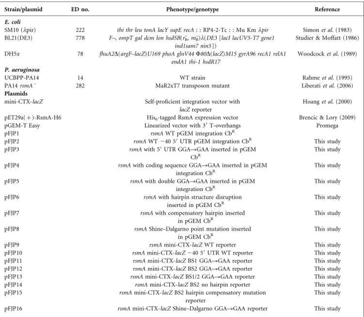
Table 1. Strains/plasmids used in this study
Strain/plasmid ED no. Phenotype/genotype Reference
E. coli
SM10 (lpir) 222 thi thr leu tonA lacY supE recA : : RP4-2-Tc : : Mu Kmlpir Simon et al. (1983)
BL21(DE3) 778 F–, ompT gal dcm lon hsdSB(rB–, mB–)l(DE3 [lacI lacUV5-T7 gene1
ind1sam7 nin5 ])
Studier & Moffatt (1986)
DH5a 78 fhuA2D(argF–lacZ)U169 phoA glnV44 W80D(lacZ)M15 gyrA96 recA1 relA1
endA1 thi-1 hsdR17
Woodcock et al. (1989) P. aeruginosa
UCBPP-PA14 14 WT strain Rahme et al. (1995)
PA14 rsmA– 282 MaR2xT7 transposon mutant Liberati et al. (2006)
Plasmids
mini-CTX-lacZ Self-proficient integration vector with
lacZ reporter
Hoang et al. (2000)
pET29a(+)-RsmA-H6 His6-tagged RsmA expression vector Brencic & Lory (2009)
pGEM-T Easy Linearized vector with 39 T-overhangs Promega
pFJP1 rsmA WT pGEM integration CbR
pFJP2 rsmA WT240 59 UTR pGEM integration CbR This study
pFJP3 rsmA with 59 UTR GGARGAA inserted in pGEM
CbR
This study
pFJP4 rsmA with coding sequence GGARGAA inserted in pGEM
integration CbR
This study
pFJP5 rsmA with double GGARGAA inserted in pGEM
integration CbR
This study
pFJP6 rsmA with hairpin structure disruption
inserted in pGEM CbR
This study
pFJP7 rsmA with compensatory hairpin inserted
in pGEM CbR
This study
pFJP8 rsmA Shine–Dalgarno point mutation inserted
in pGEM CbR
This study
pFJP9 rsmA mini-CTX-lacZ WT reporter This study
pFJP10 rsmA mini-CTX-lacZ240 59 UTR WT reporter This study
pFJP11 rsmA mini-CTX-lacZ BS1 GGARGAA reporter This study
pFJP12 rsmA mini-CTX-lacZ BS2 GGARGAA reporter This study
pFJP13 rsmA mini-CTX-lacZ BS1/2 GGARGAA reporter This study
pFJP14 rsmA mini-CTX-lacZ BS2 no hairpin reporter This study
pFJP15 rsmA mini-CTX-lacZ BS2 hairpin compensatory mutation
reporter
This study
RNA electrophoretic mobility shift assay. Fragments of rsmA mRNA were synthesized from PCR products using a T7 RNA polymerase. The primer sequences used are listed in Table S1 (available in the online Supplementary material). The T7 RNA
polymerase promoter sequence was added to the 59 portion of the
primers. The obtained PCR fragments were purified using a BioBasic (Canada) purification kit following the manufacturer’s
recommen-dations. In vitro transcription was carried out in 20ml reactions
containing 20 pmol purified PCR fragment, 2 mM rNTPs, 10mg
pyrophosphatase ml21 (Roche), 80 mM HEPES/KOH (pH 7.5),
24 mM MgCl2, 50 mM DTT, 2.5 mM spermidine, 1 U T7
poly-merase, 1 mg competitor tRNA ml21and completed with RNase-free
distilled H2O, and incubated at 37uC for a total of 3 h. Once the
transcription was completed, remaining DNA fragments were removed using 1 U RNase-free DNase I (Promega). RNA fragments were gel-purified by 12 % 8 M urea denaturing PAGE and
depho-sphorylated using 2.5 pmol RNA, 2ml 10|Antarctic phosphatase
buffer, 1 U Antarctic phosphatase enzyme ml–1 and completing to
20ml with RNase-free distilled H2O. The 59 ends of the RNA
frag-ments were radiolabelled by phosphorylation using 1 U T4
poly-nucleotide kinaseml–1and 1.85|105Bq [c-32P]ATP. Labelled RNA
fragments were gel-purified as described, resuspended in RNase-free
distilled H2O and stored at 220 uC until use. The RNA-binding
reaction consisted of the recombinant RsmA-His6dimer at various
concentrations, radiolabelled RNA transcript (0.6 pM), 10 mM Tris/
HCl (pH 7.5), 10 mM MgCl2, 50 mM NaCl, 50 mM KCl and 5 mM
DTT, and the final mixture was adjusted to 20ml with RNase-free
distilled H2O. The RNA-binding reaction was incubated at room
temperature for 30 min, mixed with 3ml loading dye (40 % sucrose,
0.05 % xylene cyanol and 0.05 % bromophenol blue) and loaded on a 10 % (29 : 1) native polyacrylamide gel using Tris/borate/EDTA as the running buffer. A Typhoon PhosphorImager FLA9500 (GE Healthcare) and ImageQuant software were used for gel scanning and analysis.
TranslationalrsmA9–9lacZ fusion constructions.Specific point mutations in the rsmA upstream intergenic region and/or coding sequence, the entire upstream intergenic region of rsmA and the first 36 nt of the ORF were synthesized by GenScript. The
resulting plasmids were cloned into E. coli DH5a. Plasmid
extractions were carried out using a Miniprep kit (BioBasic) and purity was assessed by gel electrophoresis. The various rsmA mutated alleles and the lacZ genes were synthesized by PCR directly from plasmids using different sets of primers (Table S1). A fusion PCR between the synthesized rsmA transcripts and the first 668 bp of the lacZ gene was performed, and the resulting
fragment was purified from a 1 % agarose gel. The rsmA9–9lacZ
fragments were ligated in pGEM-T Easy (Promega), the plasmids
transformed in CaCl2 thermocompetent E. coli DH5a cells and
clones selected on TSB agar plates supplemented with 100mg
carbenicillin ml21 after overnight incubation at 37uC. Positive
clones were identified by digesting Miniprep products with Eco RI
for the presence of the insert. Overnight double digestion at 37uC
of the pGEM-T Easy plasmids containing the inserts and the destination vector mini-CTX-lacZ was performed using Pst I (Fermentas) and Aat II (NEB) restriction enzymes. The released inserts were gel-purified before ligation in the destination vector. Overnight ligation between the digested inserts and vector was performed using Feldan T4 ligase following the manufacturer’s guidelines. Selection of the clones was done on TSB agar
con-taining 15mg tetracycline ml21. Positive clones were identified by
double digestion of Miniprep products. The mini-CTX-lacZ plas-mids containing the different constructions were conjugated into WT P. aeruginosa strain PA14 for integration in the unique attB site (Hoang et al., 1998), giving stable, chromosomal translational reporters.
RESULTS
RsmA represses its own expression
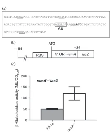
Several recent reports (and unpublished data from our lab-oratory) have shown that the major rsmA transcriptional start site during planktonic cell growth starts at position 240 before the ATG codon (Do¨tsch et al., 2012; Marden et al., 2013; Wurtzel et al., 2012). Analysis of the primary sequence of the rsmA 59 UTR showed that many GGA tri-nucleotides are present in that region (Fig. 1a). Knowing that proteins of the CsrA/RsmA family associate with a GGA trinucleotide exposed in the loop portion of a stem–loop generally located close to the ribosome-binding site on their target mRNAs that blocks their translation (Dubey et al., 2005), we hypothesized that a possible auto-regulation of RsmA on its own mRNA is possible in P. aeruginosa. Thus, to investigate if such a regulation either at the transcriptional or translational level exists,
rsmA′–′lacZ –184 RBS ATG SD +36 5′ ORF-rsmA lacZ 200 (a) (b) (c) 150 100 50 b
-Galactosidase activity (MU/OD
600
)
0
PA14 rsmA
–
Fig. 1. Expression of the rsmA9–9lacZ reporter in different
back-grounds. (a) Primary sequence analysis of rsmA. Underlined,
GGA trinucleotides; bold nucleotide, 240 transcriptional start
site; boxed, Shine–Dalgarno (SD) sequence; bold ATG codon, translational start site. (b) Fusion of the entire rsmA intergenic region and the first seven codons of its coding sequence with the
59 end of the lacZ gene. RBS, ribosome-binding site; ATG, +1.
(c) Expression of rsmA during the early growth stage using a WT reporter in various genetic backgrounds. Reporter gene activity is
shown as a ratio of Miller units (MU)/OD600. Data represent the
we monitored the expression of rsmA in various back-grounds using a chromosomal rsmA9–9lacZ reporter con-taining the whole rsmA intergenic region and the first 12 codons of its coding sequence (Fig. 1b). As shown in Fig. 1(c), the expression of rsmA was increased in a DrsmA mutant background when compared with the WT, suggesting that RsmA is, in some way, responsible for nega-tively affecting its own expression either at the transcrip-tional or translatranscrip-tional level.
RsmA binds to its own coding sequence
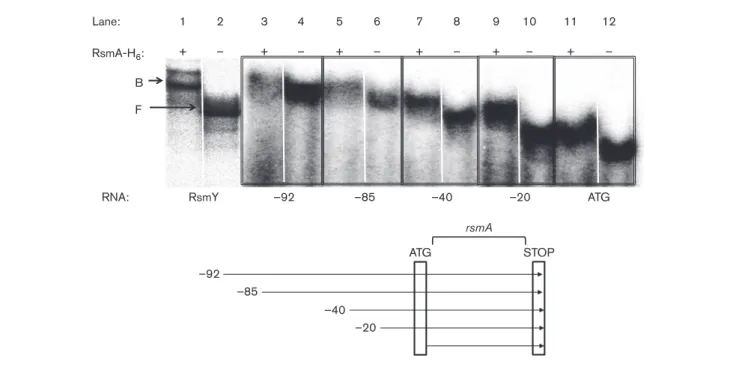
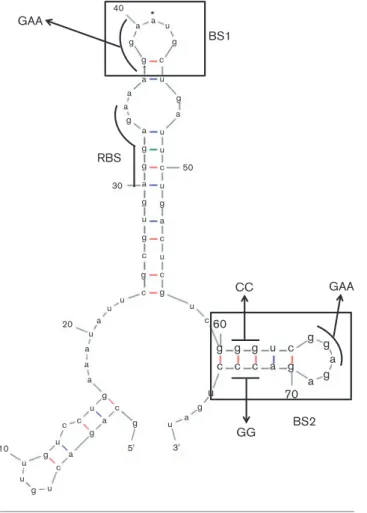
To further clarify how RsmA negatively affects its own expression, RNA mobility shift assays were carried out to determine whether or not RsmA can directly bind its own mRNA, thus promoting negative regulation specifi-cally on its own translation. Upon testing the RNA frag-ments including the presence of possible RsmA-binding sites, we found that all the tested RNA molecules were bound by RsmA, thus blocking its own translation (Fig. 2). Unexpectedly, we observed that the coding sequence of rsmA was by itself sufficient for a protein– RNA interaction (lane ATG). Regulation of target mRNAs by proteins of the CsrA/RsmA family is usually exerted on the 59 UTR of a transcript. To understand this result, we used in silico RNA folding of the full rsmA coding sequence to identify if a probable RsmA-binding site in the ORF is present that might be responsible for the observed effect. Indeed, upon analysis, a putative RsmA-binding site at position +25 after the translation
start codon was identified (Fig. 3). Given that the major rsmA transcriptional start site is located at position 240 before the AUG codon, we investigated a portion of the rsmA transcript spanning from 240 to +38 around the start codon, allowing for the formation of two possible RsmA-binding sites (BS1 and BS2), to test whether or not the observed in vitro interaction was due to these two predicted RsmA-binding sites (Fig. 3). Interestingly, the GGA trinucleotide present in the Shine–Dalgarno sequence was never found to be exposed in a loop portion of a stem– loop during in silico analyses. Thus, we decided not to focus on that portion of the mRNA. Indeed, upon testing by RNA mobility shift assays, we observed that RsmA bound the WT 240 to +38 transcript (Fig. 4a, WT). Next, to decipher which of BS1 or BS2 was responsible for this RsmA–rsmA interaction promoting negative translational regulation, we inserted point mutations only affecting the primary nucleotide sequence at various positions in the WT 240 to +38 transcript. The presence of RsmA-binding sites was confirmed by the abolition of protein– RNA interactions when GGA trinucleotides were mutated into GAA in BS1, BS2 or both (Fig. 3), when compared with the WT transcript (Fig. 4a). Looking at the effect of an alteration in the secondary RNA structure on RsmA– rsmA regulation, we disrupted the formation of the pre-dicted hairpin structure of BS2 in the early coding sequence of rsmA (Fig. 3, GGRCC). The introduction of such a mutation induced a loss of interaction between rsmA and RsmA (Fig. 4b, M4). Confirming BS2, the further introduc-tion of a compensatory mutaintroduc-tion that reinstated the
RNA: RsmY –92 –85 –40 –20 ATG
Lane: RsmA-H6: + 1 – 2 + 3 – 4 + 5 – 6 + 7 – 8 + 9 – 10 + 11 – 12 B F –92 –85 –40 –20 ATG STOP rsmA
Fig. 2. RNA mobility shift assay with purified RsmA. Determination of RsmA–rsmA interaction using radiolabelled RNA
frag-ments and purified RsmA. Odd lanes, RNA with protein; even lanes, RNA fragment only. The292, 285, 240, 220 and ATG
sites represent the 59 end of the RNA molecule relative to the ATG codon. The 39 end of each fragment is the TGA (STOP)
formation of the hairpin structure to a WT conformation in M4 (Fig. 3, CCRGG) restored the affinity for RsmA towards its own transcript (Fig. 4b, M5). These results demonstrated that not only the primary nucleotide sequence is important for protein–RNA interaction, but also the presence of secondary RNA structures is essential for RsmA–rsmA interaction.
In vivo rsmA self-regulation is driven by multiple binding sites
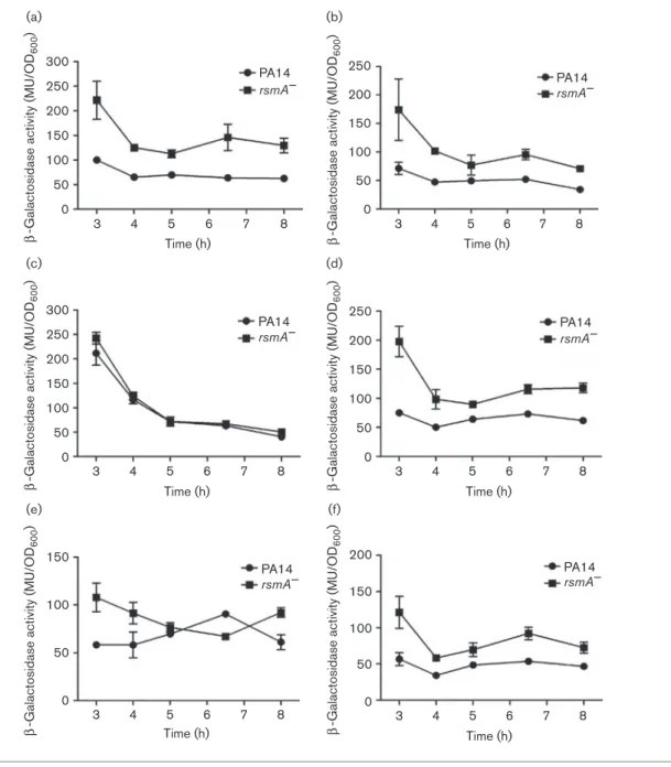
To investigate the relevance of each of the identified RsmA-binding sites (BS1 and BS2), the same point mutations used in our in vitro gel-shift assays were introduced in an in vivo setting by constructing various translational rsmA9–9lacZ reporters (Fig. S1). Compared with the WT (Fig. 5a), the substitution of a GGARGAA trinucleotide located in BS1 did not affect the capacity of RsmA to
bind to its own transcript as the translational expression of rsmA9–9lacZ was still upregulated in a DrsmA back-ground (Fig. 5b). However, the introduction of the same mutation within BS2 located in the coding sequence resulted in a complete loss of translational regulation by RsmA on itself (Fig. 5c). Surprisingly, the abolishment of both potential binding sites in BS1 and BS2 restored a WT expression pattern of rsmA9–9lacZ where inhibition of rsmA was observed (Fig. 5d). As the RsmA-binding site located within BS2 seems to be more important for the RsmA–rsmA interaction in an in vivo setting, we tested the effect of a destabilizing hairpin mutation on the translation of rsmA. We observed that the deletion of the stem–loop structure abolished the capacity of RsmA to repress its own translation as there was no significant difference in rsmA translation between the DrsmA strain and the WT (Fig. 5e). Conversely, the introduction of a compensatory mutation that restored the hairpin structure
GAA 40 g g u g g g g g g g g c 50 30 c u u u u u a c c u g g c c u u u a a a a a g g g 5' 3' u c c c g g g u u 10 u u c 20 a c a a a a a a a *a RBS CC BS1 BS2 GAA GG u u u u c c c c g g g g g g a a a 70 60 g a
Fig. 3. The240 to +38 rsmA transcript secondary RNA
struc-ture. RNA folding prediction of the rsmA240 to +38 sequence
using M-Fold. Boxed, putative RsmA-binding sites; BS1, binding site 1; BS2, binding site 2; curved lines, GGA trinucleotides; RBS, ribosome-binding site; arrows, inserted nucleotide poly-morphisms. *AUG (START) codon of rsmA.
M4 M5 RNA: Lane: RsmA-H6: + 1 – 2 + 3 – 4 RNA: WT M1 M2 M3 Lane: RsmA-H6: + 1 – 2 + 3 – 4 + 5 – 6 + 7 – 8 (a) (b)
Fig. 4. RNA mobility shift assay with purified RsmA. (a) Effect of
GGARGAA trinucleotide mutations on the 240 to +38
radio-labelled RNA fragments for RsmA binding. WT, WT240 to +38
rsmA fragment; M1, GGARGAA in BS1; M2, GGARGAA in
BS2; M3, GGARGAA in BS1 and BS2. (b) Effect of a
destabi-lizing hairpin mutation on RsmA binding. M4, rsmA fragment with no hairpin in BS2 (GGRCC); M5, rsmA fragment with
compen-satory hairpin mutation in BS2 (CCRGG). Odd lanes, RNA with
protein; even lanes, RNA fragment only. WT rsmA fragment was
allowed for the capacity of RsmA to interact with its tran-script and inhibit the translation (Fig. 5f).
DISCUSSION
Translational regulation of target genes by the CsrA/RsmA protein family generally occurs by binding of the protein to a GGA motif exposed in a loop of a stem–loop located in the Shine–Dalgarno region of the 59 UTR of a mRNA,
thus hindering the formation of the ribosomal complex (Dubey et al., 2005). In our study, the identification of several GGA trinucleotides in the primary sequence of the 59 UTR of rsmA initially suggested that an autoregula-tion of RsmA was possible, maybe similar to what was observed for CsrA in E. coli (Yakhnin et al., 2011b). Using a translational rsmA9–9lacZ reporter, we indeed observed that rsmA translation was de-repressed in a DrsmA background. However, we could not decipher at this point if the observed effect was due to direct regulation
300 PA14 rsmA– 250 200 150 100 b
-Galactosidase activity (MU/OD
600 ) 50 0 3 4 5 6 Time (h) 7 8 PA14 rsmA– 250 200 150 100 b
-Galactosidase activity (MU/OD
600 ) 50 0 3 4 5 6 Time (h) 7 8 300 PA14 rsmA– 250 200 150 100 b
-Galactosidase activity (MU/OD
600 ) 50 0 3 4 5 6 Time (h) 7 8 PA14 rsmA– 250 200 150 100 b
-Galactosidase activity (MU/OD
600 ) 50 0 3 4 5 6 Time (h) 7 8 150 PA14 rsmA– 100 b
-Galactosidase activity (MU/OD
600 ) 50 0 3 4 5 6 Time (h) 7 8 PA14 rsmA– 200 150 100 b
-Galactosidase activity (MU/OD
600 ) 50 0 3 4 5 6 Time (h) 7 8 (a) (b) (c) (d) (e) (f)
Fig. 5. Time-course of expression of rsmA determined by using a translational rsmA9–9lacZ reporter. (a) Expression of WT 240 rsmA9–9lacZ reporter. (b) Expression of rsmA containing GGARGAA in BS1. (c) Expression of rsmA with GGARGAA
in BS2. (d) Expression of rsmA with GGARGAA in BS1 and BS2. (e) Expression of rsmA with a destabilized hairpin
(GGRCC) in BS2. (f) Expression of rsmA with a compensatory hairpin mutation (CCRGG) in BS2. Reporter gene activity is
implicating RsmA or via another regulator at the transcrip-tional or translatranscrip-tional level.
To understand how RsmA affects its own expression, we used both in vitro and in vivo approaches to unravel the mechan-isms implicated in this regulation. Our initial shift assays unexpectedly indicated that the coding sequence of rsmA alone was sufficient for protein–RNA interaction, which is atypical for regulators of the CsrA/RsmA family. RNA folding predictions of the full-length rsmA ORF revealed the presence of a potential RsmA-binding site at position+25 after the start codon. Using the rsmA major transcriptional start site (position 240) that was identified in three independent studies (Do¨tsch et al., 2012; Marden et al., 2013; Wurtzel et al., 2012) and corroborated in our laboratory (data not shown), we performed shift assays with a shorter rsmA RNA molecule spanning positions 240 to +38 relative to the AUG codon, including our structure of interest. We observed that RsmA was indeed capable of binding its own mRNA, thus directly regulating its own translation nega-tively. However, a second possible, more conventional, bind-ing site located in the 59 UTR of the same RNA molecule that could be implicated in protein–RNA interaction was also identified. To investigate these BS1 and BS2 binding sites, we introduced point mutations, potentially affecting the capacity of RsmA to bind to the mRNA, in strategically located positions in the transcript by (i) modifying the GGA trinu-cleotide exposed in the loop portion of a stem–loop and (ii) destabilizing the predicted hairpin structure, both elements known to be important for RsmA binding activity, or (iii) restoring the formation of the latter. As expected, disrupting elements required for RsmA-binding activity abolished the capacity of RsmA to bind its own mRNA. Furthermore, the insertion of a compensatory stem–loop mutation restored a WT interaction, supporting the importance of the presence of a stem–loop structure for RsmA–rsmA interaction in vitro (Fig. S2). To confirm our results in vivo, we constructed various translational rsmA9–9lacZ reporters containing the same point mutations used in our RNA shift assays (Fig. S1) and containing both BS1 and BS2 identified to be important for RsmA–rsmA interaction. The determination of the rsmA9–9lacZ activity using a construct containing a GGAR GAA mutation in BS1 resulted in no difference in translational regulation when compared with the WT rsmA9–9lacZ reporter. This suggested that BS1 is not a critical regulatory binding site for RsmA self-regulation, leaving BS2 as the major RsmA target site. Indeed, when we inserted a similar mutation within the GGA trinucleotide exposed in BS2, we completely abolished the capacity of RsmA to repress its own translation, suggesting that this binding site is the major element responsible of RsmA-mediated autoregula-tion. However, the introduction of a GGARGAA mutation in both BS1 and BS2 resulted in WT rsmA regulation. If BS2 is indeed the dominant regulatory switch, the absence of these two binding sites should be somewhat similar to what was observed for rsmA9–9lacZ activity for the mutation in BS2. Still, the importance of BS2 in vivo was further sup-ported by the loss of regulation by RsmA on itself when a
destabilizing mutation of the hairpin structure was inserted. Indeed, during early cellular growth, a negative translational regulation was noticeable, but was lost during the other growth stages. Furthermore, the introduction of a compen-satory mutation allowing the formation of the hairpin struc-ture re-established a WT regulation of RsmA on itself. Overall, our results indicate that RsmA is a major regulatory factor affecting rsmA at the translational level, but that another, still unidentified, regulatory element is likely involved. RsmN, a novel RNA-binding protein different from the CsrA/RsmA protein family, could have been the mediator in such translational control. However, it has already been established that RsmN cannot directly affect RsmA trans-lation (Marden et al., 2013). To exclude the possibility of RsmA blocking its own translation through a GGA located in the Shine–Dalgarno sequence typically important for CsrA/RsmA-mediated regulation of mRNAs, we introduced a GGARGAA nucleotide substitution in that region (Fig. S1). The insertion of such a mutation did not affect the capacity of RsmA to bind to its own transcript in vitro nor did it have an effect on rsmA translation (Figs S3 and S4). Therefore, in P. aeruginosa, and conversely to what has pre-viously been reported for proteins of the CsrA/RsmA family, the presence of an RsmA-binding site in the early portion of its coding region supersedes the need for the presence of a GGA in the region of the Shine–Dalgarno for rsmA self-regu-lation itself. A recent study characterizing RsmN also noticed that RsmA can bind its own mRNA in vitro (Marden et al., 2013), but did not investigate the RsmA-binding site. Our data strongly indicate that RsmA self-regulation in P. aeruginosa diverges from the conventional protein–RNA regulation by proteins of the CsrA/RsmA family as the main binding site is located in the rsmA coding sequence. Furthermore, this regulation is more complex than initially thought and might implicate additional unknown regulatory elements acting at the translational level. This strengthens the importance of the control over rsmA through complex multiple alternative regulatory mechanisms as it is a global regulator. To the best of our knowledge, this is the first report of such a phenomenon in pseudomonads. The pre-sence of a similar unorthodox regulation mechanism has been reported in a study investigating the regulation of CsrA on the sdiA mRNA in E. coli (Yakhnin et al., 2011a). These authors demonstrated that CsrA could directly bind to two sites in the early coding sequence of that target gene, thus preventing translation by the ribosome. Thus, such an unusual regulatory mechanism exists in proteobac-teria, but has yet to be further elucidated. Regulatory RNA elements are typically not sought in coding sequences as they are considered to be mostly limited to UTRs. However, our results indicate otherwise and suggest that unexplained results previously reported in studies investigating the tar-gets of RsmA (Brencic & Lory, 2009) could be explained by other instances of the new mechanism identified here. Lastly, sequence alignment of multiple rsmA coding sequences from other P. aeruginosa strains (PAO1, PA7)
shows that the RsmA-binding site identified in this study is also present, indicating that this new mechanism is likely a fea-ture of this species. In contrast, the RsmA-binding site in the early coding sequence is absent from other Pseudomonas species (Pseudomonas stutzeri, Pseudomonas fluorescens and Pseudomonas entomophila), suggesting different mechanisms for environmental adaptation to different ecological niches as RsmA is a pleiotropic gene regulator.
ACKNOWLEDGEMENTS
We thank Dr Steve Lory for providing the pET29a(+)-RsmA-H6
expression plasmid, Mariane Se´guin for initiating this project, Marie-Christine Groleau for helpful discussions and Balasubramanian Sellamuthu for technical help. This study was supported by Canadian Institutes of Health Research operating grant MOP-97888. E. D. holds a Canada Research Chair in sociomicrobiology.
REFERENCES
Baker, C. S., Morozov, I., Suzuki, K., Romeo, T. & Babitzke, P. (2002). CsrA regulates glycogen biosynthesis by preventing translation of glgC in Escherichia coli. Mol Microbiol 44, 1599–1610.
Baker, C. S., Eo¨ry, L. A., Yakhnin, H., Mercante, J., Romeo, T. & Babitzke, P. (2007). CsrA inhibits translation initiation of Escherichia coli hfq by binding to a single site overlapping the Shine-Dalgarno sequence. J Bacteriol 189, 5472–5481.
Brencic, A. & Lory, S. (2009). Determination of the regulon and identification of novel mRNA targets of Pseudomonas aeruginosa RsmA. Mol Microbiol 72, 612–632.
Brencic, A., McFarland, K. A., McManus, H. R., Castang, S., Mogno, I., Dove, S. L. & Lory, S. (2009).The GacS/GacA signal transduction system of Pseudomonas aeruginosa acts exclusively through its control over the transcription of the RsmY and RsmZ regulatory small RNAs. Mol Microbiol 73, 434–445.
Burrowes, E., Baysse, C., Adams, C. & O’Gara, F. (2006).Influence of the regulatory protein RsmA on cellular functions in Pseudomonas
aeruginosa PAO1, as revealed by transcriptome analysis.
Microbiology 152, 405–418.
Do¨tsch, A., Eckweiler, D., Schniederjans, M., Zimmermann, A., Jensen, V., Scharfe, M., Geffers, R. & Ha¨ussler, S. (2012). The Pseudomonas aeruginosa transcriptome in planktonic cultures and static biofilms using RNA sequencing. PLoS One 7, e31092. Dubey, A. K., Baker, C. S., Romeo, T. & Babitzke, P. (2005).RNA sequence and secondary structure participate in high-affinity CsrA-RNA interaction. CsrA-RNA 11, 1579–1587.
Goodman, A. L., Kulasekara, B., Rietsch, A., Boyd, D., Smith, R. S. & Lory, S. (2004). A signaling network reciprocally regulates genes
associated with acute infection and chronic persistence in
Pseudomonas aeruginosa. Dev Cell 7, 745–754.
Goodman, A. L., Merighi, M., Hyodo, M., Ventre, I., Filloux, A. & Lory, S. (2009).Direct interaction between sensor kinase proteins mediates acute and chronic disease phenotypes in a bacterial pathogen. Genes Dev 23, 249–259.
Hoang, T. T., Karkhoff-Schweizer, R. R., Kutchma, A. J. & Schweizer, H. P. (1998).A broad-host-range Flp-FRT recombination system for site-specific excision of chromosomally-located DNA sequences: application for isolation of unmarked Pseudomonas aeruginosa mutants. Gene 212, 77–86.
Hoang, T. T., Kutchma, A. J., Becher, A. & Schweizer, H. P. (2000). Integration-proficient plasmids for Pseudomonas aeruginosa: site-specific
integration and use for engineering of reporter and expression strains. Plasmid 43, 59–72.
Lapouge, K., Schubert, M., Allain, F. H. & Haas, D. (2008).Gac/Rsm
signal transduction pathway of c-proteobacteria: from RNA
recognition to regulation of social behaviour. Mol Microbiol 67, 241–253. Liberati, N. T., Urbach, J. M., Miyata, S., Lee, D. G., Drenkard, E., Wu, G., Villanueva, J., Wei, T. & Ausubel, F. M. (2006). An ordered, nonredundant library of Pseudomonas aeruginosa strain PA14 transposon insertion mutants. Proc Natl Acad Sci USA 103, 2833–2838. Marden, J. N., Diaz, M. R., Walton, W. G., Gode, C. J., Betts, L., Urbanowski, M. L., Redinbo, M. R., Yahr, T. L. & Wolfgang, M. C. (2013). An unusual CsrA family member operates in series with RsmA to amplify posttranscriptional responses in Pseudomonas aeruginosa. Proc Natl Acad Sci U S A 110, 15055–15060.
Miller, J. H. (1972).Experiments in Molecular Genetics, Cold Spring Harbor. NY: Cold Spring Harbor Laboratory.
Morris, E. R., Hall, G., Li, C., Heeb, S., Kulkarni, R. V., Lovelock, L., Silistre, H., Messina, M., Ca´mara, M. & other authors (2013). Structural rearrangement in an RsmA/CsrA ortholog of Pseudomonas aeruginosa creates a dimeric RNA-binding protein, RsmN. Structure 21, 1659–1671.
Rahme, L. G., Stevens, E. J., Wolfort, S. F., Shao, J., Tompkins, R. G. & Ausubel, F. M. (1995). Common virulence factors for bacterial pathogenicity in plants and animals. Science 268, 1899–1902. Romeo, T., Vakulskas, C. A. & Babitzke, P. (2013). Post-transcriptional regulation on a global scale: form and function of Csr/Rsm systems. Environ Microbiol 15, 313–324.
Simon, R., Priefer, U. & Puhler, A. (1983). A broad host range mobilization system for in vivo genetic engineering: transposon mutagenesis in Gram negative bacteria. Nat Biotechnol 1, 784–791. Sonnleitner, E. & Haas, D. (2011). Small RNAs as regulators of primary and secondary metabolism in Pseudomonas species. Appl Microbiol Biotechnol 91, 63–79.
Studier, F. W. & Moffatt, B. A. (1986).Use of bacteriophage T7 RNA polymerase to direct selective high-level expression of cloned genes. J Mol Biol 189, 113–130.
Ventre, I., Goodman, A. L., Vallet-Gely, I., Vasseur, P., Soscia, C., Molin, S., Bleves, S., Lazdunski, A., Lory, S. & Filloux, A. (2006). Multiple sensors control reciprocal expression of Pseudomonas aeruginosa regulatory RNA and virulence genes. Proc Natl Acad Sci U S A 103, 171–176.
Woodcock, D. M., Crowther, P. J., Doherty, J., Jefferson, S., DeCruz, E., Noyer-Weidner, M., Smith, S. S., Michael, M. Z. & Graham, M. W. (1989). Quantitative evaluation of Escherichia coli host strains for
tolerance to cytosine methylation in plasmid and phage
recombinants. Nucleic Acids Res 17, 3469–3478.
Wurtzel, O., Yoder-Himes, D. R., Han, K., Dandekar, A. A., Edelheit, S., Greenberg, E. P., Sorek, R. & Lory, S. (2012).The single-nucleotide resolution transcriptome of Pseudomonas aeruginosa grown in body temperature. PLoS Pathog 8, e1002945.
Yakhnin, H., Baker, C. S., Berezin, I., Evangelista, M. A., Rassin, A., Romeo, T. & Babitzke, P. (2011a). CsrA represses translation of
sdiA, which encodes the N-acylhomoserine-L-lactone receptor of
Escherichia coli, by binding exclusively within the coding region of sdiA mRNA. J Bacteriol 193, 6162–6170.
Yakhnin, H., Yakhnin, A. V., Baker, C. S., Sineva, E., Berezin, I., Romeo, T. & Babitzke, P. (2011b). Complex regulation of the global regulatory gene csrA: CsrA-mediated translational repression,
transcription from five promoters by Es70 and EsS, and indirect
transcriptional activation by CsrA. Mol Microbiol 81, 689–704. Edited by: D. Demuth