O
pen
A
rchive
T
OULOUSE
A
rchive
O
uverte (
OATAO
)
OATAO is an open access repository that collects the work of Toulouse researchers and
makes it freely available over the web where possible.
This is an author-deposited version published in : http://oatao.univ-toulouse.fr/
Eprints ID : 18530
To link to this article : DOI:10.1016/j.otsr.2015.10.011
URL : https://doi.org/10.1016/j.otsr.2015.10.011
To cite this version : Accadbled, Franck and Pailhe, Régis and
Cavaignac, Étienne and Sales de Gauzy, Jérôme Bone lengthening
using fitbone(r) motorized intramedullary nail: the first experience in
France. (2016) Orthopaedics & Traumatology: Surgery & Research,
vol. 102 (n° 2). pp. 217-222. ISSN 1877-0568
Any correspondence concerning this service should be sent to the repository
administrator: [email protected]
Bone
lengthening using the Fitbone
®
motorized
intramedullary nail:
The
first experience in France
F. Accadbled
a,
R. Pailhé
b,∗,
E. Cavaignac
a,
J. Sales de Gauzy
aa Service de chirurgie orthopédique, hôpital des Enfants, CHU de Toulouse, 330, avenue de Grande-Bretagne, 31059 Toulouse cedex, France b Service de chirurgie orthopédique, hôpital Sud, CHU de Grenoble, avenue Kimberley, 38130 Échirolles, France
Keywords: Motorized nail Length discrepancy Bone lengthening Fitbone®
a b s t r a c t
Introduction: Intramedullary limb lengthening systems include mechanical systems (the Albizzia nail and the ISKD nail) as well as motorized systems with the Fitbone®(Wittenstein, Igersheim, Germany) and the Precice®(Ellipse Technologies, Irvine, CA, USA) nails. We hypothesized that limb lengthening using the Fitbone®nail was reliable, reproducible, and comfortable for the patient.
Patients and methods: Between 2010 and 2013, a prospective single-center, single-operator (FA) study was conducted on patients who had undergone limb lengthening using the Fitbone®nail. The inclusion criteria were length discrepancy of the limbs equal to or greater than 25 mm or a short stature. The exclusion criteria were indications for cosmetic reasons and/or growth plates that were still open. The lengthening parameters were assessed postoperatively and at the last follow-up. Lengthening was con-sidered achieved when the lengthening objective did not differ by more than 5 mm. All complications were noted. A statistical analysis was performed.
Results: Twenty-six Fitbone®nails were implanted in 23 patients, in the femur in 15 cases and the tibia in 11 cases. The patients’ mean age was 22.5 years (range: 15–53 years) and the mean follow-up was 3.4 years (range: 2–5.3 years). The limb lengthening targeted was obtained in 23 cases (88%) and the mean lengthening was 45.3± 18 mm (range: 20–80 mm). The mean time to healing was 277 ± 167 days (range: 86–638 days). The mean healing index was 73± 57 days/cm for the femurs and 83.5 ± 65 days/cm for the tibias. The mean complication rate was 15.4%.
Discussion: This study emphasizes the good short-term results of this motorized intramedullary lengthen-ing system. An evaluation over the longer term and with a higher number of patients remains necessary. Level of evidence: IV: uncontrolled, prospective, continuous study.
1. Introduction
Bone lengthening of the limbs is a therapeutic challenge encum-bered by complications varying from 11% to 50% depending on the study[1]. The intramedullary bone lengthening systems include mechanical systems (the Albizzia® and ISKD® nails) and more recently motorized systems with the Fitbone®(Wittenstein, Iger-sheim, Germany) and Precice® (Ellipse Technologies, Irvine, CA, USA) nails[2–4]. Use of mechanical intramedullary implants has reduced the rate of septic complications and fractures of the length-ening callus. However, control of the lengthlength-ening and the patients’ comfort remain problematic [5]. The preliminary results of the
Fitbone® system are encouraging and seem to prevent this type of complication[6–8]. However, these results stem from series of cases including fewer than ten patients for the independent studies and are often retrospective. We hypothesized that limb lengthening using the Fitbone®intramedullary nail was a reliable and repro-ducible technique that was also comfortable for the patient. In this context, the objective of this study was to provide a prospective assessment of the clinical and radiological results of lengthening the lower-limb using the Fitbone®.
2. Material and methods
Between 2010 and 2013, a single-center, single-operator (FA) prospective study was conducted on patients who underwent lower-limb lengthening with the Fitbone®nail. The inclusion crite-ria were patients presenting length discrepancy of the limbs equal to or greater than 25 mm or a short stature. The exclusion criteria
were lower-limb lengthening for cosmetic reasons and/or growth plates that were still open.
2.1. Surgical technique
The patients underwent preoperative planning with the reverse planning method described by Baumgart[9]based on a long leg film taken by EOS® low-dose radiography. Limb length discrep-ancy and the correction objective in the three dimensions were precisely defined. This also made it possible to define the level of the osteotomy line, distal metaphyseal for the femur and proximal metaphyseal for the tibia, as well as the implant position. Place-ment of blocking screws was planned if necessary. The patient was installed on a standard orthopaedic table on a plexiglas plate equipped with a metallic grid so as to evaluate the mechanical axis of the operated limb intraoperatively. Intramedullary reaming was performed through a metal working tube, thus preventing debris and ovalization of the entry point. The osteotomy was performed percutaneously with a 4-mm drill and then an osteotome according to the postage-stamp technique. The nail was connected to a recep-tor by a wire positioned subcutaneously. The mechatronic implant used, CE marked TAA®(telescope active actuator), can be used on the tibia or the femur either retrograde or antegrade. It is available in 11- and 13-mm diameters with a lengthening capacity up to 8 cm. The patients were not immobilized. Physical therapy was begun the day after surgery. The patient carried out the lengthening with an external transcutaneous command (Fig. 1) in three sessions per day corresponding to 1 mm of distraction.
2.2. Follow-up
The patients were followed-up weekly during the distraction phase during which partial weightbearing (20 kg) and immedi-ate mobilization were authorized. Weightbearing was increased monthly depending on the progression of the callous. Finally, the patients were seen 6 months after implant removal.
2.2.1. Preoperative
The general data collected before surgery comprised age, gen-der, the surgical site, etiology, the procedures correcting the associated deformity, as well as the lengthening objective. The radiological LDFA (lateral distal femoral angle) and MPTA (medial proximal tibial angle) angles were also measured.
2.2.2. Postoperative
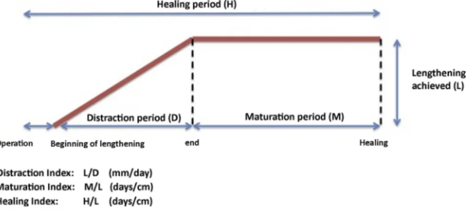
The lengthening parameters assessed postoperatively were the lengthening achieved, the duration of distraction (days), the dis-traction index (mm/day), the maturation index (days/cm), the healing index (days/cm), the length of the hospital stay (days), joint
range of movement of the lower-limb at the last follow-up, return to weightbearing, and return to walking unassisted and with complete weightbearing (Fig. 2). The lengthening was considered achieved when it did not differ by more than 5 mm from the initial objec-tive. Bone healing was defined by corticalization of at least three sides of the callus on AP and lateral X-rays. Patient comfort dur-ing the distraction phase was evaluated usdur-ing a Visual Analog Scale (VAS) and pain was scored from 0 to 10.
The Paley functional score was used for femur and tibial length-ening[10].
The postoperative LDFA and MPTA angles were also measured to assess the correction of the deformities in the frontal plane.
Intraoperative and postoperative complications were recorded, as was material removal. The complications were classified accord-ing to the Lascombes classification[11]and according to the Paley classification as a simple problem (grade 1), an obstacle (grade 2), and minor or major complications (grade 3)[12].
2.3. Statistical analysis
The descriptive analysis was performed after having verified the Gaussian distribution of the continuous variables. Chi2tests were
carried out for the qualitative variables. The subgroups were com-pared using Fisher exact tests for the quantitative variables and the Mann-Whitney U-test for the qualitative variables. The P-value indicating statistical significance was 5%. The statistical analysis was done using STATA SE v11.0 software (College Station, TX, USA). 3. Results (Table 1)
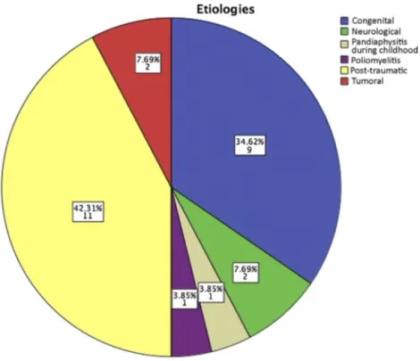
A total of 26 Fitbone®nails were implanted in 23 patients. In 15 patients, the femur was lengthened and in 11 cases the tibia. The patients’ mean age was 22.5 years (range: 15–53 years) and the mean follow-up was 3.4 years (range: 2–5.3 years). The etiolo-gies requiring the lengthening procedure were congenital in nine cases (34.6%), post-traumatic in 11 cases (42.3%), neurologic in two cases (7.7%), tumoral in two cases (7.7%), post-pandiaphysitis dur-ing childhood in one case (3.8%), and post-poliomyelitis in one case (3.8%). At the last follow-up, all the patient’s material had been removed at a mean 20± 4.2 months (range: 14–26 months) after it had been implanted. No complications, obstacles, or difficulties were encountered.
3.1. Gain in length
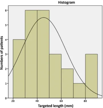
The planned limb lengthening was obtained in 23 cases (88%), for a mean gain of 45.3± 18 mm (range: 20–80 mm) (Figs. 3 and 4). Three patients presented a mean 8.6 mm of residual internal limiting membrane (ILM), which was felt clinically by one patient.
Fig. 2. Etiologies.
3.2. Distraction index and hospital stay duration
Distraction began a mean 7 days after surgery with a mean index of 0.78± 0.26 mm/day (range: 0.1–1.32 days) and a mean distrac-tion period lasting 74.1 days (range: 26–421 mm/day). During this period, the mean patient comfort evaluated by the VAS was 2.5 (range: 0–4).
3.3. Maturation (Fig. 5)
There was no statistically significant difference in terms of maturation index according to the different factors studied known to influence the results. The mean duration of matura-tion was 203± 176 days (range: 49–590 days) for the femurs and 259± 183 days for the tibias. The mean maturation index
was 59.9± 59.3 days/cm for femurs and 72.9 ± 69.3 days/cm for tibias.
3.4. Healing (Fig. 5)
The mean healing index was 73± 57 days/cm for femurs and 83.5± 65 days/cm for tibias. No statistically significant difference was found between tibias and femurs. Similarly, no statistically significant difference was found between patients presenting a pre-operative frontal deformity and the others.
3.5. Correction of the deformity
In 11 cases, the axis deformity in the frontal plane was corrected. In patients presenting a frontal mechanical axis deformity, the Table 1
Radiological results and the incomplete lengthening index.
Minimum Maximum Mean Standard deviation Target lengthening (mm) 25 80 45.3 18.1
Difference (mm) 5 10 7.50 3.5 Lengthening achieved (mm) 20 80 43.2 18.1 Preop right aLDFA (◦) 79.0 102.0 85.7 7.5
Preop left aLDFA 76.0 92.0 82.8 4.2 Preop right mMTPA 80.0 101.2 89.3 4.3 Preop left mMTPA 74.0 92.0 87.2 4.0 Postop right aLDFA 78.2 88.2 83.0 3.6 Left postop aLDFA 77.2 86.7 82.1 3.9 Postop right aMTPA 87.70 95.20 90.9 3.8 Postop left aMTPA 78.0 88.1 85.1 4.7 Duration of hospital stay (days) 3 10 5.4 2.2 Healing index (d/cm) 23.75 185.50 77.3 59.0 Maturation index (d/cm) 11.38 168.00 65.1 61.4 Distraction index (mm/d) .10 1.32 0.7 0.2 Duration of distraction (days) 26 421 74.1 89.5 Duration of maturation (days) 49 590 225.4 175.0 Duration of healing (days) 86 638 277.2 167.0 Time to total weightbearing (days) 58 305 178.5 68.2 Time to walking (days) 4 229 47.0 54.6 aLDFA: anatomical lateral distal femoral angle; aMPTA: anatomical medial proximal tibial angle.
Fig. 3. Distributions of the lengthening objectives.
mean preoperative valgus deviation was 8.7± 5.6◦(range: 4–15◦)
versus 3± 1.2◦(range: 0–5◦) postoperatively and the preoperative
varus deviation was 13± 8.1◦(range: 4–20◦) versus a postoperative
deviation of 2.1± 1.4◦(range: 0–5◦).
3.6. Joint range of movement
The functional results were excellent in 21 patients, good in one case, and poor in one case. The mean functional score was 85.4± 4.3
Fig. 5. Box plot of the maturation and healing indices (the rectangles represent 95%
of the population and the segments the extreme values; the bold line represents the median).
(range: 50–100). The knee range of movement was deemed normal
in 21 cases; one patient presented a flexion limitation at 70◦with
an extension at 0◦and another patient flessum at 10◦with flexion
at 140◦.
3.7. Complications
A single intraoperative complication was observed: an inter-condylar fracture during placement of the femoral nail via the retrograde approach. This complication, detected during the intervention, required percutaneous screw fixation (with no con-sequence on the course of the lengthening procedure).
Fig. 4. Preoperative, immediate postoperative X-rays as well as at the end of distraction and at 2 years of follow-up for one patient who underwent lengthening for 50 mm
Postoperatively, according to the Paley classification, in eight patients, five problems were observed (Lascombes grade 1), two obstacles (grade 2), and one major complication (grade 4). The five problems were two postoperative hematomas, two equinus deformities at the end of lengthening, and one complex regional pain syndrome that required physical therapy. One patient pre-sented dysfunctioning of the transmitter allowing the lengthening of the nail, which had to be replaced. One patient having under-gone several surgeries presented cutaneous necrosis requiring flap coverage. Finally, one patient decompensated an arteriovenous fis-tula of the posterior tibial artery during tibial lengthening and an embolization procedure had to be performed.
4. Discussion
This study highlights the good results of the Fitbone®
lower-limb lengthening technique with 88% reliability in terms of obtaining the planned lengthening and a 95% rate of good or excellent functional results. However, the number of complications remains high with a 15.3% rate.
This study presents a certain number of limitations. First, the number of patients included was limited (23 patients). This study did not include a group of control patients that would have made it possible to compare the results of this bone lengthening tech-nique with conventional techtech-niques. Finally, this study included the technique’s learning curve marked by the occurrence of an intra-operative intercondylar fracture that could probably have been avoided with more experience.
In addition, using an intramedullary nail presents certain techni-cal limitations. To correct the bone length in the three planes, rigid reamers are required, which makes it impossible to follow the line with the least possible resistance and therefore to guide reaming according to the position selected during the planning stage. Thus,
considering the minimum diameter of a Fitbone® TAA nail at 11
mm, this was the greatest limitation in the use of this type of sys-tem. The need to make the osteotomy line between 7 and 11 cm from the joint space to ensure solid nail locking is also a limitation. Large angular deformities with a center of rotation and angulation (CORA) very distant from the osteotomy are also geometric aspects that may make use of the lengthening systems impossible. Finally, preoperative planning requires being highly rigorous and very pre-cise because, contrary to bone lengthening systems using external fixators, no adjustment can be made postoperatively. The use of bone lengthening nails in children remains limited because of the presence of the growth plates, although some authors, in rare cases of children treated for tumors, report no complications when the implant had a smooth coating and was inserted in the central part of the physis[13,14].
The complication rate of bone lengthening procedures accord-ing to the length achieved and surgeon experience ranged from 24% to 117% for external fixation[10,12,15,16]and from 11% to 47% for intramedullary lengthening systems[3,17–19]. For external fixa-tors it has been clearly established that the complication rate varies in adults depending on patient age, the length of the procedure, and the number of wires put in place[12,15]. In children, the amount of lengthening does not seem to be a limiting factor, contrary to the correction of an angulation deformity, which seems to worsen the results when it is greater than 30◦[20]. Mechanical lengthening nails such as the ISKD®and Albizzia®require rotation movements to lengthen the limb, which can cause pain and discomfort. A con-siderable number of patients with the Albizzia nail (22–39%) have had to be rehospitalized so that the rotational movements could be done under anesthesia[17,18,21].
The complication rate in series in which the Albizzia® lengthen-ing nail was used vary from 22% to 29% if these cases of mobilization
under anesthesia are not taken into account[17,18]. As for the ISKD® nail, the mobilization rate under anesthesia is around 27% and the complication rate varies from 11% to 48% depending on the study[3,19]. The main problem with the ISKD®nail is the absence of reliable control of the lengthening and its speed[22]. Recently, a new lengthening system called the Precice® nail (Ellipse Tech-nologies, Inc, Irvine, CA, USA) was used in a series of 24 patients. Compared to the mechanical systems, this system presents the advantage of controlling lengthening with an electromagnetic sys-tem. However, other than a high material breakage rate (4%), the lengthening procedures require being supervised by the sur-geon, which is a significant deterrent to its use[23]. Moreover, the instrumentation of this implant does not include a system of working tubes guaranteeing a minimally invasive implantation and uses flexible reamers for a straight implant, which remains debatable.
Our complication rate with the Fitbone®nail was 15.4%, slightly higher than what has been reported in the literature. Krieg et al.
[6]reported a 12.5% complication rate, while Baumgart et al.[7]
reported a 13% complication rate in a large series of 150 patients. These low complication rates can be explained in part by controlled and progressive lengthening procedures. In addition, technical complications or material failure seem to be rare events with the Fitbone®nail, in both our experience and the studies reported in the literature[8,24–26]. Baumgart et al. reported a 6% rate of tech-nical incidents and a material failure rate of 3%[7]. We observed no loss of lengthening related to telescoping of the nail or fracture of the locking screws, but Krieg et al.[6]report three incidents of this type. It should be noted that in their study Krieg et al.[6]used the first generation of the Fitbone®nail, which did not provide an anti-telescoping component.
As for bone healing, intramedullary lengthening systems do not seem to disturb bone formation at the lengthening callus. Ilizarov emphasized the importance of preserving endosteal vasculariza-tion for osteogenesis of the distracvasculariza-tion calluses during lengthening procedures using external fixators [27–29]. Donnan et al. [20]
reported a mean healing index of 43.6 days/cm in children treated with external fixators. The Albizzia nail is reported to present a more rapid healing index of 35.2 days/cm[17]and the ISKD nail an index varying from 21 to 29 days/cm[19,30]. For the Fitbone®nail, the healing indices reported range from 48 days/cm to 26 days/cm
[6,7]. The slowest indices are observed in particular for the length-ening procedures involving the tibia[7]. The present study found a mean healing index of 73 days/cm for femurs and 83.5 days/cm for tibias. This difference compared to the data reported in the lit-erature can be explained partly by the fact that the mean age of our patients was much older than the age of patients in the other studies, which investigated mainly adolescents and young adults. In addition, the mean target lengthening in the present series was much longer than in other studies.
The short hospital stay and the rapid return to walking are important aspects arguing in favor of the Fitbone® long-bone lengthening systems. The absence of wires transfixing the muscles and skin make early rehabilitation possible during the distraction phase. In addition, during this phase patients can resume partial weightbearing at 20 kg and be mobilized immediately. Weight-bearing is increased monthly depending on the progression of the callus. These advantages are to be compared to the restrictions of external fixators, a source of discomfort related to bone wire care, clothing restrictions, and pain related to the transfixing wires, reducing mobilization and in the end the return to daily and occu-pational activities[5,31,32].
In conclusion, this study demonstrates the good short-term results with the Fitbone® nail from both reliability and clinical points of view. Assessment on a larger group of patients remains necessary but limited by the cost of this type of device.
Disclosure of interest
The authors declare that they have no competing interest. References
[1]Glorion C, Pouliquen JC, Langlais J, Ceolin JL, Kassis B. Femoral lengthening using the callotasis method: study of the complications in a series of 70 cases in children and adolescents. J Pediatr Orthop 1996;16:161–7.
[2]Guichet JM. [Leg lengthening and correction of deformity using the femoral
Albizzia nail]. Orthopade 1999;28:1066–77.
[3]Cole JD, Justin D, Kasparis T, DeVlught D, Knobloch C. The intramedullary skele-tal kinetic distractor (ISKD): first clinical results of a new intramedullary nail for lengthening of the femur and tibia. Injury 2001;32(Suppl 4):SD129–39.
[4]Baumgart R, Betz A, Schweiberer L. A fully implantable motorized
intramedullary nail for limb lengthening and bone transport. Clin Orthop Relat Res 1997;343:135–43.
[5]Ramaker RR, Lagro SW, van Roermund PM, Sinnema G. The psychological and
social functioning of 14 children and 12 adolescents after Ilizarov leg length-ening. Acta Orthop Scand 2000;71:55–9.
[6]Krieg AH, Lenze U, Speth BM, Hasler CC. Intramedullary leg lengthening with a motorized nail. Acta Orthop 2011;82:344–50.
[7]Baumgart R, Thaller P, Hinterwimmer S, Krammer M, Hierl T, Mutschler W. A
fully implantable, programmable distraction nail (Fitbone) – new perspectives for corrective and reconstructive limb surgery. In: Practice of intramedullary locked nails. Berlin/Heidelberg: Springer-Verlag; 2006. p. 189–98.
[8]Küc¸ükkaya M, Karakoyun Ö, Sökücü S, Soydan R. Femoral lengthening and
deformity correction using the Fitbone motorized lengthening nail. J Orthop Sci 2015;20:149–54.
[9]Baumgart R. The reverse planning method for lengthening of the lower limb
using a straight intramedullary nail with or without deformity correction. A new method. Oper Orthop Traumatol 2009;21:221–33.
[10]Paley D, Herzenberg JE, Paremain G, Bhave A. Femoral lengthening over an
intramedullary nail. A matched-case comparison with Ilizarov femoral length-ening. J Bone Joint Surg Am 1997;79:1464–80.
[11]Lascombes P, Popkov D, Huber H, Haumont T, Journeau P. Classification of
complications after progressive long bone lengthening: proposal for a new classification. Orthop Traumatol Surg Res 2012;98:629–37.
[12]Paley D. Problems, obstacles, and complications of limb lengthening by the
Ilizarov technique. Clin Orthop Relat Res 1990;250:81–104.
[13]Neel MD, Heck R, Britton L, Daw N, Rao BN. Use of a smooth press-fit
stem preserves physeal growth after tumor resection. Clin Orthop Relat Res 2004;426:125–8.
[14]Leung K-S, Taglang G, Schnettler R, Alt V, Haarman HJTM, Seidel H, et al. Practice of intramedullary locked nails. Berlin, Heidelberg: Springer Berlin Heidelberg; 2006.
[15]Tjernström B, Olerud S, Rehnberg L. Limb lengthening by callus
distrac-tion. Complications in 53 cases operated 1980–1991. Acta Orthop Scand 1994;65:447–55.
[16]Blondel B, Launay F, Glard Y, Jacopin S, Jouve J-L, Bollini G. Limb lengthening and deformity correction in children using hexapodal external fixation: preliminary results for 36 cases. Orthop Traumatol Surg Res 2009;95:425–30.
[17]García-Cimbrelo E, Curto de la Mano A, García-Rey E, Cordero J, Marti-Ciruelos R. The intramedullary elongation nail for femoral lengthening. J Bone Joint Surg Br 2002;84:971–7.
[18]Guichet J-M, Deromedis B, Donnan LT, Peretti G, Lascombes P, Bado F. Gradual
femoral lengthening with the Albizzia intramedullary nail. J Bone Joint Surg Am 2003;85-A:838–48.
[19]Leidinger B, Winkelmann W, Roedl R. [Limb lengthening with a fully
implantable mechanical distraction intramedullary nail]. Z Orthop Ihre Gren-zgeb 2006;144:419–26.
[20]Donnan LT, Saleh M, Rigby AS. Acute correction of lower limb deformity and
simultaneous lengthening with a monolateral fixator. J Bone Joint Surg Br 2003;85:254–60.
[21]Mazeau P, Assi C, Louahem D, L’Kaissi M, Delpont M, Cottalorda J. Complications of Albizzia femoral lengthening nail: an analysis of 36 cases. J Pediatr Orthop B 2012;21:394–9.
[22]Lee DH, Ryu KJ, Song H-R, Han S-H. Complications of the Intramedullary Skele-tal Kinetic Distractor (ISKD) in distraction osteogenesis. Clin Orthop Relat Res 2014;472:3852–9.
[23]Kirane YM, Fragomen AT, Rozbruch SR. Precision of the PRECICE internal bone
lengthening nail. Clin Orthop Relat Res 2014;472:3869–78.
[24]Al-Sayyad MJ. Lower limb lengthening and deformity correction using the
Fitbone motorized nail system in the adolescent patient. J Pediatr Orthop B 2012;21:131–6.
[25]Dinc¸yürek H, Kocao˘glu M, Eralp IL, Bilen FE, Dikmen G, Eren I. Functional results of lower extremity lengthening by motorized intramedullary nails. Acta Orthop Traumatol Turc 2012;46:42–9.
[26]Thaller PH, Fürmetz J, Wolf F, Eilers T, Mutschler W. Limb lengthening with fully
implantable magnetically actuated mechanical nails (PHENIX(®))-preliminary
results. Injury 2014;45(Suppl 1):S60–5.
[27]Ilizarov GA. The tension-stress effect on the genesis and growth of tissues. Part I. The influence of stability of fixation and soft-tissue preservation. Clin Orthop Relat Res 1989;238:249–81.
[28]Ilizarov GA. The tension-stress effect on the genesis and growth of tissues: part II. The influence of the rate and frequency of distraction. Clin Orthop Relat Res 1989;239:263–85.
[29]Ilizarov GA. Clinical application of the tension-stress effect for limb lengthen-ing. Clin Orthop Relat Res 1990;250:8–26.
[30]Hankemeier S, Pape H-C, Gösling T, Hufner T, Richter M, Krettek C. Improved
comfort in lower limb lengthening with the intramedullary skeletal kinetic distractor. Principles and preliminary clinical experiences. Arch Orthop Trauma Surg 2004;124:129–33.
[31]Herzenberg JE, Scheufele LL, Paley D, Bechtel R, Tepper S. Knee range of
motion in isolated femoral lengthening. Clin Orthop Relat Res 1994;301: 49–54.
[32]Young N, Bell DF, Anthony A. Pediatric pain patterns during Ilizarov
treat-ment of limb length discrepancy and angular deformity. J Pediatr Orthop 1994;14:352–7.