O
pen
A
rchive
T
OULOUSE
A
rchive
O
uverte (
OATAO
)
OATAO is an open access repository that collects the work of Toulouse researchers and
makes it freely available over the web where possible.
This is an author-deposited version published in :
http://oatao.univ-toulouse.fr/
Eprints ID : 18167
To link to this article : DOI:10.1007/s00264-015-3027-9
URL :
http://dx.doi.org/10.1007/s00264-015-3027-9
To cite this version : Cavaignac, Étienne and Pailhe, Régis and Reina,
Nicolas and Murgier, Jérôme and Laffosse, Jean-Michel and Chiron,
Philippe and Swider, Pascal Can the gracilis replace the anterior
cruciate ligament in the knee? A biomechanical study. (2016)
International Orthopaedics, vol. 40 (n° 8). pp. 1647-1653. ISSN
0341-2695
Any correspondence concerning this service should be sent to the repository
administrator:
[email protected]
Can the gracilis replace the anterior cruciate ligament
in the knee? A biomechanical study
Etienne Cavaignac1&Regis Pailhé1&Nicolas Reina1&Jérôme Murgier1&
Jean Michel Laffosse1&Philippe Chiron1&Pascal Swider2
Abstract
Purpose The purpose of this study was to determine whether a four-strand gracilis-only construct possesses the biomechan-ical properties needed to act as an anterior cruciate ligament (ACL) reconstruction graft.
Methods This was a pilot study with 32 cadaver specimens. The biomechanical properties of three types of grafts were determined using validated tensile testing methods: patellar tendon (BTB), both hamstring tendons together (GST4) and gracilis alone (G4).
Results The maximum load at failure of the G4 was 416.4 N (±187.7). The GST4 and BTB had a maximum load at failure of 473.5 N (±176.9) and 413.3 N (±120.4), respectively. The three groups had similar mean maximum load and stiffness values. The patellar tendon had significantly less elongation at failure than the other two graft types.
Conclusions The biomechanical properties of a four-strand gracilis construct are comparable to the ones of standard grafts. This type of graft would be useful in the reconstruction of the anteromedial bundle in patients with partial ACL ruptures.
Keywords ACL reconstruction . Gracilis . Short Graft . Experimental study
Introduction
The choice of grafts for anterior cruciate ligament (ACL) re-construction is not without consequences. One of the advan-tages of using the pes anserinus tendons—gracilis and semitendinosus—is that harvesting these tendons leads to lower morbidity than harvesting a bone–patellar tendon–bone (BTB) graft. Using these tendons provides sufficient strength, limits extensor mechanism weakening and lessens anterior knee pain [1–3].
However, harvesting the gracilis and semitendinosus ten-dons has its own issues, namely reduction in flexion strength and lack of control over internal rotation [4]. Recent studies have shown the semitendinosus alone can be used as a graft [5]. However, the semitendinosus muscle–tendon unit con-trols knee rotation in full extension [6]. Using the gracilis tendon alone should reduce the morbidity induced when both hamstring tendons are harvested and should preserve the semitendinosus.
We have recently shown that a four-strand gracilis con-struct (G4) meets the anatomical specifications for use as an ACL reconstruction graft [7]. The next logical step is to de-termine whether the G4 has the biomechanical properties needed to act as an ACL graft. Zamarra et al. [8] evaluated the potential use of the gracilis alone or semitendinosus alone to reconstruct the ACL. The relative laxity obtained with G4, four-strand semitendinosus (ST4) and four-strand gracilis-semitendinosus (GST4) grafts was evaluated through biome-chanical testing. The results were similar for all three graft types and each graft was able to restore normal knee kinemat-ics. These results were not unexpected because these tendons all have similar biomechanical properties [9]. Doubling the tendons appears to more than double their failure strength. A single-strand gracilis construct has a maximum strength of 925±127 N, while a two-strand construct has a maximum
* Etienne Cavaignac
1
Institut de l’appareil locomoteur, CHU Rangueil, 1, avenue Jean Poulhès TSA 50032, 31059 Toulouse Cedex 9, France
2
Laboratoire de Biomécanique, Faculté de Médecine, Toulouse, France
strength of 2,573±496 N [10]. This same study found that the maximum strength was 1,246±243 N for the native ACL and was 3,855±592 N for the patellar tendon [9].
There are no published data on the strength of a G4 con-struct. There is also no information on the maximum load that a graft in its surgical configuration can withstand before fail-ing. Several studies have reported on the strength of each individual tendon [9], but none has determined the strength of the graft in the configuration used for ACL ligament recon-struction. This led us to ask whether a G4 construct has suit-able properties to be used as the sole replacement for a rup-tured ACL.
Our null hypothesis is that the biomechanical properties of a four-strand gracilis graft are equal to those of standard ACL reconstruction grafts. The primary objective was to measure the maximum load that the G4 could withstand before failing. The secondary objective was to compare the biomechanical properties (maximum load and elongation at failure, stiffness) of three types of ACL graft: patellar tendon (BTB), combined hamstring graft (GST4) and the G4.
Materials and methods
Materials
This was a comparative biomechanical study using 32 cadaver knees from 16 donors. The donors had a mean age at death of 84 years (range, 77−90). The cadavers were stored at −20 °C and thawed overnight at 2 °C before dissection and subse-quent biomechanical analysis. All knees were free of wounds and macroscopic signs of intra-articular lesions (Outbridge> grade 3, no osteophytes in the intercondylar notch). All knees had an intact ACL and the passive joint range of motion mea-sured with a goniometer was always at least 130°.
Graft harvesting
A standard anteromedial incision was performed. The pes anserinus tendons were located at the lower part of the incision, and then harvested with an open-ended tendon stripper. The tendons were cut at the periosteum of their tibial insertions. The 10-mm wide, middle-third patellar tendon graft (BTB) was harvested with patellar and tibial bone blocks as described by Neyret et al. [11]. The cuboid-shaped patellar bone block was 15 mm long, 9 mm wide and 5 mm thick. The cuboid-shaped tibial bone block was 30 mm long, 10 mm wide and 5 mm thick. Graft preparation
The various grafts were tested in the same configuration as the one used during a surgical procedure. The surgical techniques were reproduced exactly. The hamstring tendons in the left knee
were used together. They were folded in two. This graft was named GST4. The G4 graft was prepared by folding it into four.
Graft preservation
The prepared grafts were stored at −4 °C in a cold freezing solution containing saline and 10 % dimethylsulphoxide. They were removed from the freezer the evening before test-ing and kept at room temperature (21 °C) for at least 12 hours. This process does not alter the biomechanical properties of tendons [12].
Methods Graft fixation
The grafts were fixed using validated methods [5,9,13]. The distal 15 mm of each graft was compressed between two metal clamps (Fig. 1). As a consequence, the distance between clamps (initial specimen length) varied depending on the graft’s length.
Fig. 1 Drawing of clamps used to grip the tendon specimens based on Shi et al. [13] and Handl et al. [9]
Measurement protocol
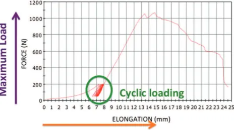
Each set of clamps was attached to a materials testing system (Instrom 3300®; Instron, Canton, MA, USA) to execute the tensile testing and measure the biomechanical properties of the graft. The measurements were performed using the sys-tem’s software (BlueHill®; Instrom SA France, Elancourt, France). Since the initial specimen length varied as a function of graft length, the length measurement sensor was reset be-fore each test. A typical load-elongation curve is shown in Fig. 2. Each graft was preloaded to 10 N, then cycled 100 times between 50 and 200 N at 0.5 Hz. A tensile test was then performed using a 10 mm/min crosshead speed until the graft failed. This sequence is a standard, validated test protocol [14]. The following structural properties were measured: (1) maximum load at failure (N), (2) maximum elongation at fail-ure (mm) and (3) linear stiffness (N.mm−1).
Statistical analysis
The statistical analysis was performed with the Excel 2011 (Microsoft, Redmond, WA, USA) and XLSTAT 2011 (Addinsoft, Paris, France) software packages. The descriptive analysis consisted of mean, median and standard deviation values. The mean values for maximum load to failure, maxi-mum elongation to failure and linear stiffness were compared between the three groups (G4, GST4 and BTB) using Student’s t-test. To ensure the conditions had been met for parametric testing, the normality of the measured variables was verified using the Shapiro-Wilk test and the homogeneity of variances was verified using Fisher’s f-test and Levene’s test. The significance threshold was set at P<0.05.
We found no published information regarding the expected maximum load at failure for G4 grafts, which made it difficult to determine how many samples were needed. We decided to
perform a pilot study with at least 30 specimens [15]. Ultimately, 32 specimens were tested.
Results
The maximum load at failure of the G4 was 416.4 N (± 187.7). The GST4 and BTB had a maximum load at failure of 473.5 N (± 176.9) and 413.3 N (± 120.4), respectively. The results for the entire series are summarised in Fig. 3. The maximum elongation at failure of the G4 was 18.0 mm (± 10.6); it was 21.2 mm (± 11.6) for the GST4 and 5.1 mm (± 4.1) for the BTB. The linear stiffness of the G4 construct was
Fig. 2 Typical force–elongation curve generated by the BlueHill® software (Instrom SA France, Elancourt, France). A cyclic preconditioning regimen (green circle) was performed before the graft was loaded to failure
Fig. 3 Box-and-whisker plot of the maximum load at failure for each graft type. BTB Bone-patellar tendon-bone, G4 four-strand gracilis con-struct, GST4 four-strand gracilis and semitendinosus construct
192.9 N.mm−1(± 41); it was 198.5 N.mm−1(± 44.9) for the GST4 and 164.6 N.mm−1(± 52.0) for the BTB.
The three groups had similar mean values in terms of the maximum load that they could withstand before failing (Table1). The BTB had the lowest elongation at failure of the three graft types; this difference was statistically signifi-cant. Since the three groups had similar mean stiffness values, no conclusions can be drawn about this comparison.
Discussion
The primary objective was to measure the maximum load that the G4 can withstand at failure to determine if its structural properties are equal to those of standard ACL reconstruction grafts. The maximum load at failure of the G4 construct was 416 N±187 (range, 242–1,069 N), which is equivalent to the reference tendon grafts, namely the patellar tendon and four-strand semitendinosus and gracilis. To our knowledge, this is the only study where the maximum failure load was measured with the grafts in their surgical configuration. Instead of mea-suring the strength of each tendon making up the graft, the strength of the fully prepared graft was measured. The goal of this study was to measure the mechanical properties of the graft itself. This aspect is novel. Most studies on this topic focus only on graft fixation methods. Few studies have report-ed the strength of the graft itself, which we felt was important information to have. Since the various grafts used for ACL reconstruction are fixed with different methods, adding the fixation variable in this study would have been a confounding factor.
We have recently shown that a G4 construct meets the anatomical specifications for use as an ACL reconstruction
graft [7]. The available length of G4 and four-strand semitendinosus grafts is always sufficient to place at least 15 mm of graft in the bone tunnels [7]. Using the G4 reduces the risk of oversizing in the middle portion of the graft, which is a problem with other types of graft [16]. An excessively thick graft can get impinged in the notch, which would disrupt its healing [17]. In addition, use of the gracilis only preserves the semitendinosus, which plays an important role in control-ling rotational stability when the knee is fully extended [6].
We have recently shown that a four-strand semitendinosus graft has better biomechanical properties (namely failure strength) than standard grafts [6]. For this reason, it is our graft of choice for ACL reconstruction. We think that the G4 is par-ticularly well-suited to being used alone during surgical treat-ment of partial ACL tears [18]. A G4 graft could be used for the isolated reconstruction of the anteromedial bundle when the pos-terolateral bundle is intact [19,20]. Its biomechanical properties are comparable to those of other types of grafts and its volume is lower [7] thus the risk of oversizing is reduced [16]. But these findings must be tempered by this study’s limitations. The tensile testing was performed with tissues that had been frozen at −4 °C and then thawed. Several studies have explored the effect of freezing and thawing tendons on their biomechanical properties [21]. Based on the results of these studies, the biomechanical properties of tendons are unaffected when fewer than three gradual freeze–thaw cycles are performed.
Only axial tension tests were performed in this study. Although this testing protocol does not reproduce the multi-axial loads experienced by the ACL in vivo, it is consistent with previous research done into graft strength and fixation [5,
13,14,22].
The fixation method is also another basic consideration, as it can affect the results of tensile tests [23]. Novel serrated jaw clamps that allow tendons to be tested in a simple and repro-ducible manner have recently been described by Shi et al. [13]. Resin-based clamps and cryoclamps are difficult to work with and have not been formally validated [24]. Pap et al. [25] recently validated a fixation method for autografts that used the serrated jaw clamps described by Shi et al. by comparing them with other types of clamps. This is the type of clamp used in the current study.
To ensure quasi-static conditions, the testing was carried out with a slow crosshead speed, so as to not bring the ten-don’s visco-elastic properties into play. The tensile strength will be lower when slower elongation speeds are used. When ligaments and tendons are loaded more quickly, the risk of damaging these structures increases [26].
The maximum load values in the current study were much lower than published values. The leading studies on this topic reported maximum load values of 1,719 N±1,167.80 (range, 456–4,546 N), which is nearly 3 times higher than the value reported here [27]. It was also surprising to see that this dif-ference did not apply to the stiffness values, which were very
Table 1 Comparison of the maximum load at failure, elongation and stiffness performed with Student’s t-test
Mean SD SE P MAXIMUM LOAD AT FAILURE
BTB–GST4 25.7 142.3 47.4 0.6 BTB–G4 7 309.8 103.3 0.9 G4–GST4 22.2 266.3 84.2 0.8 ELONGATION AT FAILURE BTB–GST4a 16.1 10.6 2.7 <0.01 BTB–G4a 12.9 12.6 3.2 <0.01 G4–GST4 3.2 15.9 3.9 0.4 STIFFNESS BTB–GST4 39.7 94 23.5 0.1 BTB–G4 34.0 117.4 29.3 0.3 G4–GST4 5.6 65 16.3 0.7 Pprobability, SDstandard deviation, SEstandard error
a
similar to ours (Table2). A review of the literature was per-formed to better understand the reasons for these differences. The first reason is related to donor age. The studies reporting the highest failure loads were also the ones with the youngest donors (20–30 years) [9,27].
A second reason relates to the method used to induce tendon failure. The study with the largest number of ACL grafts and highest published loading values had a significant bias [9]. The tensile testing system consisted of applying tension to the ten-don by dropping a weight from a set height. The maximum load and stiffness were measured using a custom, but non-validated accelerometer-based device. These methodological consider-ations bring the validity of their results into question.
The third aspect relates to the elongation speed used in the various studies (Table2). Many of these tensile tests used elon-gation rates greater than 5 mm/s (about 10 %/s). Under these conditions, the tensile tests were not being performed under static conditions, thus bringing the tendon’s visco-elastic properties into play. This could explain the higher maximum load values
reported in these studies. We performed an ANOVA test on the published data and found a relationship between the elongation rate and maximum failure load (P=0.032). However, this poten-tial tendon stiffening as the elongation rate increases must still be demonstrated with specific biomechanical studies.
This study has a certain number of limitations. The popu-lation from which the cadaver donors were taken is not repre-sentative of the population in which ACL reconstruction is typically performed. Age, BMI, gender and physical activity levels affect the biomechanical properties of tendons and lig-aments [22]. But these limitations are partially overcome by the comparative design of the study. Since all three grafts were harvested from the same individual, all grafts had the same age, BMI, gender and activity level. As a consequence, any confounding factors were evenly distributed between groups. In addition, the biomechanical testing protocol used in this study—elongation speed, grips, measurement methods—was based on published studies [13] (Table2). There is no way of knowing whether the various types of grafts would react
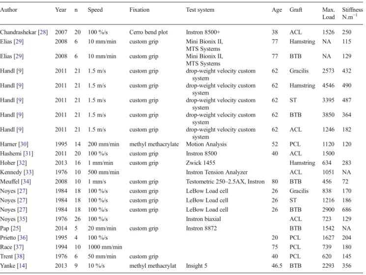
Table 2 Summary of published biomechanical study describing the structural properties of tendons used for ACL reconstruction Author Year n Speed Fixation Test system Age Graft Max.
Load
Stiffness N.m−1
Chandrashekar [28] 2007 20 100 %/s Cerro bend plot Instron 8500+ 38 ACL 1526 250 Elias [29] 2008 6 10 mm/min custom grip Mini Bionix II,
MTS Systems
77 Hamstring NA 115 Elias [29] 2008 6 10 mm/min custom grip Mini Bionix II,
MTS Systems
77 BTB NA 129 Handl [9] 2011 21 1.5 m/s custom grip drop-weight velocity custom
system
62 Gracilis 2573 432 Handl [9] 2011 21 1.5 m/s custom grip drop-weight velocity custom
system
62 Hamstring 4546 490 Handl [9] 2011 21 1.5 m/s custom grip drop-weight velocity custom
system
62 ST 3395 487 Handl [9] 2011 21 1.5 m/s custom grip drop-weight velocity custom
system
62 BTB 3850 364 Handl [9] 2011 21 1.5 m/s custom grip drop-weight velocity custom
system
62 ACL 1246 182 Harner [30] 1995 14 200 mm/min methyl methacrylate Motion Analysis 52 PCL 1120 120 Hashemi [31] 2011 20 100 %/s custom grip Instron 8500 40 ACL 1500 Hoher [32] 2013 16 1 mm/min custom grip Zwick 1455 Hamstring 634 283 Kennedy [33] 1976 10 500 mm/min Instron Tension Analyzer ACL 1051 NA Meuffel [34] 2008 10 1 mm/s custom grip Testometric 250–2.5AX, Instron 80 BTB 456 72 Noyes [27] 1984 18 100 %/s custom grip LeBow Load cell 26 Gracilis 838 170 Noyes [27] 1984 18 100 %/s custom grip LeBow Load cell 26 ST 1216 186 Noyes [27] 1984 18 100 %/s custom grip LeBow Load cell 26 BTB 2900 686 Noyes [35] 1976 26 100 %/s Instron biaxial ACL 723 129 Pap [25] 2014 5 20 mm/min custom grip Instron 8872 BTB 1542 NA Prietto [36] 1995 4 100 %/s 20 PCL 1627 204 Race [37] 1994 10 1000 mm/min 75 PCL 739 180 Trent [38] 1976 6 50 mm/min custom grip 40 PCL 620 145 Yanke [14] 2013 9 10 %/s methyl methacrylat Insight 5 46.5 BTB 2293 356 nnumber of samples, EPentrance potential, Max.maximum, ACLanterior cruciate ligament, PCLposterior cruciate ligament, STsemitendinosus
differently if one of these parameters was altered. Because the same protocol was used, the various groups could be directly compared to each other.
Conclusions
A G4 construct has the anatomical features needed to serve as an ACL reconstruction graft. Its biomechanical properties are comparable to those of the standard grafts (patellar tendon and hamstring). The G4 is particularly well-suited to serving as an augmentation graft in cases of partial ACL rupture but the four-strand semitendinosus graft remains our first choice for complete ACL reconstruction. Clinical studies of the G4 must be performed to confirm these results.
Acknowledgments The authors thank Joanne Archambault, PhD for the editorial support during preparation of this manuscript.
Compliance with ethical standards
Conflict of interest No benefits in any form have been received or will be received from a commercial party related directly or indirectly to the subject of this article.
References
1. Bonamo JJ, Krinick RM, Sporn AA (1984) Rupture of the patellar ligament after use of its central third for anterior cruciate reconstruc-tion. A report of two cases. J Bone Joint Surg Am 66(8):1294–1297 2. Goyal S, Matias N, Pandey V, Acharya K (2015) Are pre-operative anthropometric parameters helpful in predicting length and thick-ness of quadrupled hamstring graft for ACL reconstruction in adults? A prospective study and literature review. Int Orthop. doi:
10.1007/s00264-015-2818-3
3. Stevanovic V, Blagojevic Z, Petkovic A, Glisic M, Sopta J, Nikolic V, Milisavljevic M (2013) Semitendinosus tendon regeneration af-ter anaf-terior cruciate ligament reconstruction: can we use it twice? Int Orthop 37(12):2475–2481
4. Nakamura N, Horibe S, Sasaki S et al (2002) Evaluation of active knee flexion and hamstring strength after anterior cruciate ligament reconstruction using hamstring tendons. Arthroscopy 18(6):598–602 5. Pailhe R, Cavaignac E, Murgier J et al (2015) Biomechanical study of ACL reconstruction grafts. J Orthop Res. doi:10.1002/jor.22889
6. Shelburne KB, Torry MR, Pandy MG (2005) Effect of muscle compensation on knee instability during ACL-deficient gait. Med Sci Sports Exerc 37(4):642–648
7. Cavaignac E, Pailhe R, Murgier J et al (2014) Can the gracilis be used to replace the anterior cruciate ligament in the knee? A cadaver study. Knee 21(6):1014-1017. doi:10.1016/j.knee.2014.07.010
8. Zamarra G, Fisher MB, Woo SL, Cerulli G (2010) Biomechanical evaluation of using one hamstrings tendon for ACL reconstruction: a human cadaveric study. Knee Surg Sports Traumatol Arthrosc 18(1):11–19. doi:10.1007/s00167-009-0911-0
9. Handl M, Drzik M, Cerulli G et al (2007) Reconstruction of the anterior cruciate ligament: dynamic strain evaluation of the graft. Knee Surg Sports Traumatol Arthrosc 15(3):233–241. doi:10.1007/ s00167-006-0175-x
10. Sajovic M, Vengust V, Komadina R et al (2006) A prospective, randomized comparison of semitendinosus and gracilis tendon ver-sus patellar tendon autografts for anterior cruciate ligament recon-struction: five-year follow-up. Am J Sports Med 34(12):1933– 1940. doi:10.1177/0363546506290726
11. Neyret P, Demey G (2012) Reconstruction du ligament croisé antérieur: technique chirurgicale. In: Traité de chirurgie du genou. Elsevier Masson, Paris, pp 41-64
12. Huang H, Zhang J, Sun K et al (2011) Effects of repetitive multiple freeze-thaw cycles on the biomechanical properties of human flexor digitorum superficialis and flexor pollicis longus tendons. Clin Biomech (Bristol, Avon) 26(4):419–423. doi:10.1016/j. clinbiomech.2010.12.006
13. Shi D, Wang D, Wang C, Liu A (2012) A novel, inexpensive and easy to use tendon clamp for in vitro biomechanical testing. Med Eng Phys 34(4):516–520. doi:10.1016/j.medengphy.2011.11.019
14. Yanke AB, Bell R, Lee AS et al (2013) Central-third bone-patellar tendon-bone allografts demonstrate superior biomechanical failure characteristics compared with hemi-patellar tendon grafts. Am J Sports Med 41(11):2521–2526. doi:10.1177/0363546513501780
15. Hertzog MA (2008) Considerations in determining sample size for pilot studies. Res Nurs Health 31(2):180–191. doi:10.1002/nur.20247
16. Pujol N, Queinnec S, Boisrenoult P, Maqdes A, Beaufils P (2013) Anatomy of the anterior cruciate ligament related to hamstring ten-don grafts. A cadaveric study. Knee 20(6):511–514. doi:10.1016/j. knee.2012.10.006
17. Marzo JM, Bowen MK, Warren RF et al (1992) Intraarticular fi-brous nodule as a cause of loss of extension following anterior cruciate ligament reconstruction. Arthroscopy 8(1):10–18 18. Buda R, Ruffilli A, Parma A et al (2013) Partial ACL tears: anatomic
reconstruction versus nonanatomic augmentation surgery. Orthopedics 36(9):e1108–e1113. doi:10.3928/01477447-20130821-10
19. Sonnery-Cottet B, Panisset JC, Colombet P et al (2012) Partial ACL reconstruction with preservation of the posterolateral bundle. Orthop Traumatol Surg Res 98(8 Suppl):S165–S170. doi:10. 1016/j.otsr.2012.10.001
20. Pujol N, Colombet P, Potel JF et al (2012) Anterior cruciate liga-ment reconstruction in partial tear: selective anteromedial bundle reconstruction conserving the posterolateral remnant versus single-bundle anatomic ACL reconstruction: preliminary 1-year results of a prospective randomized study. Orthop Traumatol Surg Res 98(8 Suppl):S171–S177. doi:10.1016/j.otsr.2012.09.007
21. Moon DK, Woo SL, Takakura Y et al (2006) The effects of refreezing on the viscoelastic and tensile properties of ligaments. J Biomech 39(6):1153–1157. doi:10.1016/j.jbiomech.2005.02.012
22. Noyes FR, DeLucas JL, Torvik PJ (1974) Biomechanics of anterior cruciate ligament failure: an analysis of strain-rate sensitivity and mech-anisms of failure in primates. J Bone Joint Surg Am 56(2):236–253 23. Butler DL, Grood ES, Noyes FR et al (1984) Effects of structure
and strain measurement technique on the material properties of young human tendons and fascia. J Biomech 17(8):579–596 24. Cheung JT, Zhang M (2006) A serrated jaw clamp for tendon
grip-ping. Med Eng Phys 28(4):379–382. doi:10.1016/j.medengphy. 2005.07.010
25. Pap K, Hangody G, Szebenyi G, Rita K, Panics G, Hangody L (2014) An easy way to fix tendon allografts into a loading—ma-chine for biomechanical testing. Paper presented at the 16th ESSKA Congress, Amsterdam, 14-17 May
26. van Dommelen JA, Jolandan MM, Ivarsson BJ et al (2006) Nonlinear viscoelastic behavior of human knee ligaments subjected to complex loading histories. Ann Biomed Eng 34(6):1008–1018. doi:10.1007/s10439-006-9100-1
27. Noyes FR, Butler DL, Grood ES et al (1984) Biomechanical anal-ysis of human ligament grafts used in knee-ligament repairs and reconstructions. J Bone Joint Surg Am 66(3):344–352
28. Chandrashekar N, Mansouri H, Slauterbeck J et al (2006) Sex-based differences in the tensile properties of the human anterior cruciate ligament. J Biomech 39(16):2943–2950
29. Elias JJ, Rai SP, Ciccone WJ et al (2008) In vitro comparison of tension and stiffness between hamstring tendon and patella tendon grafts. J Orthop Res 26(11):1506–1511
30. Harner CD, Baek GH, Vogrin TM et al (1999) Quantitative analysis of human cruciate ligament insertions. Arthroscopy 15(7):741–749 31. Hashemi J, Mansouri H, Chandrashekar N et al (2011) Age, sex, body anthropometry, and ACL size predict the structural properties of the human anterior cruciate ligament. J Orthop Res 29(7):993–1001 32. Hoher J, Offerhaus C, Steenlage E et al (2013) Impact of tendon
suturing on the interference fixation strength of quadrupled ham-string tendon grafts. Arch Orthop Trauma Surg 133(9):1309–1314 33. Kennedy JC, Hawkins RJ, Willis RB et al (1976) Tension studies of human knee ligaments. Yield point, ultimate failure, and disruption
of the cruciate and tibial collateral ligaments. J Bone Joint Surg Am 58(3):350–355
34. Meuffels DE, Niggebrugge MJ, Verhaar JA (2009) Failure load of patellar tendon grafts at the femoral side: 10- versus 20-mm-bone blocks. Knee Surg Sports Traumatol Arthrosc 17(2):135–139 35. Noyes FR, Grood ES (1976) The strength of the anterior cruciate
ligament in humans and Rhesus monkeys. J Bone Joint Surg Am 58(8):1074–1082
36. Prietto C (1995) Anterior cruciate ligament repair in children. West J Med 163(6):567–568
37. Race A, Amis AA (1994) The mechanical properties of the two bundles of the human posterior cruciate ligament. J Biomech 27(1):13–24
38. Trent PS, Walker PS, Wolf B (1976) Ligament length patterns, strength, and rotational axes of the knee joint. Clin Orthop Relat Res 117:263–270
![Fig. 1 Drawing of clamps used to grip the tendon specimens based on Shi et al. [13] and Handl et al](https://thumb-eu.123doks.com/thumbv2/123doknet/3113403.88430/3.892.497.776.558.1066/fig-drawing-clamps-used-tendon-specimens-based-handl.webp)