Université de Montréal
Quantitative Proteomics Methods for the Analysis of
Histone Post-translational Modifications
par
Nebiyu Ali Abshiru
Département de Chimie
Faculté des arts et des sciences
Thèse présentée à la Faculté des études supérieures
et postdoctorales en vue de l’obtention du grade de
philosophiae doctor (Ph.D.) en chimie
Septembre 2015
Résumé
Les histones sont des protéines nucléaires hautement conservées chez les cellules des eucaryotes. Elles permettent d’organiser et de compacter l’ADN sous la forme de nucléosomes, ceux-ci representant les sous unités de base de la chromatine. Les histones peuvent être modifiées par de nombreuses modifications post-traductionnelles (PTMs) telles que l’acétylation, la méthylation et la phosphorylation. Ces modifications jouent un rôle essentiel dans la réplication de l’ADN, la transcription et l’assemblage de la chromatine. L’abondance de ces modifications peut varier de facon significative lors du developpement des maladies incluant plusieurs types de cancer. Par exemple, la perte totale de la triméthylation sur H4K20 ainsi que l’acétylation sur H4K16 sont des marqueurs tumoraux spécifiques a certains types de cancer chez l’humain. Par conséquent, l’étude de ces modifications et des événements determinant la dynamique des leurs changements d’abondance sont des atouts importants pour mieux comprendre les fonctions cellulaires et moléculaires lors du développement de la maladie.
De manière générale, les modifications des histones sont étudiées par des approches biochimiques telles que les immuno-buvardage de type Western ou les méthodes d’immunoprécipitation de la chromatine (ChIP). Cependant, ces approches présentent plusieurs inconvénients telles que le manque de spécificité ou la disponibilité des anticorps, leur coût ou encore la difficulté de les produire et de les valider. Au cours des dernières décennies, la spectrométrie de masse (MS) s’est avérée être une méthode performante pour la caractérisation et la quantification des modifications d’histones. La MS offre de nombreux avantages par rapport aux techniques traditionnelles. Entre autre, elle permet d’effectuer des analyses reproductibles, spécifiques et facilite l’etude d’un large spectre de PTMs en une seule analyse. Dans cette thèse, nous présenterons le développement et l’application de nouveaux outils analytiques pour l’identification et à la quantification des PTMs modifiant les histones.
Dans un premier temps, une méthode a été développée pour mesurer les changements d’acétylation spécifiques à certains sites des histones. Cette méthode combine l’analyse des histones intactes et les méthodes de séquençage peptidique afin de déterminer les changements d’acétylation suite à la réaction in vitro par l’histone acétyltransférase (HAT) de levure Rtt109 en présence de ses chaperonnes (Asf1 ou Vps75).
Dans un second temps, nous avons développé une méthode d’analyse des peptides isomériques des histones. Cette méthode combine la LC-MS/MS à haute résolution et un nouvel outil informatique appelé Iso-PeptidAce qui permet de déconvoluer les spectres mixtes de peptides isomériques. Nous avons évalué Iso-PeptidAce avec un mélange de peptides synthétiques isomériques. Nous avons également validé les performances de cette approche avec des histones isolées de cellules humaines érythroleucémiques (K562) traitées avec des inhibiteurs d’histones désacétylases (HDACi) utilisés en clinique, et des histones de Saccharomyces cerevisiae liées au facteur d’assemblage de la chromatine (CAF-1) purifiées par chromatographie d’affinité. Enfin, en utilisant la méthode présentée précédemment, nous avons fait une analyse approfondie de la spécificité de plusieurs HATs et HDACs chez Schizosaccharomyces pombe. Nous avons donc déterminé les niveaux d’acétylation d’histones purifiées à partir de cellules contrôles ou de souches mutantes auxquelles il manque une HAT ou HDAC. Notre analyse nous a permis de valider plusieurs cibles connues des HATs et HDACs et d’en identifier de nouvelles. Nos données ont également permis de définir le rôle des différentes HATs et HDACs dans le maintien de l’équilibre d’acétylation des histones. Dans l’ensemble, nous anticipons que les méthodes décrites dans cette thèse permettront de résoudre certains défis rencontrés dans l’étude de la chromatine. De plus, ces données apportent de nouvelles connaissances pour l’élaboration d’études génétiques et biochimiques utilisant S. pombe.
Mots clés : Spectrométrie de masse, histones, modifications post-traductionnelles, peptides isomérique, histones acétyltranférases, histones déacétylase.
Summary
Histones are highly conserved, basic proteins found in eukaryotic cell nuclei. They organize and package DNA strands into nucleosome core particles (NCPs), the fundamental repeating units of eukaryotic chromatin. The histones are subject to a wide variety of posttranslational modifications (PTMs) including acetylation, methylation and phosphorylation. These PTMs play an essential role in DNA-replication, transcription, and chromatin assembly. Alterations in histone PTM abundances have been implicated in several types of cancer. For example, the global loss of trimethylation at H4K20 and acetylation at H4K16 is a hallmark of human cancers. Thus, characterization of histone PTMs and their dynamics is extremely useful for elucidating normal cellular functions and molecular pathways that lead to diseases.
Traditionally, histone PTMs are analyzed using antibody-based approaches such as western blot and chromatin immunoprecipitation (ChIP) assays. These methods, however, suffer from several limitations including antibody cross-reactivity, epitope occlusion, and the cost and difficulty in producing and validating antibodies. Over the last decade, mass spectrometry (MS) has emerged as a powerful technique for the characterization and quantification of histone PTMs. MS offers several advantages over the traditional approaches including reproducibility, specificity, and ability to rapidly analyze numerous PTMs in a single experiment. In this thesis, the development and applications of novel analytical tools for the identification and quantification of histone PTMs are presented.
First, a method useful for measuring the global and site specific changes in histone acetylation is described. This method combines intact mass analysis and peptide sequencing approaches to study the global and site specific changes in histone acetylation during in vitro assays with yeast Rtt109 and its chaperone (Asf1 or Vps75). Second, a method for analysis of isomeric histone peptides is presented. This method combines a high resolution LC-MS/MS with a novel bioinformatics tool called Iso-PeptidAce to deconvolute mixed spectra of co-eluting isomeric peptides. We benchmarked Iso-PeptidAce using mixtures of synthetic isomeric peptides. We demonstrated its capability in histones isolated from human erythroleukemic (K562) cells treated with clinically relevant histone deacetylase inhibitors (HDACi) and in affinity-purified S. cerevisiae histones bound to chromatin assembly factor-1 (CAF-1). Third, by employing the above methods, an in-depth quantitative analysis of the substrate specificities of several fission yeast HATs and HDACs was assessed. We
determined the acetylation site occupancy of multiple lysines in histones isolated from a control or mutant strains lacking specific HAT or HDAC activities. Our analysis identified several known and novel HAT and HDAC target sites. Our data also defined the division of labor between the different HATs and HDACs in maintaining the steady-state level of histone acetylation. Overall, we anticipate that the methods described in this thesis will address some of the existing challenges facing the chromatin field. Moreover, the data presented will provide valuable insights for future genetic and biochemical studies involving the fission yeast.
Key Words: Mass spectrometry, histones, posttranslational modifications, isomeric peptides,
Table of Contents
Résumé ... i
Summary ... iii
Table of contents ...v
List of Tables ... viii
List of Figures ... ix
List of abbreviations ... xi
Acknowledgements ... xiv
CHAPTER 1: Introduction ...17
1.1. How is the DNA stuffed into a tiny microscopic cell nucleus? ...17
1.2. Histones ...20
1.2.1. Histone variants ... 20
1.2.2. The structure of core histones ... 22
1.2.2.1. Primary and secondary structures ... 22
1.2.2.2. Higher order structures ... 23
1.2.3. Histone post-translational modifications ... 27
1.2.3.1. Acetylation ... 28
1.2.3.2. Methylation ... 30
1.2.4. Combinatorial histone PTMs ... 32
1.2.5. Histone modifiers - ‘Writers’ and ‘Erasers’ ... 36
1.2.5.1. HATs ... 37
1.2.5.2. HDACs ... 38
1.2.5.3. HDAC inhibitors – Emerging drugs for cancer therapy ... 40
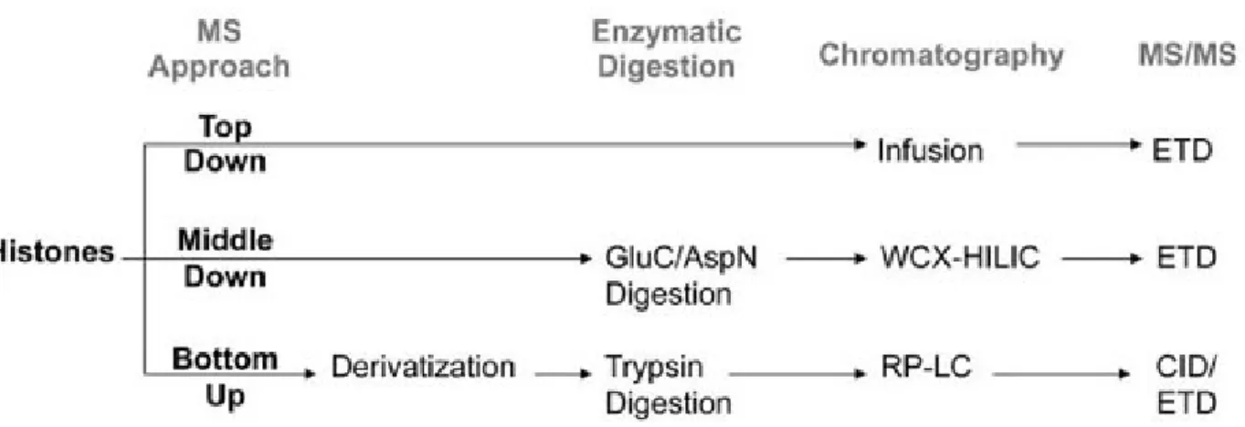
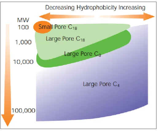
1.3. Mass spectrometry-based proteomics in histone analysis ...42
1.3.1. Basic steps in histone PTM analysis by mass spectrometry ... 46
1.3.1.1. Proteolytic digestion ... 46
1.3.1.2. RP-HPLC separation ... 47
1.3.1.3. Sample ionization... 49
1.3.1.4. Mass analysis ... 52
1.3.1.5. Tandem mass analysis... 55
1.3.1.6. Peptide identification ... 57
1.4. Thesis objectives ...62
1.5. Thesis outline ...64
1.6. References ...65
CHAPTER 2 : Chaperone-mediated Acetylation of Histones by Rtt109 Identified by Quantitative Proteomics ...75 2.1. Abstract ...76 2.2. Introduction ...77 2.3. Methods ...79 2.4. Results ...82 2.4.1. nLC/MS analysis of histones ... 82
2.4.2. Histone acetylation by Rtt109–Vps75/Asf1 complexes ... 83
2.4.3. Effect of enzyme-substrate ratios on histone acetylation ... 87
2.4.4. Peptide identification and determination of acetylation stoichiometry ... 88
2.5. Discussion ...93
2.6. Acknowledgments ...94
2.7. Supplementary data ...95
2.8. References ...100
CHAPTER 3: Discovery of Protein Acetylation Patterns by Deconvolution of Peptide Isomer Mass Spectra ...103
3.1. Abstract ...104
3.2. Introduction ...105
3.3. Methods ...107
3.4. Results ...112
3.4.1. Deconvolution of mixed MS/MS spectra by Iso-PeptidAce ... 112
3.4.2. Extraction of elution profiles and fragment ion patterns ... 114
3.4.3. Temporal profiling of histone acetylation following HDAC inhibition ... 116
3.4.4. Identification of acetylation patterns in CAF1-bound histones ... 119
3.5. Discussion ...123
3.6. Acknowledgments ...125
3.7. Supplementary data ...126
3.8. Supplementary method ...140
3.9. References ...150
CHAPTER 4: Unraveling site-specific and combinatorial acetylations of histones using high-resolution mass spectrometry in HAT and HDAC mutants of fission yeast ...154
4.1. Abstract ...155
4.2. Introduction ...156
4.3. Methods ...159
4.4. Results ...162
4.4.1. Measurement of H3 and H4 peptides by PRM- LC-MS/MS ... 162
4.4.2. Determination of acetylation site occupancies ... 162
4.4.3. Analysis of global histone acetylation in HAT mutants ... 163
4.4.4. Gcn5 and Mst2 target several H3 sites in S. pombe ... 164
4.4.5. Clr6 is specific for multiple sites on H3 ... 166
4.4.6. Methylation-dependent activity of Clr3 toward H3K14ac ... 167
4.4.7. Sir2 and Hos2 exhibit mild activity toward H3 ... 168
4.4.8. Clr6 mutation caused a major increase in combinatorial H4 acetylation ... 169
4.4.9. Hos2 mutnation cuased a major increase in H4K16 acetylation ... 169
4.4.10. Sir2 and Clr3 mutations did not alter H4 acetylation pattern ... 170
4.4.11. HDAC mutants display altered H3K9 methylation levels ... 170
4.5. Discussion ...172
4.6. Acknowledgments ...176
4.7. Main figures ...177
4.8. Supplementary data ...185
4.9. References ...195
CHAPTER 5: Conclusions and Future Perspectives ...199
5.1. Conclusions ...199
5.2. Future perspectives ...205
5.3. References ...207
Appendix A: Additional supplementary information ...210
List of Tables
Table 1.1 Histone PTM readers. ... 34
Table 1.2 Conservation of HATs between species and phyla. ... 38
Table 1.3 Classification of mammalian HDAC isotypes. ... 39
Table 1.4 HDACis currently under clinical trial or FDA-approved. ... 41
Table S1.1 Human histone variants (HIstome-The Histone Infobase). ... 210
Table S2.1 H3/Rtt109-Vps75 assay intact histone intensity data (0 to 90 min) ... 97
Table S2.2 H3/Rtt109-Asf1 assay intact histone intensity data (0 to 90 min) ... 98
Table S2.3 H3/Rtt109-Vps75 assay peptide IDs and normalized intensities. ... 99
Table S2.4 H3/Rtt109-Asf1 assay peptide IDs and normalized intensities. ... 99
Table S3.1 Lists of synthetic H3 and H4 peptides and their concentration in mM. ... 126
Table S3.2 Synthetic H4 peptide standard dilutions. ... 126
Table S3.3 Deconvolution of mixture spectra by Iso-PeptidAce. ... 128
Table S3.4 Calculated acetylation site occupancies. ... 129
Table S3.5 LC/MS intensity data for CAF-1-bound and total H3 and H4 peptides. ... 130
Table S3.6 List of known acetylation sites in selected bromodomains substrates. ... 132
Table S3.7 Raw intensity data for control and HDACi treated samples. ... 133
Table S4.1 (A) Fission yeast strains and (B) PRM inclusion list ... 185
Table S4.2 Global H3 and H4 acetylations in HAT and HDAC mutants... 187
Table S4.3 Global changes in H4 acetylation in response to HDAC depletion. ... 188
Table S4.4 H3 acetylation site occupancies in HAT mutants. ... 189
Table S4.5 H4 acetylation site occupancies in HAT mutants. ... 190
Table S4.6 H3 acetylation site occupancies in HDAC mutants... 191
Table S4.7 H4 acetylation site occupancies in HDAC mutants... 192
List of Figures
Figure 1.1 The building blocks of DNA. ... 17
Figure 1.2 Overview of eukaryotic DNA packaging. ... 19
Figure 1.3 Sequence alignment of human histone H3 variants. ... 21
Figure 1.4 Three dimensional structural models of the histone octamer/NCP ... 24
Figure 1.5 The individual core histones assemble into a histone octamer. ... 26
Figure 1.6 Mechanism of lysine acetylation by HATs. ... 28
Figure 1.7 Known histone acetylation sites. ... 29
Figure 1.8 Mechanism of lysine methylation by HMTs. ... 31
Figure 1.9 Histone tail modifications... 32
Figure 1.10 Schematic showing the reciprocal activities of ‘writers’ and ‘erasers’. ... 36
Figure 1.11 Bottom up, middle down, and top down MS approaches... 45
Figure 1.12 Recommended RP particle size versus protein MW. ... 49
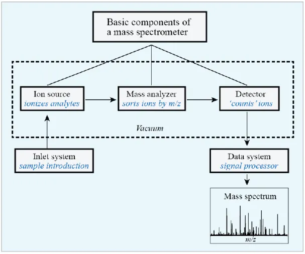
Figure 1.13 Basic components of mass spectrometry. ... 51
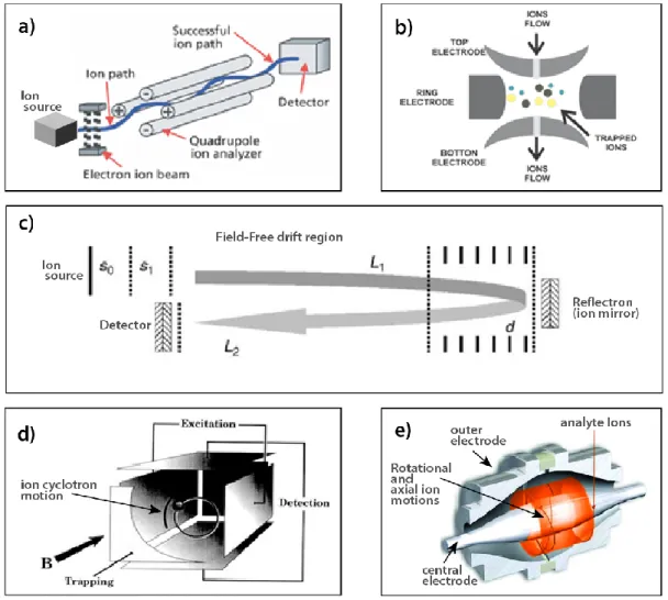
Figure 1.14 Schematics of some of the most common mass analyzers. ... 54
Figure 1.15 The Roepstorff nomenclature for peptide fragment ions. ... 56
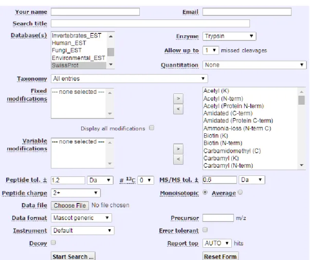
Figure 1.16 The user interface for Mascot MS/MS database search engine. ... 58
Figure S1.1 Sequence alignment of histones from various species ... 212
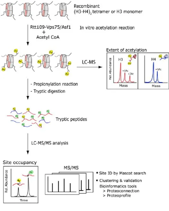
Figure 2.1 Overview of the analytical scheme for profiling histone modifications. ... 83
Figure 2.2 LC/MS analyses of intact histone H3 obtained from in vitro HAT assay. ... 85
Figure 2.3 LC/MS analyses of intact histone H4 obtained from in vitro HAT assay. ... 86
Figure 2.4 Relative proportion of histone acetylation by Rtt109–Vps75. ... 88
Figure 2.5 Analysis of acetylation stoichiometry of H3 incubated with Rtt109–Vps75. ... 90
Figure 2.6 Analysis of acetylation stoichiometry of H4 incubated with Rtt109–Vps75. ... 92
Figure S2.1 Intact mass analysis of intact proteins. ... 95
Figure S2.2 The proportion of histone H3 acetylation during 90min H3\Rtt109-Vps75 assay. ... 96
Figure 3.1 Deconvolution of co-eluting acetylated isomers by Iso-PeptidAce. ... 113
Figure 3.3 Acetylation site occupancies of CAF1-bound H3 and H4. ... 121
Figure S3.1 Peptides within the same isomeric groups exhibit very narrow retention. ... 134
Figure S3.2 MS signal responses of synthetic H3 and H4 peptides. ... 135
Figure S3.3 Co-elution and co-fragmentation of acetylated isomers. ... 136
Figure S3.4 Isomeric peptides produce distinct fragment ion patterns. ... 137
Figure S3.5 Deconvolution of di- and tri-acetylated isomers of histone H4. ... 138
Figure S3.6 Fractionation of total histones by RP-HPLC. ... 139
Figure S3.7 MS/MS spectra of H3 and H4 peptide isomer ... 213
Figure 4.1 PRM-LC-MS/MS analysis of histone peptides. ... 177
Figure 4.2 Global H3 and H4 acetylations in HAT and HDAC mutants. ... 179
Figure 4.3 Site-specific acetylation of H3 and H4 in HAT mutants. ... 180
Figure 4.4 Site-specific acetylation of H3 in HDAC mutants. ... 181
Figure 4.5 Site-specific acetylation of H4 in HDAC mutants. ... 182
Figure 4.6 Analysis of H3-K9/K36 methylation levels in HDAC mutants. ... 183
Figure 4.7 Representative histone PTM crosstalks in fission yeast. ... 184
List of abbreviations
3-D ac CoA Asf1 BCA BET bp BRD CAA CAF1 ACN CE ChIP CID csv Da DC DNA DSB DTT EI EIC ELISA ESI ETD FA FC fmol FTICR FWHM µg GC Three dimensional Acetylation Co-enzyme A Anti-silencing factor 1 Bicinchoninic AcidBromodomain and extra terminal domain Base pair
Bromodomain Chloroacetamide
Chromatin assembly factor 1 Acetonitrile
Collision energy
Chromatin immunoprecipitation Collision induced dissociation Comma separated value Dalton
Direct current
Deoxyribonucleic acid Double strand break Dithiothreitol Electron ionization
Extracted ion chromatogram
Enzyme-linked immunosorbent assay Electrospray ionization
Electron transfer dissociation Formic acid
Fold change Femtomole
Fourier transform ion cyclotron resonance Full width at half maximum
Micro-gram
GST HAT HCD HDAC HDACi HDMT HMT HP1 HPLC i.d. IAA IDs Kd µL LC-MS/MS LTQ µ/mM m/z µm MALDI me me1 me2 me3 m-H2O MS MW NAD+ NCP ND nLC PDB ppm PRM Glutathione S-transferase Histone acetyltransferase
High-energy collision dissociation Histone deacetylase
Histone deacetylase inhibitor Histone demethylase
Histone methylase
Heterochromatin protein 1
High performance liquid chromatography Internal diameter
Iodoacetamide Identifications Dissociation constant Micro-liter
Liquid chromatography tandem mass spectrometry Linear trap quadrupole
Micro-/milli-molar Mass to charge ratio Micro-meter
Matrix-assisted laser desorption ionization Methylation Mono-methylation Di-methylation Tri-methylation Milli-Q water Mass spectrometer Molecular weight
Nicotinamide adenine dinucleotide Nucleosome core particle
Not determined/detected Nano-liquid chromatography Protein data bank
Part-per-million
pr PTMs QIT QQQ QTOF RF RP Rtt109 SAHA SDC SD SILAC SRM TAP TCA TFA TIC UV v/v Vps75 WT XIC Propionylation Post-translational modifications Quadrupole ion trap
Triple quadrupole mass spectrometer
Hybrid Quadrupole-Time of flight mass spectrometer Radio frequency
Reverse phase
Ty1 transposition gene product 109 Suberoylamide hydroxamic acid Sodium deoxycholate
Standard deviation
Stable isotope labeling by amino acid in cell culture Single reaction monitoring
Tandem affinity purification Trichloroacetic acid
Trifluoroacetic acid Total ion chromatogram Ultraviolet
Volume per volume
Vacuolar Protein Sorting 75 Wild type
Acknowledgements
‘A single stick may smoke, but it will not burn’ Ethiopian proverb
It is with great pleasure that I thank all the friends, families and everyone I have worked with over the past five years who have made this work possible.
First, I would like to express my deepest gratitude to my supervisor Dr. Pierre Thibault for giving me the opportunity to pursue this graduate program and for his continuous guidance, encouragement, and support. Before coming to Montreal I did not know much about the city, which is not surprising for someone from Ethiopia (well, I understand some of you might be trying to figure out where ‘Ethiopia’ is located). Yet, I had read several outstanding scientific contributions by Pierre and his team to the field of mass spectrometry. At the time I was on the verge of completing my Master’s degree in Norway and Pierre’s work caught my attention. I immediately contacted him by email expressing my interest to join his team and luckily he accepted my application. It wasn’t easy starting life in Montreal; it was a huge cultural shift for me. Pierre’s support and encouragement along the way was very helpful. Apart from this, his capacity as one of the leading scientists in Canada- the depth of knowledge he has, his dedication and his attention to details makes him very unique. And it was an honor and a great privilege working with him over the past five years.
I would also like to thank my co-supervisor Dr. Alain Verreault who has supported me in so many ways during my study. Much of the research activities I have been involved in required a deeper understanding of biological processes though I was trained as analytical chemist. Alain helped me understand most of these processes, which made my study progress a lot more manageable. He has always been ready to share his knowledge and expertise. I also owe a big thank you to Alain’s current and former team members, a very lively and wonderful team. Especially, Roshan Elizabeth, who has been at the center of all the research works I have conducted. She has been tirelessly providing me with all the biological samples that I needed to do my work. Also, together with Alain, she has immensely improved my understanding of the role of various epigenetic processes. I’m very thankful for all this.
I would like thank Prof. Joelle Pelletier, the Chair of my thesis committee, and Prof. Martine Raymond, member of my thesis committee, for the great advice and encouragement over the
course of my study. Their guidance has been instrumental in continuously developing my knowledge and skills.
I have been fortunate enough to work with a number of wonderful labmates over the years. I would like to first thank Eric Bonneil for his great enthusiasm to share his expertise in mass spectrometry. He has always been there to train and guide me on how to operate the various instruments in the lab. Thank you, Eric! I also want to thank all the other former and current members of the lab including Christina Bell, Frederic Lamoliatte, Sibylle Pfammatter, and Chongyang Li, who are more friends than labmates. A special thank you to Frederic Lamoliatte (he likes to be called Fred) who generously accepted to translate my ‘Summary’ section into French.
I would like to thank all my friends and families in Montreal, Chicago, and Ethiopia. My parents Senait and Ali may not read this thesis but their support, encouragement and prayer have never ceased till this day. In my home country not many people go to school till they turn thirty and I guess that is why my parents have always wondered when I would stop going to classes.
This thesis would not have been possible if not for the support and encouragement of the love of my life Feben Woldemariam. She has been always close to my heart even when she is thousands of miles away. Since we married three years ago she has constantly stood by me in times of difficulties and has been a wonderful friend and wife to me. It is with great pleasure that I dedicate the Ph.D. Thesis to her. I also want to thank all my family in Chicago Bresh, Abby, Woudu, Teklu, Nate, Nuhamin, Sapphier and others for their encouragement.
“
It is a miracle that curiosity survives formal education
.”
Albert Einstein
CHAPTER 1: Introduction
1.1. How is the DNA stuffed into a tiny microscopic cell nucleus?
The DNA contains all the genetic instructions required for an organism to grow and reproduce [1]. It is composed of a phosphate group, a sugar molecule (called deoxyribose) and a nitrogen-containing base namely adenine (A), guanine (G), cytosine (C) and thymine (T). These components are linked in the order phosphate-sugar-base to form the structural unit of DNA known as the nucleotide (Fig. 1.1a). In turn, the nucleotides are joined to one another in a chain by a phosphodiester bond between the phosphate of one nucleotide and the deoxyribose of the next to form a DNA strand (Fig. 1.1b). The DNA exists in a double-stranded form whereby two adjacent strands running in opposite directions are joined together by hydrogen bonds between complementary bases: A with T, and G with C (Fig. 1.1c). In the cell, the DNA double-strand mainly exists in right-handed helix structure (Fig. 1.1d).
Figure 1.1 The building blocks of DNA.
From reference [1] with permission from Garland Science - Books)
The nucleotide is formed from a phosphate group, a deoxyribose sugar and a nitrogen-containing base (a). Successive nucleotides are linked by phosphodiester bonds to form a DNA strand (b). Adjacent DNA-strands are joined by hydrogen bonds between complementary bases to form a DNA double-strand (c). In the cell, the DNA double-strand is present as a helix structure (d).
The discovery of the DNA double-strand by James Watson and Francis Crick in 1953 [2] was indeed a major advance in the field of science. This discovery gave scientists a vital clue on how the DNA carries genetic information and how this information is transmitted between generations. But, the idea that a very long DNA double-strand can fit into a tiny microscopic cell nucleus and still be accessible for gene-regulatory processes is extremely fascinating. Just to illustrate the extent of packaging- the total length of the DNA double-strand in a single human cell is approximately 2 meters; and it is believed that the human body is comprised of tens of trillions of cells that contain this length of DNA. So, if the entire DNA sequence in all the cells were to be stretched end-to-end, it would extend to tens of billions of kilometers. Instead, the DNA sequence in each cell is condensed by about 10,000-fold and marvelously fit into a nucleus that is only about 10µm wide. The DNA undergoes various layers of organizations to achieve such high level of packaging.
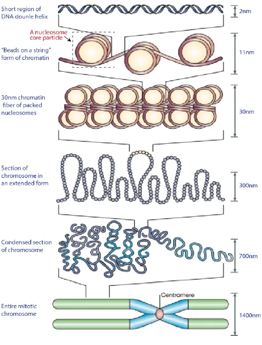
The first level of DNA packaging involves the wrapping of the DNA double-strand around a complex of proteins known as histones [3, 4]. These proteins are highly basic and contain multiple positively charged amino acid residues that form electrostatic interactions with the negatively charged phosphate group of the DNA backbone. Approximately 147 base-pairs (bps) of DNA sequences are wrapped almost twice around the histone complex to form a bead-like structure called the nucleosome core particle (NCP) [5] (see Fig. 1.2). This structure was first elucidated by electron microcopy in the mid-1970s [6]. The packaging of DNA into nucleosomes shortens the long, linear double-strand sequence by about seven-fold. Adjacent nucleosomes are then joined together by linker DNA sequences (approximately 10 to 80 bps) to form an 11 nm beads-on-a-string like structure, where the beads represent nucleosomes and the string represents the linker DNA. In the next level of DNA packaging, arrays of nucleosome cores condense into a 30 nm higher-order chromatin fiber [7]. This is achieved through a complex DNA folding processes that involve histone H1 and several non-histone proteins [8, 9].. Histone H1, also known as linker non-histone, binds to the entry and exit sites of the linker DNA sequence of the NCP.
The chromatin fiber is further compacted into different layers of higher order chromosomal domains. The two major chromosome structural domains are euchromatin and heterochromatin [9]. The euchromatin is structurally less condensed and contains a higher proportion of genes that are transcriptionally active. In contrast, the heterochromatin region is highly condensed and genes located in this region are generally less accessible for transcription. The heterochromatin is further classified into two broad structural domains.
The first type, which is called constitutive heterochromatin, is found in all cell types and comprises genes that are generally permanently inactivated [10]. This type of heterochromatin is mainly found near chromosomal regions that contain highly repetitive DNA sequences, such as centromeres and telomeres. The second type, facultative heterochromatin, refers to regions of the chromosome that are temporarily condensed [11]. Most inactive euchromatic genes exist in this form of heterochromatin.
Figure 1.2 Overview of eukaryotic DNA packaging.
1.2. Histones
Extensive research in the composition of the cell nuclei resumed in the late 1860s when Friedrich Miescher first isolated the “nuclein” (now called DNA) from human white blood cells [13]. He later extended his research to other cell types such as salmon spermatocytes, and found the DNA bound to a basic molecule, which he named “protamin”. Together, the DNA-protamin complex amounted to almost the entire mass of the sperm head. Meanwhile, Miescher was deeply interested in the DNA component and paid little attention to the protamin. Later in 1884, Albrecht Kossel isolated DNA-bound proteins from the nuclei of avian red blood cells [14, 15]. He found major similarities between the proteins and Miescher’s protamine. He named the proteins ‘histones’. Kossel later attempted to examine the physical and chemical features that distinguish histones from protamins and other nuclear proteins. In this respect he found that histones exist in complex with nucleic acids and, are highly basic in nature. Moreover, by exploiting the basic property of histones Kossel described the first protocol for acid-extraction of histones from the cell nuclei. Kossel received the Nobel Prize in Physiology or Medicine in 1910 for his major contribution in the discovery of the chemical compositions of the cell nucleus.
However, it wasn’t until the late 1950s that researchers began to investigate the different classes of histone found in eukaryotic cells. Extensive purification and characterization of histones in calf thymus and pea seedlings revealed five major classes: Histones H1, H2A, H2B, H3 and H4 [16, 17]. Histones H2A, H2B, H3 and H4 (collectively known as core histones) form the nucleosomal core around which the DNA is wrapped, whereas H1 acts as a scaffold protein for the linker DNA [18]. Early chemical cross-linking experiments showed that the nucleosome consists of two copies of each core histone assembled into an octameric structure [19]. Moreover, primary sequence analyses showed that histones contain highly basic N-terminal sequences and C-terminal fold domains that include amino acid distributions characteristic of globular proteins [20].
1.2.1. Histone variants
Each class of histone described in the previous section, except histone H4, has a number of primary sequence variants. A total of 55 histone variants have been reported in humans (Appendix A, Table S1.1) [21]. Histone H3, for instance, is present in six different variants: H3.1, H3.1t, H3.2, H3.3, H3.3C, and CenH3 (see Fig. 1.3). The first five variants share about 90% amino acid sequence similarity. The CenH3, also known as CENP-A, has longer
N-terminal domain and less than 50% amino acid sequence similarity with the other five variant.
The canonical histone H3.1 differs from H3.2 and H3.3 by one and five amino acid residues, respectively. In H3.1, the residue at position 96 is cysteine, where as in H3.2 and H3.3 the cysteine is replaced by serine (highlighted by a blue box in Fig.1.3). The other four residues of H3.1 that differ from H3.3 are alanine, serine, valine and methionine located at positions 31, 87, 89 and 90 (highlighted by a red boxes in Fig.1.3), respectively. Histones H2A and H2B have the largest number of variants, each with at least nineteen variants.
Figure 1.3 Sequence alignment of human histone H3 variants.
(Sequence alignment prepared using the ‘align’ program at http://www.uniprot.org/)
Histone variants play an important role in marking and organizing the nucleosome in specialized regions of the chromatin. For instance, CenH3 (also known as CENP-A) is incorporated specifically into nucleosomes at the centromere [22], a specialized region on a chromosome where two sister chromatids are linked. On the other hand, histones H3.1/H3.2 and H3.3 mediate distinct nucleosome assembly pathways. In vitro nucleosome assembly assays show that deposition of H3.1/H3.2 into the nucleosomes is coupled with DNA-replication processes, whereas H3.3 deposition takes place independent of DNA DNA-replication [23]. In accordance with this, studies show that the synthesis of H3.1/H3.2 is highly elevated during DNA replication in the S phase of the cell cycle, whereas the H3.3 variant is expressed at all stages of the cell cycle [24-26].
Histone H2A variants are also implicated in several DNA-associated process including DNA damage response, X chromosome inactivation and transcriptional regulation [27-29]. Histone H2A.Bbd has less than 50% amino acid sequence identity with the canonical H2A. It is believed to promote destabilization of the nucleosome core in mammals [30]. Histone H2A.X is highly linked to DNA damage response (DDR) pathways [31]. It is rapidly phosphorylated at Ser 139 (in humans) or Ser 129 (in budding yeast) following DNA damage response. The phosphorylated H2A.X is commonly referred to as ɣ-H2A.X. Accumulation of ɣ-H2A.X at DNA damage sites serves as a mark for the recruitment of several DNA repair protein complexes [32]. Recently, histone H2A.Z was also shown to have a role in DNA strand break (DSB) repair [33]. It facilitates this process by creating an open chromatin structure at the DSB site. On the other hand, despite the crucial role of histone H2B in chromatin formation, the functions of its variants remain highly understudied.
1.2.2. The structure of core histones
1.2.2.1. Primary and secondary structures
Although histones were discovered more than a century ago, analysis of their primary and higher order structure did not begin until the late 1950s. Meanwhile, significant advances were made in developing analytical strategies for the isolation and fractionation of histones from different types of cells [16]. These strategies had enabled investigators to carry out fundamental structural analysis of the core histones. Initial studies were focused on determining the primary sequences of histones. In this regard, histone H4 was the first histone for which the complete amino acid sequence was determined [34, 35]. Sequencing of tryptic peptides generated from calf thymus or pea seedling histone H4 gave rise to a total of 102 amino acid residues. The N-terminal region of H4 was found to be highly rich in positively charged amino acids such as arginine and lysine, suggesting a potential binding site for the DNA phosphate group. Histone H3 is the second histone for which a complete sequence was reported [36]. Sequencing of calf thymus H3 peptides generated by trypsin, chymotrypsin and cyanogen bromide digestion identified a total of 135 amino acid residues. Similar to histone H4, the N-terminal region of H3 contained numerous arginine and lysine residues. Moreover, histone H3 had two cysteine residues which are absent in histone H4. The complete primary sequences of histones H2A and H2B were also determined around the same period. Both histones comprised highly basic N-terminal region and a hydrophobic C-terminal domain. The primary sequences of the four core histones are highly conserved across eukaryotes,
suggesting a similar role for all. The sequence alignment for canonical histone H3 and histone H4 in five different species is illustrated in Appendix A, Figure S1.1.
Soon after sequence analyses of the core histones, researchers began a thorough investigation into how the individual histones are assembled into an octameric structure. A number of biochemical and structural analyses revealed a pairwise association between core histones [37, 38]. Equimolar mixtures of calf thymus H3 and H4 form a very stable (H3-H4)2 tetramer at high salt concentrations (~ 2M NaCl). Moreover, CD (circular dichroism) spectra analyses showed increased α-helical properties of the tetrameric structure compared to the individual histones. Similarly, fluorescence anisotropy and CD measurements indicated that histones H2A and H2B form a stable H2A-H2B dimer in high salt solutions [39]. Formation of the dimer increases the α-helical content of the H2A-H2B by about 15 residues compared to the individual histones. Moreover, CD spectra analyses of trypsin-digested chromatin showed increased secondary structure properties at the less basic (or C-terminal) regions of histones H2A and H2B [40].
The discovery of the tetrameric (H3-H4)2 and dimeric H2A-H2B structures led to the formulation of the first model for chromatin structure. As such, the chromatin repeating unit (or the nucleosome) is comprised of one (H3-H4)2 tetramer and two H2A-H2B dimers assembled into an octamer and approximately 200 base-pairs of DNA. These structural features were later tested by several research groups and were found to agree substantially with the proposed model [18, 19, 41, 42]. At high salt concentrations, the histone octamer complex is highly stable even in the absence of the nucleosomal DNA or chemical cross-linking reagents. Meanwhile at physiological salt concentrations the octamer dissociates into one (H3-H4)2 tetramer and two H2A-H2B dimers. These key physical properties were fundamental to the successful crystallization of the histone octamer in subsequent structural studies.
1.2.2.2. Higher order structures
By the late 1970s it was generally accepted that the core of the nucleosome structure is comprised of a histone octamer assembled from two copies of each core histone. Meanwhile, the physical association between the histones in the octameric complex was not fully understood. An early study by Eickbush et al. [42] suggested that the octamer is held together by two types of protein-protein interactions. The first type involves hydrophobic interactions which hold together the individual subunits of the tetrameric (H3-H4)2 and dimeric
H2A-H2B structures. The second type involves weak interactions that promote the association between one H3-H4 tetramer and two H2A-H2B dimers to form an octamer. The later type mainly involves hydrogen bond interactions between specific regions of the tetrameric and dimeric subunits. Nonetheless, these interactions were first proposed based solely on results obtained from studying the effects of temperature, urea, and pH titration on the dissociation of the octamer and hence, further structure analysis of the octamer was needed.
Initially, researchers were investigating low resolution structures of the histone octamers using techniques such as neutron and x-ray diffraction, and electron microscopy [43-47]. These studies revealed the disc-like shape of the octamer and the left handed DNA superhelix wrapped around the octamer (Fig.s 1.4a-b). Moreover, it was shown that the arrangement of histones in the octamer involves two H2A-H2B dimers flanked by one centrally located (H3-H4)2 tetramer. Meanwhile, due to the low resolution of these models accurate assessment of the secondary and tertiary structures of the individual histones could not be achieved. A more defined structure of the octamer was later determined by X-ray crystallography at 3.1 Å resolution [48]. In addition to the previously described features, the new model revealed three dimensional structural motifs (which are now called the histone fold) in each of the core histones. The motifs were later confirmed by high resolution X-ray crystallography analysis (Fig. 1.4c) of Xenopus laevis and human nucleosome core particles solved at 2.8 [49] and 2.5 Å [50], respectively.
Figure 1.4 Three dimensional structural models of the histone octamer/NCP
From references [43, 50] with permission from Nature Publishing Group and Oxford University Press, respectively. (models a & b published in 1980 and c in 2005).
(a) A 3D model of the octamer reconstructed from an electron micrograph. (b) A model showing the left handed supercoiling of the DNA around the octamer. (c) A ribbon model (top panel) and an axial view (bottom panel) of a 2.5Å crystal structure of the human NCP.
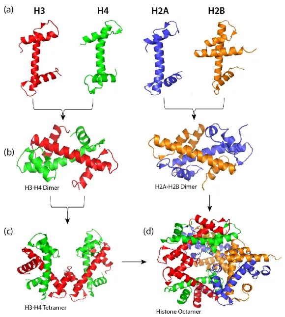
Although the four core histones have very low sequence homology, they all share the histone fold motif. This motif is characterized by a long central helix flanked on either side by a loop and a short helix (Fig. 1.5a). The histones H3 and H4 form two pairs of heterodimers (H3-H4 and H3’-H4’) mainly through the interaction of their central helices (Fig. 1.5b, left panel). The heterodimers are further assembled into a (H3-H4)2 tetramer through hydrogen bonding and weak interactions of the central helices, C-terminal helices, and the loops (Fig. 1.5c). The two H4 molecules (H4-H4’) comprising the histone octamer are aligned side by side in opposite directions (Fig. 1.5c). In contrast, the two H3 molecules in the homodimer H3-H3’ are arranged in head-to-tail or ‘handshake’ motif (Fig. 1.5c). In the same way, the H2A and H2B histone folds are joined together through several interactions to form the two H2A-H2B dimers (Fig. 1.5b, right panel). These dimers, however, do not form tetrameric structures. Instead, they each are linked to the (H3-H4)2 tetramer through the interaction of multiple residues of H2B and H4. Histone H2B residues E73 and R90 form hydrogen bonds with histone H4 residues R92 and E90, respectively. These hydrogen bonds and several other hydrophobic interactions promote the formation of the histone octamer (Fig. 1.5d).
Figure 1.5 The individual core histones assemble into a histone octamer.
A representation of (a) the helix-loop-helix-loop structural motif of histones H3, H4, H2A and H2B; (b) the structure of H3-H4 (left panel) and H2A-H2B (right panel) heterodimers; (c) (H3-H4)2 tetrameric structure; (d) the histone octamer assembled from the (H3-H4)2 tetramer and the H2A-H2B dimer. (The figures were adapted from Wikimedia Commons- https://commons.wikimedia.org, and prepared using The Pymol Molecular Graphics System, Version 1.2r 1, based on the crystal structure of Xenopus laevis nucleosome core particle (PDB entry 1AO1).
The histone fold domains contact the DNA superhelix chain at a number of sites. One of the primary contact sites involves the N-terminal tail region of the core histones [49, 51]. This region is highly flexible and as a result it produces weak electron density in X-ray diffraction experiments. A segment of about eight and five amino acids lengths of histones H2B and H3 tails, respectively, pass through the DNA superhelix minor groove channel. The side-chain of
these amino acids is involved in stabilizing the histone-DNA interactions by forming a hydrogen bond with the DNA phosphate group or by creating favorable positively charged density inside the superhelix channel. Another major histone-DNA contact site involves the amino acid side-chain of the α-helices. In this regard, a number of arginine side-chains in the histone fold were shown to interact with the DNA superhelix. Some of these arginines are H2A-R42, R77; H2B-R30, H3-R49, R63, R83; and H4-R48. Other residues are also involved in stabilizing the histone-DNA interactions. For example, the side-chains of two proline residues in the histone fold (H3-P66 and H4-P32) interact with the DNA deoxyribose moiety.
1.2.3. Histone post-translational modifications
Post-translational modifications (PTMs) are covalent modifications that occur at distinct amino acid residues in a protein [52]. Most PTMs are mediated by enzymatic activities. They are present in cellular proteins of various prokaryotic and eukaryotic organisms, ranging from bacteria to humans. PTMs play versatile roles in several cellular processes including the cell cycle, gene regulation, signal transduction, and protein-protein interactions [53-56]. Some of the most common protein modifications include acetylation, methylation, ubiquitination of lysine residues; and phosphorylation of serine, threonine or tyrosine residues. A protein can be post-translationally modified at one or more sites. When a PTM occurs on different sites of the same protein it creates several versions of that protein. Such proteins are referred to as PTM-isoforms.
While it is known that there are only four types of core histones in humans, extensive biochemical and mass spectrometry analyses have identified a number of PTM-isoforms of each type. The core histones possess multiple lysine residues on their N-terminal tail. These lysines are subject to a wide variety of modifications including acetylation, methylation, ubiquitination, butyrylation, crotonylation, and many other modifications [57]. In addition to lysines, the histone tails possess multiple arginine residues that are subject to modifications such as mono- and di-methylation, and citrullination. Phosphorylation of serine, threonine and tyrosine residues was also reported. Among the four core histones, H3 is by far the most modified. Almost all of the above PTMs have been identified in various combinations on H3. Most histone PTMs carry out their function in a combinatorial fashion although there are cases where individual histone PTMs have been shown to have important roles. One of the mechanisms by which combinatorial functions are achieved involves histone PTM ‘crosstalk’, whereby the addition or removal of a PTM at a specific site affects the
maintenance of other neighboring PTMs. In the following subsections I will briefly introduce two of the most characterized histone PTMs - acetylation and methylation of lysine residues.
1.2.3.1. Acetylation
Acetylation is one of the most widely studied histone PTMs. This modification is established by the addition of an acetyl group to the ε-amino group of lysine residues, a process catalyzed by a group of enzymes known as histone acetyltransferases (HATs) [58]. In this reaction, a conserved glutamate residue (Glu) of the HAT activates the ε-amino group of lysine for nucleophilic attack on the carbonyl moiety of acetyl-coenzyme A (CoA-SH) which acts as an acetyl donor (Fig. 1.6) [59]. The reverse reaction is catalyzed by histone deacetylases (HDAC) enzymes. The two groups of enzymes (HATs and HDACs) will be discussed in detail in sections 1.2.5.1 and 1.2.5.2.
Histone acetylation has been extensively studied in many eukaryotic organisms. Most of the identified acetylation sites are conserved from yeast to humans, suggesting a crucial role of the modifications in each species. Several studies have shown the role of histone acetylation in modulating important DNA-associated processes including replication, transcription, gene regulation and the DNA double-strand break (DSB) repair [60, 61].
Figure 1.6 Mechanism of lysine acetylation by HATs.
Adapted from reference [59] with permission from the American Society for Biochemistry and Molecular Biology.
Previous comprehensive mass spectrometry-based analyses have identified over twenty acetylation sites on the four core histones in human cells. Some of the known acetylation sites are shown in Figure 1.7a-b. The majority of these acetylations are found on the N-terminal tails, yet a small number of acetylations are also located in the histone fold domain, for example H3K64ac and H4K91ac.
Figure 1.7 Known histone acetylation sites.
(a) An illustration of the N-terminal tails of core histones and sites of acetylation; (b) lists of known histone acetylation and methylation sites, labeled ‘ac’, and ‘me’, respectively. Figures prepared using Adobe® Illustrator® CS6, version 16.0.0.
Histone acetylation is involved in the modulation of diverse chromatin-associated processes. For instance, acetylation of lysine 16 of H4 was shown to inhibit chromatin condensation and formation of higher order chromatin fiber [62]. Histone acetylation is also associated with transcriptional activity [63], although the mechanism underlying this association is still not fully elucidated. One of the proposed mechanisms by which histone acetylation could facilitate transcription is via disruption of the histone-DNA interactions [64]. According to this model, acetylation neutralizes the electrostatic interactions between the positively charged Ɛ-amino group of lysines and the negatively charged phosphate group of DNA, thereby increasing the accessibility of DNA for transcriptional machineries.
In an alternative model, increasing evidences suggest that histone acetylation may mediate transcription by acting as docking platforms for recruitment of chromatin-modifying complexes [65, 66]. This model is supported by the findings that chromatin regulators often recruit specific HATs or HDACs to promoter and transcription start sites of actively transcribing genes [67]. This has been observed in budding yeast where CHIP-ChIP assays showed elevated levels of acetylated histones in the vicinity of active genes; sites where HATs Gcn5 and Esa1 were also found to be recruited [68]. This study found a genome wide increase in acetylation of Gcn5 target sites H3K9 and H3K14, and Esa1 target sites H4K5ac,
H4K8ac, H4K12ac and H4K16ac. These modifications were found to also correlate with transcriptional rates genome-wide, suggesting the association between histone acetylation and transcriptional activity. Similar observations were made in humans [69]. ChIP-seq analysis of histone modifications in human CD4+ T cells showed elevated levels of H2BK12ac, H2BK20ac, H2BK120ac, H3K4ac, H4K5ac, H4K8ac, H4K12ac and H4K16ac in the promoter and transcription start sites of active genes, whereas the PTMs H2AK9ac, H2BK5ac, H3K9ac, H3K18ac, H3K27ac, H3K36ac, H4K91ac were mainly identified at regions surrounding the transcription start sites. It is not known whether these combinatorial acetylation patterns have distinct functions, or whether they simply reflect differences in the specificity of HATs recruited at various stages of transcription. It should be noted that transcriptional activation does not always result in increased histone acetylation. Previous studies in budding yeast have shown a dramatic decrease in histone H4 acetylation at target promoter regions upon activation by specific gene regulators [70].
1.2.3.2. Methylation
Histone methylation is catalyzed by a group of enzymes known as histone methyltransferases (HMTs) [71, 72]. The general mechanism of lysine methylation whereby an HMT catalyzes the transfer of a methyl group from the cofactor S-adenosylmethionine (SAM) to the ε-amino group of a specific lysine residue is illustrated in Figure 1.8. Lysine residues can be mono-, di-, or tri-methylated by accepting one, two or three methyl groups on their side chain. Unlike acetylation, however, methylation of lysines does not alter the overall charge state of histones. Some of the known methylation sites on the four core histones are shown in Figure 1.7b (labeled ‘me’). Majority of the methylation sites reside in the N-terminal tail. Histone H3 is by far the most methylated core histone, with abundant methylation detected at lysines 4, 9, 27, 36, and 79. The biological role of histone methylation has been implicated in several DNA-associated processes including transcription, gene silencing, and chromatin condensation [73, 74]. Histone arginine residues are also known to be mono- or di-methylated, which serve important roles in epigenetic regulation of various genes [75, 76].
Figure 1.8 Mechanism of lysine methylation by HMTs.
From reference [77] with permission from Nature Publishing Group.
Histone methylation can be associated with transcriptional activation or repression depending on the site and level of modification. Increased level of H3K4me3 was found at highly active genes, whereas H3K4me1 and H3K4m2 were found at intermediately active group of genes. [78-80]. In contrast, the levels of H3K9me2, H3K9me3, H3K27me2, H3K27me3, and H4K20me3 are highly associated with repressed or silent genes [80-83]. In contrast, however, H3K9me1, H3K27me1 and H4K20me1 are enriched at promoter regions of active genes. In a recent report it was shown that non-nucleosomal histone H3 in budding yeast contains high levels of H3K9me1, but is devoid of H3K9me2 or H3K9me3 [84], suggesting that this modification may not have a major role in transcriptional regulation. On the other hand, H3K36me3 and H3K79me3 are found abundantly at active regions, whereas H3K36me1 and H3K79me1 did not show any major preference toward either active or silent regions.
1.2.4. Combinatorial histone PTMs
Although histone PTMs correlate with various transcriptional states of genes, their precise mechanism of action is still not fully understood. As illustrated in Figure 1.9, a diverse array of closely-spaced PTMs co-exists on a short segment of the N-terminal tail of histones. While there are few instances where a specific PTM is associated with distinct cellular function [85], the majority of studies suggest that histone PTMs exert their function through combinatorial patterns [86-88]. For example, synergic interplay between H3S10ph and H3K14ac was shown to serve a major role in marking mammalian epidermal growth factor stimulation [89]. In another study, phosphorylation at H3S10 was shown to induce increased acetylation of H3K14 by the HAT Gcn5 both in vitro and in vivo [90], suggesting a strong functional relationship between the two modifications.
Figure 1.9 Histone tail modifications.
Adapted from reference [91] with permission from Nature Publishing Group).
Legends: acetylation (A, blue), methylation (M, red), phosphorylation (P, yellow) and ubiquitination (U, green).
Functional interactions between PTMs of different core histones have also been identified in budding yeast. It was shown that mono-ubiquitination of H2BK123 by Rad6 acts as a signal for activation of methylation of H3K4 by HMT complex known as COMPASS, a process termed as ‘trans-tail’ regulation of histone modifications [90]. In agreement with this observation, S. cerevisiae cells with H2B-K123R mutation lacked H3K4 methylation, whereas H2BK123 mono-ubiquitination was not affected in H3-K4R mutants [92].
Moreover, these studies showed that activation of H3K4 methylation via H2BK123 mono-ubiquitination is functionally linked with transcriptional silencing at telomere regions. The same type of PTM interactions has been recently identified in human cells [93].
Crosstalk between two different PTMs of the same residue has also been reported. Recently, it was shown that H3K4 methylation regulates the global level of H3K4 acetylation in budding yeast [94]. Deletion of specific subunits of COMPASS, the complex responsible for methylating H3K4, resulted in elevated level of H3K4ac. However, strains lacking Gcn5 or Gcn5/Rtt109, the HAT responsible for H3K4 acetylation, did not affect the level of H3K4 mono-, di- or tri-methylation. According to these results, the global abundances of H3K4me, particularly H3K4me2 and H3K4me3, affected the level of H3K4ac, whereas the converse was not true. These modifications are mutually exclusive and are enriched at promoter regions of actively transcribing genes with H3K4ac located upstream of H3K4me.
The different types of crosstalk described above represent few examples of how histone PTMs could act synergistically or in combination to regulate distinct cellular events. It was previously postulated that these type of PTM interactions form a ‘histone code’ [95]. According to this hypothesis, first, a histone code is established through a series of addition and removal of PTMs by histone modifying enzymes such as HATs, HMTs and HDACs. Second, the code is interpreted into meaningful biological outcome. The later process is facilitated by protein modules that can specifically recognize or ‘read’ a particular type of PTM or combination of PTMs on histones [65]. Some of the most common ‘reader’ modules are listed in Table 1.1.
Table 1.1 Histone PTM readers.
From reference [65] with permission from Nature Publishing Group
Bromodomains (BRDs) are protein domains that recognize acetylated lysines in histones and non-histone proteins [96]. In humans, a total of 46 BRD-containing proteins have been identified, representing various types of transcriptional co-regulators and chromatin modifying enzymes [97]. Some of these proteins possess multiple BRDs; for example, the transcription co-activator TAF1 contains double BRDs; also the yeast chromatin remodeling protein Rsc4 contains tandem BRDs. Bromodomains can recognize a single or combinations of acetylated lysines in histones. The Rsc4 tandem BRDs, for example, bind to acetylated H3K14 [98]. TAF1 (also denoted as TAFII250) have two adjacent bromodomains that efficiently recognize di-acetylated histone H4 [99]. In vitro peptide binding assays revealed that TAF1 bromodomains have stronger affinity for the di-acetylation mark H4K5_12ac (Kd = 1.4 µM) compared to H4K8_16ac (Kd = 5.6 µM), H4K5_8_12_16ac (Kd = 5.3 µM) and H4K16ac (Kd = 39 µM), and no binding affinity for the non-acetylated counterpart. These results are, however, inconclusive as the binding assays were conducted with only limited combinations of acetylated lysines derived from the N-terminal tail of H4.
The Bromodomain and ExtraTerminal (BET) family members Brd2, Brd3 and Brd4 and Brdt are additional examples of proteins that recognize multiply acetylated lysines [100, 101]. Recently, isothermal calorimetric analysis of the interactions between Brdt and acetylated histone peptides showed that Brdt recognizes tetra-acetylated H4 peptide H4K5_8_12_16ac with a Kd value of 11.4 µM [102]. This study also showed that Brdt interacts with multiply acetylated peptides through the binding pocket of only one of its two bromodomains, suggesting a combinatorial binding property of bromodomains.
On the other hand, unlike acetylated lysines, methylated lysines can be ‘read’ by more than one type of protein module including chromodomains, PHD fingers, and MBT proteins (Table 1.1). One of the well-characterized chromodomain-containing proteins is HP1 (heterochromatin-associated protein 1). Analysis of HP1-peptide binding affinity using ITC (isothermal titration calorimetry) showed that it interacts with the histone marks H3K9me3 (Kd = 2.5 µM) and H3K9me2 (Kd = 7 µM), but had no detectable affinity for the non-methylated counterpart [103]. Meanwhile, it has been shown that HP1 binding to H3K9me is diminished in the presence of phosphorylated H3S10 [104]. In addition, methylation of H3K9 was inhibited in peptides containing phosphorylated H3S10. These results suggest a negative correlation between HP1 binding or H3K9 methylation and H3S10 phosphorylation.
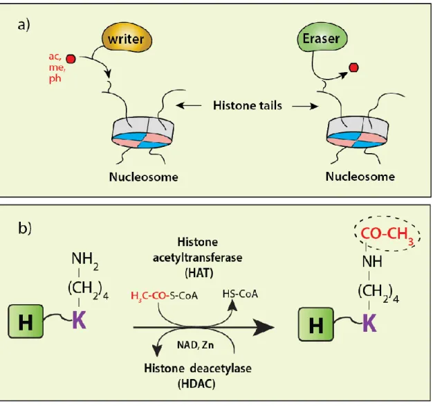
1.2.5. Histone modifiers - ‘Writers’ and ‘Erasers’
Histone modifications are generally mediated by two opposing groups of enzymes, namely ‘writers’ and ‘erasers’. The ‘writers’ are enzymes such as HATs, HMTs and kinases that modify histones by inserting respectively acetyl, methyl and phospho groups on specific residues. The ‘erasers’ are enzymes such as HDACs, histone demethylases (HDMTs) and phosphatases that reverse the activities of the ‘writers’ by removing the corresponding PTMs. Shown in Figure 1.10a is a schematic representation of the opposing activity of writers and erasers.
Figure 1.10 Schematic showing the reciprocal activities of ‘writers’ and ‘erasers’.
(a) The 'writing' and 'erasing' of histone PTMs. (c) The opposing activity of HATs and HDACs toward histone lysine residues. (ac-acetyl, me-methyl, ph-phospho). Figures prepared using Adobe® Illustrator® CS6, version 16.0.0.
1.2.5.1. HATs
HATs catalyze the transfer of an acetyl moiety from acetyl-CoA to the amino group of histone lysine residues (Fig. 1.10b). Their activity toward histones neutralizes the electrostatic interaction between the positively charged amino-group of lysines and the negatively charged phosphate group of the DNA. This process relaxes the nucleosomal DNA and increases its accessibility for transcription. HATs function in a variety of DNA-associated processes [105]. Several genome-wide analyses have shown that a large proportion of acetylation marks and the HATs that installs them are localized at promoter and enhancer regions of highly expressed genes [106-108]. They are recruited to these regions via interactions with specific activator proteins [67, 109, 110]. For example, in budding yeast, the HATs Gcn5 and Esa1 were found to be recruited to the promoter regions of active genes, leading to elevated levels of acetylation at Gcn5 target sites- H3K9 and K14, and Esa1 target sites- H4K5, H4K8, H4K12 and K16 [111]. These observations suggested a strong correlation between HATs activity and transcriptional activation. In addition to histones, HATs also catalyze the acetylation of several non-histone proteins [112, 113].
Based on their catalytic domains and sequence homology, eukaryotic HATs are grouped into three major families [114, 115]: i) Gcn5-related N-acetyltransferases (GNATs), named after the founding member Gcn5; ii) the MYST family HATs, named for the founding members of this family: MOZ, Ybf2, Sas2and Tip60; and iii) CBP/P300. Members of each family share high amino acid sequence similarity. For instance, CBP vs. P300, GCN5 vs. PCAF, MOZ vs. MORF, and TIP60 vs. MOF share respectively 87%, 89%, 87% and 65% sequence similarity [116]. The HATs Rtt109 (found in yeast) and HAT1 (found in most eukaryotes) also catalyze histone acetylation in specialized DNA-associated processes, but they share very low sequence homology with the three HAT families or with each other. TAF1, one of the components of the transcription factor TFIID, also exhibits acetyltransferase activity toward histones H3 and H4, and is required in transcriptional activation of genes. Table 1.2 shows the conservation of the different HATs between species.
Table 1.2 Conservation of HATs between species and phyla.
Adapted from reference [116], Caister Academic Press., 2011
1.2.5.2. HDACs
HDACs catalyze the deacetylation of histone lysine residues (Fig. 1.10b). Their activity is historically associated with transcriptional repression and heterochromatin formation because the removal of acetylation marks from histones often leads to condensed chromatin structure. A number of HDACs are present in association with multiple protein transcription corepressor complexes [117]. One example is the Sin3 corepressor which was found in complex with the HDAC Rpd3 in S. cerevisiae [118] and HDAC1/2 in humans [119]. Most HDAC complexes do not directly bind to nucleosomal DNA; instead they are targeted to specific genomic loci via interaction with other DNA-binding proteins. For example, in S. cerevisiae, the Sin3/Rpd3 complex interacts with the DNA-binding protein called Ume6 to target the deacetylation of histone H4K5ac and repress transcription of the adjacent genes [120, 121]. The function of HDACs is also tightly coupled with dynamic changes in the levels of histone methylation. In addition to hypoacetylation, silenced genes and heterochromatin domains are characterized by high levels of H3K9 di- and tri-methylation. It could be that the deacetylation of lysine residues by HDACs serves as a prerequisite for establishing repressive histone methylation marks.
Mammalian HDACs are classified into four major classes based on sequence similarity of their catalytic domain with the corresponding yeast HDACs [121-123]. These are: Class I: HDAC-1, -2, -3 and -8; Class-II: HDAC-4, -5, -6, -7, -9 and -10; Class III: sirtuins or SIRT 1–7; and Class IV: HDAC11. The Class I HDACs are mostly localized in the nucleus and are known to catalyze the deacetylation of histones and several non-histone proteins. In
comparison, the Class II HDACs are located in both the cytoplasm and nucleus. Their catalytic domain is located close to the C-terminus. Depending on the number of deacetylase domains they contain the Class II HDACs are divided in two subgroups, Class IIa and IIb; the latter has two deacetylase domains. HDACs Class I, II, and IV require the Zn2+ cofactor for their deacetylase activity. Class III HDACs are commonly referred to as sirtuins. Their activity requires the cofactor known as nicotinamide adenine dinucleotide (NAD+). The different classes of mammalian HDACs and their yeast counterpart are summarized in Table 1.3 below.
Table 1.3 Classification of mammalian HDAC isotypes. From reference [122] with permission from Elsevier.
1.2.5.3. HDAC inhibitors – Emerging drugs for cancer therapy
The balance between histone acetylation and deacetylation is crucial for the normal development of cells. Maintaining this balance requires continuous regulation of the interactions of HATs and HDACs with their target sites [124, 125]. Several gene deletion studies have shown that dysregulation of the acetylation equilibrium adversely affects numerous biological processes including DNA replication, transcription, and chromatin assembly [126-128]. While normal cells have dedicated control mechanisms that regulate the steady-state dynamic levels of acetylation of histones, as well as non-histone proteins, most cancer cells have been shown to possess altered global histone modifications [129-132]. For example, a global loss in H4K16ac was found to be a hallmark of human tumor cells [133]. The H4K16ac mark was previously shown to have a pivotal role in the regulation of the structure of chromatin and its functional interactions with chromatin assembly and remodeling factors [62]. The HAT known as MOF is responsible for acetylating H4K16 [134], whereas the HDAC named SirT1, the orthologue of budding yeast Sir2, is responsible for deacetylating H4K16ac [135].
Numerous studies have reported aberrations in HDAC activities in cancer cells. Increased expression of HDAC2 was found in human gastric cancer [136]. HDAC1 was shown to be upregulated in hormone refractory (HR) cancer and targets numerous transcription factors including the tumor suppressor protein p53 [137]. Increased recruitment of HDACs has also been associated with the pathogenesis of acute promyelocytic leukaemia [138]. These abnormalities in the recruitment and activities of HDACs in different types of cancers have made them strong therapeutic targets.
As such, a great deal of effort has been made in the discovery and development of new drugs that have the potential to reverse the detrimental effects of aberrant HDAC activities. These drugs are collectively known as HDAC inhibitors (HDACis). So far, numerous HDACis have been discovered and tested for antitumor activity in either cultured cells or model organisms. However, only a few were approved or are being investigated at various levels of clinical trials. The HDACis are divided into five groups depending on their chemical structure, which are, hydroxamates, cyclic tetrapeptides, benzamides, aliphatic acids, and electrophilic ketones (see Table 1.4) [139, 140]. The hydroxamates exhibit strong inhibitory activity toward HDAC I, and II, and also IV in some cases. SAHA (also known as Vorinostat) was the first to be FDA-approved for the treatment of cutaneous T-cell lymphoma [141]. It induces global hyperacetylation of histones [142], represses telomerase activity in human lung
adenocarcinoma cells [143], and arrests the growth of several types of cancer cells [144-146]. Romidepsin (FK228), inhibitor of HDAC I and II, was also FDA-approved for treatment of cutaneous T-cell lymphoma [147]. Most recently, the FDA approved a new broad-spectrum HDACi called Panobinostat (Farydak) for the treatment of patients with multiple myeloma, a type of blood cancer [148]. Panobinostat belongs to the hydroxamate HDACi group and has been shown to have activity against HDACs Class I, II, and IV [149]. Despite these successes, the three FDA-approved HDACs described above are all pan-HDAC inhibitors, meaning that they act indiscriminately on several HDAC classes [150, 151]. In addition, these drugs have been shown to have major side effects both in the clinical and experimental settings due to their inability to discriminate between normal and cancerous cells [152-155]. Thus, target-specific HDAC inhibitors are much needed for a more efficient cancer treatment.
Table 1.4 HDACis currently under clinical trial or FDA-approved.
Adapted from from reference [139]
![Figure 1.4 Three dimensional structural models of the histone octamer/NCP From references [43, 50] with permission from Nature Publishing Group and Oxford University Press, respectively](https://thumb-eu.123doks.com/thumbv2/123doknet/12250574.320008/25.892.162.735.724.1002/figure-dimensional-structural-references-permission-publishing-university-respectively.webp)