HAL Id: dumas-00998699
https://dumas.ccsd.cnrs.fr/dumas-00998699
Submitted on 2 Jun 2014
HAL is a multi-disciplinary open access archive for the deposit and dissemination of sci-entific research documents, whether they are pub-lished or not. The documents may come from teaching and research institutions in France or abroad, or from public or private research centers.
L’archive ouverte pluridisciplinaire HAL, est destinée au dépôt et à la diffusion de documents scientifiques de niveau recherche, publiés ou non, émanant des établissements d’enseignement et de recherche français ou étrangers, des laboratoires publics ou privés.
Indocyanine green fluorescence angiography for
preoperative perforator mapping and free flap
monitoring
Marine Hitier
To cite this version:
Marine Hitier. Indocyanine green fluorescence angiography for preoperative perforator mapping and free flap monitoring. Human health and pathology. 2014. �dumas-00998699�
AVERTISSEMENT
Ce document est le fruit d'un long travail approuvé par le
jury de soutenance et mis à disposition de l'ensemble de la
communauté universitaire élargie.
Il n’a pas été réévalué depuis la date de soutenance.
Il est soumis à la propriété intellectuelle de l'auteur. Ceci
implique une obligation de citation et de référencement
lors de l’utilisation de ce document.
D’autre part, toute contrefaçon, plagiat, reproduction illicite
encourt une poursuite pénale.
Contact au SICD1 de Grenoble :
thesebum@ujf-grenoble.fr
LIENS
LIENS
Code de la Propriété Intellectuelle. articles L 122. 4
UNIVERSITE JOSEPH FOURIER FACULTE DE MEDECINE DE GRENOBLE
Année 2014
Indocyanine green fluorescence angiography for
preoperative perforator mapping and free flap monitoring.
THESE
PRESENTEE POUR L’OBTENTION DU DOCTORAT EN MEDECINE DIPLÔME D’ETAT
Marine HITIER
Née le 19 avril 1986 à Paris
THESE SOUTENUE PUBLIQUEMENT A LA FACULTE DE MEDECINE DE GRENOBLE
Le 23 mai 2014
Devant le jury composé de:
Monsieur le Professeur François MOUTET, président du jury Monsieur le Professeur Georges BETTEGA, directeur de thèse Monsieur le Professeur Jean-Luc MAGNE
Madame le Docteur Alexandra FORLI Madame le Docteur Cynthia HAMOU
Professeur des Universités - Praticien Hospitalier 2013-2014
Occupation Actuelle Section.ss° CNU Discipline Universitaire
ALBALADEJO Pierre
Depuis 01/09/2008 48.01 Anesthésiologie-réanimation ARVIEUX-BARTHELEMY Catherine
Depuis de 01/09/2007 53.02 Chirurgie générale
BACONNIER Pierre
Depuis 01/10/1993 46.04 Biostat, informatique médicale et technologies de communication
BAGUET Jean-Philippe
Depuis 01/09/2006 51.02 Cardiologie
BALOSSO Jacques
Depuis 01/09/2003 47.02 Radiothérapie
BARRET Luc
Depuis 01/10/1992 46.03 Médecine légale et droit de la santé BAUDAIN Philippe
Depuis 01/05/1990 43.02 Radiologie et imagerie médicale BEANI Jean-Claude
Depuis 01/10/1992 50.03 Dermato-vénérologie
BENHAMOU Pierre Yves
Depuis 01/09/2003 54.04 Endocrinologie, diabète et maladies métaboliques BERGER François
Depuis 01/09/2001 44.03 Biologie cellulaire
BETTEGA Georges
Depuis 01/09/2013 55.03 Chirurgie maxillo-faciale et stomatologie BONAZ Bruno
Depuis 01/09/2001 52.01 Gastro-entérologie, hépatologie, addictologie BOSSON Jean-Luc
Depuis 01/01/2006 46.04 Biostat, informatique médicale et technologies de communication
BOUGEROL Thierry
Depuis 01/09/1998 49.03 Psychiatrie d'adultes
BOUILLET Laurence
Depuis 01/09/2012 53.01 Médecine interne
BRAMBILLA CHRISTIAN
Depuis 01/10/1989 51.01 Pneumologie
BRAMBILLA Elisabeth
Depuis 01/10/1993 42,03 Anatomie et cytologie pathologiques BRICAULT Ivan
CESBRON Jean-Yves
Depuis 01/09/1999 47.03 Immunologie
CHABARDES Stephan
Depuis 01/09/2010 49.02 Neurochirurgie
CHABRE Olivier
Depuis 01/09/2002 54.04 Endocrinologie, diabète et maladies métaboliques CHAFFANJON Philippe
Depuis 01/09/2005 42.01 Anatomie
CHAVANON Olivier
Depuis 01/09/2006 51.03 Chirurgie thoracique et cardio-vasculaire CHIQUET Christophe Depuis 01/09/2007 55.02 Ophtalmologie CHIROSSEL Jean-Paul Depuis 01/06/1990 42.01 Anatomie CINQUIN Philippe Depuis 01/10/1992 46.04
Biostat, informatique médicale et technologies de communication
COHEN Olivier
Depuis 01/09/2003 46.04
Biostat, informatique médicale et technologies de communication
COUTURIER Pascal
Depuis 01/09/2007 53.01 Gériatrie et biologie du veillissement CRACOWSKI Jean-Luc
Depuis 01/09/2009 48.03 Pharmacologie fondamentale, pharmacologie clinique
DE GAUDEMARIS Régis
Depuis 01/07/1992 46.02 Médecine et santé au travail DEBILLON Thierry
Depuis 01/09/2003 54.01 Pédiatrie
DEMATTEIS Maurice
Depuis 01/09/2010 48.03 Addictologie
DEMONGEOT Jacques
Depuis 01/10/1989 (46.04) Biostat, informatique médicale et technologies de communication
DESCOTES Jean-Luc
Depuis 01/09/1997 52.04 Urologie
ESTEVE François
Depuis 01/09/2004 43.01 Biophysique et médecine nucléaire FAGRET Daniel
Depuis 01/10/1992 43.01 Biophysique et médecine nucléaire FAUCHERON Jean-Luc
Depuis 01/09/2001 53.02 Chirurgie générale
FERRETTI Gilbert
Depuis 01/09/2000 43.02 Radiologie et imagerie médicale FEUERSTEIN Claude
Depuis 01/07/1992 44.02 Physiologie
FONTAINE Eric
Depuis 01/01/2006 44.04 Nutrition
FRANCOIS Patrice
Depuis 01/09/1998 46.01 Epidémiologie, économie de la santé et prévention GARBAN Frédéric
Depuis 01/09/2011 47.01 Hématologie, transfusion GAUDIN Philippe
GAVAZZI Gaetan
Depuis 01/09/2011 53.01 Gériatrie et biologie du veillissement GAY Emmanuel
Depuis 01/09/2004 49.02 Neurochirurgie
GODFRAIND Catherine
Depuis 01/09/2013 42.03 Anatomie et cytologie pathologiques GRIFFET Jacques
Depuis 01/03/2010 54.02 Chirurgie infantile
HALIMI Serge
Depuis 01/10/1990 44/04 Nutrition
HENNEBICQ Sylviane
Depuis 01/09/2012 54.05 Biologie et médecine du développement et de la reproduction
HOFFMANN Pascale Depuis 01/09/2012 54.03 Gynécologie-obstétrique HOMMEL Marc Depuis 01/09/1995 49.01 Neurologie JOUK Pierre-Simon Depuis 01/09/1997 54.05 Génétique JUVIN Robert Depuis 01/10/1993 50.01 Rhumatologie KAHANE Philippe Depuis 01/09/2007 44.02 Physiologie KRACK Paul Depuis 01/09/2003 49.01 Neurologie KRAINIK Alexandre
Depuis 01/09/2009 43.02 Radiologie et imagerie médicale LABARERE José
Depuis 01/09/2012 46.01 Epidémiologie, économie de la santé et prévention LANTUEJOUL Sylvie
Depuis 01/09/2008 42.03 Anatomie et cytologie pathologiques LECCIA Marie-Thérèse
Depuis 01/09/2002 50.03 Dermato-vénérologie
LEROUX Dominique
Depuis 01/09/1996 47.04 Génétique
LEROY Vincent
Depuis 01/09/2007 52.01 Gastro-entérologie, hépatologie, addictologie LETOUBLON Christian
Depuis 01/05/1992 53.02 Chirurgie générale
LEVY Patrick
Depuis 01/09/1997 44.02 Physiologie
MACHECOURT Jacques
Depuis 01/10/1989 51.02 Cardiologie
MORAND Patrice
Depuis 01/09/2007 45.01 Bactériologie-virologie MOREAU-GAUDRY Alexandre
Depuis 01/09/2013 46.04
Biostat, informatique médicale et technologies de communication MORO Elena Depuis 01/09/2012 49.01 Neurologie MORO-SIBILOT Denis Depuis 01/09/2005 51.01 Pneumologie MOUSSEAU Mireille Depuis 01/09/1994 47.02 Cancérologie MOUTET François
Depuis 01/10/1990 50.04 Chirurgie plastique, reconstructrice & esthétique, brulologie
PALOMBI Olivier Depuis 01/09/2011 42.01 Anatomie PARK Sophie Depuis 01/09/2013 47.01 Hématologie PASSAGIA Jean-Guy Depuis 01/09/1994 49.02 Neurochirurgie
PAYEN DE LA GARANDERIE Jean-François
Depuis 01/09/1996 48.01 Anesthésiologie-réanimation PELLOUX Hervé
Depuis 01/09/2001 45.02 Parasitologie et mycologie PEPIN Jean-Louis
Depuis 01/09/2004 44.02 Physiologie
PERENNOU Dominique
Depuis 01/04/2008 49.05 Médecine physique et de réadaptation PERNOD Gilles
Depuis 01/09/2007 51.04 Médecine vasculaire
PIOLAT Christian
Depuis 01/09/2009 54.02 Chirurgie infantile
PISON Christophe Depuis 01/09/1994 51.01 Pneumologie PLANTAZ Dominique Depuis 01/09/2003 54.01 Pédiatrie POLACK Benoît Depuis 01/09/1998 47.01 Hématologie POLOSAN Mircea
Depuis 01/09/2013 49.03 Psychiatrie d'adultes
PONS Jean-Claude Depuis 01/09/1998 54.03 Gynécologie-obstétrique RAMBEAUD Jean-Jacques Depuis 01/07/1991 52.04 Urologie REYT Emile Depuis 01/10/1992 55.01 Oto-rhyno-laryngologie RIGHINI Christian Depuis 01/09/2010 55.01 Oto-rhyno-laryngologie ROMANET J. Paul Depuis 01/10/1991 55.02 Ophtalmologie SARAGAGLIA Dominique
SCHMERBER Sébastien
Depuis 01/09/2005 55.01 Oto-rhyno-laryngologie
SCHWEBEL Carole
Depuis 01/09/2012 48.02 Réanimation, médecine d'urgence SCOLAN Virginie
Depuis 01/09/2013 46.03 Médecine légale et droit de la santé SERGENT Fabrice
Depuis 01/09/2011 54.03 Gynécologie-obstétrique
SESSA Carmine
Depuis 01/09/2005 51.04 Chirurgie vasculaire
STAHL Jean-Paul
Depuis 01/10/1992 45.03 Maladies infectieuses, maladies tropicales STANKE Françoise
Depuis 01/09/2011 48.03 Pharmacologie fondamentale TAMISIER Renaud
Depuis 01/09/2013 44.02 Physiologie
TIMSIT Jean-François 48.02 Réanimation
TONETTI Jérôme
01/09/2007 au 31/12/2010 50.02 Chirurgie orthopédique et traumatologie TOUSSAINT Bertrand
Depuis 01/09/2008 44.01 Biochimie et biologie moléculaire VANZETTO Gérald
Depuis 01/09/1999 51.02 Cardiologie
VUILLEZ Jean-Philippe
Depuis 01/09/1999 43.01 Biophysique et médecine nucléaire WEIL Georges
Depui 01/09/2011 46.01 Epidémiologie, économie de la santé et prévention ZAOUI Philippe
Depuis 01/09/2002 52.03 Néphrologie
ZARSKI Jean-Pierre
Depuis 01/09/1994 52.01 Gastro-entérologie, hépatologie, addictologie
Maître de Conférence des Universités - Praticien Hospitalier 2013-2014
Occupation Actuelle Section/ss° CNU Discipline universitaire APTEL Florent
Depuis 01/09/2012 55.02 Ophtalmologie
BOISSET Sandrine
Depuis 01/09/2012 45.01 Bactériologie, virologie BONNETERRE Vincent
Depuis 01/09/2011 46.02 Médecine et santé au travail BOTTARI Serge
Depuis 01/10/1993 44.03 Biologie cellulaire
BOUZAT Pierre
Depuis 01/09/2012 48.01 Anesthésiologie-réanimation BRENIER-PINCHART M.Pierre
Depuis 01/11/2001 45.02 Parasitologie et mycologie BRIOT Raphaël
Depuis 01/09/2009 48.04 Thérapeutique, médecine d'urgence CALLANAN-WILSON Mary
Depuis 01/09/2002 47.01 Hématologie, transfusion DECAENS Thomas
Depuis 01/09/2013 Hépato gastroentérologie
DERANSART Colin Depuis 01/09/2004 44.02 Physiologie DETANTE Olivier Depuis 01/09/2009 49.01 Neurologie DIETERICH Klaus Depuis 01/09/2012 47.04 Génétique DUMESTRE-PERARD Chantal Depuis 01/09/2004 47.03 Immunologie EYSSERIC Hélène
Depuis 01/10/2009 46.03 Médecine légale et droit de la santé FAURE Julien
Depuis 01/09/2008 44.01 Biochimie et biologie moléculaire GILLOIS Pierre
Depuis 01/09/2010 46.04
Biostat, informatique médicale et technologies de communication
GRAND Sylvie
Depuis 01/09/1995 43.02 Radiologie et imagerie édicale GUZUN Rita
Depuis 01/09/2012 44.04 Nutrition
LAPORTE François
Depuis 01/10/1991 44.01 Biochimie et biologie moléculaire LARDY Bernard
Depuis 01/09/2007 44.01 Biochimie et biologie moléculaire LARRAT Sylvie
Depuis 01/09/2009 45.01 Bactériologie, virologie LAUNOIS-ROLLINAT Sandrine
Depuis 01/09/2001 44.02 Physiologie
LONG Jean-Alexandre
MAIGNAN MaximeDepuis 01/09/2013 48.04 Médecine d'urgence MALLARET Marie-Reine
Depuis 01/08/1992 46.01 Epidémiologie, économie de la santé et prévention MARLU Raphaël
Depuis 01/09/2013 47.01 Hématologie
MAUBON Danièle
Depuis 01/09/2010 45.02 Parasitologie et mycologie MC LEER (FLORIN) Anne
Depuis 01/09/2011 42.02 Cytologie et histologie
MOUCHET Patrick
Depuis 01/10/1992 44.02 Physiologie
PACLET Marie-Hélène
Depuis 01/09/2007 44.01 Biochimie et biologie moléculaire PAYSANT François
Depuis 01/02/2008 46.03 Médecine légale et droit de la santé PELLETIER Laurent
Depuis 01/01/2006 44.03 Biologie cellulaire
RAY Pierre
Depuis 01/09/2003 47.04 Génétique
RIALLE Vincent
Depuis 01/09/2001 46.04
Biostat, informatique médicale et technologies de communication
ROUSTIT Matthieu
Depuis 01/08/1990 48.03 Pharmacologie clinique
ROUX-BUISSON Nathalie
Depuis 01/09/2012 44.01 Biochimie et génétique moléculaire SATRE Véronique
Depuis 01/09/2005 47.04 Génétique
SEIGNEURIN Arnaud
Depuis 01/09/2013 46.01 Epidémiologie, économie de la santé et prévention STASIA Marie-Josée
Depuis 01/08/1992 44.01 Biochimie et biologie moléculaire
REMERCIEMENTS
A Monsieur le Professeur François MOUTET, Qui m'a fait l'honneur de présider cette thèse.
Veuillez trouver ici l'expression de ma profonde et respectueuse reconnaissance pour m'avoir permis de réaliser ce travail et pour votre enseignement lors des semestres passés dans votre service.
A Monsieur le Professeur Georges BETTEGA, Pour avoir dirigé cette thèse.
Je te remercie de ta grande disponibilité et de ton aide tout au long de ce travail. Merci
également pour tous les conseils et enseignements que tu m'as prodigués lors de mon internat. C'est un plaisir de travailler à tes côtés.
A Monsieur le Professeur Jean Luc MAGNE,
Vous me faites l'honneur de juger ce travail, veuillez trouver ici l'expression de mon profond respect et de mes sincères remerciements pour votre grande gentillesse et pour votre
enseignement lors de mon semestre en chirurgie vasculaire.
A Madame le Docteur Alexandra FORLI,
Je te remercie pour ta gentillesse, ta bonne humeur, et ta grande disponibilité. Merci pour ces deux semestres passés à tes côtés qui m'ont beaucoup apportés, et tous ces bons moments passés au bloc en ta compagnie.
A Madame le Docteur Cynthia HAMOU,
Je te remercie pour ton précieux enseignement chirurgical, ta bonne humeur...et toutes nos séances de piscine acharnées !
A mes parents,
Je vous remercie pour tout l'amour que vous m'avez donné, pour l'éducation et le soutien que vous m'avez toujours apportés, et qui m'ont conduit jusqu'ici. Je vous aime.
A mes grands-parents Maurice et Yvette,
Merci pour votre grande affection, et pour ces séances de révision intensives qui resteront gravées dans ma mémoire !
A mes grands-parents Charles et Geneviève,
Merci pour votre grande tendresse, votre simplicité, et pour tous ces moments inoubliables passés à la Thuil !
A Clem et Arthur, ma soeur et mon frère chéris.
A Tata Claire, merci pour ta bonne humeur et pour toutes nos discussions, qui, je l'éspère, dureront encore longtemps.
A la famille Maupetit, merci pour votre énergie et pour les barbecues/piscine ! A Catherine et Jean-Philippe.
A Michaël, tu es attentionné, tu es drôle, tu es beau, tu es mon chemi, je t'aime. Merci d'être entré dans ma vie, merci pour tout l'amour que tu me donnes, merci pour ton soutien
A mes amies "grenobloises" (pas grenobloises dans l'âme :-)), ma poule Vendrell, Mathilde (et bébé ambroise bien sûr), ma coug Lorette, et Clo... à toutes nos soirées (arrosées ou plus calmes), nos brunchs, nos vacances (île d'Aix forever), nos (nombreuses) séances de
shopping... Merci pour cette belle amitié que vous me donnez, et pour votre soutien permanent. J'éspère que notre dispersion géographique future ne nous éloignera pas. Clothilde, travailler à tes côtés est un grand plaisir, et je suis ravie que tu fasses un peu de "rab" !
A tous mes copains grenoblois, Fab, Louf, Junet, Eric, Frandoni, Antoine, et tous ceux que j'oublie, et à tous les dijonnais expatriés à Grenoble...
A tous mes cointernes rencontrés lors de mon internat grenoblois; Roudet et sa mèche lyonnaise, Rudy, Belvisi, Nishal, Amélie, Albé, Aurélie, Jojo, Caro, Etienne, Boubou, Anne, Zumba... sans oublier Juliette, alias knacky ball; merci pour ta spontanéité et ta bonne humeur!
Un grand merci à tous ceux qui m'ont maudite pendant leurs gardes de CMF (surtout vers minuit) et qui ont permis la réalisation de ce travail; Juliette, Aurélie, Emma, Michael, Caro, Etienne, Junet, Rom, Zumba, Nicolas.
Au Dr. Michel Penin, merci de m'avoir donné le gôut pour la chirurgie plastique. Merci pour votre grande gentillesse et votre humour décapant.
Au Dr. Emmanuel Delay, merci pour votre riche enseignement et votre grande gentillesse à mon égard.
A Stéphanie et Pascal, merci de votre immense travail et de votre gande motivation qui m'ont permis de venir à bout de ce travail.
A toutes les équipes médicales et paramédicales avec lesquelles j'ai travaillé, en particulier les équipes de chirurgie de la main et de chirurgie plastique et maxillo-faciale du CHU de
CONTENTS
I. List of abbrevations . . . 14 II. Indocyanine green fluorescence angiography for mapping of fibular skin perforators . . . 15 - Abstract - Introduction - Methods - Results - Discussion - Conclusion - References
III. Indocyanine green fluorescence angiography for free flap monitoring: a prospective clinical trial . . . 30 - Abstract - Introduction - Methods - Results - Discussion - Conclusion - References IV. Conclusion . . . 50 The Hippocratic oath . . . .52
I. LIST OF ABBREVATIONS
CDS: Color Doppler ultraSonography
FA ICG: Indocyanine Green Fluorescence Angiography PPV: Positive Predictive Value
MRA: Magnetic Resonance Angiography CTA: Computed Tomography Angiography HHD: Hand-Helded Doppler
ICG: Indocyanine Green
DIEP: Deep Inferior Epigatric Perforator flap ALT: AnteroLateral Thigh flap
ITT: Intrinsic Transit Time FI: Fluorescence Intensity
II. Indocyanine green fluorescence angiography for
mapping of fibular skin perforators.
INDOCYANINE GREEN FLUORESCENCE ANGIOGRAPHY FOR MAPPING OF FIBULAR SKIN PERFORATORS.
M. Hitier, MD1, C. Hamou, MD1,3, C. Ochala, MD1, N. Gonnet, MD2, G. Bettega, PhD1,3
1 Plastic and maxillofacial surgery department - Hôpital A Michallon - BP217 - 38043
Grenoble cedex - France
2 Centre d'investigation clinique - Hôpital A Michallon - BP217 - 38043 Grenoble cedex -
France
3 INSERM-UJF U823 team 5 - Institut Albert Bonniot - 38706 Grenoble - France
Corresponding author: Marine Hitier, mhitier1@chu-grenoble.fr
Keywords: indocyanine green, fluorescence angiography, perforator, osteocutaneous fibular flap, colour Doppler ultrasonography
ABSTRACT
-Objective: We first evaluated the effectiveness of indocyanine green fluorescence angiography in preoperative mapping of perforators for fibular free flap. The second goal was to compare fluorescent angiographic findings with colour Doppler ultrasonographic mapping. - Background: Perforator flaps are very useful in reconstructive surgery, but the perforator anatomy is greatly variable. Preoperative perforator mapping is recommended, and several methods have been described. Laser-induced fluorescence of indocyanine green is a new method for the assessment of superficial vascularization tissue.
- Methods: 14 patients, scheduled for fibular free flap reconstruction, were included. The detection of skin perforators was performed by both colour Doppler ultrasonography (CDS) and fluorescence angiography (FA ICG), and then compared to surgical dissection findings. The sensitivity and positive predictive value (PPV) were calculated for each technique. The mean distance between anatomic and angiographic or ultrasonographic perforators was determined to evaluate the accuracy of FA ICG and compare the 2 mapping methods.
- Results: The sensitivity of both FA ICG and CDS was 75% for a precision of 3cm; the PPV was 67% for FA ICG and 48% for CDS. The sensitivity of FA ICG was 55% and 34.4% for CDS, for a precision of 1cm; the PPV of FA ICG was 50%, and that of CDS 22%. The mean distance between anatomical and FA ICG findings was 0.86 +/- 0.97 cm and significantly different from CDS, 1.45 +/- 0.88 cm (p≤0.001).
- Conclusion: FA ICG is a useful technique for the detection of fibular skin perforators and is more accurate than CDS.
INTRODUCTION
The fibular free flap is widely used for mandibular reconstruction1. It is commonly used as a
composite osteocutaneous free flap as described by Chen and Yan2 in 1983. The skin paddle
is vascularized by one or more septo- or musculo-cutaneous perforators, steming from the fibular artery. Perforator flaps have become increasingly popular due to their high plasticity and the low donor site sequellae, but the variability of perforator anatomy requires an accurate preoperative mapping of perforators3. Several methods have been described to identify
perforators in fibular flaps, such as magnetic resonance angiography4,5 (MRA), computed
tomographic angiography6,7 (CTA), colour duplex ultrasonography8,9 (CDS), or hand-held
Doppler9 (HDD). All these techniques have advantages and drawbacks, but none of them is
the "gold standard". FA ICG is a new method for assessing the superficial vascularization of tissues. Indocyanine green (ICG) is a fluorescent dye and has been used for more than 40 years. It allows measuring cardiac output10 and liver function11. It has also been used in
neurosurgery12 and in ophthalmology for choroid vessel imaging13. More recently, FA ICG
has been used to assess intestinal viability in a laparoscopic mesenteric ischemia model14. It
has been used in plastic surgery to assess flap perfusion15,16 and intraoperative flap
monitoring17.
The comparison of FA ICG with CDS for fibular flap mapping had never been made. This clinical trial had two goals:
- assessing the effectiveness of FA ICG in preoperative perforator mapping for fibular free flap.
METHODS
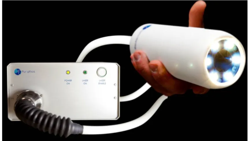
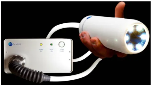
Material
We used the Fluobeam device (Fluoptics, Grenoble, France). It included a 780-nm laser light source and a camera equipped with blocking optical filters collecting only ICG-induced fluorescence (Fig. 1). The working field was set at 20 centimeters. The CCD camera recorded the ICG fluorescence induced by the near-infrared light, after the dye solution had flowed through the vessels of the illuminated field. Images were acquired with an exposure time of 100 ms. They were displayed in real time on a monitor and immediately assessed with the Fluobeam v1.47® software. They were saved on the computer hard disk for future review and analysis.
Figure 1. Fluobeam device
Monopeak Indocyanine Green (Infracyanine®, SERB laboratory, Paris, France) is a
water-soluble dye, which absorbs light in the near-infrared spectral range with a peak at 805 nm, and emits fluorescence at 835 nm. ICG has a short plasma half-life of 3.4 +/- 0.7 minutes. It is completely cleared from blood by the liver and excreted into the bile. ICG was dissolved in a
5% dextrose solution and administered intravenously. We used a dose of 0.025 mg/kg/injection.
ICG completely binds to plasma proteins after intravenous injection and is exclusively distributed in the intravascular space18. This makes it a suitable tracer for vessel perfusion.
The adverse effects of ICG are anaphylactic shock, hypotension, dyspnea, nausea, exanthema, and pruritus. All these adverse effects are rare19.
Patients
We included 14 patients in our prospective clinical trial, from July 2012 to June 2013. The inclusion criteria were reconstruction by a fibular free flap in patients more than 18 years of age. The exclusion criteria were pregnancy and a history of allergic reaction to ICG.
Methods
The Grenoble Medical Ethics Committee approved the study in February 2012.
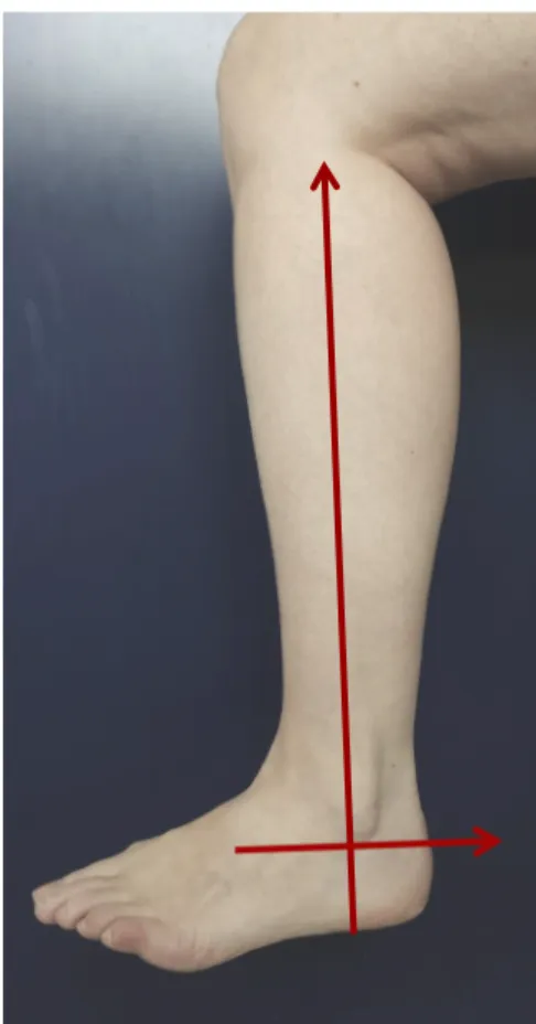
Oral and written information was given to the patient, before obtaining his informed consent. CDS was performed by a radiologist before the hospitalisation, and mapping results were recorded in the patient’s file. Perforator vessels were localized in a specific landmark defined by the posterior edge of the fibula and the perpendicular line passing on the tip of the lateral malleolus (Fig.2). The Infracyanine® injection was performed by the surgeon the day before surgery, on a supine patient. A near-infrared camera was placed opposite the lateral side of the leg, and skin perforators that appeared on monitor were marked on the leg. The perforator coordinates were measured in the same landmark and recorded in the patient's file. Skin
Figure 2. Landmarks of perforator mapping
Only perforators located between +8 cm and +30 cm from the lateral malleolus tip were considered, since the skin paddle is rarely raised outside these limits. Perforators localized by angiography or ultrasonography were matched with perforators localized surgically, when included within a range of 3 cm in the specific landmark. The sensitivity and positive predictive value were calculated for each technique. We were unable to define the number of true-negatives (perforators seen neither on FA ICG or CDS nor intra-operatively); hence it was impossible to calculate the specificity or the negative predictive value. We measured the difference between anatomic perforator localization and angiographic or ultrasonographic perforator localization to evaluate FA ICG accuracy and compare the 2 mapping methods.
Statistical analysis
Statistical analyses were performed with the Stata 11.0 software (StataCorp, College Station, Texas, USA). Qualitative parameters were expressed as number and percentage. MacNemar’s test was used to compare matched qualitative parameters. Quantitative parameters were expressed as means and standard deviation and compared with Student’s T test or Mann-Whitney’s U test if necessary. A P value of less than 0.05 was considered to be statistically significant.
RESULTS
Patients
We included 9 male and 5 female patients. Their mean age was 53.6 year (35-76) and their mean BMI was 25.6 kg/m2 (19,6-32). 13 of the 14 flaps were fibular flaps for mandibular
reconstruction, and 1 for maxillary reconstruction.
Complications
We did not record any complication or adverse effect after ICG injection.
Mapping of perforators
Patients 12 and 16 were excluded; patient 12 had anterior tibial artery perforators mapping instead of fibular due to a technical error, and the laser was turned off during recording for patient 16. We compared only FA ICG and anatomical findings for patient 10 because his
A mean of 3 perforators per patient (1-5) was observed intra-operatively, with a total of 36 perforators localized surgically (32 without patient 10). 40 were localized with FA ICG (Fig.3), and 50 with CDS.
Figure 3. Perforator mapping in patient 10. Perforators are indicated by arrows.
Results are shown in Table 1 and 2.
The sensitivity was 75% for FA ICG and CDS when within a distance of 3 cm of surgical findings, and the PPV was 67.5% for FA ICG and 48% for CDS.
The sensitivity was 55% for FA ICG and 34.4% for CDS when within a distance of 1 cm of surgical findings, and the PPV was 50% for FA ICG and 22% for CDS.
MacNemar test was performed to compare the 2 mapping techniques, after excluding patient 10. There was a significant difference between the 2 methods when within a distance of 1 cm of surgical findings (p=0.02).
FA ICG localization was accurate in a range of 0.86 +/- 0.97 cm, compared to anatomical localization. It was significantly more accurate than CDS localization 1.45 +/- 0.88 cm (p≤0.001).
Distance (cm) Number of success Sensitivity (%) IC 95% PPV (%) 3 27/36 75.0 60.8-89.1 67.5 2 24/36 66.7 51.3-82.1 60 1 20/36 55.6 39.3-71.8 50
Table 1. Comparison between FA ICG and surgical findings.
Distance (cm) Number of success Sensitivity (%) IC 95% PPV (%) 3 24/32 75.0 60-90 48 2 20/32 62.5 39.1-73.4 40 1 11/32 34.4 17.9-50.8 22
Table 2. Comparison between CDS and surgical findings.
DISCUSSION
FA ICG is a useful and safe preoperative method for fibular perforator mapping. It is more accurate than CDS.
fibula osteocutaneous flap surgery. Preoperative mapping allows adapting the flap precisely to the reconstructive requirements.
The most common technique, among the various methods described for perforator
localization, remains the hand-held Doppler (HDD). It is inexpensive, portable, non-invasive, and easy to of use. However, HDD is known to be too sensitive with a low specificity9. CDS
is non-invasive also, and offers more information on the perforator course. Hallock9 reported
a high sensitivity of CDS in detecting vessels around 0.2 mm in diameter, but its specificity was low. But CDS is highly operator dependent21 and can only be performed by a skilled
radiologist who is familiar with perforator flap surgery. CTA4,5 and MRA6,7 are newer
techniques that provide accurate details on the calibre and the course of perforators. But they have some drawbacks: exposure to radiation, need of an iodine contrast agent for CTA, and a high cost for MRA.
FA ICG is a new technique for the assessment of tissue superficial vascularization. Its use for fibular skin perforator mapping and comparison with CDS had not been described yet in a clinical trial. FA ICG has been performed on pigs and compared with x-ray angiography22. A
100% correlation was found and confirmed by perforator surgical dissection, but there was no information on the accuracy of the various measures. Azuma et al.23 evaluated the usefulness
of FA ICG for identifying perforators on 14 perforator-based island flaps, but without measurements. A mapping of perforator vessels by combining multi-detector-row computed tomography, Doppler flowmetry, and FA ICG was performed on 50 perforator flaps by Onoda et al.24. They found a PPV of 80% for CDS and of 84% for FA ICG. The sensitivity was
100% for CDS, and 76% for FA ICG. FA ICG was more accurate than CDS in perforator detection, in our study. Our FA ICG sensitivity and PPV were lower, but in Obana's study, there was no information on the degree of precision used to measure the difference between angiographic and sonographic findings with surgical findings. We noticed that the quality of
FA ICG mapping improved during the study in this preliminary trial. A learning curve is required to properly master this new perforator mapping technique, and our results will probably be improved as the study goes on.
FA ICG presents several advantages; the time needed to perform mapping ranges around 15 minutes, and this technique is non-invasive. The fluorescent dye we used did not contain iodine, minimizing the risk of adverse effects. The major advantage of FA ICG is that it is performed by the surgeon who has the knowledge of the local anatomy and of the perforator flap surgery. Moreover, the system portability could be useful for intraoperative re-evaluation, if unexpected issues are encountered.
We had to face 2 problems during the study. First the probe was not large enough to assess the whole leg in 1 time, so that we needed to sweep the probe during mapping. The second
problem was the high speed of fluorescent signal rise, that made simultaneous mapping of all perforators difficult. We developed a fluorescent ruler that was placed on the leg during video acquisition (Fig. 4), so that we could re-view the video on the monitor screen and measure precisely all the perforators.
CONCLUSION
FA ICG is a useful and safe preoperative method for fibular perforator mapping, but it
requires a learning curve to properly master the technique. Our results should be confirmed by the ongoing clinical trial and by other studies.
ACKNOWLEDGMENTS
The authors thank Gravit for financing the study, and the Grenoble University Hospital Clinical Research Department for supervising the clinical trial.
REFERENCES
1. Hidalgo DA. Fibula free flap: a new method of mandible reconstruction. Plast Reconstr Surg 1989;84:71-9.
2. Chen ZW, Yan W. The study and clinical application of the osteocutaneous flap of fibula. Microsurg 1983;4:11-6.
3. Dancey A, Blondeel PN. Technical tips for safe perforator vessel dissection applicable to all perforator flaps. Clin Plast Surg 2010;37:593-606.
4. Fukaya E, Saloner D, Leon P, et al. Magnetic resonance angiography to evaluate septocutaneous perforators in free fibula flap transfer. J Plast Reconstr Aesthet Surg 2010;63;1099-1104.
5. Akashi M, Nomura T, Sakakibara S, et al. Preoperative MR angiography for free fibula osteocutaneous flap transfer. Microsurgery 2013;33:454-9.
6. Garvey PB, Chang EI, Selber JC, et al. A prospective study of preoperative computed tomographic angiographic mapping of free fibula osteocutaneous flaps for head and neck reconstruction. Plast Reconstr Surg 2012;130:541-49.
7. Satoh T, Kimata Y, Hasegawa K, et al. The utility of multi-detector-row computed
tomography angiography for evaluating perforators of fibular osteocutaneous flaps. J Reconstr Microsurg 2011;27:29-36.
8. Futran ND, Stack BCJ, Zaccardi MJ. Preoperative color flow Doppler imaging for fibula free tissue transfers. Ann Vasc Surg 1998;12:445-50.
9. Hallock GG. Doppler sonography and color duplex imaging for planning a perforator flap. Clin Plast Surg 2003;30:347-57.
10. Fox IJ, Wood EH. Applications of dilution curves recorded from the right side of the heart or venous circulation with the aid of a new indicator dye. Proc Staff Meet Mayo Clin
1957;32:541-50.
11. Levesque E, Hoti E, Azoulay D, et al. Non-invasive ICG-clearance: a useful tool for the management of hepatic artery thrombosis following liver transplantation. Clin Transplant 2011;25:297-301.
12. Killory BD, Nakaji P, Gonzales LF, et al. Prospective evaluation of surgical microscope-integrated intraoperative near-infrared indocyanine green angiography during arteriovenous malformation surgery. Neurosurg 2009;65:456-62.
13. Ciardella AP, Prall FR, Borodoker N, et al. Imaging techniques for posterior uveitis. Cur Opin Ophtalmol 2004;15:519-30.
Real-16. Holm C, Mayr M, Höfter E, et al. Intraoperative evaluation of skin-flap viability using laser-induced fluorescence of indocyanine green. Br J Plast Surg 2002;55:635-44.
17. Holm C, Dornseifer U, Sturtz G, et al. The intrinsic transit time of free microvascular flaps: clinical and prognostic implications. Microsurg 2010;30:91-6.
18. Muckle TJ. Plasma proteins binding of indocyanine green. Biochem Med 1976;15:17-21. 19. Obana A, Miki T, Hayashi K, et al. Survey of complications of indocyanine green
angiography in Japan. Am J Ophthalmol 1994;118:749-53.
20. Miyamoto S, Sakuraba M, Nagamatsu S. Inadvertent injury of critical perforator vessels during perforator flap surgery. J Reconstr Surg 2012;28:95-8.
21. Blondeel PN, Beyens G, Verhaeghe R, et al. Doppler flowmetry in the planning of perforator flaps. Br J Plast Surg 1998;51:202-9.
22. Matsui A, Lee BT, Winer JH, et al. Image-guided perforator flap design using invisible near-infrared light and validation with x-ray angiography. Ann Plast Surg 2009;63:327-30. 23. Azuma R, Morimoto Y, Masumoto K, et al. Detection of skin perforators by indocyanine green fluorescence nearly infrared angiography. Plast Reconstr Surg 2008;122:1062-67. 24. Onoda S, Azumi S, Hasegawa K, et al. Preoperative identification of perforator vessels by combining MDCT, Doppler flowmetry, and ICG fluorescent angiography. Microsurg
III. Indocyanine green fluorescence angiography for
free flap monitoring: a prospective clinical trial.
INDOCYANINE GREEN FLUORESCENCE ANGIOGRAPHY FOR FREE FLAP MONITORING: A PROSPECTIVE CLINICAL TRIAL.
M. Hitier, MD1, C. Hamou, MD1,4, N. Gonnet, MD2, C. Righini, MD,PhD3,4, G. Bettega,
MD,PhD1,4
1 Plastic and maxillofacial surgery department - Hôpital A Michallon - BP217 - 38043
Grenoble cedex - France
2 Centre d'investigation clinique - Hôpital A Michallon - BP217 - 38043 Grenoble cedex -
France
3 ENT department - Hôpital A Michallon - BP217 - 38043 Grenoble cedex - France
4 INSERM-UJF U823 team 5 - Institut Albert Bonniot - 38706 Grenoble - France
Corresponding author: Marine Hitier, mhitier1@chu-grenoble.fr
ABSTRACT
-Objective: We evaluated the effectiveness and the tolerance of repeated fluorescent green indocyanine angiography in perforator flap monitoring, and determined the intraoperative predictive values of flap vitality.
- Background: The free flap failure rate has been significantly reduced, but free flap loss still occurs and remains a costly disaster. Repeated clinical examinations are commonly used for flap monitoring, but they can be unreliable because of their subjectivity. Laser-induced fluorescence of indocyanine green is a new method for assessing tissue perfusion.
- Methods: 20 patients undergoing microsurgical reconstruction were monitored by indocyanine green fluorescence angiography, intraoperatively, and during 4 days after surgery, with 18 injections. Monitoring was made by clinical examination, and then compared to angiographic findings.
- Results: The vascular complication rate was 15% (3/20) with 2 cases of venous thrombosis and one case of partial necrosis of the flap skin paddle. Both cases of venous thrombosis were salvaged by secondary surgery. There was no total flap loss. ICG angiography allowed detecting each intra and postoperative complication, earlier than clinical examination. The mean peroperative intensity of fluorescence was significantly lower in flaps with vascular complications (23.8 GL/ms; p=0.0087). The postoperative slope (p=0.0239) and amplitude (p=0.0335) of the fluorescent signal were both significantly lower than for uncomplicated flaps, before surgical revision. These 2 parameters came back to normal values after secondary surgery (p=0.1840; p=0.2879). There was no adverse effect of ICG despite the repeated injections.
INTRODUCTION
Microsurgery has become more routinely used; consequently, the free flap failure rate has been significantly reduced. The free flap survival rate currently ranges between 94% and 99%1. Free flap loss still occurs and remains a costly disaster, despite these improved success
rates. Free flap monitoring is of the utmost importance since the salvage rate is inversely related to the delay between the onset of ischemia and its clinical assessment. Repeated clinical examination, based on skin color, flap temperature, and capillary refill time, is commonly used for flap monitoring but is very dependent on the surgeon's experience. Various instrumental methods have been described, such as microdialysis2, implantable
Doppler probe3-4, color duplex ultrasound5, near-infrared spectroscopy6, and laser Doppler
flowmetry7. None of them are routinely used or considered as a "gold standard" method.
Fluorescence angiography with indocyanine green (FA ICG) is a new method for evaluating tissue perfusion. Indocyanine green (ICG), a fluorescent dye, has been used for more than 40 years. It allows measuring cardiac output8 and liver function9. It is also used in neurosurgery10
and in ophthalmology for imaging of the choroid vessels11. More recently, FA ICG has been
used to assess intestinal viability in a laparoscopic mesentery ischemia model12.
FA ICG has been used for intraoperative assessment of pedicled flap perfusion13, and for pre-
and intraoperative assessment of free flap perfusion14, in plastic surgery15.
The use of iterative ICG injections in postoperative flap monitoring has never been reported. Our prospective clinical trial had 2 goals:
-‐ assessing the effectiveness and the tolerance of FA ICG in free flap monitoring.
-‐ determining the intraoperative fluorescence parameters correlated with postoperative vascular flap complications.
METHODS Material
We used the Fluobeam device (Fluoptics, Grenoble, France). It included a 780-nm laser light source and a camera equipped with blocking optical filters collecting only ICG-induced fluorescence (Fig. 1). The working field was set at 20 centimeters. The CCD camera recorded the ICG fluorescence induced by the near-infrared light, after the dye solution had flowed through the vessels of the observed field. Images were acquired every 200ms with an exposure time of 100ms. They were displayed in real time on a monitor, and immediately assessed with the Fluobeam v1.47® software. They were saved on the computer hard disk for future review and analysis with the ImageJ® software.
levels. It is completely cleared from blood by the liver and excreted into the bile. ICG was dissolved in a 5% dextrose solution and administered intravenously. We used a dose of 0.025 mg/kg per injection, without exceeding a total dose of 0.5 mg/kg per patient.
ICG completely binds to plasma proteins after intravenous injection, and is exclusively distributed in the intravascular space16. This makes it a suitable tracer for vessel perfusion.
The adverse effects of ICG are anaphylactic shock, hypotension, dyspnea, nausea, exanthema and pruritus. All these adverse effects are rare17. Infracyanine® does not contain iodine,
contrary to other ICG preparations such as ICG-Pulsion®.
Patients
We included 20 patients in our prospective clinical trial, from July 2012 to June 2013. The inclusion criteria were reconstruction by one of these 3 free flaps: fibular flap, deep inferior epigastric (DIEP) flap, and anterolateral thigh (ALT) flap, in patients more than 18 years of age. The exclusion criteria were: pregnancy, and a history of allergic reaction to ICG.
Methods
The Grenoble Medical Ethics Committee approved the study in February 2012. Oral and written information was given to the patient, before obtaining his informed consent. Each patient received iterative injections of 0.025 mg/kg Infracyanine®. The first injection was
performed intra-operatively just after flap anastomosis to recipient vessels. Then the same dose was injected every 6 hours for 4 days, beginning 2 hours after the end of the surgery. Each patient received a total of 18 injections. Skin paddle color, temperature, and capillary refill time were assessed at ICG injection. The surgeon's clinical assessment data was blinded by the engineers analyzing fluorescence data and conversely. Clinical assessment was the only element allowing to diagnose complications, and to require surgical revision if needed.
All postoperative complications were recorded. FA ICG allowed for the per-operative assessment of anastomosis permeability and potential leak, and flap illumination. The measured intraoperative parameters were intrinsic transit time (ITT) and skin flap fluorescence intensity (FI). The ITT is the time needed for fluorescent dye to circulate from arterial to venous anastomosis.
The postoperative parameters were the slope of the fluorescence signal rise and the amplitude of fluorescence intensity (Fig. 2).
The data was recorded with the Fluobeam v1.47® software and analyzed secondarily by engineers.
Statistical analysis
Statistical analyses were performed with the Stata 11.0 software (StataCorp, College Station, Texas, USA). The results were expressed as means and range. The nonparametric Mann-Whitney test was used to compare data between uncomplicated and complicated flaps. A P value of less than 0.05 was considered to be statistically significant.
RESULTS Patients
We included 13 male and 7 female patients. Their mean age was 51.9 years (34-76) and their mean BMI was 25.2 kg/m2 (19.6-32). 14 of the 20 flaps were fibular flaps for mandibular
reconstruction; 4 were ALT flaps for other facial or trunk reconstruction; and 2 were DIEP flaps for breast reconstruction.
Microsurgery
The mean surgery time was 8 hours (5-10.75). FA ICG added about 5 minutes to surgery time. 1 artery was anastomosed for every flap, 1 vein in 11 flaps, and 2 in 9 flaps. An arterial and venous graft was necessary in one case.
Complications
We recorded 4 complications: 2 cases of partial necrosis of fibular flap skin paddles and 2 cases of venous thrombosis of the fibular flap pedicle. One case of partial necrosis occurred 6 days after surgery (Patient 6) the other, 14 days after surgery (patient 4). The other part of the flap (bone and muscle) was well vascularized in both cases. Necrosis was due to a local infection, caused by necrosis of the tip of the tongue stump for patient 6. Surgical debridement was necessary in both cases. The clinical diagnosis of venous thrombosis was
made at D1 H18 for patient 15, and at D1 H20 for patient 14. Revision surgery confirmed the diagnosis in both cases, justifying performing a new venous anastomosis. Both flaps were salvaged and healed secondarily. Finally, there were 3 complications of vascular origin, in patients 4, 14, and 15.
We did not observe any complication or adverse effect after iterative ICG injections.
Intraoperative results
The intraoperative clinical assessment (skin paddle coloration with capillary refill, skin paddle edge bleeding, patency test of the anastomoses) was normal for 12 patients including patients 14 and 15. It was not for 4 patients: 2 of theses were patients 4 and 6.
Patients 5, 8, 17, and 20 were excluded from the intraoperative evaluation of fluorescence angiography data because of technical problems. The laser was turned off during intraoperative measures for patients 8, 17, and 20, and the ICG was dissolved in a saline solution for patient 5.
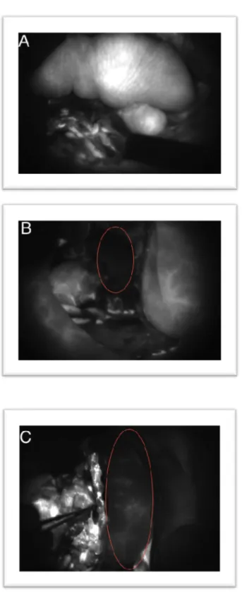
FA ICG confirmed that all anastomoses were patent without leaking. The flap skin paddle was entirely fluorescent in 9 patients (Fig 3A.) and there was no complication at follow up. The flap was not fluorescent at all and appeared dark compared to surrounding tissues in patients 4, 14, and 15 (the 3 patients with vascular complications) (Fig 3B). The flaps of the 4 remaining patients were considered partially fluorescent, as shown in figure 3C. One of these was patient 6 who presented with a local infection.
The ITT was not measured for patients 3 and 6 because of technical problems. The mean ITT was 12.8 seconds (5.7-44.6). The highest ITT was measured in patient 14. The ITT was
The mean FI was 89.6 grey levels per millisecond (GL/ms) (20.6-253.5). It was significantly lower in patients 4, 14, and 15 (23.8 GL/ms; p=0.0087).
Postoperative monitoring
Patient 4 was excluded from postoperative results because he refused injections after the second day. The typical curve of fluorescence dynamic of a well-vascularized flap is shown in Figure 2.
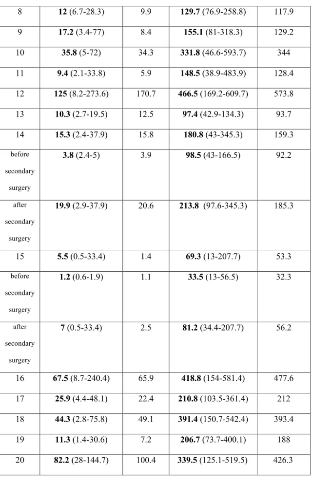
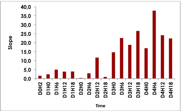
The postoperative fluorescence data is listed in Table 1. The mean value of the slope was 31 (0.5-273.6). The slope was calculated before and after surgical revision for patients 14 and 15. The mean slope before revision was 3.8 (2.4-5) for patient 14 and 1.2 (0.6-1.9) for patient 15. These values were significantly lower than those of uncomplicated flaps (p = 0.0239). The mean slope after revision was 19.9 (2.9-37.9) for patient 14, and 7 (0.5-33.4) for patient 15 with no longer any significant difference with uncomplicated flaps (p = 0.1840). The evolution of the slope for patient 14 is shown in figure 4.
The mean amplitude of the fluorescence signal was 227.8 (13-609.7). The amplitude before revision was 58.7 for patient 14 and 44.1 for patient 15; these values were significantly different from those of uncomplicated flaps (p = 0.0335). They increased after revision to 147.5 for patient 14 and 93.8 for patient 15, with no longer any difference with uncomplicated flaps (p = 0.2879).
SLOPE AMPLITUDE
Patient Mean (Min- Max) Median Mean (Min- Max) Median
1 19.3 (3.5-61.9) 12.5 285.8 (134.1-423.6) 300.5
8 12 (6.7-28.3) 9.9 129.7 (76.9-258.8) 117.9 9 17.2 (3.4-77) 8.4 155.1 (81-318.3) 129.2 10 35.8 (5-72) 34.3 331.8 (46.6-593.7) 344 11 9.4 (2.1-33.8) 5.9 148.5 (38.9-483.9) 128.4 12 125 (8.2-273.6) 170.7 466.5 (169.2-609.7) 573.8 13 10.3 (2.7-19.5) 12.5 97.4 (42.9-134.3) 93.7 14 15.3 (2.4-37.9) 15.8 180.8 (43-345.3) 159.3 before secondary surgery 3.8 (2.4-5) 3.9 98.5 (43-166.5) 92.2 after secondary surgery 19.9 (2.9-37.9) 20.6 213.8 (97.6-345.3) 185.3 15 5.5 (0.5-33.4) 1.4 69.3 (13-207.7) 53.3 before secondary surgery 1.2 (0.6-1.9) 1.1 33.5 (13-56.5) 32.3 after secondary surgery 7 (0.5-33.4) 2.5 81.2 (34.4-207.7) 56.2 16 67.5 (8.7-240.4) 65.9 418.8 (154-581.4) 477.6 17 25.9 (4.4-48.1) 22.4 210.8 (103.5-361.4) 212 18 44.3 (2.8-75.8) 49.1 391.4 (150.7-542.4) 393.4 19 11.3 (1.4-30.6) 7.2 206.7 (73.7-400.1) 188 20 82.2 (28-144.7) 100.4 339.5 (125.1-519.5) 426.3
Figure 4. Evolution of slope for patient 14, over time.
The slope and amplitude increased linearly postoperatively, day after day (Table 2).
Slope Signal amplitude
Mean SD Med Min Max Mean SD Med Min Max J0 12 18.2 5.5 0.1 72.9 178.5 150.5 151.2 3.8 603.9 J1 15.5 16.2 9.8 0.6 100.9 185 105.3 162.6 13 456.9 J2 27 33.9 15.6 0.5 180 215.2 144.4 170.2 37 573.8 J3 34.5 42.9 17.7 0.8 177.3 241.9 161.7 186.2 34.4 600.8 J4 46.8 62.6 20.8 2.7 273.6 267.1 177.7 192.8 46.6 609.7 0,0 5,0 10,0 15,0 20,0 25,0 30,0 35,0 40,0 D0H2 D1H0 D1H6 D1H12 D1H18 D2H0 D2H6 D2H12 D2H18 D3H0 D3H6 D3H12 D3H18 D4H0 D4H6 D4H12 D4H18 Sl o p e Time
The slope was steep and the amplitude was high in well vascularized flaps. The slope was low with a small amplitude for patient 15 at D0H2, before revision (Fig. 5A). The curve aspect came back to normal after revision (Figure 5B).
Figure A
Figure B
Figure 5. Patient 15 postoperative monitoring. A. Before secondary surgery, at D0H2. B. After venous thrombosis has been treated, at D4HO.
0 20 40 60 80 100 120 140 0 20 40 60 80 Fl uo re sce nc e in te nsi ty (gr ey level s/m s) Time (s) D0H2 0 20 40 60 80 100 120 140 0 20 40 60 80 Fl uo re sce nc e in te nsi ty (gr ey level s/m s) Time (s) D4H0
Vascular complications were observed on clinical assessment 18 hours after surgery for patient 14, and 20 hours after surgery for patient 15. Their fluorescence curves were abnormal before that, only 2 hours after surgery. The clinical assessment was inconclusive for 2 patients (2 and 12), but the FA ICG data was normal, and no complication was observed.
DISCUSSION
Fluorescence angiography with iterative ICG injections at a low dose is a safe and reliable technique for free flap monitoring. The intraoperative fluorescence of the flap after vessel anastomosis seems to be a predictor of postoperative flap survival. The relevant fluorescence parameters for postoperative flap perfusion assessment in our patients were the slope and the amplitude of the fluorescence signal.
Free flap intraoperative monitoring is routinely performed by clinical assessment. The color of the skin paddle, temperature, and edge bleeding depend greatly on the patient's hemodynamics. Clinical assessment is more difficult in the oral cavity, because the flap always seems pale compared to surrounding tissues. FA ICG provides intraoperative data that may allow predicting flap vascular complications. The flap illumination and the mean flap fluorescence intensity in our patients were parameters that predicted flap survival. These were abnormal in the 3 patients with vascular complications; the clinical assessment of the flap was inconclusive for only one of these 3 patients. Holm et al.18 evaluated FA ICG for free flap
intraoperative monitoring on 20 patients. They also predicted vascular complications according to the flap illumination. The same team in 201019, using microscope-integrated ICG
and its specificity 78%. The authors mentioned that multiple factors could influence the ITT: patient hemodynamics, body temperature, flap ischemia time, type of anastomosis, and venous pressure. We were not able to confirm these results. Only 1 out of 3 patients with vascular complication had a long ITT, the 2 others had very short ITTs. One of them (patient 15) might have developed venous thrombosis a few hours after surgery while perfusion was normal when ITT was measured. Muscle and bone were well vascularized after skin paddle removal for patient 4; hence, we thought that necrosis was due to a thrombosis of the sole cutaneous perforator, while the main pedicle (fibular artery and vein) was functional. We did not have enough data to conclude on the relevance and predictability of ITT for the flap outcome.
All the clinical and angiographic parameters must be taken into account to decide on intraoperative revision of anastomoses.
It is crucial to implement free flap monitoring during the first days after microsurgery, and a rapid revision after detection of flap compromise, as demonstrated in large series1,20.
The slope, fluorescence signal amplitude, and fluorescence curve profile were reliable parameters in our patients for the detection of vascular complications. These were clearly abnormal for the 2 patients who presented with venous thrombosis. Unfortunately patient 4 left the trial before the end of fluorescence monitoring. We noticed a high inter and intra-patient variability of the absolute values of slope and fluorescence signal amplitude, which was also reported by other authors 18,21. These values were patient hemodynamic dependent.
The fluorescence curve profile was a more reliable indicator of vascular complications, which reflected the trend of perfusion values. Matsui et al.22 quantified perforator flap perfusion with
ICG angiography in an experimental study on 38 pigs. The fluorescence curve did not rise when they occluded the flap artery, but stayed around the baseline level without any inflow peak. The curve remained continuously elevated after the inflow peak in case of venous
occlusion. The fluorescence curve was lower than those of uncomplicated flaps for our 2 patients presenting with venous thrombosis and remained close to the baseline level; this curve was different from the one reported by Matsui, which was obtained after 6 hours of venous occlusion. Our curve was obtained at a later stage of complication. We suggested that venous congestion could have led to a secondary decrease of arterial blood flow.
FA ICG allowed detecting all the flap complications earlier than clinical assessment, 16 hours before the diagnosis. The first postoperative angiography (2 hours after the end of surgery) was abnormal for both patients, while the clinical aspect of the flap was normal. FA ICG gave some objective arguments of vascular complications. This could help physicians lacking experienced to decide on pedicle revision. The effectiveness of FA ICG was confirmed by the rapid normalization of fluorescence parameters after flap revision.
Monitoring with iterative ICG injections had never been reported. ICG is supposed to be safe, but its safety had been assessed after a single injection only. Obana et al17 reported an adverse
effect rate of 0.34% after performing 3,774 ICG angiographies. 10 (0.26%) were mild reactions (nausea, exanthema, pruritus), 1 (0.026%) was pain along the injected vein, and 2 (0.05%) were hypotension. He had injected between 25 and 75 mg, at least 10 times more than the dose we used (0.025 mg/kg). Hope-Ross et al.23 reported similar results after
injecting between 25 and 50 mg. We did not observe any adverse effect due to ICG, after performing 18 injections per patient, as required to complete the protocol.
One of the drawbacks of FA ICG is that it does not permit continuous monitoring. Nevertheless it allowed to make an early diagnosis of vascular complications. The procedure could be improved by using a long time circulating agent and a single injection.
flap with a large device can be problematic24. A new miniaturized device called Fluostick™
has now become available for this type of clinical application.
CONCLUSION
FA ICG proved its safety and effectiveness in our study for the detection of flap vascular complications, and this earlier than clinical assessment. The number of patients and complications was too small in this preliminary clinical trial, to provide statistical proof of FA ICG effectiveness. New clinical studies should be performed to confirm our results. The next step will be developing a software giving the surgeon access to numerical data during acquisition of FA ICG images.
ACKNOWLEDGMENTS
The authors thank Gravit for financing the study, and the Grenoble University Hospital Clinical Research Department for supervising the clinical trial.
REFERENCES
1. Bui DT, Cordeiro PG, Hu QY, et al. Free flap reexploration: indications, treatment, and outcomes in 1193 free flaps. Plast Reconstr Surg 2007;119:2092-2100.
2. Hara H, M. Mihara, Lida T, et al. Blood glucose measurement for flap monitoring to salvage flaps from venous thrombosis. J Plast Reconstr Aesthet Surg 2012;65:616-19. 3. Swartz WM, Jones NF, Cherup L, et al. Direct monitoring of microvascular anastomoses with the 20-MHz ultrasonic Doppler probe: An experimental and clinical study. Plast Reconstr Surg. 1994;93:152-63.
4. Clert V, Guédon C, Cristofari JP, et al. Le micro-doppler implantable dans la surveillance des lambeaux microanastomosés en chirurgie reconstructrice maxillo-faciale. Ann Chir Plast Esthet 2013;58:82-8.
5. Few JW, Corral CJ, Fine NA, et al. Monitoring buried head and neck free flaps with high-resolution color-duplex ultrasound. Plast Reconstr Surg 2001;108:709-12.
6. Repez A, Oroszy D, Arnez ZM. Continuous postoperative monitoring of cutaneous free flaps using near infrared spectroscopy. J Plast Reconstr Aesthet Surg 2008;61:71-7. 7. Yuen JC, Feng Z. Monitoring free flaps using the laser Doppler flowmeter: Five-year experience. Plast Reconstr Surg 2000;105:55-61.
8. Fox IJ, Wood EH. Applications of dilution curves recorded from the right side of the heart or venous circulation with the aid of a new indicator dye. Proc Staff Meet Mayo Clin
1957;32:541-50.
9. Levesque E, Hoti E, Azoulay D, et al. Non-invasive ICG-clearance: a usefool tool for the management of hepatic artery thrombosis following liver transplantation. Clin Transplant 2011;25:297-301.
10. Killory BD, Nakaji P, Gonzales LF, et al. Prospective evaluation of surgical microscope-integrated intraoperative near-infrared indocyanine green angiography during arteriovenous malformation surgery. Neurosurg 2009;65:456-62.
11. Ciardella AP, Prall FR, Borodoker N, et al. Imaging techniques for posterior uveitis. Cur Opin Ophtalmol 2004;15:519-30.
12. Diana M, Noll E, Diemunsch P, et al. Enhanced-Reality Video Fluorescence: A Real-Time Assessment of Intestinal Viability. Ann Surg 2014;259:700-7.
14. Pestana IA, Coan B, Erdmann D, et al. Early experience with fluorescent angiography in free-tissue transfer reconstruction. Plast Reconstr Surg 2009;123:1239-44.
15. Liu DZ, Mathes DW, Zenn MR, et al. The application of indocyanine green fluorescence angiography in plastic surgery. J Reconstr Microsurg 2011; 27:355-64.
16. Muckle TJ. Plasma proteins binding of indocyanine green. Biochem Med 1976;15:17-21. 17. Obana A, Miki T, Hayashi K, et al. Survey of complications of indocyanine green
angiography in Japan. Am J Ophthalmol 1994;118:749-53.
18. Holm C, Tegeler J, Mayr M, et al. Monitoring free flaps using laser-induced fluorescence of indocyanine green: a preliminary experience. Microsurg 2002;22:278-87.
19. Holm C, Dornseifer U, Sturts G, et al. The intrinsic transit time of free microvascular flaps: clinical and prognostic implications. Microsurg 2010;30:91-6.
20. Genden EM, Rinaldo A, Suarez C, et al. Complications of free flap transfers for head and neck reconstruction following cancer resection. Oral Oncol 2004;40:979-84.
21. Betz CS, Zhorzel S, Schachenmayr H, et al. Endoscopic measurements of free-flap perfusion in the head and neck region using red-excited indocyanine green: preliminary results. J Plast Reconstr Aesth Surg 2009;62:1602-8.
22. Matsui A, Lee BT, Winer JH, et al. Quantitative assessment of perfusion and vascular compromise in perforator flaps using a near-infrared fluorescence-guided imaging system. Plast Reconstr Surg 2009;124:451-60.
23. Hope-Ross M, Yannuzzi LA, Gragoudas ES, et al. Adverse reactions due to indocyanine green. Ophthalmology 1994;101:529-33.
24. Betz CS, Zhorzel S, Schachenmayr H, et al. Endoscopic assessment of free flap perfusion in the upper aerodigestive tract using indocyanine green: a pilot study. J Plast Reconstr Aesthet Surg 2013;66:667-74.
Indocyanine green fluorescence angiography for preoperative perforator mapping and free flap monitoring.
Thèse soutenue par Marine HITIER CONCLUSION
Free perforator flaps have become increasingly popular due to their high plasticity and the low donor site sequellae, but the variability of perforator anatomy requires an accurate preoperative mapping of perforators. Several methods have been described to identify perforators in fibular flaps, but none of them is the "gold standard". Then, free flap monitoring is of the utmost importance since the salvage rate is inversely related to the delay between the onset of ischemia and its clinical assessment. Repeated clinical examination is commonly used for flap monitoring but is very dependent on the surgeon's experience. Laser-induced fluorescence of indocyanine green (FA ICG) is a new method for the assessment of superficial vascularization tissue.
We first evaluated the effectiveness of FA ICG in preoperative mapping of perforators for fibular free flap and compared FA ICG findings with colour doppler ultrasonography (CDS) mapping. Then we evaluated the effectiveness and the tolerance of repeated FA ICG in perforator flap monitoring, and determined the intraoperative predictive values of flap vitality. 20 patients undergoing microsurgical reconstruction were included in our prospective clinical trial. Only the 14 fibular flaps were included for perforator mapping. We used the Fluobeam device (Fluoptics, Grenoble) and Monopeak Indocyanine Green (Infracyanine®, SERB laboratory, Paris) was injected. Each patient received a total of 19 injections of 0.025 mg/kg Infracyanine®. The first injection was performed by the surgeon the day before surgery, on a supine patient. The detection of skin perforators was performed by both CDS and FA ICG, and then compared to surgical dissection findings. The sensitivity and positive predictive value (PPV) were calculated for each technique. The mean distance between anatomic and angiographic or ultrasonographic perforators was determined to evaluate the accuracy of FA ICG and compare the 2 mapping methods. The second injection was performed intra-operatively just after flap anastomosis to recipient vessels. Then the same dose was injected every 6 hours for 4 days. Monitoring was made by clinical examination, and then compared to angiographic findings. All postoperative complications were recorded. FA ICG allowed for
The sensitivity of FA ICG was 55% for a precision of 1cm, and positive predictive value was 50%. FA ICG was more accurate than CDS (p≤0.001). ICG angiography allowed detecting each intra and postoperative complication, earlier than clinical examination. The mean peroperative FI was significantly lower in flaps with vascular complications (p=0.0087). The postoperative slope (p=0.0239) and amplitude (p=0.0335) of the fluorescent signal were both significantly lower than for uncomplicated flaps, before surgical revision. These 2 parameters came back to normal values after secondary surgery (p=0.1840; p=0.2879). There was no adverse effect of ICG despite the repeated injections.
FA ICG is a safe and useful technique for the mapping of fibular skin perforators, and for the detection of free flap vascular complications. Our results should be confirmed by the ongoing clinical trial and by other studies.
SERMENT D’HIPPOCRATE
E n présence des M aîtres de cette F aculté, de m es chers condisc iples et devant l’effigie d’H IP P O C R A T E ,
Je prom ets et je jure d’être fidèle aux lois de l’honneur et de la probité dans l’exercice de la M édecine.
Je donnerai m es soins gratuitem ent à l’indigent et n’exigerai jam ais un salaire au dessus de m on travail. Je ne participerai à aucun partage clandestin d’honoraires.
A dm is dans l’intim ité des m aisons, m es yeux n’y verront pas ce qui s’y passe ; m a langue taira les secrets qui m e seront confiés et m on état ne servira pas à corrom pre les m œ urs, ni à favoriser le crim e.
Je ne perm ettrai pas que des considérations de religion, de nation, de race, de parti ou de classe sociale viennent s’interposer entre m on devoir et m on patient.
Je garderai le respect absolu de la vie hum aine.