HAL Id: dumas-01806285
https://dumas.ccsd.cnrs.fr/dumas-01806285
Submitted on 1 Jun 2018HAL is a multi-disciplinary open access archive for the deposit and dissemination of sci-entific research documents, whether they are pub-lished or not. The documents may come from teaching and research institutions in France or abroad, or from public or private research centers.
L’archive ouverte pluridisciplinaire HAL, est destinée au dépôt et à la diffusion de documents scientifiques de niveau recherche, publiés ou non, émanant des établissements d’enseignement et de recherche français ou étrangers, des laboratoires publics ou privés.
Cryoablation percutanée des tumeurs desmoides et des
sarcomes des tissus mous
Marie-Annaïg Pey
To cite this version:
Marie-Annaïg Pey. Cryoablation percutanée des tumeurs desmoides et des sarcomes des tissus mous. Médecine humaine et pathologie. 2018. �dumas-01806285�
Université de Bordeaux
U.F.R DES SCIENCES MEDICALES
Année 2018 Thèse n°3037
Thèse pour l’obtention du
DIPLOME D’ETAT DE DOCTEUR EN MEDECINE
Présentée et soutenue publiquement le 13 Avril 2018 par
Marie-Annaïg PEY
Née le 07 Août 1988 à Marseille
Discipline: Radiodiagnostic et imagerie médicale
CRYOABLATION PERCUTANEE
DES TUMEURS DESMOIDES ET DES SARCOMES
DES TISSUS MOUS
Directeur de thèse:
Monsieur le Docteur Xavier BUY
Rapporteur de thèse:
Monsieur le Professeur Antoine FEYDY
Membres du jury
Monsieur le Professeur Hervé TRILLAUD, Président
Monsieur le Professeur Jean-Michel COINDRE, Juge
Monsieur le Professeur Antoine ITALIANO, Juge
Madame le Docteur Michèle KIND, Juge
Monsieur le Docteur Eberhard STOECKLE, Juge
1 REMERCIEMENTS
A notre président du jury,
Monsieur le Professeur Hervé TRILLAUD,
Je vous remercie de nous faire l’honneur de présider ce jury de thèse.
Je vous remercie tout particulièrement pour la confiance que vous me témoignez en me choissant comme assistante dans votre service. L’apprentissage de la radiologie interventionnelle à vos côtés est pour moi un honneur.
A mon maître et directeur de thèse, Monsieur le Docteur Xavier BUY,
Tu as conforté mon goût pour la radiologie interventionnelle. Ta grande expertise et ton talent ont été des aides précieuses au cours de mon stage à l’institut Bergonié. Merci de m’avoir suggéré le thème de ce travail et de m’avoir fait confiance pour sa rédaction. Ce sujet m’a passionnée et fait énormément progresser dans la rédaction d’article scientifique. Merci enfin pour le modèle de radiologue que tu es, passionné, consciencieux, énergique et surtout très humain !
A notre rapporteur,
Monsieur le Docteur Antoine FEYDY,
Je vous remercie de m’avoir fait l’honneur d’accepter de juger ce travail et d’y apporter votre regard d’expert en imagerie ostéo-articulaire. Recevez toute ma reconnaissance et ma gratitude.
Aux membres du jury,
Monsieur le Professeur Jean-Michel COINDRE,
Je vous remercie d’avoir accepté de juger ce travail. Avoir dans son jury un si grand nom de l’anatomopathologie est un honneur. Veuillez trouver ici l’expression de mes sentiments les plus respectueux.
Monsieur le Professeur Antoine ITALIANO,
Vous m’honorez en ayant accepté de juger ce travail. Votre implication scientifique et votre expertise oncologique dans le domaine des sarcomes sont reconnues de tous. Je vous prie de croire en l’expression de mes sentiments les plus respectueux.
Madame le Docteur Michèle KIND,
Je suis très heureuse que vous ayez accepté de juger mon travail. Le jury n’aurait pas pu se constituer sans votre expertise en imagerie des tumeurs osseuses et des tissus mous. Merci pour votre disponibilité à toute épreuve pour les internes en vacation de scanner !
Monsieur le Docteur Eberhard STOECKLE,
Le jury n’aurait pas pu se constituer sans votre expertise chirurgicale dans le domaine des tumeurs des tissus mous. Je vous remercie d’avoir accepté de juger ce travail et vus prie de croire en l’expression de mes sentiments respectueux.
2
Aux Professeurs qui m’ont enseigné l’imagerie, Monsieur Nicolas Grenier, Monsieur Jean-François Chateil, Monsieur Vincent Dousset, Monsieur François Laurent, merci pour votre bienveillance et votre passion pour la formation des plus jeunes.
A mes chefs de radiologie interventionnelle: Bruno Lapuyade, c’est avec toi que j’ai fait mes premiers drainages et mes premières biopsies hépatiques. Merci tu es un modèle de calme, de patience et d’humanité avec les patients. Je vais beaucoup apprendre à tes côtés et dans la sérenité !
A Yann Lebras, la force tranquille et l’expertise du vasculaire ! Merci pour ta gentillesse et ta patience. Tu es un passionné passionnant qui ne compte pas ses heures. Comme dirait Gino, ton karma est bon, tu auras ta place au paradis !
A Jean Palussière, à qui je voue une profonde admiration pour son travail, sa modestie, son expertise, sa grande classe et surtout son humanité. J’ai beaucoup appris en radiofréquence pulmonaire à vos côtés. Merci aussi pour votre ouverture d’esprit, notre goût commun pour le voyage, partagé pendant les longues sessions de “grillage de nodules” !
A mes chefs de radiologie diagnostique et de rhumatologie: A Pierre Bessou et Sophie Misoonier, un super souvenir de mon stage en radiopédiatrie à vos côtés, merci encore. A la super équipe de rhumatologie de Pau, Laurence Lequen, Alexandre Balageas, Xavier Delbrel, Fanny Sanguinet. A Muriel Durieux…une chef en or, merci pour tout ce que tu m’as transmis. A Sandrine Molinier, merci pour ta gentillesse et ta patience à répéter des milliers de fois “hypersignaux flair de la substance blanche…” ! A Amaury Mouries et Elodie Hénot, mes 2 super CCA de HL, merci pour toutes ces vacations passées ensemble. A Delphine Gaye, merci pour ta bienveillance et ta confiance. J’ai hate de travailler avec toi. A FPP, merci de m’avoir laissé autant d’autonomie en radio interventionnelle ! A Olivier Corneloup…toujours present, toujours prêt à accepter 40 scanners et à brancarder les patients. Merci pour nos longues conversations sur mille voyages… A Gael Dournes, merci pour ta sympathie ! A tous les chefs de la Clinique du sport: Marie-Hélène, Maryse, Lionel, Nico, Eric, Sylvain, Benjamin, Philippe, Alain et Pascal. J’ai passé un de mes meilleurs semestres à vos côtés, j’ai appris, j’ai ri et… j’ai bien mange !!! Vous avez l’envie de transmettre, l’expertise de l’ostéo et la sympathie des bons copains. Merci pour tout.
A tous les manipulateurs avec qui on s’est bien marré au bloc : Thomas, Vincent, Gino et toute l’équipe de Pellegrin. A tous ceux de HL: Jojo, Caro, Rémi…j’arrive.
A mes co-internes : PF et ETCHARP, mes chouchous, merci de me faire rire autant…vous êtes des supers copains.
A la fine équipe de co-internes des USN : PF, Audrey et Antoine.
A la fine équipe de nanas de Bergo : Julia, Caroline, Carole et Nina, super stage avec vous les filles. Spéciale dédicace à Julia qui a apporté sa pierre à la thèse…t’es trop une chic fille ! Merci.
A l’équipe de neuroradio : PF, Nina et Hubert. A Adrian et Clément, dommage qu’on ait pas fait de stage ensemble!
A Jésus, Marie-Charlotte et Joseph…super co-internes!! MCH, j’ai trop hate qu’on soit co-chef… A tous les co-internes de chez Grenier: Julie, François, Louise et Florent. Traverser les topos du mercredi matin ensemble, c’est une épreuve!
A ma cointerne de Pau, Mélanie, merci de ton soutien pendant ces 6 mois, tu es adorable, faut qu’on se revoit.
A mes futurs co-chefs: MCH, Paul Schuster et Sophie Borgol.
A mes amis de médecine: ceux de Marseille, la 107, Nina, Maelle, Gilles et Antho, merci pour tous les moments partagés ensemble, les photos censurées, c’est avec vous! Spéciale dédicace à Nina, si tu pars dans une autre ville, je te suis encore ;)
Celles et ceux rencontrés à Bordeaux: Solène, Céline, Ines, Margaux, Carole et Sophie, vous êtes des chics filles, je vous adore, à quand notre premier semi?! A vos moitiés: Louis, Romain, Alex, Adri, et
3
Doudou. Merci aussi à Chloé, Thomas, Dimitri, Raph, Marie Leclère et tous les gens de l’internat du 1er semestre.
Aux amis du bon vieux temps de la coloc : Nina, Sylvain, Fx, Mélo, Emeline, Simon, j’avais l’impression d’être dans Friends parfois!
A Soumaya, à notre amitié, merci pour ton optimisme sans faille.
A Emma et Alex, les amis de Félix, devenus mes amis…et aussi à Guillaume, Amandine et Ellie! Au club des 5 + 1 du lycée : Hélène, Agathe, Chloé, Ariane et Margaux. Depuis 15 ans, rien n’a changé! Aux fidèles amis du lycée : Arnaud, Julia, Benjamin et Laure.
Aux amis d’enfance : Florent, Marie, Aurèle et Madame Grima.
A ma belle-famille : Louisette, Jef, Mélanie, Julien, Pauline, Suzanne, Thomas, Mireille, Gilles, leurs enfants, Micheline. Merci pour votre accueil toujours chaleureux, votre sens de la fête et votre sérénité.
Aux cousins : Marie et Stéphane, merci pour votre bonne humeur et votre gentillesse. Martin, Vinciane, Pauline et Mathilde, sacrés souvenirs pendant les vacances chez Papi et Mamie. A Christophe, passionnée par son métier.
A Annie, ma tante d’adoption !
A ma Mamie Renée, toujours prête à refaire le monde sur la table de la cuisine, merci pour ton écoute.
A mon Papi Louis, merci pour ta grande curiosité, ton énergie, ta bonne humeur, ta volonté de toujours faire plaisir. On veut tous vieillir comme toi.
Aux grands-parents partis trop tôt : Papi Alex et Mamie Monique.
A Pauline Salsa (du démon), on se connaît depuis le “biligou” et ça c’est plus fort que tout…
A mes soeurs chéries, vous êtes des modèles que j’ai toujours admirés et suivis. Vous serez toujours “mes grandes”soeurs, même à 70 ans! A Patrice et Swan, mes supers beaux-frères. A mes adorables neveux: Colin, Edouard, Sacha et le futur Victor!
A mes parents, merci de tout coeur d’avoir toujours cru en moi, encouragée et soutenue pendant toutes ces années.
Et enfin, à Félix, merci pour ton soutien inconditionnel, ta confiance en moi et ta patience au quotidien! J’admire ta personnalité, ta bienveillance et ta grande curiosité pour tout ce qui t’entoure... Chaque jour passé ensemble est un Bonheur.
4
TABLE DES MATIERES
I. Article 1 – Percutaneous cryoablation of desmoid tumors ... 6
1. Introduction ... 7
2. Patients, methods, and statistical analysis ... 8
3. Results ... 13
4. Discussion ... 19
II. Article 2 – Percutaneous cryoablation of soft-tissue sarcomas : early phase results ... 26
1. Introduction ... 27
2. Patients, methods and statistical analysis ... 28
3. Results ... 33
4. Discussion ... 40
III. Annexe 1 – Généralités sur la cryoablation ... 47
1. Historique ... 47
2. Principes techniques... 48
3. Aspects pratiques ... 53
4. Applications de la cryoablation percutanée (CP) en pathologies musculosquelettiques (28) ... 58
5. Avantages / inconvénients par rapport aux autres techniques de thermoablations (40,41) ... 61
6. Suivi en imagerie après cryoablation de tumeurs des tissus mous... 62
IV. Annexe 2 – Généralités sur les tumeurs desmoides ... 68
1. Définition et historique ... 68 2. Epidémiologie ... 68 3. Anatomopathologie ... 70 4. Etiologie ... 71 5. Aspect en imagerie ... 71 6. Biopsie ... 77 7. Traitement ... 77 8. Suivi en imagerie ... 80
V. Annexe 3 – Généralités sur les sarcomes des tissus mous ... 86
1. Epidémiologie ... 86 2. Anatomopathologie ... 87 3. Mode de présentation ... 88 4. Etiologie ... 88 5. Diagnostic ... 89 6. Prise en charge ... 94
5
TABLE DES FIGURES ET TABLEAUX
Article 1
Figure 1 Tumeur desmoide………9
Figure 2 Tumeur desmoide ... 100
Figure 3 Tumeur desmoide ... 122
Tableau 1 Caractéristiques des patients………14
Tableau 2 Caractéristiques des tumeurs………..15
Tableau 3 Paramètres techniques……….16
Figure 4 Kaplan-Meier plot of time-to-next treatment. ... 18
Article 2 Figure 1 Métastase de la cuisse traitée par cryoablation……….29
Figure 2 Métastase paravertébrale traitée par cryoablation……….30
Tableau 1 Caractéristiques des patients………..33
Tableau 2 Caractéristiques des tumeurs………..34
Figure 3 Rechute locale d'un liposarcome rétropéritonéal traitée par cryoablation……….32
Figure 4 Time-to-local progression (TTLP)………37
Tableau 3 Caractéristiques des patients métastatiques……….37
Figure 5 Overall time-to-next treatment………..39
Figure 6 Overall survival (OS)………40
Annexe 1 Figure 1 Effet Joule-Thomson appliqué à la cryoablation……….49
Figure 2 Lésions cellulaires directes liées à la congélation………..50
Figure 3 Températures au sein de la boule de glace………52
Figure 4 Echographie peropératoire………53
Figure 5 Cryoablation d'un hémangiome musculaire paravertébral……….54
Figure 6 Cryoablation de 2 tumeurs rénales………..55
Figure 7 Présentations des formes et dimensions des boules de glace ... 566
Figure 8 Carbodissection ... 57
Figure 9 Kyste osseux anévrysmal de L4 ... 59
Figure 10 Hémangiome intramusculaire paravertébral ... 60
Figure 11 Aspect IRM après cryoablation percutanée d’une tumeur desmoide ... 63
Figure 12 Aspect IRM post cryoablation d’une tumeur desmoide ... 63
Figure 13 Aspect IRM d’une progression tumorale locale. ... 64
Annexe 2 Figure 1 Tumeurs desmoides mutifocales………69
Figure 2 Aspect macroscopique et microscopique d'une tumeur desmoide………..70
Figure 3 Différentes présentations morphologiques et de signal………72
Figure 4 Compartiment de développement des tumeurs desmoides………..73
Figure 5 Bandes fibreuses ... 73
Figure 6 Tumeur desmoide mésentérique………..74
Figure 7 Aspect scannograpique……….75
Figure 8 Aspect échographique………76
Figure 9 Aspect échographique………76
Figure 10 Exemple de réponse tumorale………..81
Annexe 3 Figure 1 Taux d’incidence par âge, en fonction du type de sarcome. ... 86
Figure 2 Distribution des anomalies moléculaires en fonction des âges ... 87
Figure 3 Exemple de lesions bénignes………91
Figure 4 Hémangiome intramusculaire………..91
Figure 5 Synovialosarcome……….92
Figure 6 Critères de malignité dans les tumeurs myxoïdes……….92
Figure 7 Liposarcome rétropéritonéal………93
Figure 8 Métastases intramammaire traitée par cryoablation………..100
6
I. Article 1
PERCUTANEOUS CRYOABLATION OF
EXTRAABDOMINAL DESMOID TUMORS
AbstractAim: to present our five-year results of percutaneous cryoablation (PCA) in the management of
progressive and/or symptomatic extraabdominal desmoid tumors (DT).
Materials and methods: Data of patients treated by PCA for DT between December 2012 and July 2017
in our tertiary cancer center were retrospectively collected. Extraabdominal DT associated to Familial Adenomatous Polyposis (FAP) and pediatric cases were not excluded. Primary endpoint was time-to-next treatment (TNT) defined as time between first PCA procedure and modification of treatment strategy. Secondary endpoints were local tumor progression (LTP) rate, median number of PCA courses by patient and complications according to CTCAE v5.0. Variables associated with LTP rate were assessed in univariate analysis among age, sex, anatomic site, size, FAP, primary/recurrent status and mutations.
Results: thirty-four patients were included, 29 women, 5 men, with a median age of 35,5. Six patients
were affected by FAP. Median tumor size was 77mm (range 25-183). Tumors were located in trunk wall (n=26), limbs (n=4), girdle (n=3), neck (n=1). Tumors were primitive (n=26), recurrent (n=7), new (n=1). Median follow-up was 20 months (range 5-54). The 1 and 3 year estimated next-treatment rates were 4% (95%CI 0-9) and 23% (95%CI 0-48). Only 3 patients underwent modification strategy for untreatable progression at 7, 25 and 35 months. LTP rate after first PCA was 32%. Median number of PCA course by patient was 1 (range 1-4). Complications were grade 2 (n=2), grade 3 (n=1), grade 4 (n=1). Only recurrent status was associated with a higher risk of LTP after PCA (p=0.008).
Conclusion: PCA is a safe and effective treatment option for extraabdominal DT with a prolonged
local tumor control.
Abbreviations DT: Desmoid Tumor
PCA: Percutaneous cryoablation FAP: Familial Adenomatous Polyposis AZ: Ablation zone
LTP: Local tumor progression TNT: time-to-next treatment
7 1. Introduction
Desmoid tumor (DT) also named aggressive fibromatosis, is a rare monoclonal fibroblastic proliferation considered as a benign tumor. The estimated incidence is 5 to 6 per million of inhabitants (1–3) . According to World Health Classification, DT is predisposed to relapse but not to metastasize (4). It arises along fascia of muscles and may infiltrate muscles of several compartments. Histopathologic diagnosis is difficult, because DT can mimics other soft tissue tumors such as low-grade sarcomas, myofibroblastic benign lesions and reactive processes. Detection of mutations in the exon 3 of the β-catenin gene (CTNNB1) is a specific diagnostic tool (5,6). Above 90% of sporadic DT harbors those mutations leading to nuclear accumulation of β-catenin protein.
Two types of phenotype are distinguished. The first and most frequent is sporadic DT developed in young adults, women in 70%, with a peak-age ranged between 35 and 40 years (7). DT typically arises in abdominal wall, limb and trunk wall. The second type is DT associated to familial adenomatous polyposis (FAP) in Gardner’s syndrome. DT is associated to FAP in 7 to 15 % (8–10). Compared to the overall population, FAP patients have a more than 800-fold risk of developing DT. DT in FAP patients are more frequently intraabdominal tumors infiltrating mesentery, but may also arise in extraabdominal areas.
For many years standard treatment was radical surgery leading to high and variables rates of local recurrence, around 30 % according to largest and recent studies (7,11,12). Surgical margins do not seem to be associated to local recurrence although age, tumor size and tumor site have significant impact on PFS (7,12). Over the last decade, several studies advocating a conservative approach were published given that observing a first-line wait-and-see period leads to stable disease in 30%, regression in 30% and progression in 30% (7,13–15). Surgery is no long regarded as the cornerstone of treatment for primary DT (1,16). European society of medical oncology has recently edited guidelines for the management of primary and recurrent DT (17,18). After an initial wait-and-see period, surgery, systemic treatment, radiotherapy and isolated limb perfusion may be treatment options in cases of tumor progression or in symptomatic patients. Systemic treatment includes non-steroidal-anti-inflammatory drugs, hormonal therapy, chemotherapy and tyrosine-kinase inhibitors with variables degrees of efficacy and side effects.
Following the developments of percutaneous thermal ablations, interventional radiology increasingly takes part in the treatment strategy of many cancers. For soft-tissue tumor ablation, percutaneous cryoablation (PCA) is the treatment of choice as it offers several advantages compared with heat ablation techniques: visualization of the ice ball with imaging, providing an accurate control of the ablation zone (AZ); less risk of thermal damage to adjacent vulnerable structures; better tissue healing
8
after treatment (19). Three small series reported the outcome of PCA in the management of progressive and/or symptomatic extraabdominal DT in respectively 3, 13 and 18 patients with promising results (20–22).
The aim of our study is to present our five-year results of extraabdominal DT percutaneous cryoablation (PCA).
2. Patients, methods, and statistical analysis
2.1 Data collection
Data from patients who underwent PCA for extraabdominal DT between December 2012 and July 2017 in our single institution cancer center were retrospectively collected.
Extraabdominal DT in Gardner syndrome and pediatric cases were not excluded.
Patient demographics, tumors characteristics, molecular biology tests, history of prior treatments, indications for treatment, technical modalities, clinical and radiological outcomes were collected.
Indications for treatment could be: local progression despite systemic treatments, pain and functional impairment, toxicity of systemic therapy, a need to reduce tumor volume in stable disease under systemic therapy and a need to discontinue hormonal therapy.
All treatment decisions were taken by a sarcoma tumor board, from our institution or another cancer center.
After a first course of PCA, additional courses could be performed in cases of residual tumor, symptomatic and/or progressive local relapse and occurrence of a distant lesion.
When local relapse after PCA was not symptomatic or at risk for adjacent main structures, patients could be managed conservatively with a watchful waiting.
2.2 Procedures: technical modalities
PCA procedures were performed by an interventional radiologist with more than 10 years’ experience in percutaneous thermal ablation.
9
PCA were mostly performed under general anesthesia or in neuroleptanalgesia if neuromonitoring was needed.
CT-guidance was systematically used to monitor the ice ball. Combination with US-guidance could also be provided for superficial tumors, for faster needle insertion and to monitor the superficial border of the ice ball. Cryoprobes were chosen according to size and shape of the tumor (Icerod® or Icesphere ® Galil-BTG ®). Number of cryoprobes was recorded for the first course of PCA.
Thermoprotection techniques were always added including carbodissection, hydrodissection, thermocouple and warm sterile glove (figure 1, 2).
Ablation protocol included 2 freezing cycles of 10 minutes separated by a passive thawing cycle of 8 minutes.
Technical success was immediately appreciated at the end of the procedure by optimal coverage of the tumor by the ice ball in CT with at least 5 mm margin.
Radiation dosimetry (total DLP) and procedural time was recorded for each procedure. Some course of PCA were performed in two procedures because of large tumor volume.
Figure 1 8 cm-desmoid tumor in the popliteal fossa in a 44-year-old woman. MR evaluation in T2-weighted image (A),
contrast enhanced T1FS-weighted image axial (B), sagittal (C) showing the tumor close to the neurovascular popliteal bundle (white arrow, A, B). CT-guided percutaneous cryoablation (D,E,F) with 5 cryoprobes and thermoprotection by carbodissection to protect the sciatic nerve (yellow arrow, F) from the ice ball.
10 2.3 Clinical and radiological follow-up
2.3.1 Clinical assessment
Inclusion date was the day of first PCA. End of follow-up was the date of the last clinical follow-up or date of death.
Immediate post-procedural pain numerical scale and analgesic pain ladder were recorded if available. Further clinical assessment was also performed by the same radiologist. FAP-patients with mesenteric DT were followed-up by an oncologist.
In cases of a needed additional course of PCA or untreatable progression, management was always discussed by the sarcoma tumor board.
Due to the retrospective nature of study, no accurate data were available about delayed pain numerical scale, analgesic pain ladder or impairment and quality of life scale.
Early and delayed complications were collected.
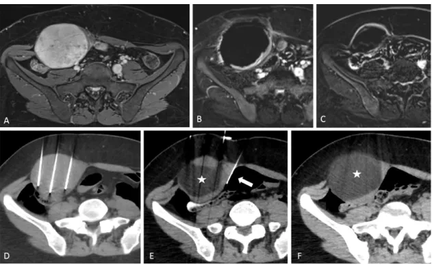
Figure 2 8 cm-desmoid tumor in the abdominal wall in a 26-year-old woman, progressive after childbirth and under hormonal
therapy. MR image before treatment, in T1FS weighted after enhancement (A). CT-guided percutaneous cryoablation using 11 cryoprobes (D, E). Cryoprobes within the tumor (D). A carbodissection and hydrodissection were performed (white arrow, E) to protect the colon (empty arrow, E). The margins of the ice ball are well-visualized in CT (white star, E, F). Complete coverage of the tumor by the ice ball is observed at the end of procedure (F). MR imaging at 1 month in T1FS weighted image after enhancement and subtraction shows a central ablation zone of necrosis, surrounded by an inflammatory rim enhancement (B). MR imaging at 9 months shows a progressive involution of the ablation zone without any evidence of local progression (C).
11 2.3.2 Tumor response – Imaging
Imaging follow-up was performed by MRI for evaluation of the ablation zone (AZ). First post-ablation contrast-enhanced MRI was performed at 1 month and considered as the baseline for further assessment. Then imaging follow-up was performed every 3 months during first year, biennially on the second year and annually. Imaging evaluation was mostly performed by MRI.
In the absence of international standardized criteria for imaging assessment after percutaneous thermal ablation, we used the updated terminology edited by Ahmed et al. for the International Working Group on Image-guided Tumor Ablation (23). Although there is no published report on MR imaging findings after PCA in soft-tissue tumors, most of knowledge arises from the experience of PCA in renal neoplasms (24,25).
First MR imaging usually shows a typical AZ of induced coagulation that appears heterogeneous, hypointense or intermediate at T2-weighted, iso- to hyperintense at T1-weighted without enhancement (figure 2, 3). Subtraction is used to evaluate the absence of enhancement. The AZ is usually surrounded by a thin rim enhancement related to the transition zone between devascularized tissue and unaffected tissue (26). This hyperemic rim is considered as a benign physiologic response to thermal injury (24). AZ size is larger than the original tumor because of safety margins encompassing the tumor.
Measurements of AZ were assessed in short and long axis diameter. In cases of additional course of PCA for residual or local recurrence on ablative margins, AZ size could increase again.
Residual unablated tumor referred to evidence of residual tumor at the ablative margin on the first MR follow-up (23).
Further MR imaging were usually performed every 3 months, demonstrating a progressive involution of the AZ. However no or minimal involution did not imply treatment failure. The rim enhancement gradually resolved over time (24).
Local tumor progression (LTP) was defined in our study as appearance of new tumor foci at the ablative margin after at least one contrast-enhanced follow-up imaging (23). It was also suspected when serial increase in AZ size was observed along follow-up imaging (24).
New lesion was defined as occurrence of a new tumor distant to prior AZ, which may be observed in some multifocal DT.
12
Although RECIST 1.1 criteria are commonly used in clinical trials, they are not appropriate for post-ablative assessment. Taking into account only size does not provide a complete imaging assessment of tumor response and may even lead to erroneous conclusions. Modified RECIST mostly concern post-ablative hepatocellular carcinoma.
Evaluation of mesenteric DT in FAP-patients was performed by abdominal CT and according to RECIST 1.1.
2.4 Endpoints
Primary endpoint was time-to-next treatment (TNT) defined by time between date of first course of PCA and date of modification of strategy. Modification of strategy could be due to untreatable progression by additional course of PCA and/or recrudescence of symptoms. Modification of strategy included introduction or switch of a systemic treatment and introduction of local treatment other than PCA (surgery, radiotherapy and isolated limb perfusion).
Dead patients were excluded of the TNT analysis.
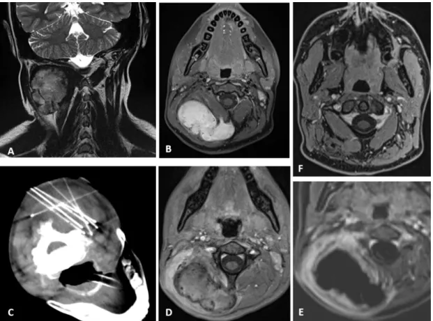
Figure 3 6 cm-desmoid tumor within the neck in a 14-year-old girl. MR images before treatment: in coronal T2 (A) and
T1FS after contrast (B). Perprocedure CT images, patient in ventral decubitus, cryoprobes within the tumor (C). First MR imaging at 1 month, axial T1FS after enhancement (D), with subtraction (E), demonstrating the induced hemorrhagic necrosis surrounded by an expected hyperemic rim. MR follow-up at 12 months, axial T1FS after contrast demonstrating a residual thin scar in the right paravertebral muscles (F).
13
Secondary endpoints were local tumor progression rate after first course of PCA and mean number of PCA courses by patient.
Complications were also assessed according to the Common Terminology Criteria for Adverse Events (CTCAE) v5.0 of the National Cancer Institute. Immediate complications occurred within the 24 hours after procedure, periprocedural complications within 30 days and delayed complications greater than 30 days after ablation according to the International Working Group on Image-guided Tumor Ablation. (23).
An univariate analysis was conducted using Fisher and Chi₂ tests for the screening of local progression prognostic factors. Variables included in univariate analyses were: age, sex, Gardner syndrome, anatomic site, tumor size, nature of treated tumor (primitive/recurrent) and mutation type.
Statistical analysis: TNT was assessed by the Kaplan Meier method, with XLSTAT and PRISM software. All statistical tests were two-sided and a p-value of 0.05 was considered statistically significant.
3. Results
3.1 Patient characteristics (table 1)
Thirty-four patients were treated by PCA for extraabdominal DT, 29 women (85%) and 5 man (15%) with a median age of 35, 5 (range 13-72). Six patients (17%) were affected by FAP. A history of diagnosis after childbirth and after local trauma/surgery were reported respectively in 4 and 6 patients.
Histological diagnosis was proven by percutaneous biopsy in 28 patients (82%) and by surgical resection in 6 patients (18%).
Prior treatments were conservative management in 24 patients (71%), surgery in 8 patients (24%), NSAI drugs in 28 patients, hormonal therapy in 9 patients, chemotherapy in 9 patients, molecular-targeted therapy in 10 patients, radiotherapy in 2 patients.
When previous surgical resection was performed, margin status was R1 in 3 patients, R2 in 1 patient, unknown in 4 patients.
Indications for PCA were local progression despite systemic treatments in 25 patients, pain and functional impairment without progression in 4 patients, toxicity of systemic therapy (Sorafenib and Methotrexate-Vinblastine) in 2 patients, to decrease tumor volume in stable disease under systemic therapy in 2 patients, to stop hormonal therapy for future pregnancy in 1 patient.
14 3.2 Tumors characteristics (table 2)
Median tumor size was 77 mm (range 25-183). All were deep tumors, located in trunk wall in 26 patients, in limbs in 4 patients, in girdle in 3 patients and in neck in 1 patient. Tumors were multifocal in 8 patients, half occurring in FAP-patients.
At the time of first PCA course, tumor nature was primitive in 26 patients, recurrent in 7 patients, and other location in 1 patient.
Molecular biology tests found mutations in the βcatenin gene CTNNB1 in 21 patients (61%) and in the APC gene in 6 patients (affected by FAP). One patient did not harbor any mutation (wild-type) and it was confirmed by many expert pathologists. Mutations data were not available in 6 patients. Mutations in the exon 3 of CTNNB1 were T41A in 12 patients, S45F in 4 patients, S45P in 4 patients and one patient had an insertion in CTNNB1 of undetermined significance.
Table 1. Patient characteristics
Patient characteristics Parameters
n % Gender
Female 29 85
Male 5 15
Age at inclusion time
Minimum 13 __ Maximum 72 __ Median 35,5 __ Mean 36,9 __ Gardner syndrome Yes 6 17 No 28 83 Prior treatments Conservative 24 71 Surgery 8 24 NSAI drugs 28 82 Hormonal therapy 9 26 Chemotherapy 9 26 Molecular-targeted-therapy 10 30 Radiotherapy 2 6
Indications for treatment
Local progression 25 73
Symptoms 4 11
Toxicity of systemic therapy 2 6
Decrease tumor volume 2 6
15
Table 2. Tumors characteristics, * US= Undetermined Significance
3.3 Percutaneous cryoablation procedures: technical parameters (table 3)
Fifty-four courses of PCA were performed between 2012 and 2017 with a total of 58 procedures. Three courses of PCA were performed in 2 procedures because of a large tumor volume and 1 procedure was repeated because of initial technical failure.
Number of treated tumor by procedure was 1, 2 and 3 in respectively 27 patients, 6 patients and 1 patient. Tumors characteristics N % Anatomical location Abdominal wall 12 35 Paraspinal 6 17 Thoracic wall 5 15 Lumbar wall 3 9 Girdle 3 9 Inferior limb 2 6 Superior limb 2 6 Neck 1 3 Largest diameter (mm) Minimum 25 __ Maximum 183 __ Median 77 __ Mean 83 __ Multifocal Yes 8 24 No 26 76 Nature of treated tumor Primitive 26 76 Recurrent 7 21 Other location 1 3 Mutations CTNNB1 T41A 12 36 S45F 4 12 S45P 4 12 US* 1 3 Mutation APC Yes 6 17 Wild-type Yes 1 3
Mutations Missing data
16
Procedures were performed under general anesthesia in 32 patients and under neuroleptanalgesia in 2 patients.
All procedures were CT-guided and US-guidance was added in 11 patients. Median number of used cryoprobes was 8 (range, 2-12).
Thermoprotection techniques were always provided: hydrodissection in 24 patients, carbodissection in 14 patients, electrostimulation in 1 patient and thermocouple in 1 patient.
Procedures were at risk for skin in 28 patients, for main peripheral nerves in 6 patients, medulla in 2 patients and bowel in 13 patients.
Median procedural time from initial to final CT imaging was 130 minutes (range, 60 – 240).
Median total DLP for all PCA courses was 1780 mgy.cm (range, 618 – 5898). In cases of a single course, median DLP was 1459 mgy.cm (range 618 – 2589).
Table 3. Technical parameters of first PCA course
Technical parameters N % Treatment intent curative 27 79 symptomatic 7 21 Type of anesthesia general anesthesia 32 94 neuroleptanalgesia 2 6 Number of cryoprobes minimum 2 __ maximum 12 __ median 8 __
Proximity of vulnerable structures
skin 28 85
bowel 13 38
peripheral main nerve 6 17
medulla 2 6 Thermoprotection hydrodissection 24 80 carbodissection 14 47 electrostimulation 1 3 thermocouple 1 3
Procedural time (min)
minimum 60 maximum 240 median 130 Total DLP (mgy.cm) minimum 618 maximum 2589 median 1459
17 3.4 Outcomes
3.4.1 Immediate follow-up
Median hospitalization stay was 2 days (range, 1-6).
Median immediate post-procedural pain numerical scale was 2 (range 0-8). After the first PCA procedure, 11 patients received a strong opioid analgesic, 10 patients received a weak opioid analgesic, and 5 patients received a non-opioid analgesic.
3.4.2 Delayed follow-up
Median follow-up was 20 months (range 5-54 months).
At 1 month after first procedure, only 28% of patients still had pain.
In cases of a single course of PCA with at least 12-months follow-up, median of AZ size decrease was: 60,6 % in short axis diameter (range -100% to -30,2%) and 37,1% in long axis diameter (range -100% to -6,2%).
3.4.3 Primary endpoint
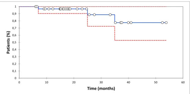
TNT is represented by the Kaplan Meier curve (figure 4).
The 1 and 3 year estimated next treatment rates were respectively 4% (95% CI 0 – 9%) and 23% (95% CI 0 - 48). Median TNT is not achieved.
Three events “next treatment” occurred during follow-up:
- One patient presented a local recurrence in scapular girdle close to brachial plexus, 7 months after 1 course of PCA and went into a clinical trial (DESMOPAZ).
- One patient presented a new lesion in the inferior limb, close to the sciatic nerve in the thigh, 25 months after 1 course of PCA and went into a clinical trial (DESMOPAZ).
- One patient presented a local recurrence 35 months after 2 courses of PCA, close to the sciatic nerve in the popliteal fossa and went into a clinical trial (DESMOPAZ).
18
Figure 4 Kaplan-Meier plot of time-to-next treatment. Probability of next treatment in the selected population in blue, with
a 95% confidence interval in red dotted lines. Circles are censored patients.
3.4.4 Secondary endpoints
LTP rate after first PCA course is 32 % with a median time to LTP of 18, 7 months (range 11,9-26,6). Among the 11 patients with recurrence, 8 patients were treated by a second course of PCA, 1 patient underwent a surgery, 1 patient was included in a clinical trial for systemic treatment and 1 patient was managed with a conservative approach. Median size of local recurrence was 24 mm (range 6-42 mm). Median number of PCA course by patient was 1 (range 1-4).
Median time between 2 courses of PCA was 15, 4 months (range 6 - 26, 6 months).
Indications for additional course of PCA were local recurrence in 8 patients, occurrence/progression of a new distant lesion in 3 patients and residual unablated tumor in 2 patients.
Ten patients underwent 1 additional course of treatment, 2 patients underwent 2 additional courses and 2 patients underwent 3 additional courses.
In univariate analysis only one factor was associated with local progression: nature of treated tumor. The LTP rates were respectively 27% and 100% in the primitive and recurrent tumors (OR=0.02 [0.001-0.5], p=0.008). The LTP rates were respectively 41% and 75% in patients with T41A/S45P mutations and S45F mutation, but the difference was not significant (p=0.2).
0 0,1 0,2 0,3 0,4 0,5 0,6 0,7 0,8 0,9 1 0 10 20 30 40 50 60 Pati e n ts (% ) Time (months)
19
Two per-procedure complications occurred: 2 skin thermal burns on the freezing site medically treated, grade 2 according to the CTCAE v5.0.
Two immediate complications occurred: one brachial plexus stretch injury related to patient positioning during general anesthesia progressively resolved (grade 2), one rhabdomyolysis treated with overhydration and prolongation of hospitalization (grade 3).
One periprocedural complication occurred: one pleural effusion which had needed a drainage (grade 2).
One delayed complication occurred: one necrosis exteriorization through the cryoprobes orifices which had needed a surgical debridement (grade 4).
Death occurred in 2 FAP-patients:
- one died of a mesenteric DT in a Gardner Syndrome
- one died of an undifferentiated neuroendocrine carcinoma of high grade malignancy diagnosed on hepatic metastases.
4. Discussion
Our results are very promising, demonstrating the ability of PCA to provide a prolonged local tumor control in extrabadominal DT. The estimated 1 and 2 year TNT rates are both 4% (95% CI 0 – 9) and the estimated 3-year TNT rate is 23% (95%CI 0 – 48).
Comparison with other reported series of DT treated by PCA
Although the efficacy of PCA has already been reported in literature (20–22), this study reports the largest series. PCA is technically successful in majority of extraabdominal DT with an achievable complete ablation. Our results are difficult to compare to those of Schmitz et al. who reported a series of 18 patients treated by PCA for extraabdominal DT in 2016 (22). Only 39% of patients had no evidence of residual viable tumor on follow-up imaging, but standard definitions were different. Residual viable tumor was defined as new or persistently enhancing soft-tissue at ablation site, which actually represents residual unablated tumor and local tumor progression in our study. However they reported a degree of volume decrease in 95.7% of patients. In a series of 13 patients, Havez et al. found a median change of - 37.6 % on the last imaging with a mean follow-up of 11.3 months (21). This is consistent with our results. In our series median of AZ size decrease 12 months after PCA was 60.6% in short axis diameter and 37.1% in long axis diameter. We observed a higher involution of the AZ in its short-axis diameter compared to long-axis diameter, which is not reported in literature.
20
PCA is also a safe technique in majority of cases. In our experience 3 moderate complications occurred. Two minor complications were skin burns medically treated. The frequent infiltration of dermis by DT and proximity to skin often lead to a real challenge in the treatment of DT by PCA. Tumors in our series were highly close to skin in 85% of the cases. Two major complications occurred in a patient with a large thoracic paravertebral DT of 17 cm treated in 2 procedures. A rhabdomyolysis occurred immediately after the first procedure and was resolved by overhydration. It may be considered as an expected aggravated post-ablative syndrome in cases of large ablated volume. A necrosis exteriorization through a cryoprobe orifice occurred few months after completion of treatment, due to the large volume of AZ. Schmitz et al. reported only 3 mild complications in his series (22). Tumor volumes were larger in our series with a median size of 77 mm (range 25 to 183 mm) against 64 mm (range 17 to 140 mm) by Schmitz et al. (22) and 53 mm by Havez et al. (21). This can explain a higher rate of complications. Recently Ghanouni et al. reported a multicenter retrospective series of 15 patients with extrabadominal DT treated with MR-guided focused Ultrasound (27), in which median decrease of viable targeted tumor volume was 63%. Complications occurred in 10 patients and were skin burns (n=8), nerve injury (n=3) and off-target heating (n=3).
High advantage of PCA in the management of extraabdominal DT is that procedures may be repeated safely in cases of LTP or occurrence of new lesion. It allows a prolonged local tumor control with a low number of courses by patient (median=1, range 1- 4). Moreover, in cases of additional courses of PCA, median time between two courses is tolerable (15,4 months, range 6 - 26, 6 months).
PCA in FAP-patients
Six patients with FAP were treated in our series. Extrabadominal DT in FAP-patients are often multifocal leading to symptoms. PCA allows a curative or symptomatic treatment. Ablation of multiple symptomatic or progressive extrabadominal DT is achievable. Conversely, mesenteric DT are not allowed to be treated by PCA because of high risk of bowel perforation and necrosis. The prognosis of FAP-patients is poorer due to colorectal cancer and mesenteric DT. In our study death occurred in 2 FAP-patients, one with a mesenteric DT and one of an undifferentiated neuroendocrine carcinoma diagnosed on hepatic metastases.
Comparison with others treatment options
The European Society of Medical Oncology edited updated guidelines for the management of DT (17). After a Wait-and-See frontline approach of 1-2 years, treatment options in cases of progression are: surgery/medical therapy (including hormonal therapy) in second line, medical therapy/radiotherapy/isolated perfusion limb in third line and investigational treatments in last line. Those indications depend on anatomic location of the tumor.
21
In a study evaluating 771 patients of the prospectively maintained french database of sarcomas and DT, Penel et al. assessed the outcomes of surgery versus non-surgical approach (11). After initial surgery the local recurrence rate was 31.7%. In a retrospective study of 426 patients with DT, Salas et al. reported a local recurrence rate of 44.3% in 323 patients treated by surgery and/or radiotherapy (7). Our results are not inferior, with a LTP rate of 32%. In addition to equal local recurrence rates, PCA may represent an alternative to surgery in cases of expected morbidity or functional impairment.
In a retrospective study evaluating 147 patients with abdominal wall DT managed conservatively (102 patients) or surgically (41 patients), Bonvalot et al. found a modification of treatment strategy of respectively 33% and 41% at 1 and 3 years (13). Our results are not inferior with estimated 1 and 3 year TNT rates of 4% (95% CI 0-9) and 23% (95% CI 0-48).
A phase II pilot study evaluating radiotherapy at a dose of 56 gy for inoperable progressive DT reported a 3-year local control rate of 81.5% (28). It suggests that probability of “next treatment “at 3 years was 18.5%. Complete response rate was 13.6%. Lymphedema was observed in 10 patients.
Medical therapies show various degree of tumor response, symptoms relief and toxicity. Today, none of these treatments could be considered as a standard of care, as available data have only come from retrospective studies with a limited number of cases or non-randomized phase II studies (29). A retrospective series of 134 patients showed that 85% of patients with DT treated with an association of Sulindac plus antiestrogen achieved progression arrest (30). In cases of hormonal therapy failure with an aggressive growing tumor, chemotherapy using a low dose regimen with methotrexate and vinblastine/vinorelbine is an option. Palassini et al. found an objective response rate of 99% with symptomatic relief in 80% of symptomatic cases (31). There is a prospective, uncontrolled evidence for the activity of the tyrosine kinase inhibitor in progressive DT. Most of published studies report high rates of stabilization (60-80%) despite low response rates (6-16%) (32,33). Toxicity of tyrosine kinase inhibitors usually includes mild hematologic and digestive adverse events. A randomized phase II trial evaluating pazopanib versus methotrexate plus vinblastine is ongoing in France with a completion of inclusion in 2020 (DESMOPAZ).
Tumor size decrease is difficult to compare with other types of treatment because involution of the AZ after PCA describes the process by which the body eliminates the zone of induced coagulation over weeks and months (23). Compared with medical therapies or radiotherapy, PCA do not only provide a tumor progression arrest but also a progressive involution of the AZ. It is a high advantage in cases of functional impairment or major cosmetic issue.
22
Prognostic factors of local progression after PCA
In univariate analysis, only the “recurrent” nature of treated DT was associated with a higher risk of local recurrence after PCA. In a retrospective series comparing primary versus recurrent DT, Mueller et al. found local recurrence rates of respectively 33% and 35% in both groups (34). In the prognostic nomogram by Crago et al., recurrent status was not statistically associated with a higher risk of local recurrence (12). This result could be explained by a higher aggressiveness of recurrent tumors addressed for PCA. There was also a trend for more frequent local progression after PCA in patients with S45F mutation. Even if it was reported in literature (35), this effect remains non-significant in our study probably due to small sample size.
Nevertheless, prognostic factors for local recurrence are less clinically relevant for PCA, because PCA can be repeated in recurrent DT. Prognostic factors of TNT would be more interesting to assess, because the event “next treatment” implies a loss of tumor control. In our series, only 3 patients underwent a modification strategy because of anatomic constraints leading to untreatable progression. With a larger sample size and a longer follow-up, prognostic factors of modification strategy should be achievable.
Limits of PCA
PCA of extrabadominal DT is achievable in majority of anatomic site. The main limitation to manage a complete, prolonged and safe ablation is the proximity of vulnerable structures such as peripheral nerves and skin. In our series, 3 patients experimented a modification of strategy with introduction of a systemic treatment because of an untreatable local progression of DT respectively closed to the brachial plexus, femoral and sciatic nerve.
Even if patients with large DT of 17cm were treated in our series, the tumor volume may be also a limitation to manage a complete and safe ablation.
Other disadvantage is the exposure to radiation in young patients because PCA was always CT-guided in our institution. However median total DLP for a single course of treatment was 1459 mgy.cm, equivalent to a multiphasic CT. In few centers, MR-guidance is available for thermal ablation with a higher contrast resolution and without any radiation.
23
Limits of our study
As a weakness our study is retrospective, monocentric, with a small sample size and a short follow-up. Due to its retrospective nature, accurate data on clinical assessment were not available. Quality of life, impairment and pain numerical scales were not available over follow-up. A prospective multicenter phase II trial is ongoing to confirm the efficacy of PCA in terms of local tumor control and symptoms relief in progressive extraabdominal DT after 2 lines of systemic treatments (CRYODESMO). Execution by a single interventional radiologist do not avoid to assess the reproducibility of our results. However DT are rare tumors which required management in expert reference centers.
Conclusion
PCA has proven its efficacy and safety in the management of extrabadominal DT with a prolonged local tumor control. After an initial watchful waiting, it is an alternative option to surgery when morbidity and functional impairment of a resection is expected. PCA may also be an alternative to medical therapies in aggressive and fast-growing tumors with symptoms and cosmetic issue. It may also be considered as an option prior to pregnancy, which may lead to tumor progression.
The role of PCA remains to be defined between the different treatment options available in cases of progressive or remained symptomatic extraabdominal DT.
References
1. Broekhoven DLM van, Grünhagen DJ, Bakker MA den, Dalen T van, Verhoef C. Time Trends in the Incidence and Treatment of Extra-Abdominal and Abdominal Aggressive Fibromatosis: A Population-Based Study. Ann Surg Oncol. 2015 Sep 1;22(9):2817–23.
2. Reitamo JJ, Häyry P, Nykyri E, Saxén E. The desmoid tumor. I. Incidence, sex-, age- and anatomical distribution in the Finnish population. Am J Clin Pathol. 1982 Jun;77(6):665–73.
3. Penel N, Coindre J-M, Bonvalot S, Italiano A, Neuville A, Le Cesne A, et al. Management of desmoid tumours: A nationwide survey of labelled reference centre networks in France. Eur J Cancer Oxf Engl 1990. 2016 May;58:90–6.
4. Jo VY, Fletcher CDM. WHO classification of soft tissue tumours: an update based on the 2013 (4th) edition. Pathology (Phila). 2014 Feb;46(2):95–104.
5. Le Guellec S, Soubeyran I, Rochaix P, Filleron T, Neuville A, Hostein I, et al. CTNNB1 mutation analysis is a useful tool for the diagnosis of desmoid tumors: a study of 260 desmoid tumors and 191 potential
morphologic mimics. Mod Pathol Off J U S Can Acad Pathol Inc. 2012 Dec;25(12):1551–8.
6. Colombo C, Bolshakov S, Hajibashi S, Lopez-Terrada L, Wang W-L, Rao P, et al. “Difficult to diagnose” desmoid tumours: a potential role for CTNNB1 mutational analysis. Histopathology. 2011 Aug;59(2):336–40. 7. Salas S, Dufresne A, Bui B, Blay J-Y, Terrier P, Ranchere-Vince D, et al. Prognostic factors influencing progression-free survival determined from a series of sporadic desmoid tumors: a wait-and-see policy according to tumor presentation. J Clin Oncol Off J Am Soc Clin Oncol. 2011 Sep 10;29(26):3553–8.
24
8. Nieuwenhuis MH, Casparie M, Mathus-Vliegen LMH, Dekkers OM, Hogendoorn PCW, Vasen HFA. A nation-wide study comparing sporadic and familial adenomatous polyposis-related desmoid-type fibromatoses. Int J Cancer. 2011 Jul 1;129(1):256–61.
9. Fallen T, Wilson M, Morlan B, Lindor NM. Desmoid tumors -- a characterization of patients seen at Mayo Clinic 1976-1999. Fam Cancer. 2006;5(2):191–4.
10. Koskenvuo L, Ristimäki A, Lepistö A. Comparison of sporadic and FAP-associated desmoid-type fibromatoses. J Surg Oncol. 2017 Jun 1;
11. Penel N, Le Cesne A, Bonvalot S, Giraud A, Bompas E, Rios M, et al. Surgical versus non-surgical approach in primary desmoid-type fibromatosis patients: A nationwide prospective cohort from the French Sarcoma Group. Eur J Cancer Oxf Engl 1990. 2017 Sep;83:125–31.
12. Crago AM, Denton B, Salas S, Dufresne A, Mezhir JJ, Hameed M, et al. A prognostic nomogram for prediction of recurrence in desmoid fibromatosis. Ann Surg. 2013 Aug;258(2):347–53.
13. Bonvalot S, Ternès N, Fiore M, Bitsakou G, Colombo C, Honoré C, et al. Spontaneous regression of primary abdominal wall desmoid tumors: more common than previously thought. Ann Surg Oncol. 2013 Dec;20(13):4096–102.
14. Fiore M, Rimareix F, Mariani L, Domont J, Collini P, Le Péchoux C, et al. Desmoid-type fibromatosis: a front-line conservative approach to select patients for surgical treatment. Ann Surg Oncol. 2009
Sep;16(9):2587–93.
15. Colombo C, Miceli R, Le Péchoux C, Palassini E, Honoré C, Stacchiotti S, et al. Sporadic extra abdominal wall desmoid-type fibromatosis: surgical resection can be safely limited to a minority of patients. Eur J Cancer Oxf Engl 1990. 2015 Jan;51(2):186–92.
16. Fiore M, MacNeill A, Gronchi A, Colombo C. Desmoid-Type Fibromatosis: Evolving Treatment Standards. Surg Oncol Clin N Am. 2016 Oct;25(4):803–26.
17. Kasper B, Baumgarten C, Garcia J, Bonvalot S, Haas R, Haller F, et al. An update on the management of sporadic desmoid-type fibromatosis: a European Consensus Initiative between Sarcoma PAtients EuroNet (SPAEN) and European Organization for Research and Treatment of Cancer (EORTC)/Soft Tissue and Bone Sarcoma Group (STBSG). Ann Oncol Off J Eur Soc Med Oncol. 2017 Oct 1;28(10):2399–408.
18. Kasper B, Baumgarten C, Bonvalot S, Haas R, Haller F, Hohenberger P, et al. Management of sporadic desmoid-type fibromatosis: a European consensus approach based on patients’ and professionals’ expertise - a sarcoma patients EuroNet and European Organisation for Research and Treatment of Cancer/Soft Tissue and Bone Sarcoma Group initiative. Eur J Cancer Oxf Engl 1990. 2015 Jan;51(2):127–36.
19. Callstrom MR, Kurup AN. Percutaneous ablation for bone and soft tissue metastases--why cryoablation? Skeletal Radiol. 2009 Sep;38(9):835–9.
20. Kujak JL, Liu PT, Johnson GB, Callstrom MR. Early experience with percutaneous cryoablation of extra-abdominal desmoid tumors. Skeletal Radiol. 2010 Feb;39(2):175–82.
21. Havez M, Lippa N, Al-Ammari S, Kind M, Stoeckle E, Italiano A, et al. Percutaneous image-guided cryoablation in inoperable extra-abdominal desmoid tumors: a study of tolerability and efficacy. Cardiovasc Intervent Radiol. 2014 Dec;37(6):1500–6.
22. Schmitz JJ, Schmit GD, Atwell TD, Callstrom MR, Kurup AN, Weisbrod AJ, et al. Percutaneous Cryoablation of Extraabdominal Desmoid Tumors: A 10-Year Experience. AJR Am J Roentgenol. 2016 Jul;207(1):190–5.
23. Ahmed M, Solbiati L, Brace CL, Breen DJ, Callstrom MR, Charboneau JW, et al. Image-guided tumor ablation: standardization of terminology and reporting criteria--a 10-year update. Radiology. 2014
25
24. Kawamoto S, Solomon SB, Bluemke DA, Fishman EK. Computed tomography and magnetic resonance imaging appearance of renal neoplasms after radiofrequency ablation and cryoablation. Semin Ultrasound CT MR. 2009 Apr;30(2):67–77.
25. Wile GE, Leyendecker JR, Krehbiel KA, Dyer RB, Zagoria RJ. CT and MR imaging after imaging-guided thermal ablation of renal neoplasms. Radiogr Rev Publ Radiol Soc N Am Inc. 2007 Apr;27(2):325-339; discussion 339-340.
26. Garnon J, Koch G, Caudrelier J, Tsoumakidou G, Cazzato RL, Gangi A. Expanding the borders: Image-guided procedures for the treatment of musculoskeletal tumors. Diagn Interv Imaging. 2017 Sep;98(9):635–44. 27. Ghanouni P, Dobrotwir A, Bazzocchi A, Bucknor M, Bitton R, Rosenberg J, et al. Magnetic resonance-guided focused ultrasound treatment of extra-abdominal desmoid tumors: a retrospective multicenter study. Eur Radiol. 2017 Feb;27(2):732–40.
28. Keus RB, Nout RA, Blay J-Y, de Jong JM, Hennig I, Saran F, et al. Results of a phase II pilot study of moderate dose radiotherapy for inoperable desmoid-type fibromatosis--an EORTC STBSG and ROG study (EORTC 62991-22998). Ann Oncol Off J Eur Soc Med Oncol. 2013 Oct;24(10):2672–6.
29. Penel N, Chibon F, Salas S. Adult desmoid tumors: biology, management and ongoing trials. Curr Opin Oncol. 2017 Jul;29(4):268–74.
30. Quast DR, Schneider R, Burdzik E, Hoppe S, Möslein G. Long-term outcome of sporadic and FAP-associated desmoid tumors treated with high-dose selective estrogen receptor modulators and sulindac: a single-center long-term observational study in 134 patients. Fam Cancer. 2016 Jan;15(1):31–40.
31. Palassini E, Frezza AM, Mariani L, Lalli L, Colombo C, Fiore M, et al. Long-term Efficacy of Methotrexate Plus Vinblastine/Vinorelbine in a Large Series of Patients Affected by Desmoid-Type Fibromatosis. Cancer J Sudbury Mass. 2017 Apr;23(2):86–91.
32. Kasper B, Gruenwald V, Reichardt P, Bauer S, Rauch G, Limprecht R, et al. Imatinib induces sustained progression arrest in RECIST progressive desmoid tumours: Final results of a phase II study of the German Interdisciplinary Sarcoma Group (GISG). Eur J Cancer Oxf Engl 1990. 2017;76:60–7.
33. Penel N, Le Cesne A, Bui BN, Perol D, Brain EG, Ray-Coquard I, et al. Imatinib for progressive and recurrent aggressive fibromatosis (desmoid tumors): an FNCLCC/French Sarcoma Group phase II trial with a long-term follow-up. Ann Oncol Off J Eur Soc Med Oncol. 2011 Feb;22(2):452–7.
34. Mueller C, Croner R, Klein P, Grützmann R, Vassos N. Primary and recurrent sporadic desmoids: Prognostic factors influencing recurrence-free survival after complete gross resection. Int J Surg Lond Engl. 2016 Jul;31:63–70.
35. Colombo C, Miceli R, Lazar AJ, Perrone F, Pollock RE, Le Cesne A, et al. CTNNB1 45F mutation is a molecular prognosticator of increased postoperative primary desmoid tumor recurrence: an independent, multicenter validation study. Cancer. 2013 Oct 15;119(20):3696–702.
26
II. Article 2
PERCUTANEOUS CRYOABLATION
OF SOFT-TISSUE SARCOMAS:
EARLY PHASE RESULTS
Abstract
Purpose: The aim is to report our preliminary outcomes of percutaneous cryoablation (PCA) in the
management of locally recurrent and selected advanced soft-tissue sarcomas (STS).
Patients and methods: Data of patients treated by PCA for locally recurrent STS (group A) or soft-tissue
metastasis of STS (group B) between December 2012 and March 2017 in our tertiary cancer center were retrospectively collected. Primary endpoint was time-to-local progression (TTLP) in group A and time-to-next treatment (TNT) in group B. Second endpoint was overall survival (OS), third endpoint was complications according to Common terminology Criteria for Adverse events v5.5 (CTCAE).
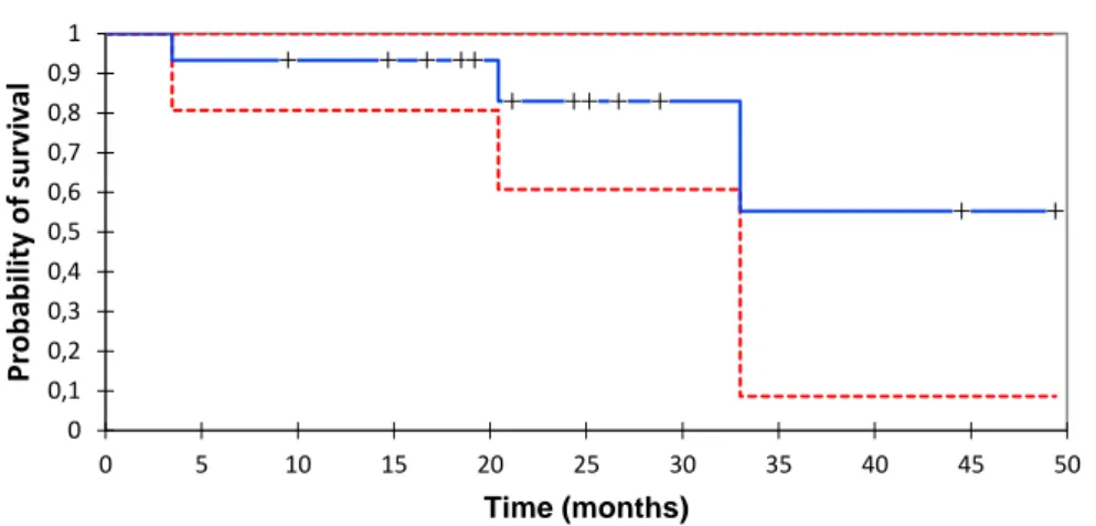
Results: Fifteen patients were successfully treated by PCA, 10 in group A, 5 in group B. Median
follow-up is 21.1 moths (range 3.4-49.4). Median of tumor size is 43 mm (range 15-80). The median TTLP in group A was not achieved. The 1-year local-progression rate is 22% (95% CI 0-48). The median TNT in group B was 10.6 months (range 1.7-24.4). The Median OS was not achieved. The 1 and 2-year OS rates are respectively 93% (95%CI 80-100) and 83% (95%CI 60-100). One immediate adverse event related to PC occurred: a transient hand paresis in a patient treated for a locoregional relapse closed to the brachial plexus (grade 2).
Conclusion: PCA is a safe, effective and minimally-invasive treatment. In locally recurrent non-surgical
STS it allows a salvage treatment with a low morbidity. In selected metastatic patients it allows an increased TNT delaying new line of systemic therapy.
Abbreviations
PCA: percutaneous cryoablation STS: soft-tissue sarcoma
TNT: Time-to-next treatment TTLP: time-to-local progression OS: overall survival
PTA: percutaneous thermal ablation AZ: ablation zone
27 1. Introduction
Soft tissue sarcomas (STS) are a heterogeneous group of uncommon tumors arising from mesenchymal cells, representing less than 1% of malignant tumors. STS are relatively rare with an estimated incidence of 4-5 per 100 000 per year in Europe (1). They may arise in soft tissue, skin or various organs and show a broad range of differentiation (2,3). Prognosis varies greatly according to histologic type, FNCLCC grade, tumor size and primary site (4,5). In Europe the 5-year overall survival (OS) rate is 58% (1,6). A multidisciplinary approach is required and management should be provided in reference centers (7,8).
Therapeutic strategies are different according to the stage of disease. Focal treatments may play a role all along the course of disease.
In case of local recurrence, conservative surgery is often complex with a higher risk of second local recurrence and morbidity, especially when prior treatment included radiotherapy (9). In limb and trunk wall tumors, alternative salvage strategies are amputation, isolated limb perfusion, intra-operative interstitial brachytherapy (10,11). In retroperitoneal sarcomas, local recurrence and/or its treatment is the most common cause of death (12). The probability of cure depends on type of recurrence (solitary locoregional or multifocal), grade/histologic subtype, disease-free interval, local recurrence growth rate and expected risk of mortality and morbidity after re-resection (13,14).
In case of oligometastatic disease, many studies have reported the potential oncologic benefit of local treatment of metastasis (15,16). In STS, published evidence on local treatment of oligometastasis mainly focused on pulmonary metastasis, including surgery, radiotherapy and radiofrequency (16–19). In advanced disease, standard treatment is chemotherapy based on a single-agent doxorubicin as first-line but polychemotherapy as a firstfirst-line is suggested to be highly associated with an increased overall survival (20,21). Others molecular agents such as olaratumab, pazopanib and pembrolizumab demonstrated various results in clinical trials (22–24). In a recent retrospective series of 1575 patients with metastatic STS, Savina et al. found that locoregional treatment of metastases were significantly associated with an increased overall survival (21).
Following the developments of percutaneous thermal ablations (PTA), interventional radiology increasingly takes part in the treatment strategy of many cancers. For soft-tissue tumor ablation, percutaneous cryoablation (PCA) is the treatment of choice as it offers several advantages compared with heat ablation techniques: visualization of the ice ball with imaging, providing an accurate control of the ablation zone (AZ); less risk of thermal damage to adjacent vulnerable structures and better tissue healing after treatment. PCA has already been evaluated in the management of desmoid tumors
28
and soft-tissue metastasis of variable primary tumors with promising results (25,26). However, little data are available about its potential role for salvage treatment of locally recurrent STS non-eligible to surgery. Only one study recently reported the role of PCA as a salvage treatment of locally recurrent retroperitoneal sarcomas with promising results (27). There are also little data specifically on local treatment of soft-tissue metastases of STS.
The aim of our study is to describe our early-phase results of PCA of soft-tissue tumors in two different scenarios: locally recurrent STS and soft tissue metastases of STS.
2. Patients, methods and statistical analysis
2.1 Data collection
Data from patients treated by PCA for STS in our single institution cancer center between December 2012 and March 2017 were retrospectively collected. Patients treated for bone sarcomas or visceral metastases of STS were not included.
Patient demographics, histology, grade, site, size measurements, stage, history of prior treatments, technical modalities, clinical and radiological outcomes were collected.
Patients were divided in 2 groups.
Group A included patients with locoregional recurrence of STS after treatment of the primary tumor, non-eligible to surgery. PCA was performed in a salvage intent.
Group B included patients with advanced disease: oligometastatic or metastatic.
Oligometastatic state was defined by the presence of one to five simultaneous metastases in a limited number of sites according to Palma et al. (28–30). Oligoprogression indicated progression of a limited number of metastasis while others were controlled with systemic therapy (30).
In this group, PCA was performed in various scenarios. In cases of oligoprogression, PCA was performed to control progression of a single metastasis while others were controlled with systemic therapy. It was also performed in cases of metastatic occurrence that appeared after the completion of a systemic treatment. PCA could also be performed in a palliative intent for pain relief or to reduce tumor burden. All cases were selected by the sarcoma tumor board.
29
In cases of local or distant soft-tissue recurrence after a PCA course, additional course could be performed if estimated to be clinically relevant by the sarcoma tumor board (figure 1).
2.2 Procedures: technical modalities
PCA procedures were performed by an interventional radiologist with more than 10 years’ experience in PTA. PCA were mostly performed under general anesthesia or in neuroleptanalgesia if neuromonitoring was needed.
CT-guidance was systematically used to monitor the ice ball. Combination with US-guidance could also be used for superficial tumors, for faster needle insertion and to monitor the superficial border of the ice ball. Cryoprobes were chosen according to size and shape of the tumor (Icerod® or Icesphere ® (Galil-BTG®) (figure 2).
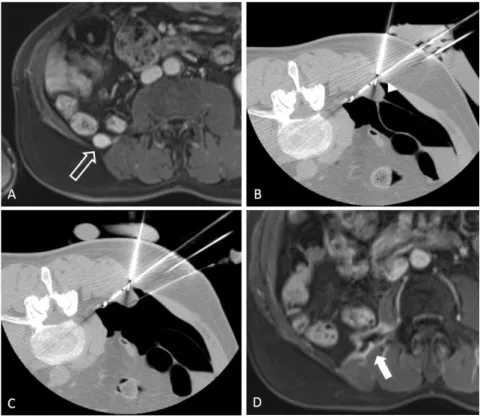
Thermoprotection techniques were always used including carbodissection, hydrodissection, thermocouple and warm sterile glove (figure 3).
Ablation protocol included 2 freezing cycles of 10 minutes separated by a passive thawing cycle of 8 minutes.
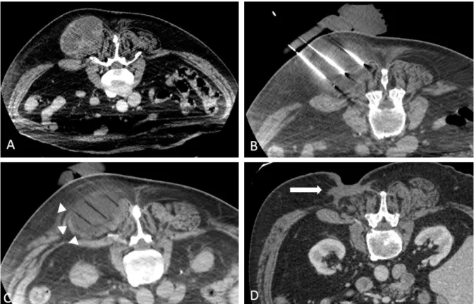
Figure 1 CT images of a new metastasis within the inferior limb (white star, A, B) in a patient previously treated for a paravertebral metastasis of a grade 3 pleomorphic liposarcoma (figure 2). CT-guided PC, showing the cryoprobes within the lesion (C), and the well-differentiated ice ball compared with surrounding tissue (D). CT images follow-up at 6 months demonstrating the cryoablation zone without appearance of new foci at the ablative margins (black star, E). Systemic treatment is still discontinued since first PCA.
30
Technical success was immediately appreciated at the end of the procedure by optimal coverage of the tumor by the ice ball in CT, with at least 5 mm margin.
Radiation dosimetry (total DLP) was recorded for each procedure.
2.3 Clinical and radiological follow-up
2.3.1 Clinical Assessment
Inclusion date was the day of the first PCA. End of follow-up was the date of the last clinical follow-up or date of death.
Immediate post-procedural pain numerical scale and analgesic pain ladder were recorded. Early and delayed complications were collected.
Further clinical assessment was performed by an oncologist.
Figure 2 CT image of a soft-tissue paravertebral metastasis of a grade 3 pleomorphic liposarcoma in a 73-year-old men, before
procedure (A), perprocedure showing cryoprobes within the lesion (B). The ice ball is well-differentiated from the surrounding tissue (arrowhead, C). CT image follow-up at 16 months demonstrating the scar of the cryoablation zone and wound debridement.
31 2.3.2 Tumor response - Imaging
Imaging follow-up was usually performed by MRI for focal evaluation of the AZ. First post-ablation contrast-enhanced MRI was performed at 1 month and considered as the baseline for further local assessment.
In the absence of international standardized criteria for imaging assessment after percutaneous thermal ablation, we used the updated terminology edited by Ahmed et al. for the International Working Group on Image-guided Tumor Ablation (31). Although there is no published report on MR imaging findings after PCA in soft-tissue tumors, most of knowledge arises from the experience of PCA in renal neoplasms (32,33).
First MR imaging usually shows a typical AZ of induced coagulation that appears heterogeneous, hypointense or intermediate at T2-weighted, iso- to hyperintense at T1-weighted without enhancement. Subtraction is used to evaluate the absence of enhancement. The AZ is usually surrounded by a thin rim enhancement related to the transition zone between devascularized tissue and unaffected tissue (34). This hyperemic rim is considered as to be a benign physiologic response to thermal injury (figure 3) (32). AZ size is larger than the original tumor because of the safety margins encompassing the tumor. Measurements were assessed in short and long axis diameter.
Residual unablated tumor indicated evidence of residual foci at the ablative margin on the first MR follow-up (31).
Further MR imaging were usually performed every 3 months and demonstrated a progressive involution of the AZ. However no or minimal involution did not imply treatment failure. The rim enhancement gradually resolved over time (32).
Local tumor progression (LTP) was defined in our study as appearance of new tumor foci at the ablative margin after at least one contrast-enhanced follow-up imaging (31). It was also suspected when serial increase in AZ size was observed along follow-up imaging (32).
Although RECIST 1.1 criteria are commonly used in clinical trials, they are not appropriate for post-ablative assessment. Only taking size into account does not provide a complete imaging assessment of tumor response and may even lead to erroneous conclusions. Modified RECIST criteria mostly concern post-ablative hepatocellular carcinoma.