Université de Montréal
Stress oxydatif, fonction mitochondriale et maladie inflammatoire de l’intestin
par Rame Taha
Département de Nutrition Faculté of Médecine
Thèse présentée à la Faculté de Médecine en vue de l’obtention du grade de PhD
en Nutrition
Aout, 2011
Université de Montréal
Faculté des études supérieures et postdoctorales
Cette thèse est intitulée :
Stress oxydatif, fonction mitochondriale et maladie inflammatoire de l’intestin
Présentée par : Rame Taha
a été évaluée par un jury composé des personnes suivantes :
Dr Dominique Garrel, président-rapporteur Dr Emile Levy, directeur de recherche
Victor Gavino, membre du jury Charles Ramassany, examinateur externe Christiane Malo, représentante du doyen de la FES
Résumé
CONTEXTE: Bien que la dysfunction mitochondriale et le stress oxydant jouent des rôles prépondérants dans plusieurs conditions pathologiques, ils n’ont pas été étudiés de façon extensive au niveau du tube digestif qui est constamment exposé aux oxydants (provenant de l’alimentation) et à divers agents pathogènes. L’ingestion simultanée de sels ferreux et d’acide ascorbique peut causer le dommage des macromolécules par oxydation. Le ‘’Nuclear factor erythroid 2 related factor’’ (Nrf2) est un important facteur de transcription sensible au potentiel redox et qui protège contre le stress oxydant en induisant des gènes anti-oxydants et de detoxification par sa liaison à l’élément de réponse antioxydante (ARE). Les fonctions anti-oxydantes et anti-inflammatoires de Nrf2 ont été décrites dans une variété de types cellulaires et de tissus. Cependant son rôle est très peu connu au niveau du tube digestif. OBJECTIFS: Les objectifs sont d’évaluer comment la peroxydation lipidique médiée par le fer/ascorbate (FE/ASC) affecte les fonctions mitochondriales dans les cellules Caco-2/15, et de déterminer l’ampleur de l’implication de Nrf2.
MÉTHODES: Le stress oxydant a été induit dans les cellules Caco2/15 en les traitant
avec 0.2mm/2mm de FE/ASC. L’augmentation de l’expression de Nrf2 a été obtenue suite au prétraitement des cellules Caco2/15 avec 50 µM d’Olitpraz (OPZ), un puissant activateur. L’invalidation du gène de Nrf2 a été réalisée dans les cellules par transfection avec un vecteur lentiviral contenant un shRNA contre Nrf2. RÉSULTATS: Nos résultats montrent que le traitement des cellules Caco-2/15 avec du FE/ASC (0.2 mm/2 mm) augmente les niveaux du malondialdehyde (MDA), réduit la production d’ATP, entraîne une surcharge mitochondriale de calcium, active l’expression protéique du cytochrome C et de l’AIF (apoptotic inducing factor), réduit l’activité des complexes I, II,III et IV de la chaîne respiratoire mitochondriale, augmente les niveaux de 8-OHdG, un marqueur des dommages à l’ADN mitochondrial, diminue la DNA glycosylase, et altère les expressions génique et protéique des facteurs de transcription mitochondriaux (mtTFA, mtTFB1, mtTFB2).
De plus, nos observations montrent que l’induction et l’activation de Nrf2 dans les cellules Caco-2/15 résultent en: une augmentation des enzymes anti-oxydantes endogènes (catalase, glutathion peroxydase, et superoxyde dismutase), une réduction du facteur nucléaire NFκβ et de TNF-α, une augmentation de la production d’ ATP et de l’activité des complexes respiratoires (I, II, III, IV) et de PGC-1α, et une régulation des niveaux de la prohibitine mitochondriale, du Bcl-2 anti-apoptotique et de l’occludine.
CONCLUSION: Dans l’ensemble, nos résultats montrent que l’exposition aigüe des
cellules Caco-2/15 à la peroxydation par le FE/ASC entraîne des effets pathologiques sur les fonctions mitochondriales et l’intégrité de l’ADN, qui sont abolis par l’induction de Nrf2. Il en ressort que Nrf2 joue un rôle majeur dans la protection de l’épithélium intestinal contre le stress oxydant.
Abstract
Background:
Although mitochondrial dysfunction and oxidative stress are key mechanisms in various pathological conditions, they have not been extensively studied in the gastrointestinal tract, which is known to be constantly exposed to luminal oxidants from ingested foods and pathogens. Key among these is the simultaneous ingestion of iron salts and ascorbic acid, which can cause oxidative damage to macromolecules. The protein ‘’Nuclear factor-erythroid 2- related factor’’ (Nrf2) is an important redox-sensitive transcription factor, which protects against oxidative stress by inducing antioxidant and detoxifying genes through binding with antioxidant response element (ARE). Many of Nrf2 antioxidant protective and anti-inflammatory functions have been established in various cells and tissues. However, limited information is available on its role in the gastrointestinal tract.
Objectives:
The objectives are to evaluate how iron-ascorbate (FE/ASC)-mediated lipid peroxidation affects mitochondrion functioning in Caco-2/15 cells, and to mechanistically determine the role of Nrf2.
Methods:
Caco2/15 cells were treated with 0.2mm/2mm of FE/ASC to induce oxidative stress. To
increase Nrf2 expression, cultured Caco2/15 cells were pre-treated with 50 µM Olitpraz (OPZ). To down regulate the Nrf2 function, Nrf2 gene was knocked down by transfecting Caco-2/15 cells with a pGFP-RS lentiviral vector containing shRNA against Nrf2.RESULTS:
Our results show that the treatment of Caco-2/15 cells with FE/ASC (0.2 mm/2 mm): increased the levels of malondialdehyde (MDA), a marker of oxidative stress; reduced ATP production; raised mitochondrial calcium content; regulated the protein expression of cytochrome C and apoptotic inducing factor (AIF); decreased mitochondrial respiratory chain complexes I, II, III and IV activity; prevented mtDNA damage as illustrated by the raised levels of 8-OHdG; lowered DNA Glycosylase, and altered the gene expression and protein mass of mitochondrial transcription factors (mtTFA, mtTFB1, mtTFB2).
Furthermore, our observations indicate that the induction and activation of Nrf2 in Caco2/15 cells resulted in an augmentated endogenous antioxidants enzymes (catalase, glutathione peroxidase, and superoxide dismutase), a reduction of nuclear factor-kappaB (NFκβ) and Tumor Necrosis Factor- Alpha (TNF-α), an increase in the ATP production, mitochondrial respiratory complexes (I, II, III, VI), PGC1α , and a regulation of the mitochondrial Prohibitin, anti-apoptotic Bcl-2 protein, and Occludin level.
CONCLUSION:
Findings indicate that acute exposure of Caco-2/15 cells to FE/ASC-catalyzed peroxidation produces pathological effects on mitochondrial functions and DNA integrity, which were diminished by Nrf2 induction. It appears that Nrf2 plays a critical cytoprotective role in intestinal epithelial cells against oxidative stress.
Table des matières
1 INTRODUCTION ... 13
1.1 Oxidative stress ... 13
1.1.1 Reactive oxygen species generation and oxidative damage ... 14
1.1.2 Iron over-consumption induce oxidative stress... 19
1.1.3 Antioxidant definition and classification ... 20
1.1.4 Oxidative stress and intestinal inflammation ... 23
1.1.5 Intestinal permeability and tight junction proteins... 26
1.2 Mitochondria ... 28
1.2.1 Mitochondrion structure and function... 28
1.2.2 Electron Transport Chain Complexes and ATP production ... 29
1.2.3 Mitochondrial function and oxidative stress... 30
1.2.4 Mitochondrial DNA damage related to oxidative stress... 31
1.2.5 Oxidative stress induce mitochondrial calcium overload ... 33
1.2.6 Mitochondrial dysfunction and apoptosis ... 34
1.2.7 Prohibitin protects mitochondrial dysfunction... 37
1.2.8 Nucleus-mitochondrial cross talk, role of PGC-1 α... 38
1.2.9 Mitochondrial dysfunction related to oxidative stress ... 39
1.2.10 Implication of oxidative stress and mitochondrial dysfunction in Inflammatory bowel diseases... 41
1.3 Nuclear transcription factor (Nrf2)... 47
1.3.1 Nrf2 structure and functions ... 47
1.3.2 Activation of keap1-Nrf2 –ARE pathway, Anti-oxidant role of Nrf2 signalling... 47
1.3.3 Anti-inflammatory role of Nrf2 signalling... 50
2 RESEARCH PROJECT... 53 2.1 Hypothesis ... 53 2.2 Objectives... 53 3 ARTICLE 1... 55 4 ARTICLE 2... 56 5 ARTICLE 3... 57 6 DISCUSSION ... 58 7 CONCLUSION... 70 8 REFERENCES... 72
Liste des figures
Figure 1: Oxidative Stress... 13
Figure 2: Major sources of free radicals in the body and the consequences of the free radical damage. Young IS J Clin Pathol. 2001 ... 14
Figure 3: Mild (physiological) and severe (pathological) oxidative stress... 15
Figure 4: Oxidative Stress affects various compartments with macromolecules damage ... 17
Figure 5: Antioxidant defenses against free radical attack 21
Figure 6: Inflammation and Oxidative Stress vicious cycle ... 24
Figure 7: Mitochondrial structure ... 29
Figure 8: Summary of protein subunits of the five Respiratory chain complexes encoded by nuclear and mitochondrial genes... 30
Figure 9: Caspase-depended (Extrinisc) pathway and caspase- independed (intrinsic) pathway ... 36
Figure 10: Mitochondrial dysfunction events related to oxidative stress ... 40
Figure 11: IBD risk factors ... 42
Figure 12: Pathopghysiology of IBD ... 44
Liste des abbreviations
ATP Adenosine-5'-Triphosphate Ca2+ Calcium
CD Crohn’s Disease
DNA Deoxyribonucleic Acid IBD Inflammatory Bowel Disease IL Interleukin GSH; Glutathione MDA Malondialdehyde Mt Mitochondrial NF-κβ Nuclear factor-kappa B 8-OHdG 8-hydroxy-deoxy-guanosine OGG1 8-oxo DNA Glycosylate ROS Reactive Oxygen Species SOD Superoxide Dismutase CAT Catalase
GPx Glutathione Peroxidase
TNF-α Tumor Necrosis Factor- Alpha UC Ulcerative Colitis
Nrf2 Nuclear factor erythroid 2 related factor ARE Antioxidant Response Element (ARE) (O2–•) Superoxide Anion
TJ; Tight Junction
PGC-1α Peroxisome Proliferation Activator Receptor γ-coactivator 1α OXPHOS Oxidative Phosphorylation
PHB Prohibitin (OH-) Hydroxyl Radical (H2O2) Hydrogen Peroxide
PUFA Polyunsaturated Fatty Acids ETC Electron Transport Chain Fe2+ Iron
Mt TFA Mitochondrial Transcription Factor A Mt TFB1 Mitochondrial Transcription Factor B1 Mt TFB2 Mitochondrial Transcription Factor B2
Mt Mitochondria
AIF Apoptosis Inducing Factor ER Endoplasmic Reticulum DSS Dextran Sulfate Sodium OS Oxidative Stress
Remerciements
I would like to thank my Director, Dr. Emile Levy, for his exceptional support and motivation throughout my thesis research. While always encouraging me to work my hardest, he showed a constant commitment to providing me with the best up-to-date training opportunities and every chance for success.
I have been fortunate to work with Carole Garofalo. The work in this thesis could not have been done without her help. I would like to thank her for her essential contributions and guidance to see the work done.
I would also like to thank Ms. Spahis Schohraya (Zola) for her support, helpful discussions, and preparation related to manuscripts and thesis. I gratefully acknowledge her involvement of time and technical supports.
I am especially grateful to Alain Sane for his crucial contributions in terms of providing me with scientific guidance, experimental assistance and for his encouragement.
I would especially like to thank my colleagues, Thierry Ntimbane and Louis-Philippe Precourt, for being available for experimental guidance and scientific discussions.
I would also like to thank Dr. Levy’s Lab team (Maurice Bouity-Voubou, Marie-Claude Denis, Alain Montoudis, Emilie Grenier, Lea Emonnot and Sabrina Yara) for their professional support and assistance. I am especially grateful to Danielle Saint-Cyr for her technical support.
My thanks also go to Dr. Basam Al-Hasoo for proof-reading and checking the language of the Abstract, Introduction and the Discussion of my thesis.
frustrations, and above all my caring and loving wife Ban for her patience and faithful support during all stages of this Ph.D.
1 INTRODUCTION
1.1 Oxidative Stress
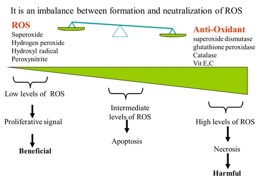
Oxidative stress can be defined as an imbalance between Reactive Oxygen Species (ROS) production and anti-oxidant defense as seen in (Fig.1).
Oxidative stress
Low levels of ROS Proliferative signal
Intermediate levels of ROS
Apoptosis
High levels of ROS Necrosis
Beneficial
Harmful
It is an imbalance between formation and neutralization of ROS
ROS Superoxide Hydrogen peroxide Hydroxyl radical Peroxynitrite Anti-Oxidant superoxide dismutase glutathione peroxidase Catalase Vit E,C
Figure 1: Oxidative stress
The presence of potent cellular detoxification systems minimizes radical generation, terminates radical processes, and repairs damaged macromolecules. However, continued overproduction of ROS and free radicals can overwhelm antioxidant defense and become deleterious to cellular biological processes and tissue functions [1]. Oxidative stress has been implicated in a number of diseases that include atherosclerosis, cancer, and
neurodegenerative diseases, Parkinson’s disease, multiple sclerosis, aging and gastro-intestinal. [1-4].
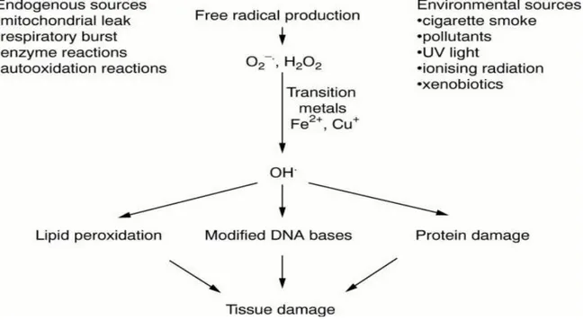
1.1.1 Reactive oxygen species generation and oxidative damage
ROS are free radicals with one unpaired electron derived from molecular oxygen. The most important free radicals are oxygen derivatives, particularly superoxide anion (O2–•),
hydroxyl radical (OH-) and hydrogen peroxide (H2O2), as well as reactive nitrogen species
such as nitric oxide and peroxynitrite. Radical formation in the body occurs through several mechanisms, involving both endogenous and exogenous sources, such as environmental factors that lead to tissue damage as illustrated in (Fig. 2) [3, 4].
Figure 2: Major sources of free radicals in the body and the consequences of the free radical damage. Young IS J Clin Pathol. 2001
Under normal physiological conditions, ROS are generated by immune cells as a non-specific defense mechanism against invading organisms, a low level of ROS constitutes inter- and intra-cellular signals, which are vital to maintain proper cellular function [3, 5]. In addition, immune cells, structural cells like epithelial cells, are also involved in the ROS production
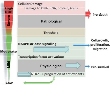
However, under pathological conditions, progressive and irreversible generation of ROS increases to a point where the antioxidant system cannot effectively counteract them, and results in impairing physiological functions [4-6]. Thus, depending on their cellular concentrations and duration, ROS can act as either beneficial or harmful agents, as shown in (Fig.3). Pathological Physiological S ever e M ild
High ROS concentrations (pathological condition) can affect various subcellular compartments that lead to oxidize mitochondrial enzyme complexes, plasma membrane, nucleic acids (nuclear and mitochondrial DNA), calcium homeostasis, lipid and protein damage as projected in Fig.4 [1, 3, 6].
Maintaining the protein homeostasis via the regulation of protein unfolding and aggregation is crucial since it shares close relationship with other intracellular pathways, including oxidative stress and mitochondrial dysfunction to impact cell survival and death pathway. Under pathological conditions, the level of abnormal proteins may exceed the ability of the cell to degrade them, allowing aggregation to proceed. Increased oxidative stress results in the accumulation of aggregated proteins that have been suggested to underlie the loss of cellular function.
Oxidative damage modifies a large variety of proteins, including mitochondrial protein and proteins involved in signal transduction, which is often irreversible due to impairment in protein turnover and the need to be degraded by the ubiquitin protein system. However, these damaged proteins are prone to aggregate, poorly degraded and become toxic to the cell [7, 8].
Oxidative damage to mitochondrial and nuclear nucleic acids causes modification of pyrimidine and purine bases and commonly mitochondrial and nuclear DNA damage, measured as increased in 8-hydroxydesoxyguanosine (8-OxO) [9]. Mitochondrial DNA is highly susceptible to ROS induced damage because it is located in close proximity to the production site of ROS, and the mt DNA repair mechanisms are limited. ROS induced mt DNA deletion leads to a decline in the mitochondrial function and enhances ROS production [9, 10].
Oxidative stress and subcellular
compartments
Plasma membrane
Mitochondria Endoplasmic reticulum Nucleus • lipid peroxidation •receptor/transport activity disrupted • membrane signaling altered • generation of ROS
• impact on different pathways •• Accumulation of mis-folded proteinsLoss of Ca2+ homeostasis
•oxidative DNA damage • disrupted DNA binding & gene transcription
Figure 4: Oxidative stress affects various compartments with macromolecules damage
Furthermore, oxidative attacks to polyunsaturated fatty acids (PUFA) in mitochondrial and cellular membranes may trigger lipid peroxidation, resulting in oxidative breakdown of cellular membranes [6, 11].
Mitochondria are a major source of ROS generation. In the mitochondria, O2–• can be
produced by respiratory complexes, enzymes on the outer mitochondrial membrane, inner mitochondrial membrane and in the matrix [7]. Non enzymatic production of O2–• occurs
mainly in the mitochondria, particularly at complex I and III of the mitochondrial electron transport chain (ETC) [12].
Outside of mitochondria, several enzymatic systems are the major sources of O2–• that are
responsible for the production of the intracellular ROS, including NADPH oxidases [13], xanthine oxidase [14] and cytochrome P450-dependent oxygenases [15].
Any condition that results in over-consumption of oxygen can lead to the production of free radicals. It is estimated that up to 1-5% of the oxygen that is passed through the ETC inside the mitochondria results in O2–• production [16, 17].
O2 + e- → O2- Superoxide radical
O2- + H2O → HO2 + OH- Hydroperoxyl radical
HO2 + e- + H → H2O2 Hydrogen Peroxide
H2O2 + e- → OH + OH- Hydroxyl Radical
Under pathological conditions, O2–• can react with either luminal or mucosal iron through
the Fenton reaction, to produce toxic and highly reactive OH•, leading to a more free radicals production. OH• is also formed from H2O2 through the Fenton reaction [18] or
from O2–• through another transition metal-dependent reaction, called the iron-catalyzed
Haber–Weiss reaction [19] and is considered to be the most reactive ROS [20].
H2O2 is generally considered as a relatively weak ROS that might directly damage proteins
and enzymes containing reactive thiol groups. It has the ability to react with partially reduced metal ions, such as Fe2+ or Cu+, to form OH• in Fenton reaction [18].
1.1.2 Iron over-consumption induce oxidative stress
Free iron is a potential source of oxidative stress because it catalyses the conversion of H2O2 into highly OH•. Iron, like oxygen, is essential for life, but together they can form a
highly ROS [21, 22]. The mucosal iron concentration has been observed to be significantly increased in the presence of inflammation, at least in part due to overproduction of free radicals via increased levels of free haemoglobin from mucosal ulceration and bleeding [23]. Subsequent increased in iron may trigger a self perpetuating cycle, resulting in further tissue damage and more inflammation [24].
High concentrations of iron decrease oxidative phosphorylation (OXPHOS) and the total electron transport proteins in the mitochondria. This disturbance of electron movement can lead to increased mitochondrial H2O2 production [25]. The combination of increased iron
and the physiological concentrations of H2O2, generated in the mitochondria during
OXPHOS might result in DNA damage [22, 26]. Thus, accumulation of mtDNA damage and subsequent decline in mitochondria function could be a primary cause in various chronic inflammatory diseases.
Free radicals interact with iron and can potentially enhance intestinal inflammation through propagation of lipid peroxidation whereas ascorbic acid can amplify the oxidative potential of iron by promoting metal ion-induced lipid peroxidation [21, 23, 25]. Ascorbic acid, an essential micro-nutrient for the physiological metabolic function, has a significant antioxidant role by scavenging active oxygen species. On the other hand, the interaction of
ascorbic acid with free, catalytic active metal ions could participate in oxidative damage by releasing hydroxyl and alkoxyl radicals. This is further highlighted in patients receiving ferrous as a supplement, as ascorbic ferrous negatively affects the function of intestinal cells.
Iron salts and ascorbic acid, frequently consumed together in multiple-vitamin preparations or ingested foods, form reactive hydroxyl radicals. Clinical observations suggest that oral iron supplementation is not well tolerated and may increase gastrointestinal symptoms and exacerbate disease activity [21, 23, 27]. Thus the cross-talk between iron homeostasis and intestinal inflammation will yield new insights into the pathogenesis of chronic inflammatory diseases and may suggest new therapeutic approaches for these diseases.
Under normal condition, transition metal ions, such as iron and copper, are kept sequestered in cytosol by a number of chelating proteins like ceruloplasmin, ferritin and transferrin given their capacity to aggravate ROS generation. This large amount of chelating iron is released in response to oxidative stress [22]. At the same time, mitochondria are taken up the released chelating iron from cytosol, which may play a critical role in augmenting oxidative stress.
1.1.3 Antioxidant: definition and classification
Antioxidants have been defined as substances that are able,at relatively low concentrations, to significantly reverse the increase of ROS. Antioxidants cannot prevent ROS generation; they rather bring into balance the effects of ROS function.
The physiological role of antioxidants is to prevent cellular damage arising as a consequence of free radicals generation. In many disease states, however, mechanisms that prevent or limit ROS damage may become inadequate. In general, antioxidants prevent free radical-induced tissue damage by preventing the formation of radicals, scavenging them or promoting their decomposition [4, 28].
The most primary antioxidants counteracting free radicals are either enzymatic such as intracellular Superoxide Dismutase (SOD), Catalase (CAT) , or non-enzymatic antioxidants that include vit C, E, and A [3, 7] as seen in Fig. 5
There are three forms of SOD in the mammalian tissues, each with a specific subcellular location and different tissue distributions:
(1) Copper zinc superoxide dismutase, found in the cytoplasm.
(2) Manganese superoxide dismutase, located in the mitochondria.
(3) Extracellular superoxide dismutase, expressed on the cell surface [29, 30].
There are several dietary compounds with antioxidant properties, non-enzymatic antioxidants, normally originating from natural sources, such as fruits, vegetables and plant extracts that play a key role in the host defense and cell survival. In particular, certain minerals (e.g. Zinc), vitamins (C and E) and the flavonoids found in these extracts are considered to be of prime interest [4,31- 35]. Furthermore, the human gut naturally contains a variety of non-enzymatic antioxidant defences. These include water-soluble agents, such as glutathione and ascorbic acid (vitamin C), as well as lipid-soluble defences, such as α-tocopherol (vitamin E) and ubiquinol (reduced co-enzyme Q10) [2, 4, 33-36].
Glutathione (GSH) is a significant intracellular peptide with multiple physiological functions, including antioxidant defensive actions as well as regulatory mechanisms that are able to promote intracellular processes. Additionally, GSH important functions include: (A) The reduction of various oxidative insults.
(B) The maintenance of the basic structure of protein thiols. (C) Maintaining of the cysteine reserve.
(D) The modulation of DNA synthesis and immune function. (E) An anti- apoptotic function [31, 32]
Although evidence shows that antioxidant treatment results in beneficial effects, the clinical benefit from antioxidants is still under wide debate. This remains an important area for future investigation, as researchers attempt to augment intracellular antioxidant, either by dietary supplementation of antioxidants or by overexpressing genes or transcription factors such as Nrf2 encoding antioxidant enzymes, which I will discuss in details in the 2nd part of my thesis.
1.1.4 Oxidative stress and intestinal inflammation
The intestine represents a key defense barrier against luminal toxic agents. Thus, in addition to being exposed to luminal nutrients, the intestinal mucosa is constantly challenged by diet-derived oxidants, mutagens, and carcinogens as well as by endogenously generated free reactive radical [23, 24].
The primary event of the induction of cellular oxidative stress is the inflammatory cascade of neutrophil adherence to vascular endothelial cells, disruption of the endothelial barrier, and subsequent infiltration of inflammatory cells into the intestinal wall, where oxidants and proteases are released to produce mucosal injury [37].
Oxidative stress appears to be a primary causal factor in generating and maintaining a chronic intestinal inflammation, which is characterized by the activation of neutrophils and macrophages, as well as generation of numerous pro-inflammatory mediators, including IL-1β , tumour necrosis factor alpha (TNF-α) , IL-8 and IL-6 [38-40]. It is well known that TNF-α is involved in ROS production; ROS in turn activate nuclear factor- kappa B
(NF-κβ), which then enhances further TNF-α production, making a vicious cycle of excessive oxidative stress production [41-43] as appears in (Fig.6 ).
Oxidative stress Inflammation
Inflammation-Oxidative stress vicious cycle
Pro inflammatory cytokines
• TNF-α • IL-6 • IL-1β • IL-8
Reactive oxygen species
•Superoxide • Hydrogen peroxide • Hydroxyl radical • Peroxynitrite
Figure 6: Inflammation and oxidative stress vicious cycle
There is an evidence that ROS and pro-inflammatory cytokines work synergistically as activators of NF-κβ and activator protein-1, thereby modulating their activity [44]. However, activation of NF-κβ has been specificallyimplicated in maintaining inflammation by the activation of a variety of inflammatory genes [45].
Enhanced NF-κβ signalling in the gut epithelium initiates the primary events of intestinal inflammation and leads to defects in the intestinal epithelial barrier. A recent study by Banan et al suggests that the loss of intestinal epithelial barrier function caused by the exposure to oxidants is lessened by inhibition of NF-κβ activity [27, 40]. There is an
increasing awareness of a role for cellular thiol redox status in NF-κβ activation and gene expression. Staal et al showed that low thiol concentrations promote NF-κβ activation, suggesting that intracellular thiol status has a key role in regulating gene activation [46].
Studies on TNF-α -mediated inflammation have linked TNF-α induced mitochondrial oxidant production with induced NF-κβ activation [29]. Interestingly, Cogswell et al have found that NF-κβ and IκΒ-α are localized in the mitochondria and can negatively regulate mitochondrial gene expression in response to cellular TNF-α stimulation. Furthermore, mitochondrial exposure to TNF-α has confirmed the loss of expression of cytochrome C oxidase and cytochrome b mRNA, which down-regulates mitochondrial biogenesis [47].
TNF-α−induced oxidative stress and mitochondrial dysfunction in cell lines and tissues are evident in several patho-physiological conditions. In cardiac tissues, Maria pan N et al have shown that TNF-α induction leads to defects in mitochondrial permeability transition pore (PTP) proteins, resulting in pore opening, cytochrome C release and subsequently leading to apoptosis and mitochondrion dysfunction in cardiac tissues [48, 49].
The various mechanisms whereby activation of TNF- α modulates chronic inflammation and mitochondrial damage in gastro-intestinal diseases remain to be elucidated. Such ROS/cytokine-/transcription factor regulatory network loops may contribute to the perpetuation and exacerbation of chronic inflammation and tissue damage, particularly when the local immune response fails to successfully down-regulate the immune reaction.
Increased levels of TNF-α have been found in serum, mucosa and stool of patients with inflammatory bowel diseases (IBD) [50, 51]. Several clinical trials have shown that anti-TNF-α antibody is effective in the treatment of intestinal inflammation, supporting the importance of TNF-α in the ongoing regulation of epithelial barrier functions in association with increased apoptosis [52, 53].
1.1.5 Intestinal permeability and tight junction proteins
The mucosal barrier is established by the single layer of intestinal epithelial cells. The intestinal epithelium is a polarized monolayer of columnar epithelial cells, which forms a barrier between the luminal contents and the mucosa. Its role is to achieve an efficient absorption of nutrients, water and ions, while preventing the uptake of noxious antigens, microbes and toxins. Epithelial permeability is influenced by the integrity of the epithelial cell layer and the basement membrane, as well as by the surface mucus layer and by autonomic nervous system functions [54, 55].
In intestinal inflammation, lipid peroxidation causes disruption of mucosal barrier function, increased permeability and a reduction of tight junction proteins functions, which ultimately leads to alterations in cellular metabolism [56]. Epithelial barrier dysfunction and inflammation are major contributors to the pathogenesis of gastro-intestinal diseases. However, much remains unknown about how these two processes contribute independently to disease initiation.
The movement of particles in both small and large intestinal parts occurs through two pathways: paracellular and/or transcellular. Numerous stimuli, including microbial components, smoking, anti-inflammatory drug use and proinflammatory cytokines, can regulate paracellular permeability [54]. The integrity of epithelial barrier is dependent on the function of tight junctional proteins (TJs) located near the apical poles of two adjacent epithelial cells. TJs are composed of multiple proteins (claudin family, occludin, zonula occludin-1) that are involved in establishing the epithelial barrier, and they selectively determine which molecules are able to traverse the paracellular space [57-59].
Occludin was the first identified transmembrane protein of this intercellular junction. TJs seem to interact together as networks and it appears that occludin interacts, directly or indirectly, with claudin. Conceivably, occludin has increased electrical resistance by influencing the extracellular conformation of Claudins [57, 58].
The TJs are dynamic structures that are rate limiting for passive absorption of hydrophilic molecules in the intestine, which should be determined by intestinal permeability [59]. Thus, TJs dysfunction may be an important source of the overall intestinal barrier defects, leading to increased permeability very often seen in patients with chronic intestinal inflammation. In vitro experiments examining the effects of inflammatory cytokines on model intestinal epithelial cells suggest that disruption of the epithelial barrier is associated with internalization of transmembrane TJ protein such as occludin [55, 60, 61].
1.2 Mitochondria
1.2.1 Mitochondrion structure and function
Human mitochondrion contains multiple copies of a small double-stranded DNA genome that encodes 13 proteins (subunits of complexes I, III, and IV and the ATP synthase complex), 2 Ribosomal Ribonucleic Acids (RNAs), and 22 transfer RNAs that are needed for mitochondrial DNA translation. Therefore, these organelles represent a critical intracellular target for oxidative damage, which may lead to lethal injury through the loss of electron transport, mitochondrial membrane potential and ATP generation [12, 62, 63].
The mitochondrial genome is transcribed by machinery that includes RNA polymerase, the mitochondrial transcription factor A (mt TFA) and two mitochondrial transcription factors, mt TFB1 or mt TFB2 [64]. Although mitochondria have their own genome, most of the proteins and enzymes that reside in mitochondrial membranes are nuclear gene products. Mitochondria possess both an outer and inner membrane, the latter of which has a larger surface area, is impermeable to all molecules and contains the enzymes responsible for OXPHOS and ATP production as seen in (Fig. 7)
Figure 7: Mitochondrial structure
Mitochondria are multifunctional organelles that receive, integrate and transmit signals [65] and are involved in numerous metabolic activities, including ATP production, (OXPHOS), ROS generation and detoxification, calcium (Ca2+) homeostasis and regulation of apoptosis pathways [65-67].
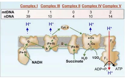
1.2.2 Electron Transport Chain Complexes and ATP production
Mitochondria produce 90% of the required energy necessary for cellular function via adenosine-5'-triphosphate (ATP) production either by anaerobic glycolysis or by mitochondrial OXPHOS. During OXPHOS, electrons from reduced substrates are transferred to O2 through (ETC), including complex I, III, and IV, which in turn generate a
phosphorylation in the matrix to generate ATP [69]. It appears that any blockage in the ATPproduction result in a severe impairment of one ormore of these complexes. Excess electrons, however, may react with oxygen to form more ROS production that plays a key role in creating oxidative stress [70] as shown in (Fig.8).
Figure 8: Summary of protein subunits of the five Respiratory chain complexes encoded by nuclear and mitochondrial genes
1.2.3 Mitochondrial function and oxidative stress
Mitochondria produce the energy needed for normal cellular function and metabolic homeostasis by OXPHOS and serve as biosensors for oxidative stress. Mitochondria constitute a major source of free radicals in cells, resulting in oxidative stress, but are also targets to oxidative stress action. Although the molecular mechanisms responsible for mitochondrion-mediated diseases are not fully elucidated, yet oxidative stress appears to be
a pivotal player. Hence, it is apparent that mitochondrial damage may lead to the impairment of various aspects of tissue functioning.
It is now appreciated that the reduction of mitochondrial oxidative stress may prevent or slow down the progression of many mitochondria dysfunctions related to oxidative stress diseases, including aging, obesity, diabetes and neurodegenerative disorders [10, 70-73]. However, if mitochondria are the major source of intracellular ROS and mitochondria are most vulnerable to oxidative damage, then it would be ideal to deliver the antioxidant therapy to mitochondria [74].
Dietaryantioxidants are widely used to ameliorate excessive oxidativestress, but scientific proof of their efficacy is poor. The currently existing antioxidants are not very effective in overcoming oxidative stress-mediated diseases. One of the reasonable answers for the failure of antioxidants to show clear therapeutic effects is their incapacity to reach mitochondria. There are currently growing efforts in developing mechanisms for the targeted delivery of antioxidants to mitochondria [74, 75]. More recently, confocal imaging studies in Caco-2 cells show that intracellular distribution of SS-19 tetrapeptidase antioxidant resembles that of MitoTracker, which localizes in mitochondria [76].
1.2.4 Mitochondrial DNA damage related to oxidative stress
Increased Oxidative Stress may contribute to alterations in the abundance of mitochondria as well as the copy number and integrity of mt DNA in human cells, which is vulnerable to
particular since mitochondria lack protective histones and have much more reduced base excision repair mechanisms [73].
Oxidative stress has been one of the risk factors that induce gut malignancy. If oxidative stress is irreversible, these damages lead to mutagenesis and carcinogenesis [77]. The accumulation of mt DNA mutations cause the loss of mitochondrion ability to produce sufficient energy to meet cellular needs and serves as a trigger for mitochondrial dysfunction and apoptosis [78]. Mutated mtDNA shows ~10- fold more mutation rates than nuclear DNA in relationship with increased free radical production, thereby leading to a vicious cycle that progressively stimulates the rise of oxidative stress and leads to impaired mitochondrial function.
Oxidative DNA damage was measured by the production of 8-hydroxy-deoxy-guanosine (8-OHdG). This molecule (8-OHdG), which is more specific for mt DNA, is of practical importance because it is easily measurable and has therefore been proposed as a useful marker of oxidative stress [9, 79]. Human cells have developed different repairing enzymes, the most important being 8-oxo DNA Glycosylase (OGG1) that protects against the effects of oxidized DNA bases and preferentially removes 8-OHdG opposite cytosine [79, 80]. In mitochondria, OGG1 is thought to prevent activation of the intrinsic apoptotic pathway in response to oxidative stress by augmenting DNA repairing mechanisms [73].
It has been proposed that mt DNA damage can lead to inhibition of ETC, increased ROS production, loss of mitochondrial membrane potential, and released signals for cell death, such as cytochrome C and AIF [62, 81, 82]. Therefore, mt DNA damage represents an important target for intervention and a biomarker in the course of many human diseases.
1.2.5 Oxidative stress induce mitochondrial calcium overload
Mitochondria may have an impact on Ca2+ signals and functions as a buffer to stabilize calcium concentrations within the cell [83, 84]. Thus, mitochondrial Ca2+ is considered as a physiological regulator to balance mitochondrial ATP output and cellular ATP demand.
A growing body of evidence suggests that Ca2+ channels, which control Ca2+ efflux from the endoplasmic reticulum (ER) in response to different biochemical signals, are sensitive to small changes in ROS concentrations, suggesting that these Ca2+ channels serve as physiological redox sensors [67, 84]. Under physiological conditions, Ca2+ released from the ER during cell activation is taken up by mitochondria to promote OXPHOS.
During oxidative stress, there is a rapid increase in cytosolic Ca2+, which is followed by mitochondrial Ca2+ over loading [85]. At its turn, Ca2+ overload can lead to induced ROS production and another potential vicious cycle develops, thereby triggering mitochondrial (PTP) opening and cytochrome C release and resulting in apoptosis.
The exact mechanism of mitochondrial Ca2+ induced ROS generation is unclear, although it
may involve changes in the three-dimensional conformation of the respiratory complexes [72]. Specifically complex results in an increase of H2O2 production and inhibits OXPHOS,
which may provoke an irreversible reduction in the energy status, thereby initiating patho-physiological processes in certain cells [86, 87].
A recent study suggests that disruptions in intracellular Ca2+ mobilization may contribute to the dysmotility of colonic smooth muscles in murine dextran sulfate sodium (DSS)-colitis
by enhancing NF-κβ activity [88]. Furthermore, Di Sabatino A et al have provided evidence that the suppression of Ca2+ overloading may inhibit pro-inflammatory cytokine
release ininflamed gut. Therefore, Ca2+ channel inhibitors might reduce mitochondrial Ca2+ overload, decrease mitochondrial ROS accumulation, improve mitochondrial energy production and have the potential to ameliorate mitochondrial oxidative stress-mediated diseases [89].
1.2.6 Mitochondrial dysfunction and apoptosis
Apoptosis, known as programmed cell death, plays a vital role in all stages of cell development. Mitochondria play a checkpoint of apoptotic signalling and integrate various types of proapoptotic endogenous signals incoming from other organelles, including nucleus, cytosol and lysosomes as well as exogenous factors including specific viral proteins or xenobiotics [62, 81].
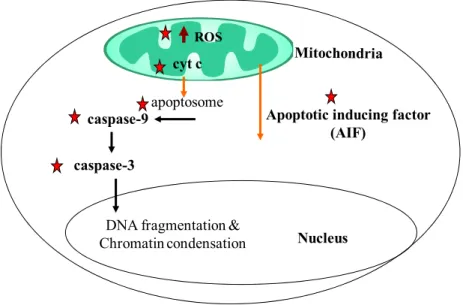
Apoptosis may be triggered by extracellular signals (extrinsic pathway) or by intracellular processes (intrinsic pathway). An increased mitochondrial formation of ROS triggers the intrinsic pathway by increasing permeability of the outer mitochondrial membrane through the opening of PTP, leading to collapse of mitochondrial membrane potential, along with release of apoptogenic mitochondrial proteins into the cytoplasm, which triggers a cascade of events, leading to apoptosis [62, 90] as seen in (Fig. 8).
The release of different proteins from the mitochondria is a critical early event in mitochondrial mediated apoptotic cell death. Cytochrome C is such a protein that, upon releasing into the cytosol, forms a complex with procaspase-9, resulting in the activation of the caspase cascade and ultimately in apoptosis [26, 62, 82]. Early release of cytochrome C would be the early sign of apoptosis and suggests the occurrence of mitochondrial dysfunctions along with apoptosis development [76]. The gradual loss of cytochrome C from the intermembrane space during apoptosis favours the mitochondrial formation of O2–
• in two ways: (1) cytochrome C is a scavenger of O2–• and (2) as cytochrome C is released,
the electron flows between Complex III and Complex IV, slows down, and the respiratory chain becomes more reduced [82, 91].
The mechanism of cytochrome C release connects the upstream event of cytochrome C (i.e., dissociation from cardiolipin). Hence, cytochrome C must first dissociate from the inner membrane in order to escape mitochondria. Therefore, oxidation of cardiolipin may be one mechanism by which cytochrome C is solubilized in the intermembrane space [26, 76].
Nucleus caspase-3
DNA fragmentation & Chromatin condensation
caspase-9
cyt c
apoptosome
ROS
Apoptotic inducing factor (AIF)
AIF and Cytochrome C protein level
Figure 9: Caspase-depended (Extrinsic) pathway and caspase- independed (intrinsic) pathway
Apoptosis is also initiated by mitochondrial intermembrane space protein called AIF, involved in initiating a caspase-independent pathway of apoptosis (positive intrinsic regulator of apoptosis) through DNA fragmentation and chromatin condensation [62]. AIF normally stabilizes mitochondrial membrane permeability and supports OXPHOS. However, if released through the outer membrane into the cytosol, AIF can produce terminal damage to n DNA [92].
Apoptotic pathways contain counter balancing concentrations of anti-apoptotic and pro-apoptotic Bcl-2 family [93]. These proteins are normally found in the cytosol but can be induced to target mitochondria. All of these Bcl-2 family proteins are encoded within the nuclear genome, transported into mitochondria and stored in the space between the inner
and outer membranes [94]. Under physiological conditions, anti-apoptotic Bcl-2 remains bound to the outer membrane and prevent the mitochondrial PTP. In contrast, when pro-apoptotic factor like Bax is induced and translocated to mitochondria, it initiates the apoptosis process [91]. The relative ratio of anti- and pro-apoptotic Bcl-2 family proteins dictate the ultimate sensitivity of cells to various apoptotic stimuli, including oxidants and Ca2+ overload [62, 91].
Bcl-2 family proteins regulate the release of cytochrome C, AIF and certain caspases (caspase-2, -3, and -9) from mitochondria. Pro-apoptotic Bcl-2 enhances release of this caspase-activating protein and anti-apoptotic members of the family, which stops cytochrome C release [62, 81]. Furthermore, it has been suggested that the anti-apoptotic functions of Bcl-2 may be (at least in part) associated with depletion of ER Ca2+, thus indirectly reducing mitochondrial Ca2+ uptake [67].
1.2.7 Prohibitin protects mitochondrial dysfunction
Prohibitins (PHB) are ubiquitous chaperone proteins forming a ring-like, high-molecular-mass complexes that are mainly localized in the inner mitochondrial membrane, help in the stabilization of mitochondrial respiratory enzymes and mitochondrial membrane proteins, and are implicated in mitochondrial biogenesis, mitochondrial function, mitochondrial morphogenesis and apoptosis [95]. Deficiency of the PHB complex results in increased ROS production or sensitivity to free radicals [96]. Abnormal PHB levels have been reported in mitochondrial dysfunction related to oxidative stress diseases, including obese
Interestingly, it has been also found that PHB expression is down-regulated in intestinal diseases in experimental colitis in vivo and during oxidative stress in vitro. Furthermore, Theiss et al have shown that TNF-α decreases PHB expression in intestinal epithelial cells, and restoration of PHB expression in these cells can protect against the various effects of TNF-α and NF-κβ on intestinal barrier function [99, 100]. More recently, it has been shown that PHB is a regulator of Nrf2 expression in intestinal epithelial cells during oxidative conditions that prevent inflammation-associated oxidative stress and injury through sustained activation of Nrf2. The above findings emphasize the importance of the PHB complex in maintaining mitochondrial homeostasis that is crucial for human health.
1.2.8 Nucleus-mitochondrial cross talk, role of PGC-1 α
Mitochondrial functions must rely on an orchestrated cross-talk between nuclear and mitochondrial genes and it clearly appears that the nucleus has a dominant role in the regulation of mitochondrial activity. Nuclear transcriptional factors control the activity of mitochondrial genome and coordinate the expression of both nuclear and mitochondrial genes encoding mitochondrial proteins. Nonetheless, nuclear gene expression can be influenced by signals derived from mitochondria, through retrograde communication, so that the regulation of mitochondrial activity is exposed to a bidirectional flow of information [101, 102].
Crosstalk between nucleus and mitochondrial genes contribute to mitochondrial protection against oxidative stress via enhancing antioxidant defense through the Nrf2 pathway that
acts on the genes coding for constituent subunits of the OXPHOS system and mtDNA replication or by stimulating mitochondrial biogenesis through the activation of peroxisome proliferation activator receptor γ-coactivator 1α pathway (PGC-1 α) [68, 103]. PGC-1α may serve as an adaptive set-point regulator, capable of providing an accurate balance between metabolic requirements and cytotoxic protection. Therefore, its dual activities of inducing mitochondrial biogenesis and suppressing ROS make PGC-1α an almost ideal target protein for the control damage associated with mitochondrial dysfunction.
PGC-1α interacts with Nrf-2 to transactivate a number of genes involved in mitochondrial functions such as OXPHOS regulation, protein import and heme biosynthesis. It also mediates mt DNA transcription and replication through two nuclear-encoded genes, the mt TFA and B (mt TFB) [103, 104]. It is suggested that the Nrf-2 pathway could be affected at the levels of transcription, translation or protein levels.
Surprisingly, a more recent study has shown that mice with a targeted disruption of PGC-1α are viable and show no changes in mitochondrial abundance or morphology in liver or brown fat [104]. The retrograde or compensatory pathway has been proposed: free radicals generated from respiratory chain are involved in the signalling pathway from mitochondria to the nucleus in order to enhance the expression of nuclear genes involved in mitochondrial biogenesis such as Nrf2 [101, 102].
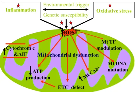
Mitochondrial dysfunction has gained more attention in recent years, whereby electrons leaking from the ETC generate ROS that in turn damage ETC components and mitochondrial DNA, which leads to an additional increase in intracellular ROS levels and defects in mitochondrial functions [48, 105, 106]. Because dysfunctional mitochondria will produce more ROS, a feed-forward loop is set up whereby ROS-mediated oxidative damage to mitochondria favours more ROS generation, then resulting in a vicious cycle. This mitochondrial dysfunction causes cell damage and death by compromising ATP production, disruption of calcium homeostasis, damage of mt and nuclear DNA, increases in cytochrome C and AIF release, impairment in various proteins and lipid peroxidation (Fig 10): Oxidative stress Inflammation Genetic susceptibility Environmental trigger Mt TF modulation Mt DNA mutation Cytochrom c &AIF ETC defect ATP production
Mitoc
hondrial dysfunctionI
ROS
Since mitochondria are attractive targets for drug-delivery strategies, it would be ideal to deliver the antioxidant therapy directly to mitochondria [75]. The mitochondrial outer membrane is permeable to small molecules, and thus the inner membrane represents the major barrier for drug delivery to mitochondria. However, mitochondria-targeted antioxidants research is still in infancy and needs sufficient time to yield more reliable data.
1.2.10 Implication of oxidative stress and mitochondrial dysfunction in inflammatory bowel diseases
Inflammatory Bowel Diseases (IBD) are idiopathic, chronic and relapsing inflammatory conditions of the gastrointestinal tract. They are recognized as important causes of gastro-intestinal diseases in children and adults and are common in highly industrialized western countries [107, 108]. Crohns Disease (CD) and Ulcerative Colitis (UC) are the two main clinic-pathological subtypes of IBD [109, 110].
Although the incidence and prevalence of IBD are beginning to stabilize in high-incidence areas such as North America and Northern Europe, they continue to rise in low-incidence regions such as Southern Europe, Asia, and much of the developing world [111, 112]. There are about 201,000 Canadians living with IBD: 112,500 with CD and 88,500 with UC. Canada is among the highest reported prevalence and incidence of IBD in the world [113]. Economic costs in 2008 for IBD are estimated at $1.8 billion per year in Canada [114, 115]. There has been a significant increase in IBD morbidity that exerts enormous economic burden and makes IBD a public health issue [114, 116].
No precise aetiology has been identified for IBD. Epidemiological studies have stressed the involvement of environmental and genetic factors and especially anomalies in the immune system in response to microbial infections [108, 110] as seen in Fig.11.
Environmental Trigger Genetic Susceptibility Luminal Microbial infection Immune response IBD Pathophysiology of IBD
Figure 11: IBD risk factors
The strongest environmental factors being identified are smoking and diet [113, 117, 118]. It is clearly established that smoking is the most consistently environmental risk factor reported to be associated with CD. A meta-analysis suggests that smokers are more than twice likely to develop CD compared to non-smokers [119, 120]. Several other studies have considered the role of dietary factors in the pathogenesis of IBD, while immunological mechanisms have been suggested to link food antigens to the development of intestinal inflammation [121].
It is postulated that the higher incidence of IBD seen in developed countries may be associated with dietary habits. Persson et al suggest that frequent fast food intake and increased simple sugar consumption confer a 3-to-4-fold greater risk for IBD [122]. Whether other suggested factors such as western life-style, social and occupational status, psychological stress, sanitation, appendectomy, drugs and exposure to infections play a role in the expression of IBD remains unclear [123-126] .
Previous findings suggest that IBD incidence appears linked to race. A strong genetic component in IBD aetiology is most likely related. Some epidemiological data support this hypothesis: (1) the heterogeneity in the geographical distribution of IBD, (2) the existence of familial forms of IBD and (3) the high rate of concordance in monozygotic twins [127-130].
Various mechanisms have been discussed concerning immune dysfunction in IBD, including defects in immune response to normal luminal components, and/or defective mucosal barrier to luminal antigens such as diet and enteric bacteria [131, 132] as seen in Fig. 12.
Pathophysiology of IBD
-Summary
Pathophysology of IBD
Oxidative Stress IL-8, IL-1 TNF-α, IL-6 ROSFigure 12: Pathopghysiology of IBD
The intestinal epithelium is the first host defense against invading pathogens and food antigens acting as key mediators of communication between the intestinal lumen and mucosal immune system. The gastrointestinal mucosa is constantly exposed to luminal oxidants from ingested foods that might contain iron salts and ascorbic acid [21, 131].
The ingestion and/or occurrence of peroxides may have significant implications in human health in the long term. In this regards, ROS metabolites have been suspected to provoke injury to intestinal mucosa in various diseases, including intestinal ischemia and subsequent reperfusion, as well as IBD [133, 134].
Oxidative stress is currently considered among the most plausible explanations for IBD in human [3, 35, 134, 135]. Markers for oxidative damage can be measured in plasma, urine,
or post-mortem tissue by means of HPLC and immunohistochemistry [4, 33]. The increased in malonyldialdehyde (MDA), CH2 (CHO)2, levels found in plasma and colonic
biopsies from CD patients provide evidence for excess lipid peroxidation reactions [134, 136]. Furthermore, the increased breath ethane and pentane excretion in CD patients, which are non-invasive markers of lipid peroxidation, have been correlated with disease activity [35].
The oxidative stress parameters in CD will be as follow: 1. Breath ethane output (pmol•kg-1•min-1)
2. Breath pentane output (pmol•kg-1•min-1) 3. F2-isoprostane (ng/L)
4. MDA
There are many studies supporting the notion that a decline in antioxidant activity occurs with CD. Lih-Brody et al found a decrease in SOD in the mucosa of CD patients [137], which was correlated to the activity of the disease [30]. Furthermore, Buffinton GD et al observed a reduction of total glutathione in inflamed mucosa of patients with CD compared to normal tissue areas [138].
Interestingly, reduced levels of PHB during intestinal inflammation may be one underlying factor that contributes to oxidant-induced mucosal barrier and intestinal permeability dysfunction [99, 100]. Finally, in a more recent report, Beltran B et al have demonstrated that ROS are directly implicated in the oxidative damage that occurs in CD patients. Additionally, mitochondrial membrane potential of CD cells is significantly inhibited compared to control cells and correlates significantly with markers of inflammation [139].
Surprisingly, even if oxidative stress occurs in the intestinal mucosa, limited studies have explored the response of mitochondria to oxidative stress in the intestinal tissue in relation with the key cellular regulatory processes, including (ATP) production, intracellular calcium regulation, cell signaling, ROS generation and apoptosis.
A number of findings have suggested that mitochondrial pathology related to OS may be associated with CD. Mitochondrion morphological changes have been observed in epithelial cells in tissues resected from patients with CD and in animal models of gut diseases [140, 141]. Farhadi et al discussed that mitochondrial damages were noted in intestinal epithelial cells and mucosal protein oxidation in stressed CD patients [100]. Furthermore, Soderholm et al. observed the presence of numerous swollen and irregular mitochondria in the epithelium of colonic segments from stressed rats [39]. O'Morain et al, on the other hand, showed mitochondrial damage through examining rectal biopsies obtained from CD patients [142].
Furthermore, Plasma levels of 8-OHdG was found to be increased in CD patients compared to controls [143, 144]. It has also been shown that 8-OHdG levels were significantly higher in active CD, inactive CD and CD patients in remission compared to controls [139]. A recent report provides evidence on the association between intestinal inflammation, oxidative stress, DNA repair enzyme OGG1 and carcinogenesis. OGG1 (-/-) mice developed a significantly higher number of adenocarcinomas in the DSS colitis model compared to wild type mice [80]. Finally, a recent case report on a young girl with CD disclosed impaired OXPHOS along with abnormalities in Complexes III and IV [103].
1.3 Nuclear transcription factor (Nrf2)
1.3.1 Nrf2 structure and functions
Nrf2, a basic leucine-zipper motif transcription factor, plays a key role in the regulation of phase II genes by binding to the ARE element in their promoters in conjuction with small Maf proteins [145, 146]. Nrf2 is one of the redox sensitive transcription factors, which gets activated in response to the increase in ROS. It attempts to restore the redox haemostasis by transcribing antioxidant proteins. Nrf2 is found in most tissues but is mostly abundant in the brain, liver, kidney and systems that are exposed to external environmental stresses like the gastrointestinal tract and skin [146]. Nrf2 has various cytoprotective functions and acts as antioxidant and anti-inflammatory factor capable to regulate mitochondrial biogenesis, mitochondrial functions and apoptosis process [145-148].
1.3.2 Activation of keap1-Nrf2 –ARE pathway, Anti-oxidant role of Nrf2 signalling
In normal conditions, Nrf2 is sequestered by Keap1 which is sensitive to oxidative stimuli owing to the presence of reactive cysteines. Keap1 is a key regulator of the Nrf2 signalling pathway and serves as a molecular switch to turn on and off the Nrf2-mediated antioxidant response [149].
In mild (physiological) oxidative stress, when oxidative modification of one of the Keap1 cysteines occurs, Nrf-2 escapes from this proteolytic pathway, then translocates to the nucleus where it dimerizes with a small Maf protein and binds to DNA sequences of Phase
II antioxidant genes [150, 151]. In severe (pathological) oxidative stress, there is a dissociation of Keap-1 from Nrf-2 in the cytoplasm exposing Nrf2 for ubiquitination. Degradation of Nrf-2 will take place once it is released from Keap-1 to the cytoplasm [150-152] as seen in Fig.13. NRF2 ARE Keap1 NRF2
Keap1-NRF2-ARE pathway
nucleusPhysiological oxidative stress
Keap1 NRF2
Pathological oxidative stress
Cytoplasm Cytoplasm ARE nucleus DJ 1 DJ 1
Figure 13: keap1-Nrf-2-ARE pathway
Two mechanisms have been proposed for the activation of Nrf2-Keap1 pathway. First, Keap1 contains reactive cysteines that form protein-protein cross links following reaction with electrophiles leading to disruption of the Keap1-Nrf2 interaction and release of Nrf2. The high cysteine content of Keap1 suggested that this would be an excellent candidate
acting as a sensor for oxidative stress [150, 153]. The second mechanism involves the activation of protein kinase signaling pathways resulting in phosphorylation of Nrf2 and enhanced release of Nrf2 from Keap1 [151].
To date, activators of the Nrf2-ARE pathway are widely available and have proven to be well tolerated and have the ability to cross the blood brain barrier. Exposure to a number of these activators leads to dissociation of Nrf2 from Keap1, thereby rescuing Nrf2 from proteasomal degradation and allowing it to enter the nucleus. They include both endogenous activators (ROS and lipid aldehydes) and exogenous agents (heavy metals and electrophilic Xenobiotics) [36]. Various structurally-related plant polyphenolic compound activators have been used to activate Nrf2 like curcumin, caffeic acid phenyl ester, a tocopherol and synthetic antioxidants (ethoxyquin, OPZ, phorbol esters) [36].
OPZ, 4-methyl-5-pyrazinyl-3H-1,2-dithiole-3-thione, isa chemo-preventive compound that counteracts the insults of ROS in an Nrf2-dependet manner, induces antioxidantprotection (eitherby a mechanisminvolving an increase in the production of ROS, which may lead to an increase in antioxidant defense [105]), or disturbs the Keap1-Nrf2 complex by modifying cysteine thiol groups and/or via the phosphorylation of Nrf2-keap1 complex by protein kinases residues of keap1, ultimately leading to Nrf2 release [154, 155]. The liberated Nrf2 then accumulates in the nucleus and, in combination with Maf, transactivates the ARE of many cytoprotective genes, as well as that of Nrf2 itself. The gene families regulated by Nrf2 pathway include phase 2 enzymes (NQO-1, HO-1), antioxidants (SOD, CAT, GPx), NADPH generating enzymes and DNA repair enzymes. Overall, these
antioxidant genes commonly play an important role in cellular membrane protection against oxidative stress, DNA repair and protein damages [151]
Genomic analyses indicated that gene families affected by Nrf2 provide direct antioxidant, increase levels of GHS synthesis, enhance NADPH synthesis, induce the recognition, repair and removal of damaged proteins, regulate expression of other transcription factors, growth factors and molecular chaperones, and inhibit cytokines-mediated inflammation [149, 151-153, 156].
In vitro studies have established the importance of the Nrf2-regulated signalling pathway in the protection against oxidative stress. For example, mouse embryonic fibroblasts from Nrf2 deficient mice had increased sensitivity to superoxide anion relative to mouse embryonic fibroblasts cells from Wild Type (WT) mice [148, 150].
Gong and Cederbaum demonstrated that the knockdown of Nrf2 in a variant line of HepG2 cells, a human liver cell line exposed to increased ROS, resulted in decreased cell viability and failed to induce genes responsible for protection against oxidative stress [157]. Additionally, in a recent study, Khor et al. reported that the aggravation of DSS colitis in Nrf2−/− mice was associated with decreased expression of endogenous antioxidant such as HO-1, NQO-1 and GST and increased sensitivity to carcinogenesis [158, 159].
Inflammation-mediated oxidative stress represents a potential stimulus for the activation of Nrf-2-regulated cytoprotective response. The evidence is overwhelming that Nrf2 plays a key role in cellular modulation of the inflammatory response. Nrf2 deficient mice have shown to exhibit increased susceptability to DSS-mediated colitis, cancer, allergen-driven airway inflammation mediated asthma , tobacco smoking and elastase mediated chronic obstractive pulmonary disease compared to WT mice [148, 159, 160]. Additionally, an enhanced expression of Nrf2 and decreased expression of pro-inflammatory mediators, such as IL-1β, IL-6 and TNF-α, were detected in the colons originating from WT but not mice exposed to DSS, a model of colitis [150, 158].
It has been proposed that the protective effect of Nrf2 modulates inflammation by inhibiting the NF-κβ pathway as documented in the case of Nrf2–/– mice. As increased ROS levels activate an NF-κβ signalling pathway (leading to pathogenic states like traumatic brain injury and septic shock), Nrf2 displays ability to limit ROS levels leading to the inactivation of redox sensitive pro-inflammatory NF-κβ pathway, thereby maintaining redox homeostasis [160, 161].
Severe mucosal damages as a consequence of increased infiltration of inflammatory cells, up-regulation of pro-inflammatory cytokine signaling and raised oxidative input, was detected in the colons derived from Nrf2-deficiencient mice but not WT mice during DSS-mediated inflammatory components. These results demonstrated that the Nrf2 signaling pathway is a protective factor against inflammation-associated tumorigenesis and illustrates a potential strategy for chemoprevention of inflammation-associated carcinogenesis. Taken
together, these studies show that the Nrf2 signalling pathway can effectively attenuate pro-inflammatory stimuli leading to decreased inflammation and pro-inflammatory damage [158]. Many studies have provided strong evidence that Nrf2 exerts significant anti-oxidant and anti-inflammatory effects in protecting a variety of tissues (lung, liver, intestine) [160, 162-164]. However; the mechanisms by which Nrf2 contributes to protection of intestinal epithelial cells are only partially understood.
2 RESEARCH PROJECT
2.1 Hypothesis
1. Oxidative stress may affect various mitochondrial functions, including ATP production, Ca2+ homeostasis, cellular redox state, apoptosis and mtDNA integrity in intestinal epithelial cells.
2. The Nrf2 pathway maintains cellular redox homeostasis in intestinal epithelial cells, thereby improving mitochondrial functions and cell survival.
2.2 Objectives
We will address the mechanisms whereby increasing the ROS generation leads to mitochondrial dysfunction in intestinal epithelial cells. We will also evaluate the role of Nrf2 as an antioxidant and anti-inflammatory agent with the capacity to maintain cellular redox homeostasis in intestinal epithelial cells, which ultimately results in improving mitochondrial functions and cell survival.
More specifically, we will
1. Characterize the interaction between oxidative stress and mitochondrial dysfunction in the Caco-2/15 cell line using the Iron-Ascorbate (FE/ASC) Oxygen radical-generating system, which participates in lipid peroxidation and represents a powerful tool in our hands
for the initiation of highly reactive hydroxyl radicals and for the down-regulation of endogenous antioxidants
2. Evaluate the anti-oxidant and anti-inflammatory functions of Nrf2 in relationship with oxidative stress, inflammation, apoptosis, intestinal permeability and mitochondrial functions in Caco-2/15 cells.
The role of oxidative stress and mitochondrial dysfunction in Crohn’s disease
Rame Taha1, Ernest Seidman2,3, Schohraya Spahis1 , François Boudreau3, Fernand-Pierre Gendron3, Jean-François Beaulieu3, Daniel Ménard3, Edgard Delvin4, Amre DK5, Emile
Levy1,3
Research Centre, CHU-Sainte-Justine, Departments of 1Nutrition, 4Biochemistry and 5Pediatrics, Université de Montréal, Montreal (Quebec), Canada, H3T 1C5.
2Research Institute, McGill University, Campus MGH, C10.148.6, Montreal (Quebec), Canada
3Group on the Functional Development and Physiopathology of the Digestive Tract, Canadian Institute of Health Research and Department of Cellular Biology, Faculty of Medicine, Université de Sherbrooke, Sherbrooke, Quebec J1H 5N4, Canada
Running title: oxidative stress in Crohn’s disease Address correspondence to:
*Dr. Emile Levy Centre de Recherche CHU-Sainte-Justine 3175 Côte Ste-Catherine
Montréal, Québec, Canada H3T 1C5 Tel.: (514) 345-4626
ABSTRACT
Inflammatory bowel diseases (IBD), comprising Crohn’s disease (CD) and ulcerative colitis (UC), are common disorders characterized by chronic inflammation of the gastrointestinal tract. CD can affect any region of the gastrointestinal tract and is characterized by a dysregulated mucosal immune response, whereas UC appears as a condition in which the inflammatory response and morphological changes remain confined to the colon . No precise etiology has been identified for CD, and complex interactions between genetic, microbial, immune, and environmental risk factors are thought to underlie the pathogenesis. Very limited evidence is available on the mechanisms that generate the chronicity and relapsing/remitting nature of the disease. A key factor relevant to chronic intestinal inflammation is oxidative stress. Reactive oxygen species, produced in large amount by inflammatory cells, are a major tissue-destructive force in most organ systems, including the gastro-intestinal tract. We have hypothesized that mitochondria are central to the regulation of oxidative stress and play a paramount role in the pathogenesis of IBD. In this review, we highlight the importance of mitochondria and discuss the potential mechanisms linking mitochondrial function to chronic inflammation in CD.
Key words:
Abbreviations:
ATP; Adenosine-5'-Triphosphate Ca2+; Calcium
CD; Crohn Disease
DNA; Deoxyribonucleic Acid IBD; Inflammatory Bowel Disease IL; Interleukin
MDA; malondialdehyde Mt; Mitochondrial
NF-κB; Nuclear factor-kappa B 8-OHdG; 8-hydroxy-deoxy-guanosine OGG1; 8-oxo DNA glycosylase ROS; Reactive Oxygen Species SOD; Superoxide dismutase
TNF- Tumor Necrosis Factor- Alpha UC; Ulcerative Colitis