Université de Sherbrooke
Influence of age and comorbidities on the level of high sensitive cardiac troponin T (hs-cTnT) in the geriatric population
By:
Seyed Mahdi Sédighi M.D
Mémoire présenté à La Faculté des lettres et sciences humaines en vue de l’obtention de grade de maître ès arts (M. A.) en gérontologie
Membres du jury d'évaluation
Pr. Abdelouahed Khalil, Ph. D. Département de médecine, FMSS Pr. Tamàs Fülöp, M.D, Ph. D. Département de médecine, FMSS
Pr. Ghassan Bkaily, Ph. D. Département d'anatomie et de biologie cellulaire, FMSS Pre.Dominique Lorrain Ph.D. Département de psychologie, FLSH
Sherbrooke, Québec, Canada Mai 2018
RÉSUMÉ - Objectif de l’étude : Nous avons déterminé la valeur prédictive de la troponine T cardiaque de haute sensibilité (TnTc-hs) chez les patients âgés et très âgés afin de diagnostiquer plus précisément les événements coronariens aigus, en particulier chez les patients atteints de maladies concomitantes.
Matériels et méthodes:
Nous avons évalué rétrospectivement 6 977 dossiers médicaux de patients âgés de ≥ 65 ans admis au CHUS CIUSSS-Estrie ayant eu une mesure en série TnTc-hs. Le premier échantillon sanguin pour la mesure de TnTc-hs qui a été recueilli au moment de l'admission a été pris en compte. Les patients âgés ont été regroupés en trois groupes d’âge : les patients âgés de 65 à 74 ans (jeunes âgés), les patients âgés de 75 à 84 ans (âgés) et les patients âgés de 85 ans et plus (très âgés). Ensuite, ils ont été divisés en 3 catégories selon le tertile TnTc-hs concentration avec tertile 1 (0-14 ng / L = niveau bas), tertile 2 (15-31 ng / L = niveau modéré) et tertile 3 (≥32 ng / L = niveau élevé). Dix-sept comorbidités ont été identifiées ultérieurement dans notre échantillon. Les patients ont été regroupés en quatre catégories selon la présence de comorbidités avec : quartile 1 (une ou deux comorbidités), quartile 2 (trois comorbidités), quartile 3 (quatre à cinq comorbidités) et quartile 4 (≥ 6 comorbidités).
Résultats :
Trois mille quatre cent trente-neuf patients de sexe masculin (50,4%) ont été inclus dans l'étude. Deux mille quatre cent quatorze patients ont eu six comorbidités ou plus (35,4%). Pour notre cohorte, dont l'âge moyen était de 78,3 ans, le taux TnTc-hs était de 79,9 ng / L. Chez les deux sexes, la valeur de la troponine dans tous les groupes d'âge, avec tous les types de comorbidités, était remarquablement élevée. En outre, l’odd ratio (OR) de la dose élevée et de la dose modérée de troponine a été trouvéle plus faible chez les groupes jeunes âgés et âgés (p < 0,001). L'augmentation de l'âge et le nombre de comorbidité pourraient augmenter les chances d'avoir des taux élevés de TnTc-hs (p <0,001),mais étonnamment, en ce qui concerne l’OR ajusté, si l'on considère une année de vieillissement et une comorbidité en continu, les troponines surélevées sont influencés plus significativement par la comorbidité, comparativement au vieillissement (p <0,001)
Conclusion :
En ce qui concerne l’étude actuelle, une élévation globale des valeurs de TnTc-hs dans tous les groupes de comorbidités a été détectée. De plus, bien que l’âge avancé puisse être associé à une élévation de TnTc-hs (OR =1,07, p<0,001); en revanche, l’élévation de la troponine
cardiaque résulterait plus de comorbidités préexistantes (OR=1,31 pour le groupe jeune âgé et OR=1,22 pour le groupe âgé, p <0,001). Par conséquent, une valeur élevée de TnTc-hs devrait être considérée comme étant d’origine pathologique et l’éthologie spécifique devrait être recherchée
Abstract. – A high level of troponin correlates significantly with the risk of death or recurrence of myocardial infarction. However, most of these studies have been obtained in middle-aged people. It is considered that ageing is associated with increased troponin levels. This can be a major drawback for the stratification and diagnostic of acute coronary syndrome in elderly patients. Our study was designed to determine the predictive value of high-sensitivity cardiac troponin T (Hs-cTnT) in the elderly and very elderly patients and mainly in the presence of concomitant diseases.
Materials and Methods: We retrospectively evaluated 6 977 medical records of patients aged ≥ 65 years and admitted for patients admitted to the hospital for chest pain. Three age groups were formed: patients aged 65 to 74 years (young-old), patients aged 75 to 84 years (old) and patients ≥85 years old (old-old). Three categories were formed according to the Hs-cTnT levels: 0-14 ng/L, 15-31 ng/L and ≥32 ng/L. Seventeen comorbidities were identified and patients were grouped into four categories according to the number of comorbidities: 1 or 2 comorbidities, 3 comorbidities, 4-5 comorbidities and ≥ 6 comorbidities.
Results:3 439 male patients (50. 4%) were included in this current study among which 2 414 patients had six or more comorbidities (35.4%). For our cohort, whose average age was 78.3 years, the Hs-cTnT level was 79.9 ng/l. In both sexes, the troponin value across all age groups, with any types of comorbid disease excluding any cardiac diseases, was remarkably high compared to the normal troponin values (p<0.05). Our results also demonstrated that the Hs-cTnT levels increased in the presence of comorbidities independently of their number (p<0.05). In the old-old group the troponin levels decreased even when comorbidities were present suggesting that age is not the determinant factor in the troponin increase.
Conclusion: Advanced age could not be associated to an elevation of Hs-cTnT; in contrast, cardiac troponin elevation was the result of pre-existed comorbidities independently of their number. Increased troponin level in elderly should always be considered as pathological and a specific etiology searched.
Je dédie mon mémoire à: Mes parents. C'est leur amour qui m’aura porté si loin.
Ma chère épouse, Elmira, et à mon adorable fille, Hasti:
Aucune dédicace ne saurait exprimer tout l’amour que j’ai pour vous, Votre joie et votre gaieté me comblent de bonheur.
Remerciements
Tout d'abord, je tiens à remercier bien sincèrement mon directeur Pr Abdelouahed Khalil et mon codirecteur Pr Tamàs Fülöp pour l’orientation offerte au cours de mon projet, pour la gentillesse et la spontanéité avec lesquelles ils ont bien voulu diriger ce travail et également pour leur excellente coopération et leur attitude ouverte.
Je voudrais remercier également Pr Michel Nguyen pour ses précieux conseils.
Ensuite, je remercie toute particulièrement Pre. Véronique Provencher, responsable de la maîtrise et du doctorat en gérontologie, pour ses aimables collaborations dans le cadre de mes études à la maîtrise.
Enfin, je remercie sincèrement Pr Ghassan Bkaily, et Pre Dominique Lorrain, les membres du jury.
TABLE DES MATIÈRES
Résumé ... ii
Abstract………. ... iv
Table des matières ... vi
Liste des tableaux ... vii
Liste des figures ... viii
Liste des abréviations ... ix
Introduction... 1
The main issues that emerge... 4
Physiopathology of ageing... 5
Cause-specific mortality in elderly... 10
The main causes of death in elderly in Canada... 12
Epidemiology of acute coronary syndrome ... 13
The economic burden of acute coronary syndrome in elderly... 16
Risk factors for acute coronary syndrome... 17
Biomarkers of acute coronary syndromes in the elderly... 20
The characteristics of an ideal biomarker for acute coronary events ... 21
History of cardiac biomarkers, from past to present ... 22 Biology of the Troponin Complex in Cardiac Myocytes... 24
Behavior of troponin in the elderly ... 28
Sensitivity and specificity of troponin... 29
Causes of increased cardiac troponin values ... 32
Acute Coronary Syndromes ... 33
Main clinical presentations ... 34
Diagnostic evaluation ... 35
Identification of ACS in older patients... 35
Acute Myocardial Infarction ... 36
AMI in the elderly Scope of the Problems ...37
Research question ...39
Literature review... 40
Objectives ...42
The principal objective Hypotheses Methodology ...43 Results... 45 Odd ratio... 53 Statistical Conclusion... 58 Discussion ...60 Conclusion ...62 Study strengths Study Limitations Future directions References ...64
LIST OF TABLES
Table I. Demographic and clinical characteristics of the study cohort, page 45
Table II. General distribution of Hs-cTnT of the study cohorts, according to sex, page 46 Table III. General distribution of Hs-cTnT of the study cohorts, according to age and comorbidity, page 48
Table IV .the median distribution of Hs-cTnT of the study cohorts, according to age and comorbidity, page 52
Table V. Odd Ratio in men, by considering age groups and comorbidities, page 54 Table VI. Odd Ratio in women, by considering age groups and comorbidities, page 55 Table VII- Adjusted Odds Ratio for one comorbidity and for a year of ageing, page 56 LIST OF FIGURES
Figure 1 Number of people aged 60 or over; World, developed, and developing
country, 1950 – 2050,page 1
Figure 2 Population aged 60-79 years and aged 80 years or over by development
group, 2000, 2015, 2030 and 2050,page 2
Figure 3 The expected percentage change in the world's elderly population, by category, from 2010 to 2050, page 3
Figure 4 Population aged 80 years or over observed (1981 to 2009) and projected (2010 to 2061) according to three scenarios, Canada, page 4
Figure 5 Arterial and cardiac changes that occur with aging in healthy humans, page 8 Figure 6 The 10 main global mortality causes in people aged 60-69 years, in 2015,
page 10
Figure 7 The 10 main global mortality causes in people aged 70 years and over, in 2015, page 11
Figure 8 Leading contributors to burden of disease in people aged 60 years and older in 2010,page 12
Figure 9 Percentage distribution for the five leading causes of death in people aged 65 years and over in Canada, 2013, page 13
Figure 10 Prevalence of current diseases in the United States, page 14
Figure 11 Prevalence of coronary heart disease in the US by age and sex, page 15 Figure 12 A cross-sectional distribution of left coronary artery bifurcation, page 17
Figure 13 Rupture of the fibrous cap, page 18
Figure 15 Tropomyosin, troponin and actin filaments behaviour,page24
Figure 16 Interaction between the actin and myosin filaments,page 24
Figure 17 Mechanism of release of cardiac troponin after ischemic cardiac injury, page 25
Figure 18 Detection range of various cardiac troponin assays, page 26
Figure 19 Time courses (hours) for elevation of various biomarkers after the onset of symptoms of AMI,page 29
Figure 20 Time courses (days) for elevation of various biomarkers after the onset of symptoms of AMI,page 30
Figure 21 ECG of unstable angina,page 32
Figure 22 Acute Coronary Syndrome, unstable angina and non-ST elevation myocardial
infarction,page 33
Figure 23 Chest Pain and Acute Coronary Syndrome, page 34
Figure 24 Presentation of AMI according to patient age, page 36 Figure 25
Figure26 Figure 27
High-sensitive cardiac troponin T level in all male age groups,page 47 High-sensitive cardiac troponin T level in all female age groups,page 47 Hs-cTnT value of age groups in men with different commodities, page 49 Figure 28 Hs-cTnT value of age groups in women with different commodities, page 50 Figure 29 Hs-cTnT value of age groups in men with different comorbidities,page51 Figure 30
Figure 31
Figure 32
Hs-cTnT value of age groups in women with different comorbidities, page 51 Hs-cTnTvalue of age groups in men with different commodities with regards to median, page 53
Hs-cTnTvalue of age groups in women with different commodities with regards to median, page 53
LISTE DES ABRÉVIATIONS
FMSS Faculté de Médecine et des Sciences de la Santé
HS-cTnT High-Sensitivity Cardiac Troponin T
HS-cTnI High-Sensitivity Cardiac Troponin I
AMI Acute Myocardial Infarction
ACS Acute Coronary Syndrome
UA Unstable Angina
ACC/AHA American College of Cardiology/American Heart Association
CAD Coronary Artery Disease
CHD Coronary Heart Disease
ECG Electrocardiogram
NSTEMI Non-ST Elevation Myocardial Infarction
cTn cardiac Troponin
cTnT cardiac Troponin T
cTn I Cardiac troponin I
CHUS Centre Hospitalier Universitaire de Sherbrooke
COPD Chronic Obstructive Pulmonary Disease
RI Renal Insufficiency
AHTN Arterial Hypertension
NCD Neurocognitive Disorders
PHTN Pulmonary Hypertension
PE Pulmonary Embolism
CVA Cerebrovascular Accident
ASVD Atherosclerotic Vascular Disease
SAH Subarachnoid Hemorrhage
CVD Cardiovascular Disease
CHF Congestive Heart Failure
CIUSSS Centre intégré universitaire de santé et de services sociaux
NVSS National Vital Statistics System
PHAC Public Health Agency of Canada
NHLBI National Heart, Lung, and Blood Institute
ARIC Atherosclerosis Risk In Communities
INTRODUCTION
Over the next thirteen years, the world's population is estimated to rise from 7.6 billion (in mid-2017) by more than one billion people, reaching 8.6 billion in 2030, rising to 9.8 billion in 2050, and 11.2 billion in 2100(UN, 2015). The number of people 60 years old and over is estimated to be 962 million, 13% of the total population in mid-2017. The annual growth rate for this age group has been estimated approximately three percent(UN, 2015).According to the United Nations report, the world’s population is dramatically continuing to become old. Almost, all countries have shown an increase in number and percentage of elderly in their population. In other words, the number of people aged 60 years and older has increased significantly in most countries and regions in recent years and is expected to increase rapidly in the coming decades. The fastest growth rate of the elderly between 2000 and 2015 has been demonstrated in high-income countries and is expected to show the same between 2015 and 2030(UN, 2015). It is expected that the growth in number of elderly will increase rapidly between 2015 & 2030, in upper-middle-income countries too.
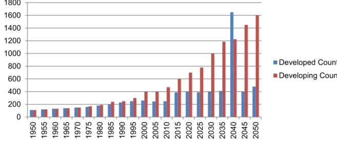
Considering this projection, between 2017 and 2030, the number of aged people 60 years and over will increase by 56 per cent, from 962 million to 1.4 billion, and by 2050, the world population of elderly is projected to more than double its size in 2017 (Fig. 1), reaching nearly 2.1 billion, and will reach to 3.1 billion in 2100.
Figure 1. Number of people aged 60 or over; World, developed, and developing country, 1950 – 2050.Source: UNDESA, World Population Ageing 2011 (2012; forthcoming), based on UNDESA Population Division medium projection scenario, World Population Prospects: The 2010
Revision. 0 200 400 600 800 1000 1200 1400 1600 1800 19 50 19 55 19 60 19 65 19 70 19 75 19 80 19 85 19 90 19 95 20 00 20 05 20 10 20 15 20 20 20 25 20 30 20 35 20 40 20 45 20 50 Developed Countries Developing Countries
This report also projects that, the growth rate of population aged 80 or over, who is called the ″oldest-old″ persons, will increase faster than the rate of older population (Figure 2)
Figure 2. Population aged 60-79 years and aged 80 years or over by development group, 2000, 2015, 2030 and 2050, Data source: United Nations (2015). World Population Prospects: The 2015 Revision
In this way, the oldest-old population, from 137 million in 2017 will reach to 425 million in 2050, and it is estimated that in 2050 having more than tripled in numbers compared to 2017 (Fig.3).
Figure3. The expected percentage change in the world's elderly population, by category, from 2010 to 2050.Reconstructed from:United Nation, world population prospect,Available
at:https://www.un.org/development/desa/en/news/population/world-population-prospects-2017.html
The number of people reaching 80 or over will exceed 909 million in 2100, showing the increase by almost seven times its value in 2017.
In the United States of America (USA), the percentage of people aged 60 and over in 2015 has been calculated as 24.6%, and for years 2050 and 2100, it has been projected as 36.2% and 44.1% of the population respectively(UN,2015).
For Canada, the proportion of the population aged 65 and over grew from 14.1% in 2006 to 16.6 % in 2016(Statistic Canada,2017). For the first time, the number of Canadian seniors aged 65 and older surpassed the persons under 15 years of age in 2015 (Statistic Canada, 2015). The projected growth scenario is expected to be further increasing the future population of the elderly
0% 200% 400% 600% 800% 1000% 1200% 0-64 65+ 85+ 100+ Age Group
in Canada. According to the United Nations, the percentage of aged people for years 2050 and 2100 in Canada will reach to 43% and 50.4% respectively(UN, 2015).
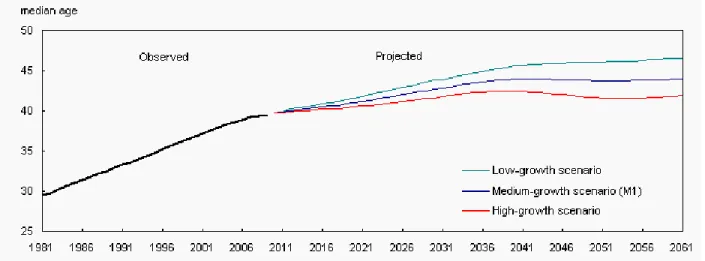
According to three demographic projections named low-growth, medium growth & high growth scenarios, referring to population growth rate that has been presented by Statistic Canada (Fig.4), both overall numbers & proportions of people aged 80 years and over are rising rapidly in Canada in the near future.
Figure 4.Median age observed (1981 to 2009) and projected (2010 to 2061) according to three scenarios, Canada. Available at:http://www.statcan.gc.ca/pub/91-520-x/2010001/ct010-eng.htm. The main issues that emerge
Obviously, dramatic growth in numbers and proportions of the senior population, not only puts a heavy pressure on health systems and requires further demand for medical services, but also increases the risk of diseases associated with age.
The main reasons assigned for accompanying of the growth of the ageing population and increase of life expectancy can be concluded as: (1) Prevention, treatment and control of the transmission of communicable diseases as a result of advances in medicine, adoption of modern technology in public health and public health services development. (2) Implementation of successful strategies for pandemic control. (3) Possible elimination of large-scale wars. (4) Improvement of standards of living and (5) Revolution in favor of agricultural systems (food production and food security), (Giannitsis,et al., 2010).
It is acknowledged that the elderly patients are poorly represented in clinical studies. It is estimated that the older individuals were excluded up to 35% in the published studies(Shenoy et
Harugeri,2015,Lee, ,Alexander, Hammill, Pasquali, Peterson,2001 ,Masoudi, et al., 2003), which contributes to lack of knowledge and understanding of the process that may contribute in developing age-related diseases that undermines the diagnostic criteria for this group. The cost of ignoring old people, particularly very old people from research trials, not only threatens their well-being by lacking of knowledge and understanding of the process that may contribute in developing age-related diseases that undermines the diagnostic criteria for this age group, but it can significantly influence physician's decision making process.
Physiopathology of ageing
Although there are no tools to define ageing precisely from the medical perspective, it has been defined as a sum of all changes that occur in living organisms, which appears over time, and inevitably lead to senescence (Levine, 2012).Actually, aging is defined as a subtle, progressive and irreversible process which appears slowly during years with different rate among people (Roger, 2007) that result from a continuous biological accumulation of many different types of damages caused by molecular defects that augment in cells and tissues.
Although many theories exist that explain the aging, there is no a single theory that defines the ageing process reasonably & comprehensibly (Hayflick, and Moorhead, 1961)Amongst different possible theories that explain the mechanism of ageing, Dr. Hayflick et al., suggested a phenomenon, which refers to a restriction for cellular division in human, known as ″The Hayflick Limit Theory″(Hartman,1956). According to this theory, this limitation may result from several different factors including genetic misconstruction as telomere shortening, activation of different oncogenes as well as aberrations in genetic pathways, release of free radicals during oxidative stress conditions that raise with aging that leads to intracellular damages, abnormally elevated inflammatory markers and increase in apoptosis(programmed cell death). In 1956, Dr. Denham Harman(Erbas and Sekerci, 2011)proposed the free radial theory of ageing, considering ageing as a consequence of imbalance between the productions of Reactive Oxygen species (ROS) or oxygen free radicals and antioxidant protection systems. A free radical is defined as any highly reactive atom or molecule that contains at least a single unpaired electron in an outer shell mainly produced as mitochondrial respiration (Cheeseman and Slater, 1993; Wennberg, 1999). Based on this theory, advanced accumulation of oxidative DNAcauses cellular damage that is a synergistic factor leading to aging.
However, ageing-induced changes could be observed in essentially tissues, organs and organ systems. These changes not only lead to a variety of consequences in tissue and organ dysfunction but also influence clinical presentation of diseases.
Considering the genetic and environmental influence on human aging, there are no specific ages at which people become exactly aged or very aged. By tracking a sample of subjects from birth,it can be proven that the risk of developing a disease would vary as the birth cohort aged, thus, confirming the role of aging as an important and non-modifiable contributor in many diseases. It can be derived that it is inevitable to define ageing according to chronological models. Therefore, it has been accepted as a convention, an individual aged 65 year or more be called as the elderly(Batchelor et al., 2000; WHO, 2012).
Age related changes could be aggravated by specific comorbidities or pre-existingconditions. Consequently, the presence of comorbidities with ageing may affect functioning, quality of life and mortality. These conditions potentially can increase the risk when comparing with the condition that might be expected in the younger adults(CIHI, 2011).In other words, comorbidities that mostly accompany in aged people can mainly lead to a worse prognosis and consequently increase of both mortality and morbidity of acute heart conditions in elderly. Therefore, ageing and comorbidities are two conditions that have strong relation in medical practice.
Almost three quarters of Canadians and Americans seniors over 65 have at least one to three chronic conditions respectively (Alami,Fanf,Song,Nacamuli,2003; LeRoyet al.,2014).
A number of different processes are more associated with ageing, and have direct impact on cardiovascular health, function, and clinical decision making relative to heart conditions. Ageing process involves progressive and often functional impairments in across multiple organ systems. As a result, this can significantly lead to major morbidity and mortality and affect the pharmacokinetics and pharmacodynamics of medication in the elderly(Kajstura, 1996; Schwartz, 2007).
The main changes in the heart structure with ageing result in loss of cardiomyocytes, due to necrosis or apoptosis (Lie et al.,1988), which stimulates the hypertrophy of the remaining cardiomyocytes, increase of the connective tissue accumulation (fibrosis), and amyloid deposition in very old individuals (Gerstenblith,1977). Loss of cardiomyocytes can cause excessive cells stretching and atrial wall stiffening impose an increased vascular load, leading to left ventricle walls thickness(Lakatta, 2007).
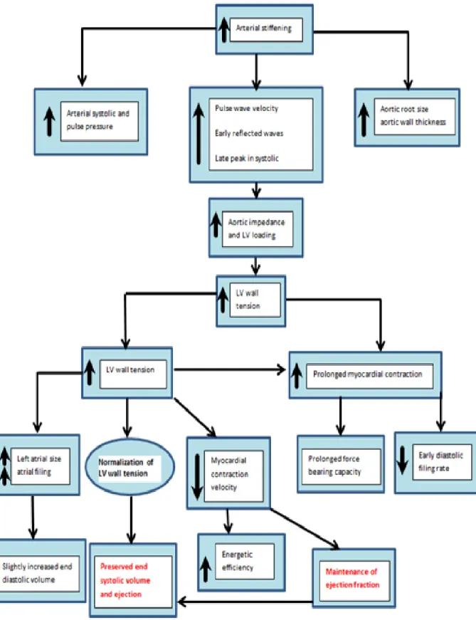
In the elderly, due to an increased amounts and concentration of collagen and decrease levels of elastin in the arterial wall, arterial compliance is normally decreased (Yu and Chung, 2001; Maruyama, 2007).In the above population, the blood vessels, the endothelial function is also abnormal because the production of nitric oxide (NO) is reduced by decreasing NO-dependent dilation. In addition, other bimolecular changes such as increases in specific matrix metalloproteinase, transforming growth factor-beta 1, and angiotensin II, contribute to endothelial dysfunction (Martz et al., 2000). Moreover, NO bioavailability decreases with aging(Van der loo et al., 2000).Subsequently, it has mostly been assumed that the reduction of bioavailable nitric oxide is secondary to increased oxidative stress during ageing (Pugh,et al., 2001; Webb, et al., 2005). The structural and functional changes in the heart with aging have shown in figure 5.
Stiffening of the great arteries may promote an increase in systolic blood pressure, a decrease in diastolic pressure and widening pulse pressure(Yu, B. P., and Chung, 2001). Meanwhile, the effect of aging on the other systems may affect cardiovascular system that could be exemplified by aged-induce decrease in the testosterone production of the endocrine system resulting in changes to distribution of the cardiac contractile proteins (Tsang,et al., 2002).
Figure 5.Ageing induced changes in cardiovascular system in healthy subjects.Modified from Lakatta, EG: Cardiovascular regulatory mechanisms in advanced age. Physiol. Rev 73: 413– 465, 1993
The main age-associated changes in the cardiovascular system that contribute in morbidity and mortality could be summarizedas follows:
➢ The cardiac mass in elderly usually is increased(Pearson, ,et al.,1997) ➢ Endothelial dysfunction which enhances vasoconstriction
➢ Heart rhythm disorders (Hees, et al.,2004)
➢ Increased systolic blood pressure, even if they are normotensive (Beck,2000) ➢ Left ventricular hypertrophy as excess thickening of the left ventricle
➢ Arterial stiffness as an increase in both systolic blood pressure & pulse pressure
➢ Diastolic ventricular dysfunction (early diastolic filling of the ventricles is reduced), that could deteriorate diastolic filling abnormalities (Yazdanyar et al.,2009)
➢ Decreased cardiac reserve (the heart cannot achieve its maximum capacity when needed)
➢ The heart pumping would be in an arrhythmic fashion because the cardiac action potentials are prolonged
➢ Cardiac-induced renal dysfunction(WHO,2010)(inability to maintain composition and volume of body fluid)
The effect of ageing on the heart could be exemplified by referring to increased occurrence of acute coronary syndrome (ACS) or acute myocardial infarction (AMI) inthe elderly. In spite of the fact that aging alone should not necessarily be considered linked to ACS or AMI, the majority of epidemiological studies concerning AMI or ACS have been significantly demonstrated that ageing is associated with a sharp rise of these cardiac diseases(Orimo, et al., Pal Yu, et al.,Yu,2006;Roger,2007).
The correlation between the physiological processes of aging and age-related pathological processes has been demonstrated (Extermann, et al., 2005, Carroll and Miller, 2010).Consequently, aging may alter the clinical manifestation, response to treatment, and outcomes of diseases. Therefore, the observations regarding the performance of a clinical trial in accordance with the young population may not apply in elderly patients.
It is important to identify with precision the major causes of death in the elderly and the extreme elderly as well, in order to change mortality rates among the older population.Given the differences in physiological reserves, comorbidities, functional capacity, and geriatric
syndromes, the elderly reveal a heterogeneous population(HALE project, 2004).These criteria may influence the clinical symptoms, even may modify the clinical consequences. As a result, it seems inappropriate to assume that the elderly benefits of the same clinical approach as for the younger population. Furthermore, elderly patients may not represent the same predetermined outcomes in paraclinical investigations.
In conclusion, risk of developing morbidity and mortality of ACS or AMI has been significantly increased with aging (WHO,2015). The impact of ACS or AMI among older adults has been integrated by reducing homeostatic reserves, increasing prevalence of comorbidity, increasing polypharmacy and creating more complex social issues.
Cause-specific mortality in elderly
The global outbreak of non-communicable diseases is mainly related to aging. Although there is a worldwide difference to present the secondary causes of death in the elderly among stroke, cancers or respiratory diseases, there is a global consensus regarding the role of Ischemic heart diseases (IHD) as a main cause leading to death among older people (HALE project, 2004,WHO,2015).
Figure6. The 10 main global mortality causes in people aged 60-69 years, in 2015,
IHD=Ischemic Heart Disease, COPD=Chronic Obstructive Pulmonary Disease,L&URTC=Lower and Upper Respiratory Tract Cancers,DM=DiabetesMellitus, LRI=Lower Respiratory Infection, TB=tuberculosis, LC=Lung cancers, RD=Renal Diseases,Modified
Figure 7. The 10 main global mortality causes in people aged 70 years and over, in
2015,IHD=Ischemic Heart Disease, COPD=Chronic Obstructive Pulmonary Disease, LRI=Lower r respiratory Infection, DM=Diabetes Mellitus L&URTC= Lower and Upper Respiratory Tract Cancers, HHD=Hypertensive Heart Disease, RD=Renal Diseases, CRC=Colorectal
CancerModified from
http://www.who.int/gho/mortality_burden_disease/causes_death/top_10/en/
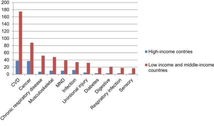
CHD is the most common cause of death worldwide, in both men and women(Scarborough, Wickramasinghe, Bhatnagar, and Rayner, 2011; Lloyd-Jones, Gersh. 2008; Mozaffarian, et al., 2015). Despite recent progress in the prevention and treatment of cardiovascular disease with adopting a healthy lifestyle such as regular exercise and physical activity, developing healthy eating habits or a balanced diet to have a healthy weight, that could help to reduce the rate of CHD in elderly (WHO,2008;Statistics Canada,2015;Ibanez, et al,2017). However, cardiovascular diseases are reported as the main cause of death in the elderly in all economic levels (low, middle, & high income) of many countries (Statistics Canada,2012).
Figure 8.Leading contributors to burden of disease in people aged 60 years and older in 2010— DALYs (million) by cause and World Bank income.DALYs=disability-adjusted life years.
CVD=cardiovascular and circulatory diseases. MND=mental and neurological disorders, combining the IHME GBD mental and behavioral disorders and neurological disorders groups. The main causes of death in elderly in Canada
Given that the Canadian population is aging, it can be expected that in the near future an increasing number of people with heart disease will be observed. In Canada, the highest incidence of heart disease has been reported with the groups aged 65-79 and 80 and older, 15% and 24% respectively (Alexander, et al., 2005; WHO, 2017). The age adjusted incidence of acute myocardial infarction is 10 times higher in aged group 65-74 compared to aged group 35-44, amounting to 70,000 acute myocardial events yearly. According to statistics Canada, heart diseases are the second leading cause of death for people aged 65 years and over (Fig.9).
0 20 40 60 80 100 120 140 160 180 200 High-income contries
Low income and middle-income countries
Figure 9.Death Database, CANSIM Table 102–0561., HD= Heart Diseases, CLRD=Chronic Lower Respiratory Disease, Modified from: Vital Statistics. Modified
from:http://www.statcan.gc.ca/pub/82-625-x/2017001/article/14776-eng.htm
Epidemiology of acute coronary syndrome
About 33% of all ACS events happen in patients aged over 75, and they give reason for almost 60% of all-cause mortality (Savonitto, Morici, and De Servi, 2014).The worldwide fatality rate of ACS in 2015 has been estimated 7.6 million deaths per year (Braunwald, and Bonow, 2014).Elderly patients, who are suffering from ACS, make a prominent part of hospitalized patients; this will be significantly increased in the near future(Saunderson, et al.2014). ACS accounts for 60% of hospital admission and 85% of deaths in patients aged over 65 years (Roger,et al., 2012). ACS-induced mortality rate in patients aged over 85 years is at least three folds higher than the age group under age of 65 years (Arnold, et al., 2005).
28% 21% 6% 5% 3% 37%
Figure10.Prevalence of current diseases in the united states,data are percentages.AF(Atrial Fibrillation),BP(blood pressure),CVD(cardiovascular diseases),PAD(Peripheral Artery
disease),From:Douglas L. Mann,et al.Braunwald’s Heart Disease : A Textbook of Cardiovascular Medicine ,2015,ISBN: 978-1-4557-5133-4
Coronary heart disease (CHD) accounts for more than half of all cardiovascular events in both men and women, under the age of 75 in the United States(Lloyd-Jones, et al.,2009). Regardless of race or gender, the elderly will face with significant increases in incidence of CHD with advancing age (WHO,2011).In women compared to men, not only the incidence of CHD takes place 10 years later, but also the occurrence of more life-threatening conditions like sudden death or acute MI delay for at least 20 years(Yusuf, Reddy, Ounpuu, and Anand,2001).
Figure 11.Prevalence of coronary heart disease in the US by age and sex (National Health and Nutrition. Examination Survey: 2009–2012).Source: National Center for Health Statistics and National Heart, Lung, and Blood Institute
AMI is recognized for 40% and 50% of the 17 million annual causes of CVD death in the world (PHIC, 2009; Fitchett, et al., 2011). Advanced age notably associated with increased incidence of AMI. In the USA, more than 60% of hospital admissions are due to AMI in people who are 65 and older(Harman, 1956). Incidence of AMI is increased 10-fold greater in patients 65 to 74 years of age compared to those 35 to 44 years of age, and continuously have higher death rates in patients over the age of 65 years (Hayflick, 1961, Orimo,2006). Up to the age of 80, the prevalence of AMI appears in both sexes in equal frequency, but then it will be more common in women (Roger,2012).
In Canada, ACS is responsible for 19,000 deaths annually, whereas it has been estimated the AMI prevalence is almost 70,000 per year (Welsh, Travers, Huynh, and Cantor, 2009). Among Canadian adults aged 65 to 74 years, the prevalence rates of heart disease have been estimated to be 14.8%, it reaches 22.9% over aged 75 years (Benjamin, 2017; Mozaffarian, et al.,2016).
Mortality of ACS and AMI, in persons who experience it for the first time is estimated 34% and 15% respectively, and it is projected that, every 42 seconds, an American will suffer from MI (Mozaffarian, et al., 2015). Coronary heart disease accounts for 51% of all cardiac death in the USA (NHLBI, 2007).
In 2013, CHD was accounted for one-seventh of all deaths among the Americans. It has been computed that one American is attacked every 34 seconds by acute coronary events and one of these patients dye every 84 seconds (ARIC,2004-2009). With increasing age, the prevalence rate of AMI, appears to be rising, so that in adult aged 65-74 years compared to 35-44 age group, it is approximately seven times higher (Goodman, et al.,2009). The estimated median age at first MI in men and women is 65.1 and 72.0 years respectively (Kolansky, 2009).
The economic burden of acute coronary syndrome in elderly
The impact of age on ACS has made its significant economic burden in elderly (76).
Hospitalization rates for ischemic heart disease are increased with advancing age. ACS as the most common condition related to ischemic heart disease in the USA, is significantly common condition associated with heart disease leading to hospitalization in the USA(PHAC,2009;Yusuf, Reddy,Ounpuu, and Anand,2001;Mozaffarian,2015).In General, the economic burden of ACS has a direct impact in the increase cost of health care. It has been reported the US spends more than 150 billion dollars annually for their total direct medical expenditures(CIHI,2017). It has been shown that the elderly with AMI are the biggest users of professional health services(Maton,1997;Cohen,2014). In Canada, the average cost of AMI in elderly patients, in a 6 year care-period, has been estimated $28,169 per patient (Ross,1993). There is a significant need to distinguish the ACS and AMI utilizing cardiac risk- stratification in order to reduce the cost of the health care system.
Risk factors for acute coronary syndrome
Coronary heart disease is usually caused by atherosclerosis, which creates a plaque resulting from migration and accumulation of macrophages (foam cells) inside arterial walls (chronic inflammatory response(Hansson, and Hermansson, 2011). Atherosclerosis induces intimal smooth muscle cell proliferation (intimal hyperplasia) constructing a bump called atheromatous (fibro-fatty) plaque (Libby, 2002; Lind, 2003). The growing bump on the arterial walls, with decreasing cross-sectional area of the vessels, contributes to partial or complete blockage of the narrow coronary arteries (Wagenknecht, et al., 2009; Kim, et al., 2011), this leads to disturbances in coronary circulation and therefore insufficient oxygen supply to the heart muscle. In other words, the atherosclerotic plaque may develop slowly and encroach into the arterial lumen, or turn vulnerability into thrombosis yielding obstruction.
The established atherosclerotic plaque consists of two main parts (Wang, and Bennett, 2012): 1) A fibrous cap that is composed of vascular smooth muscle cells and their major secretory products (such as collagen and elastin), inflammatory cells (such as macrophages, T lymphocytes, dendritic cells, and mast cells)
2) A "necrotic" core which is surrounded by fibrous cap, and includes intra cellular and extracellular lipids, foam cells, and debris.
A developed atherosclerotic plaque contains high concentrations of calcium salts as well (Roger, et al., 2012).
Figure12. A cross-sectional distribution of left coronary artery bifurcation that illustrated severe atherosclerosis. A fibrous plaque in the left circumflex (Left),a complicated plaque with a nonocclusive thrombosis in the left marginal artery, a branch of the circumflex artery which called obtuse branch, (Right).Abbreviations: C:contrast in the lumen, Ca: calcification, T: thrombosis. From: Pierre Théroux, Acute Coronary Syndromes, 2nd edition, A Companion to Braunwald’s Heart Disease,2011,C H A P T E R 6,page 42
Although atherosclerosis plaque stability results from cap thickening and inflammation degree of the capsule, plaque instability and or its rupture refers to vascular smooth muscle cell apoptosis, that result in cap thinning, segregation of collagen and extracellular matrix (Eggers,et al.,2008) [ as structural and biochemical supporters of surrounding cells].
The vascular endothelium has a crucial role to maintain vascular integrity. Therefore, age associated vascular endothelial dysfunction; vascular stiffness and inflammation contribute to rise in the incidence and prevalence of CHD with advancing age in both sexes(Virmani,et al., 2006). The role of atherosclerosis has been proven over many years in clinical medicine as a single largest suspect and main cause of coronary artery diseases that predispose to death and disability with the passing of time (Berenson,et al., 1998).
It has been demonstrated that there are two different fundamental mechanisms for thrombosis on the plaques as superficial erosion of the endothelial monolayer and deep endothelial fissuring which involves the developed plaque(Bolton, and Rajkumar, 2011).
Figure 13. Rupture of the fibrous cap (left) triggers two-thirds to three-quarters of all cases of fatal coronary thromboses. Superficial erosion (right) takes place in one-fifth to one-quarter of fatal coronary thromboses. (Modified from:Libby (2008).The molecular mechanisms of the thrombotic complications of atherosclerosis
Coronary atherosclerosis has been predisposed as an underlying condition that is developing during childhood and adolescence(Maton, et al., 1994). In cardiac myocytes, with developing atherosclerosis, cellular senescence will be observed (Gale, et al., 2011).
As a result, most individuals with age progression have evidence of coronary atherosclerosis. Today, it is well recognized that atherosclerosis has an early and long phase of development, which begins in infancy (Fox, et al.,2005). In other words, ACS in elderly compared to younger counterparts generally has a poorer prognosis (Eagle, Lim, and Dabbous,2004;Rosengren,et al., 2006; Elbarouni,et al., 2009). Moreover, ageing as a verified risk factor has been determined for CHD development that is remarkable to predict short term and long term mortality in the most inclusive of ACS risk models (Wilson, et al.,1998;Gale, et al.,2008;Farhat,et al.,2008;Kozieradzka,et al.,2011).Consequently, it can be deduced that the prevalence of coronary artery diseases increases with advanced age. However, it is now well recognized that among symphony orchestra players that create atherosclerosis, ageing has a non-modifiable role(Niemann, et al.,2011) that makes a prominent ear-splitting noise in this orchestra.
Among the identified atherosclerotic risk factors including: LDL cholesterol, HDL cholesterol, triglycerides, systolic blood pressure, male sex, family history of early MI, diabetes mellitus, and smoking, ageing has a significant rolethat independently promotes the development of atherosclerotic disease even if all other mentioned risk factors could be controlled (Saunderson,
et al.2014,Mueller,1991).Considering that age is included in the main risk-related fashions, consequently older people are put as high risk based on age alone (Naylor,2003).
Biomarkers of acute coronary syndromes in the elderly
Biomarkers or biological markers are defined as cellular, biochemical, molecular or anatomic modifications that are empirically measurable in order to detect the process related to normal or particular health conditions. These tests can assist in understanding the evolution of diseases, risk factors of diseases, monitoring responses to therapeutic interventions in a culture medium such as blood serum or tissue extract (Abernethy, et al., 2001, Morrow, & De Lemos, 2007; Wallace, et al., 2008). In other words, in clinics, the use of biomarkers are a practical and dynamic approach in order to study the disease characteristics with referring to screening, diagnosis, prognosis and monitoring medical interventions.
The characteristics of an ideal biomarker for acute coronary events
There has been serious debate among researchers, clinical chemist and physicians to introduce advanced screening strategies. In order to identify patients who are initially free of CHD manifestations but at risk of acute coronary events, the clinicians require safe, accurate, affordable, and reliable biomarker.
A novel and standard cardiac biomarker has to be measured easily, with added new information that can be useful in patient management (Rosalki, 2004). In addition, the main feature of each biomarker has to consist of a release kinetic model from specialized or individual cells, i.e. with specificity and sensitivity.
According to the FDA (Food and Drug Administration) (Lippi, 2015), the feature of an ideal cardiac marker is as follows:
1. Specific:
high serum ratio in myocardium
not even pathologically found in non-cardiac tissue
Show appropriately a clear distinction for different pathogenesis of cardiac involvement (acute to chronic, necrosis, hypertrophy, rhythm)
2. Sensitive:
Zero measurement baseline (standard)
indicator of early onset and reversible cardiac injury instantaneous release if there is an injury
3. Predictive:
Long serum half-life in circulation (indicates that it is reversible) degree of release is proportional to degree of injury
4. Performance:
must be accurate, simple, inexpensive and fast detection in all clinically relevant markers
5. Provide a clinical and paraclinical bridge 6. Readily available and noninvasive
History of cardiac biomarkers,from past to present
Despite the fact that there has been a great biomedical debate for decades over the ACS diagnosis (Dolci, & Panteghini, 2006), the acceleration of cardiac biomarkers development has come to an almost complete agreement to introduce a relatively perfect cardiac biomarker (LaDue, Wroblewski, & Karmen, 1954; Karmen, Wroblewski, & LaDue, 1955).
Traditional cardiac biomarkers of acute coronary syndromes
The primary biomarkers applied to identify acute cardiac ischemia consist of aspartate aminotransferase and lactate dehydrogenase isoenzymes.
1. Aspartate aminotransferase (AST)
In 1954, aspartate aminotransferase (AST) or serum glutamic oxaloacetic transaminase (SGOT) was presented as first cardiac biomarkers to determine predictors of coronary events (Henry, ChiamoriI, Golub, Berkman, 1960;Wilkinson, Baron, Moss, & Walker, 1972), technically improved in years later (Cabaud, Leeper, & Wroblewski, 1956; Wroblewski, Ruegsegger, & Ladue, 1956), and for many years, that played the most important role to predict acute coronary events (Ruegsegger, Nydick, Freiman & Ladue, 1959;Ladenson, 2007). Increased AST levels can be observed 3–4 hours after acute coronary injury in circulation provides the maximum blood levels in 15–28 hours and then returns to normal levels during 5 days(Wroblewski, 1955). Although AST has a great sensitivity to detect acute coronary injury, it is not an ideal cardiacbiomarker due to its existence in the liver, skeletal muscles, brain and kidneys, that gives significantly a low specificity (Lee, 1986, Saunderson, 2014).
2. Lactate dehydrogenase (LDH)
Elevated level of lactate dehydrogenase (LDH) and its isoenzyme (LDH-1) have been demonstrated among patients with AMI(Rozberg, 1962; Penttilä, et al., 2000), in 1956. Increased blood level of LDH and LDH-1(LD-1) are observed 5–10 hours after AMI, reaches maximum at 2.5 to 6 days and returns to baseline in 12 days (Blomberg, Kimber,& Burke, 1975).
3. Creatine Kinase (CK)
In 1960s, the Creatine Kinase (CK) presented as a better cardiac specific marker to confirm acute myocardial damages (Ishikawa, Saffitz, Mealman, Grace, Roberts, 1997). CK, as a dimeric molecule, consist of two different subunits known as M and B, which split into three distinct isozymes as CK-MB (found predominantly in myocardium), CK-MM (found predominantly in skeletal tissue), and CK-BB (found predominantly in the brain tissue) (Adams, Abendschein, & Jaffe, 1993). It has been determined that the release of CK-MB is particularly obvious following myocardial necrosis (not under ischemic condition)(Wu, 1998), so it was most commonly applied in identifying myocardial injury. It can be typically detected in circulation 3–6 hours following the onset of acute myocardial necrosis, reaches its maximum in 12-24 hours and begins to return to normal level within 48-72 hours (Galarraga, 2003). Although sensitivity of CK-MB is high enough, its extensive tissue distribution will strongly yield poor specificity. In other words, CK-MB elevated levels could be observed in various forms of musculoskeletal damage or myopathies, diseases of small bowel, uterus, prostate, and diaphragm, renal failure, hepatobiliary system disease, non-cardiac surgery, chest trauma, asthma, pulmonary embolism, head trauma, hyperventilation, hypothyroidism, during peripartum period and in substance abuse like cocaine and alcohol (WHO, 1979; Lee, 1987). Consequently, it has been suggested that in order to improve CK-MB specificity, calculating CK-MB relative index as CK-MB/total CK, whereas the ratio greater than 2.5 will be associated with myocardial injury (Mair, et al., 1992). Thus, the World Health Organization (WHO) confirmed the use of serum AST, LDH, CK, and CK-MB levels in presence of clinical and electrocardiographic manifestations to identify acute myocardial infarction, in 1979(WHO, 1979).
4. Myoglobin (MYO)
Myoglobin (MYO) was introduced as a primary indicator of myocardial damages in 1978(Kolendorf, Pedersen, Christiansen, & Gad, 2009). It is a globular oxygen-carrying protein which found in myocardial tissue and striated skeletal muscle (Ohman, et al., 1990). The first report was published in 1975, confirmed the association between elevated serum levels of MYO and AMI (Varki, Roby, Watts, & Zatuchni, 1978). Myoglobin can be detected within 1 hour after the initiation of myocardial necrosis, reaches its maximum in 4-12 hours and returns to normal level within 24-36 hours (Ebashi, 1963). A high concentration of myoglobin in skeletal muscles is the reason why MYO has a poor specificity (Galarraga, 2003;Jaffe, 2012). In other words, though myoglobin is one of the earliest serum markers for AMI, there are various medical
conditions that could increase myoglobin blood levels in the absence of acute coronary occlusion.
5. Cardiac troponin(cTn), as a biomarker in the current standard of care
In 1963, it was revealed that a new myofibrillar protein called troponin is integral to muscle contraction in skeletal and cardiac muscle and absent from smooth muscle(Takeda, Yamashita, Maeda, & Maeda, 2003).
Biology of the Troponin Complex in Cardiac Myocytes
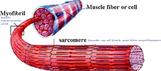
Troponin is a complex of three subtypes (Katus, Remppis, Scheffold, Diederich, & Kuebler, 1991): Troponin I (Tn I) which binds to actin with Inhibitory role, Troponin T (Tn T) which binds to tropomyosin, Troponin C (TnC) which binds to calcium ions and all of them are located on the actin filament in sarcomere.
Sarcomere is the basic contractile unit of muscle cell composed of thin ball shaped filament, called actin, and thick filament, called myosin, includes tail, hinge and head as dual- golf club shaped. Each muscle fiber or myofibril (a long, cylindrical, multinucleated cell) contains hundreds of sarcomeres (Brogan, et al., 1997).
Figure 14. Structure of musculoskeletal contractile cell (Modified from: Marieb,et al.,2010). Anatomie et physiologie humaines)
Tropomyosin attaches longitudinally to the actin filaments. Each actin molecule has a myosin-binding site. If calcium ion is unavailable, the myosin-binding site of myosin to actin will be blocked by tropomyosin-troponin complex.
Figure 15. A schematicview:Tropomyosin binds lengthwise along actin filaments and, in striated muscle, is associated with a complex of three troponins: troponin I (TnI), troponin C (TnC), and troponin T (TnT). In the absence of Ca2+, the tropomyosin-troponin complex blocks the binding of myosin to actin. Binding of Ca2+ to TnC shifts the complex, relieving this inhibition and allowing contraction to proceed. (Cooper, G. M., & Hausman, R. E. (2009). The cell: a molecular approach. Washington, DC: ASM Press; Sunderland, MA: Sinauer Associates, c2009.)
By binding of calcium ion to the troponin C, tropomyosin moves to lateral side, that causes exposed binding side on actin for myosin(dark area), then, results from ATP-hydrolysis, some changes occurs in myosin head that promote to bind myosin head into exposed site(Figures 15 &16)
Figure 16. Interaction between the actin and myosin filaments ,from:In Fuster, V., In Harrington, R. A., In Narula, J., & In Eapen, Z. J. (2017). Hurst's The Heart, 14e. New York, NY: McGraw-Hill Education LLC
It has been assumed that in addition to the structural sources of troponin, there is at least one other pool that is called cytosolic pool (Wu, 1998; Thygesen, 2010).The cytosolic or early releasable pool for cTnI and cTnT accounts for almost 3.7% (Wu, 1998) and 5% (Thygesen, 2010) of total released troponin respectively. Consequently, it may justify the long persistence of cardiac troponins in blood circulation following an AMI. Besides, cytosolic pool is responsible for the biphasic kinetics of troponin as a rapid release of free cytoplasmic troponin and then a gradual structural troponin release(Sribhen, Phankingthongkum, & Wannasilp, 2012).
Figure17. Mechanism of release of cardiac troponin after ischemic cardiac injury, available at: http://ja.ma/1NKufTm #AMI #heart attack)
Although the biochemical behavior of cTnI is quite clear, that means, cTnT has never been detected neither during neonatal development nor in pathologic conditions of all the organs and body tissues, except the heart (Tiwari, 2012), the situation of cTnT becomes more complicated.
Apparently, the elevated levels of cTnT are detectable in some patients who suffer from musculoskeletal disorders or renal failure (Tucker, 1997). Therefore, cTnT could be prone to false-positive elevation in the absence of significant coronary artery disease.
Elevated levels of cTnT and cTnI are detectable within 3-4 h after of the onset of ischemia and typically reach its maximum after 12-48 h (Bertinchant, 1996) and stay elevated for 4-10 days that refers to a gradual decline in myofibril-bound troponin complex (Jaffe, 2011;Twerenbold, 2012). Cardiac troponin assays are based on high-affinity antibodies thatare specific for both cTnT and cTnI, but because of the uniform amino acid sequences of troponins in both myocardial and musculoskeletal origins, measuring cTnC has not been proposed (Tiwari,2012). Novel high-sensitivity cardiac troponin assays (hs-cTn), compared to previous generation, in order to detect myocardial necrosis with respect to their sensitivity have been developed (Jaffe, 2011; Twerenbold, Jaffe,Reichlin, Reiter,&Mueller, 2012).Hs-cTn assays provided an opportunity to detect cardiac troponin levels which is 10- fold lower in previous assays, hence they are able to measure the lowest troponin values in healthy individuals (Apple, & Collinson, 2011). Each assay has a unique, incomparable and assigned cut off value that refers to the dissimilarity of the antibodies and the different matrix compositions of the assays ( Ferrieres, 1998; Perry, 1999; Panteghini, 2001). In addition, the various components of circulating troponins are distinguished by various assaysin different ways (Venge, Johnston, Lindahl, & James, 2009). Furthermore, cardiac troponin can be detected by the new highly sensitive assay in healthy individuals that indicates unique variation inside or interindividuals that could refer to a normal cardiac cells turnover or an unidentified mechanism (Sandoval, & Apple, 2013; Thygesen, Alpert, Jaffe, & White, 2015,).
Figure 18, Detection range of various cardiac troponin assays (from Hochholzer et al. Am Heart J 2010).
The green line refers to normal turnover of cTn in all human beings. The orange line refers to a slight but rapid increase of cTn that reflects either an early-release of cytosolic pool of cardiac troponin or myocardial microinfarction.The red line represents a steep rise in cTn levels, 2 -6 hours after extensive myocardial infarction. In addition to elevated cardiac troponin monitoring, hs-cTn assays are designed to detect lower levels of troponin caused by ischemia/micro necrosis and the normal turnover as well. While the first generation of cardiac troponin assays only detect a significant increase of cardiac troponin.
Although increased serum troponin level is defined as the value of 99th percentile upper reference limit (URL) by including 99% of all troponin values of the given population (Apple, & Collinson, 2014), URL values for the same assay could vary significantly in different reference groups (Clerico, et al., 2008,Olivieri, 2012).Cardiac troponins assays would be considered as highly sensitive tests if: their coefficients of variance are less than 10% at the 99th percentile value of the healthy reference population, and the concentrations above the test detection limit can be measured in more than 50% healthy population. (Olivieri, 2012).
Behavior of troponin in the elderly
Both age and gender significantly have an effecton hs-cTn assays (Missov,& De Marco, 1999;Olivieri,2012), as it decreases in women and increases with advancing age, this may be due to the myocardial ageing, in both sexes, particularlyageing causes a remarkable increase of
serum troponin. Consequently, the interpretation of serum troponin in elderly, exceptionally in old-old with presence of comorbidities needs further consideration.
Sensitivity and specificity of troponin
Although the interpretation of diagnostic tests is a clinical process that have own lexicon with regards to its science, the efficiency of a laboratory test is usually presented in terms of sensitivity and specificity.
Both cardiac troponin T and I isoforms are distinctly found in myocardium (Jeremias, 2010),so it is a cause of high cardiac specificity for both of them. It is hypothesized that the normal baseline troponin values should be 0.1–0.2 ng/L, caused by persistent loss of cardiomyocytes throughout life (Panteghini, Pagani, & Bonetti, 1999).Despite the cardiac troponins sensitivity and its predictive value have improved over time, its specificity does not change notably over time (Balk, 2001).
Sensitivity of cTnT in hospital outpatients varies between 25-65 percent (time zero), increments to 59-90 percent at 2 to 6 hours after their admission, and reaches 100 percent within 6 to 12 hours after admission (Bertinchant,1996;Eggers,, Nordenskjöld, & Lindahl, 2004;Jaffe, Babuin, Apple,2006). For cTnI, its sensitivity in time zero is at most 45 percent, increases to 69–82 percent at 2 to 6 hours after admission, and the same as cTnT, achieves to 100 percent within 6 to 12 hours after admission (Eggers, Oldgren, Nordenskjöld, & Lindahl, 2004;Jaffe, Babuin, Apple,2006;Twerenbold, Jaffe, Reichlin, Reiter, & Mueller, 2012). As a result, the maximum sensitivity of cTn assays could be observed longer than 6 hours after myocardial cell death occurs (Christenson, 2007; Hammarsten, 2012). Thus, it is recommended to measure cTn blood levels at time zero and at least 6-9 hours after patient admission in order to improve the accuracy of the diagnosis of myocardial infarction (Koerbin, Tate & Hickman, 2010).
The positive predictive value, for cTnI, at time zero and 12 hours after admission is estimated to be 25% and 89% respectively, whereas for cTnT, is estimated to be 35% and 57% respectively (Twerenbold, , Jaffe, Reichlin, Reiter, & Mueller, 2012). The negative predictive value of cTnI at time zero and 12 hours after admission is predicted to achieve 85% and 98% respectively, whereas for cTnT, it is estimated to be 88% and 99% respectively(Twerenbold, , Jaffe, Reichlin, Reiter, & Mueller, 2012).
The specificity of cTnI and cTnT has been reported to be in the range of 83 - 98 percent and 86– 98 percent respectively (Twerenbold,et al., 2012).
Although few studies have been reported that the level of cTn is increased with advancing age (Ferri, 2010;Reiter, Reichlin, Twerenbold, & Mueller, 2011;Anderson, 2011), reasons of which still remains obscure, even with explanations like presence of more comorbidities or silent heart diseases in elderly, and age dependent phenomenon. In other words, the troponin alterations in elderly, particularly among the old-old, are not inclusive.Emerging serum cardiac biomarkers is summarized in following graphs in the first hours (figure 19) and days (figure 20) after the onset of acute myocardial infarction. High-sensitivity assays are capable of detecting cTn in blood circulation more accurately even at a concentrations 10 times lower than conventional assays (Liu, et al,2017).Therefore, use of high sensitivity assays of troponin could reduce the risk of being misdiagnosed with ACS compared with conventional assays.
Figure 19.Time courses (hours) for elevation of various biomarkers after the onset of symptoms of AMI.( Source : Michael L. Bishop, et al. Clinical chemistry, 6th edition, 2010, chapter 25, cardiac function, page 551, ISBN 978-0-7817-9045-1)
Figure 20.Time courses (days) for elevation of various biomarkers after the onset of symptoms of AMI. Reprinted from:Goodier, J. (2009). Lippincott Williams & Wilkins anesthesiology, creatine kinase-MB to troponin: the adoption of a new standard.
As it is explained, reference values for cardiac troponin encompass the values of 99% of a healthy population, the results vary between different laboratories, so at present, there is still no certain standard consensus on which cut-off point value should be used (Jaffe,2000).The baseline of troponin level has been suggested as 0-0.4 ng/ml (negative), 0.05-0.49 ng/ml and ≥0.05 ng/ml for intermediated risk and strong risk of acute myocardial infarction respectively (Lippi, Sanchis-Gomar, & Cervellin,2016).
In conclusion, to this date, among available cardiac biomarkers, cardiac troponin is the biomarker of choice to detect acute coronary events (Sanchis-Gomar, Perez-Quilis, Leischik, & Lucia, 2016).
Causes of increased cardiac troponin values
Although cardiovascular disease (CVD) has been defined as a group of diseases that involve both the heart and blood vessels(Fitchett, et al.,2011),at some point, there is an ambiguity in defining its related-conditions such as coronary heart disease (CHD), coronary artery disease (CAD), and acute coronary syndrome (ACS).
CHD refers to heart diseases such as angina pectoris, MI and silent myocardial ischemia, but CAD typically apply to indicate pathologic changes in the coronary arteries that usually caused by atherosclerosis. In other words, CAD is a condition which characterized by atherosclerosis in coronary arteries, could refer to pathologic process and can remain symptomless (Lippi, & Cervellin, 2016). Instead, ACS nearly always becomes symptomatic and it is a life threatening condition, irrespective of the presence of CAD (Roffi,2015).
There are different approaches to classify the clinical conditions other than myocardial infarction that cause cardiac troponin to rise. One of the more detailed classifications related to cTn increase is presented as (Apple, & Collinson, 2014):
1. Troponin leak primary to myocardial ischemic injury: Plaque rupture, Intraluminal coronary artery thrombus formation
2. Troponin leak secondary to myocardial oxygen supply-demand imbalance: tachy-/brady-arrhythmias, aortic dissection or severe aortic valve disease, hypertrophic cardiomyopathy, shock (cardiogenic, hypovolemia, or septic), severe respiratory failure, severe anemia, hypertension with or without left ventricular hypertrophy, coronary spasm, coronary embolism or vasculitis, and coronary endothelial dysfunction without significant CAD
3. Troponin leak not associated with myocardial ischemia: cardiac (contusion, surgery, ablation, pacing, or defibrillator), shocks, rhabdomyolysis with cardiac involvement, myocarditis, cardiotoxic agents (e.g. Anthracyclines, Herceptin)
4. Multifactorial or undefined myocardial insults: heart failure, stress (Takotsubo) cardiomyopathy, severe pulmonary embolism or pulmonary hypertension, sepsis and critically ill patients, renal failure, severe acute neurological diseases (e.g. stroke, subarachnoid hemorrhage), Infiltrative diseases (e.g. amyloidosis, sarcoidosis), strenuous exercise
Consequently, detection of elevated serum concentrations of cardiac troponin (>99th percentile URL) in the presence or absence of ischemic heart disease symptoms, or other diagnoses associated with myocardial damage have to be ruled out.
Acute Coronary Syndromes (ACSs)
ACS - as a subset of CHD addresses a range of conditions that are compatible with unstable angina (UA), non ST-segment elevation myocardial infarction (NSTEMI), and ST-segment elevation myocardial infarction (STEMI) - causes a decrease levels in coronary artery blood flow (Bertrand, 2002).
As discussed above, the underlying etiology of ACSs is the atherosclerosis, which is characteristically composed of a vulnerable plaque with a thin layer of fiber and a large lipid core. Sooner or later, this plaque tears, causes platelet accumulation and activation, which lead to coronary thrombus formation, narrows the coronary arteries and ultimately reduces the flow of oxygen-rich blood to the section of heart muscle that is fed by the coronary artery.
It can be difficult to distinguish UA from NSTEMI (Mega, 2005). UA is defined as myocardial ischemia that occurs at rest or even with slight physical exertion without any presence of cardiomyocytes necrosis, so that is distinguishable from NSTEMI by the pain frequency-severity-duration to cause myocardial necrosis (Cannon, 1997). ECG findings associated with UA can be found in 30-50 percent of the patients, including normal morphology, Ssegment depression, T-wave inversion, or due to a combination of all these factors, may vary depending upon the severity of clinical presentations (Ottani, 1999, Antman, 2004). Although small amounts of cTn could be traced in the serum of patients suffering from UA, it has not been shown any prognostic value in predicting future coronary events (McCarthy,, Wong, & Selker, 1990).
Figure 21. ECG of unstable angina, (available at: http://nstemi.org/unstable-angina/, accessed: July 5, 2017)
STEMI is typically the result of an acute and sudden complete interruption of blood flow to part of the myocardium which could be displayed as an unusual pattern by ECG with ST-segment
elevation, whereas NSTEMI mostly results from a partial occlusion which develops to become total blockage of coronary artery as well, but does not exhibit ST segment elevations in ECG(Figure 22). STEMI is the most severe type of ACS with the highest mortality rates (Goodacre, 2002).
Figure 22. Acute Coronary Syndrome, unstable angina and non-ST elevation myocardial infarctionModified from: Ange, R. A., & Hillis, L. D. (2015). Acute Coronary Syndrome.
Goldman's Cecil Medicine. Acute Coronary Syndrome, unstable angina and non-ST elevation myocardial infarction, Goldman-Cecil Medicine, pages 432-440
Main clinical presentations
A typical symptoms of ACS include chest pain (most common), referred pain (arm, the jaw, the neck, the back, or the abdomen), nausea, vomiting, dyspnea (may be as a sudden onset), diaphoresis, light-headedness (Alexander, 2007). Referred or radiating pain to the shoulder, left arm, or both arms may increase the probability of ACS (Avezum, 2005).
Figure 23. Chest Pain and Acute Coronary Syndrome,Reprinted from:https://jamanetwork.com/journals/jama/fullarticle/2468893
Diagnostic evaluation
The main step for assessment of a patient with ACS is to distinguish the probability of coronary-artery disease as causing any symptoms. The American College of Cardiology/American Heart Association (ACC/AHA) proposed guidelines include, among the factors associated with the high probability of ACS, prior history of angina, AMI or CHF, History of established CAD by angiography, new ECG changes, and elevated cardiac biomarkers (Jaffe, et al.,2000).
Identification of ACS in older patients
Seniors often experience a different range of symptoms, so the diagnosis of ACS among them is challenging. Although chest pain is still the most frequent symptom in older patients, autonomic