HAL Id: hal-03159575
https://hal.archives-ouvertes.fr/hal-03159575
Submitted on 4 Mar 2021
HAL is a multi-disciplinary open access
archive for the deposit and dissemination of
sci-entific research documents, whether they are
pub-lished or not. The documents may come from
teaching and research institutions in France or
abroad, or from public or private research centers.
L’archive ouverte pluridisciplinaire HAL, est
destinée au dépôt et à la diffusion de documents
scientifiques de niveau recherche, publiés ou non,
émanant des établissements d’enseignement et de
recherche français ou étrangers, des laboratoires
publics ou privés.
Distributed under a Creative Commons Attribution| 4.0 International License
Materials on Biofouling in the Marine Environment
Mahmoud Hayek, Marie Salgues, Jean-Claude Souche, Etienne Cunge, Cyril
Giraudel, Osanne Paireau
To cite this version:
Mahmoud Hayek, Marie Salgues, Jean-Claude Souche, Etienne Cunge, Cyril Giraudel, et al.. Influence
of the Intrinsic Characteristics of Cementitious Materials on Biofouling in the Marine Environment.
Sustainability, MDPI, 2021, 13 (5), pp.2625. �10.3390/su13052625�. �hal-03159575�
Sustainability 2021, 13, 2625. https://doi.org/10.3390/su13052625 www.mdpi.com/journal/sustainability
Article
Influence of the Intrinsic Characteristics of Cementitious
Materials on Biofouling in the Marine Environment
Mahmoud Hayek
1,*, Marie Salgues
1, Jean-Claude Souche
1, Etienne Cunge
2, Cyril Giraudel
2and Osanne Paireau
21 LMGC, IMT Mines Alès, University of Montpellier, CNRS, 30100 Alès, France;
marie.salgues@mines-ales.fr (M.S.); jean-claude.souche@mines-ales.fr (J.-C.S.)
2 ARTELIA, 6 rue de Lorraine, 38432 Echirolles, France; etienne.cunge@arteliagroup.com (E.C.);
cyril.giraudel@arteliagroup.com (C.G.); osanne.paireau@arteliagroup.com (O.P.) * Correspondence: mahmoud.hayek@mines-ales.fr
Abstract: Coastal marine ecosystems provide essential benefits and services to humanity, but many
are rapidly degrading. Human activities are leading to significant land take along coastlines and to major changes in ecosystems. Ecological engineering tools capable of promoting large-scale restoration of coastal ecosystems are needed today in the face of intensifying climatic stress and human activities. Concrete is one of the materials most commonly used in the construction of coastal and marine infrastructure. Immersed in seawater, concretes are rapidly colonized by microorganisms and macroorganisms. Surface colonization and subsequent biofilm and biofouling formation provide numerous advantages to these organisms and support critical ecological and biogeochemical functions in the changing marine environment. The new challenge of the 21st century is to develop innovative concretes that, in addition to their usual properties, provide improved bioreceptivity in order to enhance marine biodiversity. The aim of this study is to master and clarify the intrinsic parameters that influence the bioreceptivity (biocolonization) of cementitious materials in the marine environment. By coupling biofilm (culture-based methods) and biofouling (image-analysis-based method and wet-/dry-weight biomass measurement) quantification techniques, this study showed that the application of a curing compound to the concrete surface reduced the biocolonization of cementitious materials in seawater, whereas green formwork oil had the opposite effect. This study also found that certain surface conditions (faceted and patterned surface, rough surface) promote the bacterial and macroorganism colonization of cementitious materials. Among the parameters examined, surface roughness proved to be the factor that promotes biocolonization most effectively. These results could be taken up in future recommendations to enable engineers to eco-design more eco-friendly marine infrastructure and develop green-engineering projects.
Keywords: cementitious materials; intrinsic parameters; marine environment; biofilm/biofouling;
bioreceptivity; ecological engineering
1. Introduction
The management and conservation of the world’s oceans require synthesis of spatial
data on the distribution and intensity of human activities and the overlap of their impacts
on marine ecosystems [1]. Human activities are leading to significant land take along
coastlines [1] and to major changes in ecosystems [2]. “Sprawl” in natural marine areas is
leading to a loss of spatial connectivity [3] and erosion of biodiversity [4] in this
Anthropocene epoch [5]. The recent global assessment report by the Intergovernmental
Science-Policy Platform on Biodiversity and Ecosystem Services (IPBES) on biodiversity
and ecosystem services (May 2019) [6] clearly identifies five direct drivers of change in
nature, first among them being changes in land and sea use due to urbanization, industrial
Citation: Hayek, M.; Salgues, M.;
Souche, J.-C.; Cunge, E.;
Giraudel, C.; Paireau, O. Influence of the Intrinsic Characteristics of Cementitious Materials on Biofouling in the Marine
Environment. Sustainability 2021, 13, 2625. https://doi.org/10.3390/ su13052625
Academic Editor: Claudio Favi
Received: 27 January 2021 Accepted: 24 February 2021 Published: 1 March 2021
Publisher’s Note: MDPI stays
neutral with regard to jurisdictional claims in published maps and institutional affiliations.
Copyright: © 2021 by the authors.
Licensee MDPI, Basel, Switzerland. This article is an open access article distributed under the terms and conditions of the Creative Commons Attribution (CC BY) license (http://creativecommons.org/licenses /by/4.0/).
complexes, roads and mining. The challenge of the 21st century must be to find new
solutions in the construction field. One such solution is to combine engineering techniques
and ecological understanding to provide cost-effective ways to maintain or enhance
biodiversity [7–9].
In the marine environment, urban, coastal and offshore structures provide an
important protective function, but can also have unintended ecological consequences,
such as the loss or modification of habitat and the alteration of hydrological and ecological
flows [3,10–13]. The challenge for managers now is to design marine infrastructure in a
way that minimizes its ecological impact and increases its bioreceptivity (the ability to be
colonized by living organisms) in order to enhance marine biodiversity [9,14,15]. The
results of this paper are intended for technical applications in the area of concrete
protection for breakwaters.
In the last decade, concrete has been one of the materials most widely used for the
construction of marine infrastructure such as ports and coastal defenses [16,17]. It has
proved to be a reliable structural material with very good durability when properly used
[18]. However, the durability of a concrete structure is influenced by the exposure
environment [19]. In seawater, chemical, physical and biological actions affect the aging
and durability of a concrete structure [20–23]. Influences of a physical and chemical nature
are generally well studied, and are subject to standards and recommendations [24,25].
Several scientific publications state that actions of a chemical nature such as chloride and
magnesium sulfate attack are the main cause of concrete structure deterioration in the
marine environment [18,22,26,27]. However, the actions of a biological nature (e.g.,
colonization of concrete by marine organisms) have been less studied, and less is known
about them [24]. The effect of these actions on the durability of concrete in the marine
environment remains unclear, but most scientists agree that marine organisms adhered to
the concrete surface have a protective effect (bioprotection) against chemical attack in
seawater [7,23,28–34]; they form a physical barrier that reduces surface permeability. The
decrease in surface permeability leads to less-efficient diffusion of aggressive ions (Cl
−,
Mg
2+and OH
−), which can increase the durability of a concrete structure in the marine
environment [23,29,35,36]. Understanding and mastering the biological actions affecting
concrete in this environment is therefore an essential step toward meeting the new
challenge of designing sustainable maritime structures that enhance the regeneration of
marine biodiversity [36].
Actions of a biological nature result from the interaction between the material and
the marine organisms that colonize the surface, leading to biofouling. Biofouling in the
marine environment is described as the colonization of any solid surface, living or dead,
natural or artificial, by micro- and macroorganisms. Biofouling formation is hence a
phenomenon common to the majority of materials submerged in seawater [37–39]. It is
generally divided into two main stages: microfouling and macrofouling (Figure 1). The
submerged material is quickly colonized by marine bacteria, which form a bacterial
biofilm on the surface (microfouling). This bacterial biofilm facilitates colonization by
other micro- and macroorganisms such as cyanobacteria, fungi, diatoms, barnacles, algae
and protozoa (macrofouling) [40–45].
Figure 1. Schematic representation of marine biofouling formation [45].
The stages of cementitious material biocolonization in the marine environment are
illustrated in Figure 1. After immersion, organic and mineral molecules are quickly
adsorbed at the surface. This adsorption, known as “conditioning film formation” or
“surface conditioning,” modifies the physicochemical properties of the surface and makes
it favorable to the stable adhesion of bacteria [45,46]. One or more pioneer bacterial species
then adhere to the surface and form a bacterial biofilm (microfouling). Mature bacterial
biofilms have complex three-dimensional structures, composed of one or more bacterial
species adhered to the surface and enclosed in a matrix of extracellular polymeric
substances (EPS) consisting of proteins, glycoproteins, glycolipids, extracellular DNA and
polysaccharides [45,47–49]. A few days to a few months later, other micro- and
macroorganisms such as diatoms, algae and larva adhere to the surface, leading to the
formation of macrofouling [50,51]. Biofouling in seawater is hence the process whereby
fouling organisms collect and grow on a surface, and their morphology is characterized
by their thickness, bioadhesive strength and weight [52].
Generally speaking, the factors that influence the bioreceptivity (biofouling
formation) of cementitious materials are the nature and the physicochemical properties of
the surface [53,54]: chemical composition [55–58], roughness [59,60], porosity [60–62],
hydrophobicity [56,63–65] and pH [60,66]. However, these factors are less well known in
the marine environment [9,67,68]. We showed in a previous study that the bacterial
colonization of cementitious materials in seawater is influenced by the pH and the type of
cement [69,70]. In this present paper, the effect of several parameters in the two main
stages (microfouling and macrofouling) of cementitious material biocolonization in the
marine environment is tested. The main factors studied are:
The surface conditions: smooth surface (SS); faceted and patterned surface (FPS),
which describes an irregular and patterned surface mimicking natural rocks at the
surface of the ECOPODE™ unit; and rough surface (RS), which allows a biomimetic
rocky surface to be obtained.
The use of two products during concrete production: a curing agent and a “green”
formwork oil.
The type of cement: ordinary Portland cement (OPC) CEM I and slag cement CEM
III.
The aim of this study is to master and clarify the intrinsic parameters that influence
the bioreceptivity of cementitious materials in the marine environment, in the context of
coastal infrastructure construction. It is also to identify the parameters that influence the
biocolonization of marine structures such as breakwaters most effectively, in order to help
engineers in their new challenge of designing marine infrastructures using green concrete.
The results of this study will be used directly by Artelia (an international engineering
company specializing in the field of marine infrastructure construction) to enhance
biocolonization of its armor-facing blocks.
2. Materials and Methods
The choice of materials and the techniques used to prepare the surfaces of the
cementitious materials used in this study were inspired by the existing artificial concrete
units developed by Artelia for use in breakwater armor layers. Several types of concrete
armor units have been developed by Artelia since 1953, among them there are two
single-layer systems called ACCROPODE™ (smooth surface) and ECOPODE™ (faceted and
patterned surface).
This study was carried out using two types of specimen—mortar and concrete—
dedicated to the quantification of microfouling (bacterial biofilm) and macrofouling
(algae, etc.), respectively. These specimens were subsequently immersed in a seawater
basin for 5 months, during which time micro- and macrofouling were measured
periodically.
2.1. Preparation of Mortar Specimens
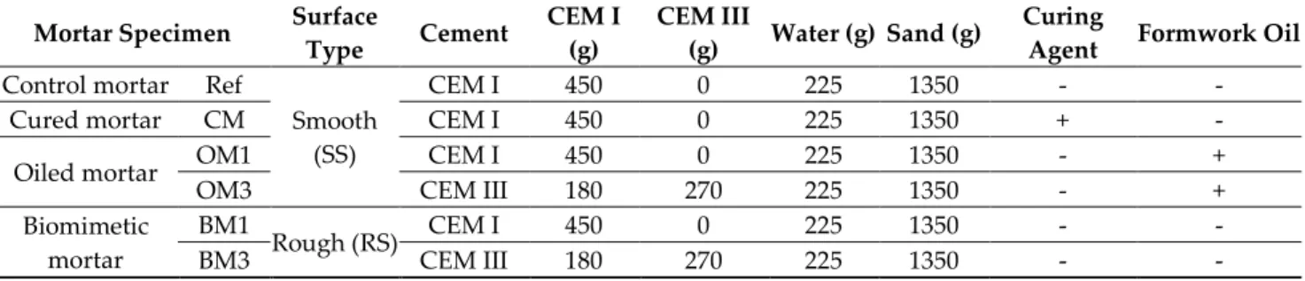
OPC CEM I cement 52.5 N CE CP2 NF “SB” (provided by Ciments Calcia) and
blast-furnace slag cement CEM III (composed of 60% ground granulated blast-blast-furnace slag NF
EN 15167-1, provided by Ecocem, CAS no.: 65996-69-2) were used in this study to produce
six types of mortar specimens (Table 1). The mortar had a water/cement ratio (w/c) of 0.5
and was composed of 450 g cement and 1350 g sand (sand 0/4 EN 196-1).
Table 1. Types and compositions of mortar specimens investigated in this study.
Mortar Specimen
Surface
Type
Cement
CEM I
(g)
CEM III
(g)
Water (g) Sand (g)
Curing
Agent
Formwork Oil
Control mortar
Ref
Smooth
(SS)
CEM I
450
0
225
1350
-
-
Cured mortar
CM
CEM I
450
0
225
1350
+
-
Oiled mortar
OM1
CEM I
450
0
225
1350
-
+
OM3
CEM III
180
270
225
1350
-
+
Biomimetic
mortar
BM1
Rough (RS)
CEM I
450
0
225
1350
-
-
BM3
CEM III
180
270
225
1350
-
-
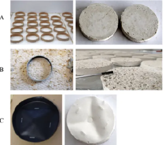
After mixing, the mortars were cast in cylindrical molds measuring 2.7 cm in
diameter and 2.9 cm high. After 24 h of hardening, the mortar specimens were removed
from the molds and placed in a laboratory room at 20 °C for 30 days (Figure 2).
Figure 2. Preparation of mortar specimens. (A) Preparation of SS mortar specimens (mortars in
molds); (B) SS mortar specimens obtained after stripping; (C) preparation of RS mortar specimens on rough silicone skin; (D) RS mortar specimens obtained after stripping.
Formwork oil (vegetable oil, BIODEM SI 3) was spread on the molds, using absorbent
paper to avoid any surplus, before the mortar was poured. Curing compound (SikaCem®
Cure) was added to the top of the specimens in accordance with the supplier’s instructions
1 h after stripping. The rough mortars were prepared with a rough silicone skin; no release
agent was needed. The cylindrical molds filled with mortar were poured on the silicone
skin and held in place with a weight (Figure 2).
2.2. Preparation of Concrete Specimens
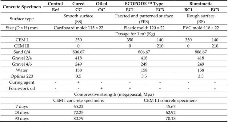
The same types of cement used for the mortar specimens were used to produce seven
types of concrete. Table 2 presents an overview of the specimens investigated, their size,
their surface condition, the various components used and their compressive strength.
The concrete specimens were prepared with a water-to-cement ratio of 0.45, using
cement (CEM I, CEM III), sand (0/4), a natural limestone gravel (2/4 and 4/6) and a
superplasticizer with very good water-reducing properties (CHRYSO®Fluid Optima 220).
The size of these specimens varied slightly depending on the type of mold used during
preparation (Table 2).
Table 2. Size, surface condition, compressive strength and components used to prepare the concrete specimens.
Concrete Specimen
Control
Cured
Oiled
ECOPODE ™ Type
Biomimetic
Ref
CC
OC
EC1
EC3
BC1
BC3
Surface type
Smooth surface
(SS)
Faceted and patterned surface
(FPS)
Rough surface
(RS)
Size (D × H) mm
Cardboard mold: 115 × 22
Plastic mold: 120 × 22
PVC mold:118 × 22
Dosage for 1 m
3(Kg)
CEM I
350
350
140
350
140
CEM III
0
0
210
0
210
Sand 0/4
806.67
806.67
806.67
Gravel 2/4
418
418
418
Gravel 4/6
249
249
249
Water
158
158
158
Optima 220
3.5
3.5
3.5
Curing agent
-
+
-
-
-
-
-
Formwork oil
-
-
+
+
+
-
-
Compressive strength (megapascal, Mpa)
CEM I concrete specimens
CEM III concrete specimens
7 days
65.22
45.67
28 days
72.25
62.92
90 days
80.79
70.13
All the concrete specimens were incubated in their molds for 7 days (hardening
phase). After hardening, the concrete specimens were removed from the molds and
placed in a laboratory room at 20 °C for 30 days. As with the mortar specimens, formwork
oil was added to the molds before the concrete was poured, and the curing agent was
added as soon as possible after stripping.
Smooth concrete specimens (control, CC and OC) were cast after mixing in circular
cardboard molds fixed with silicone on a glass table. ECOPODE™ concrete specimens
(EC1 and EC3) were cast after mixing in plastic cylindrical molds (12 cm in diameter and
2.2 cm high) representing an ECOPODE™ surface (Figure 3).
Figure 3. Photo of an ECOPODE™ concrete block [71].
In addition, biomimetic concrete specimens (BC1 and BC3) were cast in circular
cardboard molds fixed on a rough silicone skin (Figure 4).
Figure 4. Preparation of concrete specimens. (A) Preparation of SS concrete specimens; (B)
preparation of RS concrete specimens; (C) preparation of FPS concrete specimens.
2.3. Immersion Conditions
An immersion test in seawater was carried out using flat-bottomed basins (polyester,
6 m long, 0.6 m high and 2 m wide) located at the IFREMER station (biology of exploited
marine organisms research unit in Palavas, France). The basins featured a seawater inlet
and outlet allowing for an open seawater circuit (Figure 5). To ensure that the experiment
would be completed smoothly and avoid contamination of any type, the basins were
cleaned before the cementitious material specimens were placed in them.
Figure 5. Basins used during this study.
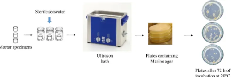
2.4. Quantification of Bacterial Biofilm
Quantification of the bacterial biofilm adhered to the surface of the mortar specimens
was performed using “culture-based methods” (Figure 6) [72–74]. This quantification was
carried out after 0, 1, 2, 6, 8, 10, 15, 24, 26 and 28 days of immersion in seawater. This
method involves three main steps: (i) recovering the bacterial biofilm from the surface, (ii)
cultivating the solution obtained on a bacterial culture medium and (iii) counting the
bacterial colonies after sufficient incubation time for bacterial growth. With this method,
the culture medium has a major impact on bacterial growth. In this study, we used marine
agar (MA) medium. MA is widely used for the cultivation of marine bacteria [75–77].
Figure 6. Culture-based method used in the quantification of bacterial biofilm formed on the
surface of mortar specimens.
After each incubation period, three specimens of each mortar type were placed in
three sterile tubes containing 10 mL of sterile seawater. Bacteria adhering to the mortar
surface were detached by immersing the tubes in an ultrasonic bath (Bandelin
SONOREX™) for 10 min at 20 °C. The solution obtained was diluted using sterile
seawater, then 100 µL of diluted solution was spread on plates containing MA (Dutscher,
490614). The plates were then incubated at 20 °C, and a colony count was performed at
least 72 h after incubation. The results are expressed as colony-forming units per cm
3of
mortar (CFU/cm
3).
2.5. Evaluation and Quantification of Marine Fouler Organisms (Macro-Fouling)
Various techniques were used in this study to quantify the marine fouler organisms
(adhered to the surfaces of the concrete specimens) and assess their visual appearance: an
image-analysis-based method [61,64,78,79], and wet-/dry-weight biomass measurement
[80–82] (Figure 7).
Figure 7. Techniques used to quantify macrofouling.
For the image-analysis-based method, photographs of the concrete specimens were
taken using a Nikon D5300 camera. The photographs were taken under constant lighting
using a photography lab. The images were subsequently processed using the ImageJ 1.38x
software [83]. To distinguish the biofouling from the noncolonized surface of the concrete,
a threshold color was used in the CIE Lab color space. Afterward, the images were
processed with a binary system (Figure 8) to obtain white pixels (noncolonized area) and
black pixels (colonized area, biofouling) from the initial photograph. These data were
compiled and used to calculate the biofouling coverage for each concrete type as a
percentage of surface area.
Figure 8. Example of the analyses performed on a concrete surface image. (A) Original picture
obtained with the Nikon D5300 camera. Image processed using ImageJ; (B) the black and white pixels indicate the fouled and unfouled surfaces, respectively.
This method was applied at 0, 8, 15, 22, 29, 43, 57, 71, 78, 85, 99, 106 and 113 days of
immersion using 15 replicates of each concrete type.
For the dry-weight biomass measurement technique, the first step was to recover the
marine fouler organisms from the concrete surface. The recovered cells were placed in
tubes and dried at 60 °C for 24 h [80]. The weight of dry biomass was then measured using
scales (accuracy 0.01 g). This quantification method was applied at 71, 78, 85, 99, 106 and
113 days of immersion using 3 replicates of each concrete type per time; the data are
reported as grams of biomass (g) per cm
2of concrete surface (g/cm
2).
The principle of the wet-weight biomass measurement technique was the same as
that of the dry biomass quantification technique, i.e. quantifying the weight of the marine
fouler organisms adhered to the concrete specimens. To apply this technique, the weight
of 15 concrete specimens was measured before they were immersed using scales (Sodipro,
300619, accuracy 1 g). Since concrete is a porous material, the immersed specimens absorb
water and increase in weight. Then, the weight of these specimens after 15 days of
immersion was taken as W0 (concrete + absorbed water weight), and the weight of
biomass was subsequently calculated by subtracting the weight obtained after X days of
immersion (Wx) from W0. Before the evaluation of W0 and Wx, the concrete specimens
were drained for 15 min. This quantification method was applied at 0, 8, 15, 22, 29, 43, 57,
71, 78, 85, 99, 106 and 113 days of immersion using 15 replicates of each concrete type; the
data are reported as grams of biomass (g) per cm
2of concrete surface (g/cm
2).
2.6. pH Measurement
The alkalinity of the medium and the surface of a submerged cementitious material
can affect biocolonization [70,84,85]. Therefore, the pH of the seawater and the surface of
the mortar specimens were measured in triplicate throughout the immersion period using
a pH electrode (Hanna instrument, HI1230, accuracy 0.1 pH unit) and pH indicator paper
(Whatman, 0.0 to 14.0, accuracy 1 pH unit), respectively, according to the protocol
described by Hayek et al. [69]. The pH of the mortar specimens was verified using
phenolphthalein indicators (which go from colorless to pink at pH ≥9). However, the use
of phenolphthalein and pH indicator paper gives a fairly good-quality estimate of the pH
of the surface, but does not accurately quantify the pH of the material [86].
2.7. Hydrophobicity Evaluation
The hydrophobicity of the mortar surface during bacterial colonization was
evaluated using a drop-shape analyzer (KRÜSS, DSA 30), which measures the angles and
diameters of contact between a drop of water and a surface [70,87]. After each period of
incubation in seawater, 3 mortar specimens were drained for 2 h. Then, a drop (3 µL) of
water was placed on mortar surface. Absorption of the drop was monitored using a
camera (8 images per second). The contact angle between the drop and the material was
measured using the Advance machine software, and a minimum of three tests were
carried out at different points.
2.8. Statistical Analyses
In order to evaluate the significance of the various results obtained, statistical
analysis was performed using GraphPad Prism 5 (GraphPad Software, San Diego, CA,
USA) and t-test, one-way and two-way ANOVA tests. Statistical significance was
accepted by p value <0.05 obtained using Bonferroni or Tukey’s multiple comparison post
hoc tests.
3. Results and Discussion
3.1. Preliminary Result: Seawater pH and Temperature Measurement
Marine environmental conditions such as seawater temperature and alkalinity
influence bacterial growth and biofilm formation [88–90]. In order to ensure that
environmental conditions were favorable to the growth of marine bacteria during this
study, the temperature and the alkalinity of seawater used in this experiment were
measured throughout the immersion period (Figure 9).
Figure 9. The pH and temperature of seawater during the quantification of bacterial biofilm on
mortar specimens.
Figure 9 shows that the seawater temperature remained between 16 and 23 °C
depending on variations in the weather at IFREMER’s outdoor experimental platform in
Palavas-les-Flots over the course of the experiment. During the subsequent process of
bacterial-biofilm quantification, the seawater temperature was optimal for the growth of
most marine bacteria [75,91–93].
Moreover, the seawater pH remained constant at an average of 8.16 throughout the
experiment (Figure 9). This value was consistent with the literature; the pH of seawater
varied between 7.5 and 9.0, with an average of around 8.2 [69,94,95]. Seawater alkalinity
was hence unaffected by the immersion of cementitious material specimens (the release
of Ca(OH)
2and KOH resulting from the leaching reaction) and remained optimal for the
growth of marine bacteria [96–98].
3.2. Examination of the Mortar Specimens
When immersed in seawater, mortar and concrete surfaces are rapidly colonized by
marine bacteria. These bacteria form on the surface a highly complex dynamic and 3D
biofilm structures [47,48,99,100].
To study the influence of intrinsic parameters on the bacterial colonization of
cementitious materials in the marine environment, six types of mortar specimen (Table 1)
were immersed in seawater under natural light. The bacterial biofilm formed on the
mortar surfaces was quantified after 0, 1, 2, 6, 8, 10, 15, 22, 24 and 28 days. At the same
time, the pH of the seawater and mortar surfaces were determined.
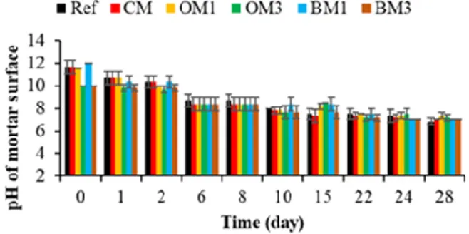
3.2.1. pH of Mortar Surfaces
Cementitious materials have a very high surface alkalinity (pH ~13) [101,102]. Due to
this very basic pH, their constituent materials have been cited as inhibitors of the
recruitment of marine biota [36]. In addition, a decrease in pH from 13 to around 9 has
been reported as necessary for bacterial colonization of cementitious materials submerged
in seawater [69,103]. Therefore, the pH of the mortar surfaces was evaluated throughout
the experiment in order to discern whether the pH was indeed responsible for inhibiting
bacterial colonization when obtained.
Figure 10 shows that the pH of the CEM I mortar specimens (Ref, CM, OM1 and BM1)
at T0 was around 11.7. The pH was also verified using phenolphthalein indicators, which
gave a pink coloration after contact with the surface of the specimens, indicating a pH
higher than 9. Therefore, the surface pH measured was lower than expected (pH ~13)
[101,102]. This might be due to the 30 days of contact between air and mortar specimens
before the start of the immersion test. In such conditions, mortar specimens undergo a
carbonation reaction due to the presence of carbon dioxide. Carbon dioxide from air reacts
with calcium hydroxide and calcium silicate hydrate in the mortar to form calcium
carbonate and water. This carbonation reaction decreases the pH of mortar specimens
[104,105].
Figure 10. Evaluation of the pH of mortar surfaces. Each evaluation was performed in triplicate,
and the error bars present the standard deviation from the values obtained. Ref = control mortar; CM = cured mortar; OM1 = oiled mortar prepared with CEM I; OM3 = oiled mortar prepared with CEM III; BM1 = biomimetic mortar prepared with CEM I; BM3 = biomimetic mortar prepared with CEM III.
Moreover, when CEM I was replaced by CEM III, the pH of the CEM III mortar
specimens (OM3 and BM3) at T0 was around 10 (due to the carbonation reaction). This
difference in pH between the CEM I and CEM III specimens was probably one reason why
the cementitious materials prepared with CEM III were more bioreceptive than those
formulated using CEM I, according to [69,70,106].
After immersion, the pH of the CEM I and CEM III mortar specimens decreased
quickly and reached a value of 8.3 at T6. Then, the pH decreased gradually and reached a
value of 7.3 at T28. This drop in pH can be explained by the action of the leaching reaction
in seawater. In contact with water, alkali metals such as potassium, calcium and
hydroxide ions will leach out of the mortar specimens and thus reduce the pH of the
surface [107,108].
Therefore, the pH values measured were lower than 9 from 6 days of immersion for
both the CEM I and the CEM III mortar specimens. This pH was favorable for the adhesion
and growth of marine bacteria [98,109].
3.2.2. Quantification of Bacterial-Biofilm Formation
The bacterial colonization of cementitious materials and biofilm formation are
complex processes that are affected by many factors, including the environmental
conditions, the bacterial properties and the physical/chemical characteristics of the
material surface [109–111]. In this study, the mortar specimens were incubated under the
same environmental conditions (pH and temperature) in the presence of the same marine
bacteria. Therefore, only the physicochemical properties of the mortar surfaces could
generate a different rate of bacterial colonization. The results concerning bacterial
colonization of the six types of mortar specimen used in this study are presented in Figure
11.
Figure 11 shows that the bacterial colonization of mortar surfaces (all types of mortar)
started with a latency phase, followed by a phase of cell growth and accumulation on the
surfaces and ending with a plateau phase. These colonization kinetics were also observed
in most of the studies quantifying bacterial-biofilm formation on cementitious-material
surfaces or on other surface types [60,61,69,112–114]. Moreover, a decrease in the bacterial
colonization of mortar surfaces was observed between 6 and 10 days. This decrease could
be justified by the drop in water temperature (from 19 to 16 °C) observed during the
immersion period [115–117].
Figure 11. Quantification of bacterial-biofilm formation on mortar specimens. Each quantification
was performed in triplicate using the culture-based method, and the error bars present the standard deviation from the values obtained. Ref = control mortar; CM = cured mortar; OM1 = oiled mortar prepared with CEM I; OM3 = oiled mortar prepared with CEM III; BM1 = biomimetic mortar prepared with CEM I; BM3 = biomimetic mortar prepared with CEM III.
Therefore, based on the rate of bacterial colonization, the mortar types can be
classified from less to more bioreceptive in the following order: CM < Ref < OM1 < OM3 <
BM1 < BM3. Then, surface chemical characteristics, chemical composition (type of cement)
and surface roughness influenced the bacterial colonization of cementitious materials
submerged in seawater. Among these parameters, surface roughness seemed to be the
parameter that promoted the bacterial colonization of cementitious materials most
effectively in the marine environment.
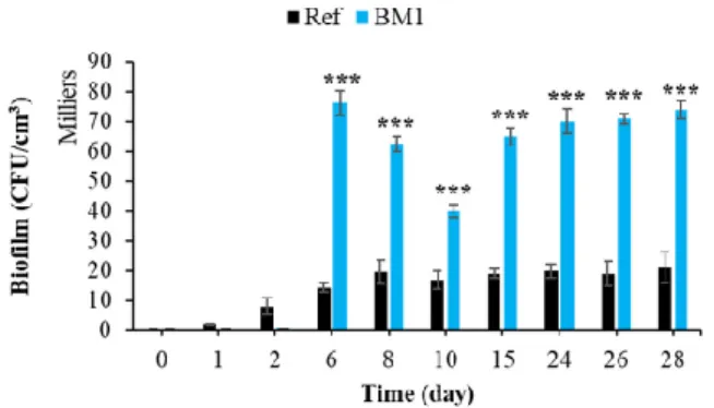
3.2.3. Effect of Surface Roughness on Bacterial Colonization
Surface roughness is one of the main physical factors influencing the bioreceptivity
of materials. The effect of roughness has been determined for different types of material
and in several types of environment [118–122]. Figure 12 shows that, in the marine
environment, a rough mortar surface significantly increased bacterial colonization in
comparison with a smooth surface (Ref). This influence of surface roughness also was
identified in several studies concerning the bacterial colonization of cementitious
materials [123,124].
Figure 12. Quantification of bacterial biofilm on smooth and rough mortars. Each experiment was
performed in triplicate, and the error bars present the standard deviation of the values obtained. Ref = control mortar; BM1 = biomimetic mortar prepared with CEM I. The experiments highlighted with asterisks differed significantly from the control (Bonferroni; ***: p < 0.001) at the indicated time.
Moreover, nanoscale bumps on the cementitious surface can act as anchoring sites
and micro-refuges for the installation of microorganisms [125]. These microrefuges
protect the microorganisms from the hydrodynamic forces that tend to detach them
during the adhesion phase [125,126]. Therefore, surface roughness increased the bacterial
colonization (bioreceptivity) of cementitious materials in the marine environment.
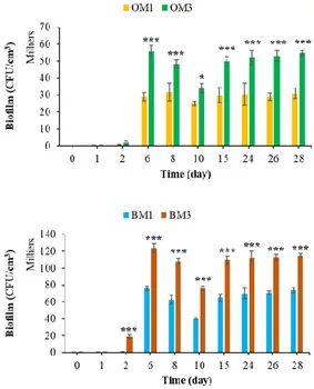
3.2.4. Effect of Cement Type on Bacterial Colonization
Chemical composition has a significant influence on the biocolonization of
cementitious materials [36,54]. Figure 13 shows that the type of cement influenced the
bacterial colonization of cementitious materials in the marine environment. The formation
of bacterial biofilm was much greater in the case of CEM III mortars, regardless of the
surface topography or whether formwork oil was applied. These results were in keeping
with the literature, in which a similar effect of cement type on the bacterial colonization
of cementitious materials has been reported [63,69,127]. Therefore, the use of CEM III
binder increased the bioreceptivity of cementitious materials in the marine environment.
Figure 13. Quantification of bacterial biofilm on CEM1 and CEM3 mortar specimens. Each
experiment was performed in triplicate, and the error bars present the standard deviation of the values obtained. Ref = control mortar; OM1 = oiled mortar prepared with CEM I; OM3 = oiled mortar prepared with CEM III; BM1 = biomimetic mortar prepared with CEM I; BM3 = biomimetic mortar prepared with CEM III. The experiments highlighted with asterisks differed significantly from the control (Bonferroni; *: p < 0.05, ***: p < 0.001) at the indicated time.
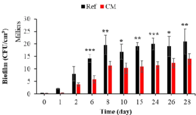
3.2.5. Effect of Curing Compound on Bacterial Colonization
The curing agent is a liquid applied to the concrete or mortar surface to protect the
material from water evaporation and give it greater aesthetic and mechanical durability,
preventing early-age surface cracking [128–130].
To the best of our knowledge, the effect of a curing product on the biocolonization of
cementitious materials in seawater has not been studied. Figure 14 shows that the curing
compound significantly inhibited the bacterial colonization of mortar submerged in
seawater. Cells accumulated and grew faster and more extensively on an untreated
surface.
Figure 14. Quantification of bacterial biofilm on treated and untreated mortars with curing
compound. Each experiment was performed in triplicate, and the error bars present the standard deviation of the values obtained. The experiments highlighted with asterisks differed significantly from the control (Bonferroni; *: p < 0.05, **: p < 0.01, ***: p < 0.001) at the indicated time.
In the marine environment, one of the anti-biofouling strategies used to inhibit
bacterial adhesion and the adhesion of other organisms is the treatment of the material by
a hydrophobic surface coating [99,131–135]. To verify the effect of the curing film on the
hydrophobicity of the mortar surface, the contact angle with a drop of water was
measured using a drop-shape analyzer (Figure 15). From their preparation until the start
of the immersion test, the mortars treated with the curing compound presented a contact
angle greater than 110°, indicating a hydrophobic surface [136,137]. After immersion and
under the action of seawater, the contact angle gradually decreased with time and became
equal to that of the control mortar at 28 days of immersion (data not shown).
Figure 15. Contact angle of a water drop on the surface of untreated mortar and mortar treated
with a curing compound. Photos were taken two seconds after contact between the water and the surface.
Moreover, the curing compound used in this study contained a mixture of alkyl
(C14-C18) bis (2-hydroxyethyl) amine, 5-chloro-2H-isothiazol-3-one and
2-methyl-2H-isothiazol-3-one (FDS Sikacem cure). These elements or their derivatives have been
cited as anti-biofouling molecules that inhibited the formation of bacterial biofilm and the
biofouling process [138–140].
Therefore, we propose that the curing compound inhibited the formation of bacterial
biofilm on mortar surfaces in seawater because of its chemical composition
(anti-biofouling) and its effect on surface hydrophobicity. Therefore, surface treatment with this
type of curing compound reduced the bacterial colonization of cementitious materials in
the marine environment.
3.2.6. Effect of Formwork Oil on Bacterial Colonization
Formwork oil is a mold-release agent that is applied to a wall of a mold to ensure
easy separation of the hardened concrete from the mold by reducing the adhesion forces
at the concrete/mold interface [141,142]. Two types of formwork oil with different
chemical compositions are mentioned in the literature: mineral and green (plant-based)
[143]. Mineral formwork oils have been reported to cause environmental pollution and
health problems for construction workers [144]. For this reason, plant-based formwork oil
(which is nontoxic and biodegradable) is the release agent used most widely today to
prevent damage to the hydraulic concrete surface during mold-stripping operations [143–
145]. This oil forms a well-adsorbed and stable “lubricant monolayer” on the surface of
cementitious materials, leading to improved release performance [144]; the free fatty acids
from the oil react in a basic medium with calcium hydroxides and produce a layer of
calcium carboxylates (soaps) and glycerol [144,146]. Van der Waals cohesion forces
stabilize the glycerol and make the layer more resistant to stress [144].
To the best of our knowledge, the effect of formwork oil on the biocolonization of
cementitious materials in seawater has not been studied to date. Figure 16 shows that the
colonization process was significantly increased if the surface was treated with formwork
oil. The composition of the green formwork oil used is not given in this study. Detailed
information is given neither in the technical product information nor in the bibliography.
Therefore, given that green formwork oil is biodegradable, we propose that this oil
applied on the surface of the mortar specimens was used as a carbon source by marine
bacteria, according to the study and the results of [147–150]. Therefore, the presence of a
biodegradable oil as an additional source of nutrients increased the biofilm formation on
oiled mortars; Dusane et al. showed through a laboratory test that the carbon sources
differentially affected the formation of biofilms. Lactic acid, erythritol, glycerol, glucose
and edible oils increase this process [151]. Therefore, surface treatment with this type of
formwork oil increased the bacterial colonization of cementitious materials in the marine
environment.
Figure 16. Quantification of bacterial biofilm on untreated mortars and mortars treated with
formwork oil. Each experiment was performed in triplicate, and the error bars present the standard deviation of the values obtained. The experiments highlighted with asterisks differed significantly from the control (Bonferroni; *: p < 0.05, **: p < 0.01, ***: p < 0.001) at the indicated time.
3.3. Quantification of Marine Fouler Organisms on the Concrete Specimens
After the colonization of immersed materials by marine bacteria, biocolonization
continues through the adhesion and growth of macroorganisms such as algae and larvae.
This adhesion is known as macrofouling, and is the second main stage of biofouling in the
marine environment [40,41,43–45]. The first part of this study showed that intrinsic
parameters such as surface treatment with curing compound or formwork oil, chemical
composition and surface condition (SS, RS) influence the bacterial colonization of
cementitious materials in the marine environment. To test the effect of these parameters
on the macrofouling, seven types of concrete specimens were immersed in seawater under
the same conditions (the same incubation period) as those used for the mortar specimens.
The marine fouler organisms adhered on the concrete surfaces were quantified after 0, 8,
15, 22, 29, 43, 57, 71, 78, 85, 99, 106 and 113 days.
3.3.1. Quantification of Marine Fouler Organisms Using an Image-Analysis-Based
Method
Image analysis is a quantification technique used widely in performing biofouling
tests on cementitious materials [61,64,78,79].
Figure 17 shows that the colonization of concrete surfaces (all types of concrete) by
marine fouler organisms started with a latency phase, followed by a phase during which
cells accumulated and grew on the surfaces (from 43 days to 113 days). These kinetics of
the colonization process were also observed in most of the other studies quantifying
biofouling on cementitious-material surfaces [61,79,106,152].
Figure 17. Quantification of marine fouler organisms using an image-analysis-based method. Ref = control concrete; CC =
cured concrete; OC = oiled concrete; EC1 = ECOPODE™ concrete prepared with CEM I; EC3 = ECOPODE™ concrete prepared with CEM III; BC1 = biomimetic concrete prepared with CEM I; BC3 = biomimetic concrete prepared with CEM III. Each experiment was performed on 15 replicates, and the error bars present the standard deviation of the values obtained.
As was the case with bacterial colonization, curing compound inhibited the adhesion
of marine fouler organisms to the surface of concrete (Table 3). This inhibition may have
been due to a toxic chemical composition and the hydrophobicity of the material surface
treated with the curing agent [131–135,138,140].
Table 3. Statistical comparison (GraphPad Prism 5) of the macrofouling quantification results obtained using an
image-analysis-based method. Ref = control concrete; CC = cured concrete; OC = oiled concrete; EC1 = ECOPODE™ concrete prepared with CEM I; EC3 = ECOPODE™ concrete prepared with CEM III; BC1 = biomimetic concrete prepared with CEM I; BC3 = biomimetic concrete prepared with CEM III. (Bonferroni; ns: not significant, p > 0.05, *: p < 0.05, **: p < 0.01, ***: p < 0.001).
Time (days) Ref vs. CC Ref vs. OC OC vs. EC1 OC vs. EC3 EC1 vs. EC3 Ref vs. BC1 Ref vs. BC3 BC1 vs. BC3
0 ns ns ns ns ns ns ns Ns 8 ns ns ns ns ns ns ns Ns 15 ns ns ns ns ns ns ns Ns 22 ns ns ns ns ns ns ns Ns 29 ns ns ns ns ns ns ns Ns 43 ns *** * *** * *** *** Ns 57 ns *** *** *** ** *** *** Ns 71 *** *** *** *** *** *** *** Ns 78 *** ns *** *** *** *** *** Ns 85 *** ns ns ns ns *** *** Ns 99 *** ns ns ns ns *** *** Ns 106 *** ns ns ns ns *** *** Ns 113 *** ns ns ns ns *** *** Ns
To assess the effect of other parameters on macrofouling, a statistical analysis of the
results was performed using GraphPad Prism 5 software (Table 3). This analysis showed
that:
Treating the surface with formwork oil only supported greater biocolonization
during the first days of macrofouling (Ref vs. OC); after 71 days, the quantity of
marine fouler organisms on the oiled surface was equal to that on the control surface.
Surface topography (FPS surface) and the use of CEM III cement did not affect
biocolonization after 78 days of immersion (OC vs. EC1, OC vs. EC3 and EC1 vs.
EC3).
Surface roughness supported greater biocolonization from the start of macrofouling
(43 days) until the end of our study at 133 days of immersion (Ref vs. BC1).
The effect of surface roughness on macrofouling dominated the effect of chemical
composition (use of CEM III cement). The biocolonization was significantly different
when the control concrete and the rough concrete were compared (Ref vs. BC1, Ref
vs. BC3), whereas no significant differences were observed when biomimetic
concretes prepared using CEM I or CEM III were compared (BC1 vs. BC3).
Therefore, surface-chemical characteristics, chemical composition (type of cement)
and surface roughness influenced the macrofouling of cementitious materials submerged
in seawater.
Based on the persistence of their significant effect, the intrinsic parameters that
supported a greater biocolonization of cementitious materials in the marine environment
can be classified from less to more effective in the following order: surface treatment with
formwork oil < surface topography (FPS surface) < chemical composition (type of cement)
< surface roughness (biomimetic surface). Therefore, as was the case with bacterial
colonization, surface roughness was the parameter that promoted the macrofouling of
cementitious materials in the marine environment most effectively. We suggest that
surface roughness was the most important factor in the design of bioreceptive concrete in
the marine environment.
These results and conclusions are in keeping with those from the study performed
by Hsiung et al., which showed that the decrease in pH did not affect the long-term
bioreceptivity of cementitious materials in the marine environment. At the end of their
study, Hsiung et al. suggested that manipulation of the physical structure (surface
roughness) of the concrete was a more effective eco-engineering approach to enhancing
ecological value and species diversity in the marine environment [102].
3.3.2. Quantification of Marine Fouler Organisms Using Wet-Weight Biomass
Measurement
The second method used to quantify macrofouling on concrete surfaces is wet-weight
biomass measurement. This technique is quick and easy to apply. It allows a rapid
assessment of biocolonization in the marine environment. To the best of our knowledge,
this is the first study in which this technique has been applied in this way (see the
Materials and Methods section) to quantify macrofouling on concrete surfaces in a marine
environment.
The results obtained with this method (Figure 18) were in keeping with those
obtained using the image-analysis-based method. The statistical analysis of the results
(data not shown) was similar and the conclusions were the same—surface roughness was
the most important factor in the design of bioreceptive concrete in the marine
environment.
Figure 18. Quantification of marine fouler organisms using wet-weight biomass measurement. Ref = control concrete. CC
= cured concrete; OC = oiled concrete; EC1 = ECOPODE™ concrete prepared with CEM I; EC3 = ECOPODE™ concrete prepared with CEM III; BC1 = biomimetic concrete prepared with CEM I; BC3 = biomimetic concrete prepared with CEM III. Each experiment was performed on 15 replicates, and the error bars present the standard deviation of the values obtained.
3.3.3. Quantification of Marine Fouler Organisms Using Dry-Weight Biomass
Measurement
To be sure that the lack of significance in concrete surface biocolonization after 85
days of immersion was not due to a detection limit inherent to the method applied, we
used dry-weight biomass measurement to quantify the marine fouler organisms on the
surfaces of control, cured, oiled and ECOPODE™ concrete [80–82]. Cured concrete was
used as a control during this experiment.
Statistical analysis of obtained results (data not shown) and Figure 19 show that no
significant difference was observed after 85 days of incubation, which confirmed the
results obtained using wet-weight biomass measurement and image analysis. Surface
treatment with formwork oil and surface topography only influenced the initial
macrofouling of concrete in the marine environment.
Figure 19. Quantification of marine fouler organisms using dry-weight biomass measurement. Ref = control concrete; CC
= cured concrete; OC = oiled concrete; EC1 = ECOPODE™ concrete prepared with CEM I; EC3 = ECOPODE™ concrete prepared with CEM III. Each experiment was performed in triplicate, and the error bars present the standard deviation of the values obtained.
4. Conclusions
In summary, this study indicates that the intrinsic parameters of cementitious
materials influence the biocolonization and the biofouling formation in the marine
environment. Regarding the parameters tested in the preset work, we can summarize that:
(1) the surface design (smooth surface, faceted and patterned surface, rough surface) affect
the bacterial and macroorganism colonization of cementitious materials in seawater; (2)
the chemical composition such as the type of binder (ordinary Portland cement CEM I,
slag cement CEM III) significantly influences the biocolonization of cementitious
materials; (3) work practices such as the use of a curing agent and/or formwork oil have
an impact on biocolonization.
Using mortar specimens and culture-based methods, we have shown that in the
marine environment: (1) surface roughness seems to be the factor enhancing the bacterial
colonization of cementitious materials most effectively; (2) the use of slag cement CEM III
increases the bioreceptivity of cementitious materials; (3) the application of a curing
compound to the surface reduces the bacterial colonization of cementitious materials,
whereas green formwork oil has the opposite effect.
Using concrete specimens, an image-analysis-based method and wet-/dry-weight
biomass measurements, we have shown that the surface design, the chemical composition
(type of cement) and the chemical characteristics of the surface (curing compound or
formwork oil) have a similar effect to that observed with mortar on the macrofouling of
cementitious materials submerged in seawater.
Based on the persistence of their significant effect, the intrinsic parameters that
support greater biocolonization are classified from more to less effective in the following
order: surface design (roughness of biomimetic specimens) > chemical composition (slag
cement CEM III) > faceted and patterned surface > chemical characteristics (formwork oil).
Therefore, at the material scale, surface roughness is the most effective factor in
designing bioreceptive concrete that enhances marine biodiversity [9,14,15]. Nevertheless,
faceted and patterned surfaces seem to improve the initial colonization stages, maybe
because they induce a local hydrological change in environmental conditions. This
characteristic should be investigated in detail, especially in real conditions, at the
macroscopic scale.
In regard to improving construction techniques used in marine ecosystems, this
conclusion will be used directly by an engineering and project-management company
specializing in the field of marine-infrastructure construction to design a new innovative
concrete block that promotes marine-infrastructure colonization more effectively. The use
of CEM III cement has a positive effect in terms of both durability and biocolonization of
the concrete by marine structures. This research must be pursued further at a 1:1 scale, on
real works, to develop new concrete-block designs and study the macroscopic aspects of
the structure of concrete armor used to protect breakwaters.
Author Contributions: Writing—review and editing, M.H., M.S., J.-C.S., E.C., C.G. and O.P. All
authors have read and agreed to the published version of the manuscript.
Funding: This work was supported by Artelia, an international, multidisciplinary consultancy,
engineering and project-management company specializing in the field of marine-infrastructure construction.
Institutional Review Board Statement: Not applicable. Informed Consent Statement: Not applicable. Data Availability Statement: Not applicable.
Acknowledgments: The authors thank Emmanuel Rezzouk (IFREMER, Biology Research Unit for
exploited marine organisms) for receiving them at the Palavas-les-Flots oceanographic station and thank LIB industry for the supply of the silicone matrix and their assistance. The authors also thank IMT Mines Ales and University of Montpellier 3 for their participation and support. The authors wish to acknowledge the help provided by Florian Stratta, Habiba Lharti, Léa Capitan-Fernandez, Thomas Davignon, Oumaïma Saâdoune, Claire Desoubry, Emma Guillot and Mathilde Metges.
Conflicts of Interest: The authors declare no conflict of interest.
References
1. Halpern, B.S.; Walbridge, S.; Selkoe, K.A.; Kappel, C.V.; Micheli, F.; D’Agrosa, C.; Bruno, J.F.; Casey, K.S.; Ebert, C.; Fox, H.E.; et al. A Global Map of Human Impact on Marine Ecosystems. Science 2008, 319, 948–952, doi:10.1126/science.1149345.
2. Bulleri, F.; Chapman, M.G. The Introduction of Coastal Infrastructure as a Driver of Change in Marine Environments. J. Appl.
3. Bishop, M.J.; Mayer-Pinto, M.; Airoldi, L.; Firth, L.B.; Morris, R.L.; Loke, L.H.; Hawkins, S.J.; Naylor, L.A.; Coleman, R.A.; Chee, S.Y.; et al. Effects of Ocean Sprawl on Ecological Connectivity: Impacts and Solutions. J. Exp. Mar. Biol. Ecol. 2017, 492, 7–30, doi:10.1016/j.jembe.2017.01.021.
4. Barnosky, A.D.; Hadly, E.A.; Bascompte, J.; Berlow, E.L.; Brown, J.H.; Fortelius, M.; Getz, W.M.; Harte, J.; Hastings, A.; Marquet, P.A.; et al. Approaching a State Shift in Earth’s Biosphere. Nat. Cell Biol. 2012, 486, 52–58, doi:10.1038/nature11018.
5. Corlett, R.T. The Anthropocene Concept in Ecology and Conservation. Trends Ecol. Evol. 2015, 30, 36–41, doi:10.1016/j.tree.2014.10.007.
6. Bai, X.; Geschke, A.; Molnár, Z.; Jaureguiberry, P.; Hendry, A.; Liu, J.; Martin, A.; Midgley, G.F.; Scheffran, J.; Seppelt, R.; et al. IPBES Global Assessment on Biodiversity and Ecosystem Services Chapter 1. Assessing a Planet in Transformation: Rationale and Approach of the IPBES Global Assessment on Biodiversity and Ecosystem Services. In IPBES Global Assessment on
Biodiversity and Ecosystem Services; IPBES Secretariat: Bonn, Germany, 2019; p. 69.
7. Pioch, S.; Relini, G.; Souche, J.; Stive, M.; De Monbrison, D.; Nassif, S.; Simard, F.; Allemand, D.; Saussol, P.; Spieler, R.; et al. Enhancing Eco-Engineering of Coastal Infrastructure with Eco-Design: Moving from Mitigation to Integration. Ecol. Eng. 2018,
120, 574–584, doi:10.1016/j.ecoleng.2018.05.034.
8. Martins, G.M.; Thompson, R.C.; Neto, A.I.; Hawkins, S.J.; Jenkins, S.R. Enhancing Stocks of the Exploited Limpet Patella Candei D’Orbigny via Modifications in Coastal Engineering. Biol. Conserv. 2010, 143, 203–211, doi:10.1016/j.biocon.2009.10.004. 9. Souche, J.-C.; Pioch, S.; Salgues, M.; De Weerdt, K.; Agostini, A.; Hayek, M. De la Conception à L’éCo-conception Des Ouvrages
Maritimes : Intégrer la Nature AU Projet D’Aménagement Maritime. Rev. Paralia 2019, 12, n01.1–n01.26, doi:10.5150/revue-paralia.2019.n01.
10. Heery, E.C.; Bishop, M.J.; Critchley, L.P.; Bugnot, A.B.; Airoldi, L.; Mayer-Pinto, M.; Sheehan, E.V.; Coleman, R.A.; Loke, L.H.; Johnston, E.L.; et al. Identifying the Consequences of Ocean Sprawl for Sedimentary Habitats. J. Exp. Mar. Biol. Ecol. 2017, 492, 31–48, doi:10.1016/j.jembe.2017.01.020.
11. Dugan, J.E.; Airoldi, L.; Chapman, M.G.; Walker, S.J.; Schlacher, T. 8.02—Estuarine and Coastal Structures: Environmental Effects, A Focus on Shore and Nearshore Structures. In Treatise on Estuarine and Coastal Science; Wolanski, E., McLusky, D., Eds.; Academic Press: Waltham, MA, USA, 2011; pp. 17–41 ISBN 978-0-08-087885-0.
12. Powell, E.J.; Tyrrell, M.C.; Milliken, A.; Tirpak, J.M.; Staudinger, M.D. A Review of Coastal Management Approaches to Support the Integration of Ecological and Human Community Planning for Climate Change. J. Coast. Conserv. 2019, 23, 1–18. doi:10.1007/s11852-018-0632-y.
13. Waltham, N.J.; Dafforn, K.A. Ecological Engineering in the Coastal Seascape. Ecol. Eng. 2018, 120, 554–559, doi:10.1016/j.ecoleng.2018.07.028.
14. Dafforn, A.K.; Glasby, T.M.; Airoldi, L.; Rivero, N.K.; Mayer-Pinto, M.; Johnston, E.L. Marine Urbanization: An Ecological Framework for Designing Multifunctional Artificial Structures. Front. Ecol. Environ. 2015, 13, 82–90, doi:10.1890/140050. 15. Browne, M.A.; Chapman, M.G. Ecologically Informed Engineering Reduces Loss of Intertidal Biodiversity on Artificial
Shorelines. Environ. Sci. Technol. 2011, 45, 8204–8207, doi:10.1021/es201924b.
16. Moradllo, M.K.; Shekarchi, M.; Hoseini, M. Time-Dependent Performance of Concrete Surface Coatings in Tidal Zone of Marine Environment. Constr. Build. Mater. 2012, 30, 198–205, doi:10.1016/j.conbuildmat.2011.11.044.
17. Oleson, J.P.; Brandon, C.; Cramer, S.M.; Cucitore, R.; Gotti, E.; Hohlfelder, R.L. The ROMACONS Project: A Contribution to the Historical and Engineering Analysis of Hydraulic Concrete in Roman Maritime Structures. Int. J. Naut. Archaeol. 2004, 33, 199– 229, doi:10.1111/j.1095-9270.2004.00020.x.
18. Costa, A.; Appleton, J. Case Studies of Concrete Deterioration in a Marine Environment in Portugal. Cem. Concr. Compos. 2002,
24, 169–179, doi:10.1016/s0958-9465(01)00037-3.
19. Pandey, A.; Kumar, B. Investigation on the Effects of Acidic Environment and Accelerated Carbonation on Concrete Admixed with Rice Straw Ash and Microsilica. J. Build. Eng. 2020, 29, 101125, doi:10.1016/j.jobe.2019.101125.
20. Song, H.-W.; Lee, C.-H.; Ann, K.Y. Factors Influencing Chloride Transport in Concrete Structures Exposed to Marine Environments. Cem. Concr. Compos. 2008, 30, 113–121, doi:10.1016/j.cemconcomp.2007.09.005.
21. Collepardi, M. Concrete Durability in a Marine Environment. In Proceedings of the Canmet/Aci International Conference on Advances in Concrete Technology in the Middle East, Dubai, United Arab Emirates, 15 October 2008; pp. 19–20.
22. Yi, Y.; Zhu, D.; Guo, S.; Zhang, Z.; Shi, C. A Review on the Deterioration and Approaches to Enhance the Durability of Concrete in the Marine Environment. Cem. Concr. Compos. 2020, 113, 103695, doi:10.1016/j.cemconcomp.2020.103695.
23. Chlayon, T.; Iwanami, M.; Chijiwa, N. Combined Protective Action of Barnacles and Biofilm on Concrete Surface in Intertidal Areas. Constr. Build. Mater. 2018, 179, 477–487, doi:10.1016/j.conbuildmat.2018.05.223.
24. Jacob, C.; Buffard, A.; Pioch, S.; Thorin, S. Marine Ecosystem Restoration and Biodiversity Offset. Ecol. Eng. 2018, 120, 585–594, doi:10.1016/j.ecoleng.2017.09.007.
25. Ting, M.Z.Y.; Wong, K.S.; Rahman, M.E.; Meheron, S.J. Deterioration of Marine Concrete Exposed to Wetting-Drying Action. J.
Clean. Prod. 2021, 278, 123383, doi:10.1016/j.jclepro.2020.123383.
26. Song, H.-W.; Shim, H.-B.; Petcherdchoo, A.; Park, S.-K. Service Life Prediction of Repaired Concrete Structures under Chloride Environment Using Finite Difference Method. Cem. Concr. Compos. 2009, 31, 120–127, doi:10.1016/j.cemconcomp.2008.11.002. 27. De Rincón, O.T.; Sánchez, M.; Millano, V.; Fernández, R.; De Partidas, E.; Andrade, C.; Martinez, I.; Castellote, M.; Barboza, M.;
Irassar, F.; et al. Effect of the Marine Environment on Reinforced Concrete Durability in Iberoamerican Countries: DURACON project/CYTED. Corros. Sci. 2007, 49, 2832–2843, doi:10.1016/j.corsci.2007.02.009.
![Figure 1. Schematic representation of marine biofouling formation [45].](https://thumb-eu.123doks.com/thumbv2/123doknet/11571789.297581/4.918.277.823.125.327/figure-schematic-representation-marine-biofouling-formation.webp)