Interventional 3D Augmented Reality in Orthopedic,
Trauma and Vascular Surgery
A
UGMENTED REALITY C-ARM FLUOROSCOPY IN ORTHOPEDIC AND TRAUMA SURGERIESIt is expected that by 2050, the number of patients undergoing hip and spine surgery will reach 40 million, which will make up a global market share estimated to reach $30 billion (1,2). Hip and spine surgery data show that 90% of these occur in older adults (ages 65 and older), usually as a consequence of osteoporosis or a low-energy mechanism such as a fall from a standing height (1,2). All the more alarming is that hip and spine surgeries account for most of the total radiation exposure in all orthopedic and trauma types of procedures, with the risk of contracting cancer being up to eight times more for a surgeon and their patients when compared to other workers having a non-clinical background (3). The reason is that surgeons often place emphasis on acquiring the ‘perfect picture’ using C-arm fluoroscopes (X-ray devices) in order to complete surgery efficiently, which may lead to excess from a standing height (1,2). All the more alarming is that hip and spine surgeries account for most of the total radiation exposure
in all orthopedic and trauma types of procedures, with the risk of contracting cancer being up to eight times more for a surgeon and their patients when compared to other workers having a non-clinical background (3). The reason is that surgeons often place emphasis on acquiring the ‘perfect picture’ using C-arm fluoroscopes (X-ray devices) in order to complete surgery efficiently, which may lead to excesive use of X-rays. As an alternative to C-arm fluoroscopes, 3D navigation systems are commercially available. These systems use pre-operative CT scans, external optical tracking systems, and tracked markers as reference landmarks affixed onto both the medical instruments and the patient (4). The 3D navigation system then provides information during the surgery on the spatial relation between the medical instruments, patient, and the pre-operative CT scan which, in theory, facilitates the completion of many surgical tasks without X-ray acquisition (4). However as of 2016, it is now documented by major medical policy makers (Blue Cross Blue Shield Association, United HealthCare, etc.) that these systems are costly, are common in only some The Medical Education, Training and Computer Assisted Interventions (METRICS) Laboratory aims to integrate novel mixed-reality technologies with application in computer assisted interventions. We showcase two technologies with specific aims at optimizing surgical workflow and minimizing radiation exposure in orthopedic, trauma, and vascular surgeries. The first is an Augmented Reality C-arm fluoroscope, which provides intuitive real-time visualization by accurately overlaying X-ray to video images. The second is a ‘Desired-views’ user interface which resolves the challenges involved in the optimal control of C-arm fluoroscopes for their constant repositioning during surgery by either the interventionalist or the surgical team.
Sheila Esmeralda Gonzalez-Reyna
1, Pascal Fallavollita
11University of Ottawa, Faculty of Health Sciences, Interdisciplinary School of Health Sciences, Ontario, Canada
ABSTRACT
Le laboratoire d’éducation médicale, de formation et d’intervention assistée par ordinateur (METRICS) vise à intégrer de nouvelles technologies à réalité mixte à des interventions assistées par ordinateur. Nous présentons deux technologies ayant des objectifs spécifiques pour optimiser le flux de travail chirurgical et minimiser l’exposition aux rayonnements dans les chirurgies orthopédiques, traumatologiques et vasculaires. La première est un fluoroscope C-arm à réalité augmentée, qui fournit une visualisation intuitive en temps réel en superposant avec précision les rayons X aux images vidéo. La seconde est une interface utilisateur «Desired-views» qui résout les problèmes liés au contrôle optimal des fluoroscopes C-arm pour leur repositionnement constant pendant la chirurgie, soit par l’interventionniste, soit par l’équipe chirurgicale.
surgeries, showed no increased benefit to patient outcomes when compared to traditional C-arm fluoroscopes, and have yet to find a niche in orthopedic and trauma operating rooms worldwide. Consequently, surgeons fall back to the traditional C-arm devices to perform surgeries (4).
The President of the Radiation Safety Institute of Canada has reported that cancers of various types are a potential outcome of occupational overexposure to radiation. The Health Economics Review published a report in early 2017 titled “Costs of Productivity Loss Due to Occupational Cancer in Canada (…)”, which highlighted the estimated total cost of cancer to the Workers’ Compensation System in Canada, due to productivity losses alone, to be $1.2 billion (5).
With respect to risks associated to healthcare providers, reports have documented the dosage of radiation among interventionalists as the greatest registered among any medical staff working with C-arm fluoroscopes (6). As highlighted by Picano et al. (7), cumulative doses after 30 years of working life are in the range of 50 to 200 mSv, corresponding to a whole-body dose equivalent of 2,500 to 10,000 chest X-rays. In the case of patients, the benefits of a proper usage of C-arm fluoroscopy devices outweighs the experienced radiation risks, especially in the older age groups (8). The patient is exposed to primary radiation, namely radiation between the X-ray source and the image intensifier. Short-term risks are radiation induced skin damages (erythema, epilation and even dermal necrosis), which result from acute radiation doses beyond 2 Gy (9). Thus, there is an urgent need to develop technologies that rectify radiation exposure in the surgery room.
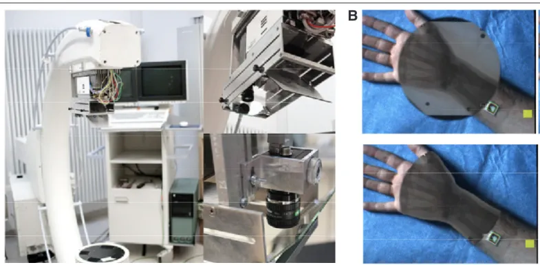
The METRICS lab (Medical Education, Training, And Computer Assisted Interventions) aims to break new ground on optimizing the current surgical workflows in orthopedic and trauma surgeries while alleviating the effects of radiation exposure. An Augmented Reality (AR) C-arm fluoroscope provides intuitive real-time visualization for surgeons by overlaying the 2D X-ray images to the 2D video images obtained by affixing one camera (or multiple depth sensors) on the C-arm device (10,11). Thanks to a one-time calibration of the AR C-arm, the camera centre and the X-ray source centre are virtually aligned, thus providing a geometrically correct 2D/2D overlay (12). The precision of the overlay is less than 0.5mm (11). Figure 1a shows the AR C-arm fluoroscope and Figure 1b shows an example of the 2D X-ray overlaying a 2D video image. Advanced multi-modal visualization is also a possibility when applying machine
learning algorithms to the X-ray and video data (Figure 2). Our publications conclusively demonstrate that the video image overlay has the benefit of facilitating many surgical tasks without the acquisition of an X-ray (13–19).
As mentioned previously, it is clinically accepted that some C-arm device positions may generate up to six times more radiation exposure to highly sensitive body-parts such as thyroid, hands, eyes and head, and should be avoided (20). Modeling of X-ray photon propagation is complex and not often possible ton achieve analytically. To visualize the amount of radiation dosage during surgery, we consider Monte Carlo (MC) simulations, which are algorithmic methods that approximate radiation exposure by simulating the X-ray imaging process, and tracking the resulting scattered particles towards clinicians and patients during surgery (20,21). Optimizing the position of any C-arm device (to minimize radiation exposure) is challenging since the context, the imaging parameters, and the patient’s and staff’s positioning need to be considered in the MC simulations. As in Bott et al. (22), the real C-arm imaging parameters (tube kilovoltage, filtration, aperture angle and beam projection), together with the X-ray photon scattering process, are all considered by the simulation. These can be extracted from the C-arm device application programming interface (API). The patient and surgeon’s positioning is known from the camera or multiple sensors affixed to the C-arm. The representation of the surgeon and patient is created and their surface (tessellation) is visualized in 3D in real-time. The radiation dose at the surface is visualized using color coding. The color values for each triangle corner of both the surgeon and patient tessellation are computed by interpolating the dose distribution information of the dose accumulation (Figure 3). With the advent of modern mixed-reality head-mounted displays (HMDs), the typical challenges encountered by mobile AR technologies have now been solved, and commercial devices suitable for medical applications are now available. Indeed, a recent study showed that the Microsoft’s HoloLens HMDs are now suitable enough in terms of contrast perception, task load and frame rate, during surgical interventions (23). Thus, the HMD not only provides an accurate feedback about the current radiation exposure to surgeon and patient, but also correlates such an exposure to the underlying workflow tasks taking place. Collecting the dose generated at each step of the surgery can help generate exposure statistics. Such statistics will be computed by considering each person’s record of previous exposures to radiation, which would help to reduce the risks of long-term negative effects of exposure. To conclude,
Figure 1. Augmented Reality Fluoroscope. A) A C-arm augmented by video camera, and B) 2D X-Ray images correctly aligned to 2D Video images.
A
Figure 3. Radiation exposure visualization. Red colors demonstrate areas of high dosage whereas blue colors demonstrate areas of low dosage.
Figure 2. Example visualizations between X-ray and video images demonstrating center punching, drilling and clamping tasks.
the development of the AR C-arm was necessary as it has the potential of transforming orthopedic and trauma surgeries at a relatively low cost (i.e. equivalent to the cost of affixing
a camera or several RGB-D sensors on the C-arm grantry) in order to maximize patient outcomes and minimize radiation exposure.
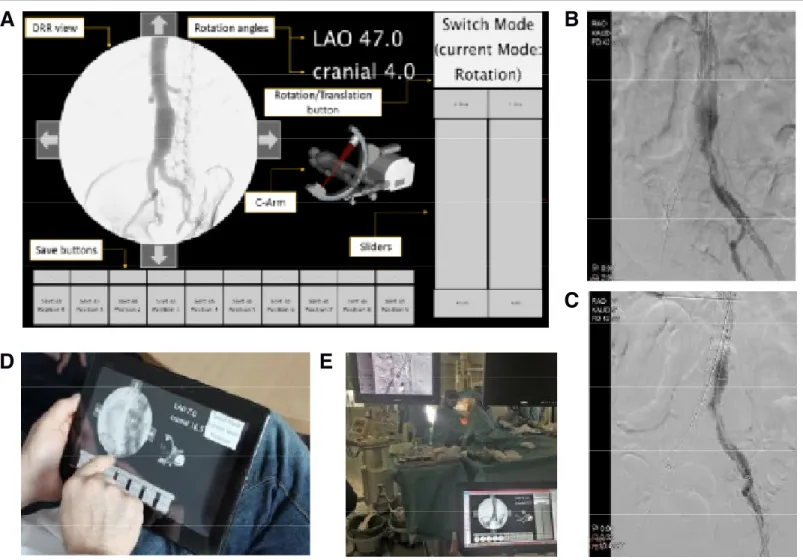
Figure 4. “Desired-views” user interface. A) The user interface, B) Iliacs optimal view vs. C) Iliacs non-optimal view, D) Defining optimal view before surgery, and E) Using predefined optimal view.
A
B
E
C
D
A preliminary and unpublished study was undertaken to evaluate this interface in a real clinical scenario. Twelve patients underwent EVAR surgery at our clinical partner site. The study consisted of comparing the planned C-arm positions using our ‘Desired-views’ interface prior to intervention, to the ones during
intervention when navigating through the left and right iliacs of each patient. For all patient cases, we received the relevant intra-operative X-ray images, which enabled us to identify any deviations of the intra-operative C-arm parameters from the pre-operatively planned ‘desired-views’. Results demonstrated that for all twelve patient surgeries the C-arm was positioned optimally on the first try, which leads us to conclude that the proposed work has potential to increase the accuracy while decreasing radiation exposure, contrast intake, and duration of future patient surgeries.
CONCLUSIONS
The METRICS lab hosts many junior and senior researchers aiming to redefine the design and development of physiological sensing, virtual and augmented reality immersion, and advanced user interactions. This is made possible with the collaboration of clinicians, computer scientists and industry, for the effective translation of our new developments inside the operating rooms of tomorrow. Two key projects involve the development and validation of an Augmented Reality C-arm device and a ‘Desired-views’ user interface in orthopedic, trauma, and vascular surgery setting. The Augmented Reality C-arm device will be evaluated during the next five years as the principle investigator is the recipient of an Early Career Research Award titled: Interventional 3D Augmented Reality. Additionally, the validation of the ’Desired-views’ user interface will continue with partners in the Faculty of Surgery upon ethics board approval.
REFERENCES
1. Kannus P, Parkkari J, Sievänen H, Heinonen A, Vuori I, Järvinen M. Epidemi-ology of hip fractures. Bone. 1996 Jan;18(1):S57–63.
2. Gebhard FT, Kraus MD, Schneider E, Liener UC, Kinzl L, Arand M. Does Computer-Assisted Spine Surgery Reduce Intraoperative Radiation Doses? Spine (Phila Pa 1976). 2006;31(17).
3. Mastrangelo G, Fedeli U, Fadda E, Giovanazzi A, Scoizzato L, Saia B. In-creased cancer risk among surgeons in an orthopaedic hospital. Occup Med (Chic Ill). 2005;55(6):498–500.
4. UnitedHealthcare. Medical Policy Update Bulletin: Medical Policy, Drug Policy & Coverage Determination Guideline Updates. 2015.
5. Wranik WD, Muir A, Hu M. Costs of productivity loss due to occupational cancer in Canada: estimation using claims data from Workers’ Compensa-tion Boards. Health Econ Rev. 2017;7(1).
6. Roguin A, Goldstein J, Bar O, Goldstein JA. Brain and neck tumors among physicians performing interventional procedures. Am J Cardiol. 2013;111(9):1368–72.
7. Picano E, Andreassi MG, Piccaluga E, Cremonesi A, Guagliumi G. Occu-pational risks of chronic low dose radiation exposure in cardiac cath-eterisation laboratory: the Italian healthy cath lab study. EMJ Int Cardiol. 2013;1(1):50–8.
8. Roguin A. Radiation in cardiology: can’t live without it! Eur Heart J. 2014;35:599–604.
9. Miller DL. Interventional Fluoroscopy: Reducing Radiation Risks for Patients and Staff. Vol. 20, Journal of Vascular and Interventional Radiology. 2009. 10. Navab N, Bani-Kashemi A, Mitschke M. Merging visible and invisible: Two
camera-augmented mobile C-arm (CAMC) applications. In: Augmented Reality, 1999(IWAR’99) Proceedings 2nd IEEE and ACM International Work-shop on. 1999. p. 134–41.
11. Navab N, Heining S-M, Traub J. Camera augmented mobile C-arm (CAMC): calibration, accuracy study, and clinical applications. IEEE Trans Med Imag-ing. 2010;29(7):1412–23.
12. Wang L, Traub J, Heining SM, Benhimane S, Euler E, Graumann R, et al. Long bone X-ray image stitching using Camera Augmented Mobile C-arm. In: International Conference on Medical Image Computing and Computer-Assisted Intervention. 2008. p. 578–86.
13. Wang L, Fallavollita P, Zou R, Chen X, Weidert S, Navab N. Closed-form inverse kinematics for interventional C-arm X-ray imaging with six de-grees of freedom: modeling and application. IEEE Trans Med Imaging. 2012;31(5):1086–99.
14. Wang L, Fallavollita P, Brand A, Erat O, Weidert S, Thaller P-H, et al. Intra-op measurement of the mechanical axis deviation: an evaluation study on 19 human cadaver legs. In: International Conference on Medical Image Com-puting and Computer-Assisted Intervention. 2012. p. 609–16.
15. Pauly O, Diotte B, Habert S, Weidert S, Euler E, Fallavollita P, et al. Relevance-based visualization to improve surgeon perception. In: Lecture Notes in Computer Science. 2014. p. 178–85.
16. Diotte B, Fallavollita P, Wang L, Weidert S, Euler E, Thaller P, et al. Multi-mod-al intra-operative navigation during distMulti-mod-al locking of intramedullary nails. IEEE Trans Med Imaging. 2015;34(2):487–95.
17. Londei R, Esposito M, Diotte B, Weidert S, Euler E, Thaller P, et al. The “aug-mented” circles: A video-guided solution for the down-the-beam position-ing of im nail holes. In: Lecture Notes in Computer Science. 2014. p. 100–7. 18. Leucht N, Habert S, Wucherer P, Weidert S, Navab N, Fallavollita P. [POSTER] Augmented Reality for Radiation Awareness. In: Mixed and Augmented Re-ality (ISMAR), 2015 IEEE International Symposium on. 2015. p. 60–3. 19. Fallavollita P, Brand A, Wang L, Euler E, Thaller P, Navab N, et al. An
aug-mented reality C-arm for intraoperative assessment of the mechanical axis: a preclinical study. Int J Comput Assist Radiol Surg. 2016;11(11):2111–7. 20. Koukorava C, Carinou E, Ferrari P, Krim S, Struelens L. Study of the
parame-ters affecting operator doses in interventional radiology using Monte Carlo simulations. In: Radiation Measurements. 2011. p. 1216–22.
21. Bert J, Perez-Ponce H, Bitar Z El, Jan S, Boursier Y, Vintache D, et al. Geant4-based Monte Carlo simulations on GPU for medical applications. Phys Med Biol. 2013;58(16):5593–611.
22. Bott OJ, Wagner M, Duwenkamp C, Hellrung N, Dresing K. Improving educa-tion on C-arm operaeduca-tion and radiaeduca-tion proteceduca-tion with a computer-based training and simulation system. Vol. 4, International Journal of Computer Assisted Radiology and Surgery. 2009. p. 399–407.
23. Qian L, Barthel A, Johnson A, Osgood G, Kazanzides P, Navab N, et al. Com-parison of optical see-through head-mounted displays for surgical inter-ventions with object-anchored 2D-display. Int J Comput Assist Radiol Surg. 2017;12(6):901–10.
24. Yuman F, Giulianotti PC, Lewis J, Koerkamp BG, Reiner T. Imaging and Vi-sualization in the Modern Operating Room: A Comprehensive Guide for Physicians. 2015.